The Anaesthesia Science Viva Book - part 2 ppt
Bạn đang xem bản rút gọn của tài liệu. Xem và tải ngay bản đầy đủ của tài liệu tại đây (490.75 KB, 35 trang )
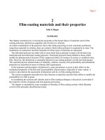
The anatomy of the trachea and bronchi
Commentary
Anatomy of these areas is of self-evident importance both in anaesthesia and inten-
sive care. You may be given the opportunity to describe every bronchopulmonary
segment, but because the terminology is cumbersome, with considerable duplication,
it is more likely, once you have demonstrated that you know the key points (such as
the origin of the right upper lobe bronchus), that the viva will move onto more
applied clinical aspects.
The viva
You will be asked to describe the anatomy.
●
Trachea: The trachea is a tube of cartilage with a membranous lining which is
continuous inferiorly with the larynx. The trachea proper is 10–11 cm long, and
extends downwards from the cricoid cartilage at the level of the sixth cervical
vertebra, as far as the sixth thoracic vertebra (in full inspiration). It then divides
into left and right main bronchi. Its diameter in the adult is around 20 mm.
In the first year of life its diameter is 3 mm or less, and increases thereafter by
about 1 mm year
Ϫ1
of age until it attains adult dimensions. It comprises 16–20
C-shaped cartilages attached vertically by fibro-elastic connective tissue, which
helps explain the mobility of the structure. Through most of its course the
trachea lies in the midline although at the bifurcation it is displaced slightly
rightwards by the arch of the aorta.
●
Anterior relations: In the upper part of the neck these are confined to skin and
fascia, with the isthmus of the thyroid overlying the second to fourth tracheal
rings. In its lower cervical course the trachea is partly overlain by the
sternohyoid and sternothyroid muscles, and by the jugular arch connecting the
anterior jugular veins. In its thoracic course the manubrium sterni lies anteriorly,
as do the remnants of the thymus, the inferior thyroid veins and the
brachiocephalic artery.
●
Posterior relations: Posteriorly lies the oesophagus, and in grooves between
trachea and oesophagus run the recurrent laryngeal nerves.
●
Lateral relations: In the upper neck it is related to the lobes of the thyroid and to
the carotid sheath. In its lower course it is related on the right to the lung and
pleura, to the brachiocephalic artery and veins, to the azygos vein and to the
superior vena cava. On the left it is related to the arch of the aorta and the left
common carotid and subclavian arteries.
●
The right and left main bronchi: The main bronchi are formed at about the level
of T
5
. The right is shorter (3 cm long), wider and angled more vertically than the
left, which means that foreign bodies and tracheal tubes are more likely to enter
its orifice than the left. The left main bronchus is more obliquely placed and is
some 5 cm in length. Important relations on the right are the pulmonary artery
which lies first below and then anterior to it, with the azygos vein above; while
on the left side the main bronchus lies below the arch of the aorta with the
descending aorta behind and the left pulmonary artery lying in fr
ont. In children
the angles of the bronchi at the carina are equal.
●
Bronchopulmonary segments – right lung: Within about 2.5 cm of the
bifurcation the right main bronchus gives off the right upper lobe bronchus
(which divides in turn within 1 cm into apical, anterior and posterior segments).
It is this right upper lobe bronchus that is most at risk from inadvertent
occlusion by a tracheal tube or a right-sided double-lumen endobronchial tube.
The right main then gives off the middle lobe bronchus, which is directed
downwards and forwards (before bifurcating into medial and lateral lobes). Just
below the origin of the middle lobe bronchus and opposite to it, is the bronchus
of the apical segment of the lower lobe. This directs posteriorly, before dividing
CHAPTER
2
The anaesthesia science viva book
28
into superior, anterior basal and lateral basal segments. The medial, anterior,
lateral and posterior basal segments arise in due course from the main stem of
the lower lobe bronchus, which continues in its downward direction.
●
Bronchopulmonary segments – left lung: The longer left main bronchus gives
off the left upper lobe bronchus after about 5 cm, and this then divides into a
superior division fr
om which arise apical, posterior and anterior segments of the
upper lobe, and a lingular bronchus from which arise the superior and inferior
lingular segments. The anatomy of the left lower lobe is similar to the right, in
that the left lower lobe bronchus gives off superior, anterior basal, lateral basal
and posterior basal segments. The medial basal bronchopulmonary segment
usually arises in common with the anterior basal, however, which means
technically that there are only four rather than five bronchopulmonary segments
on the left.
Direction the viva may take
You may be asked whether this anatomy allows you to predict which parts of the
lung may be contaminated following an episode of aspiration.
●
Pulmonary aspiration of gastric contents: The anatomy of the lobes and
bronchopulmonary segments influences zonal contamination should pulmonary
aspiration occur. If the patient is supine it is more likely that the apical segments
of the lower lobes will be affected because of the direct posterior projection of the
bronchus of the apical segment. If they are in the lateral position then aspiration
is more likely to affect the upper lobes. If prone, the right middle lobe and
lingula will be the site of the problem because of their downward and forward
orientation, and if the patient is sitting, it will be the posterior or lateral basal
segments of the lower lobes that are contaminated.
Further direction the viva could take
You may be asked about double-lumen tubes
●
Double-lumen endobronchial tubes: These are used when one lung needs to be
isolated so that the other can be collapsed to allow surgery. Such procedures
include pulmonary resection, oesophago-gastrectomy, surgery on the thoracic
aorta, anterior spinal fixation and thorascopic sympathectomy. A left-sided tube
is almost always favoured because this avoids the risk of occluding
inadvertently the origin of the right upper lobe bronchus. Problems with
malpositioned tubes are an important cause of mortality and morbidity (see
One-lung anaesthesia, page 107). A double-lumen tube is positioned correctly
when the upper surface of the bronchial cuff lies immediately distal to the
bifurcation of the carina. The position of the tube should be checked
endoscopically.
CHAPTER
2
Anatomy and its applications
29
The stellate ganglion
Commentary
Stellate ganglion block is a common procedure in the chronic pain clinic, is simple to
perform, and has significant potential complications. You may well not have carried
out this block yourself, but as one of several procedures in the neck undertaken by
anaesthetists (others include interscalene block, deep cervical plexus block and internal
jugular cannulation), its anatomy is of some relevance.
The viva
You will be asked to describe the anatomy.
●
The cervical sympathetic chain lies either side of the vertebral column in the
fascial space: posterior lies the fascia over the prevertebral muscles, anteriorly is
the carotid sheath.
●
The area where the inferior cervical and the first thoracic ganglia meet, either in
close proximity or fusion, is referred to as the stellate ganglion.
●
The ganglion extends from the neck of the first rib where its lower part is
covered anteriorly by the dome of the pleura, to the transverse process of C
7
where anterior lies the vertebral artery
. By the level of C
6
the vertebral artery has
moved posteriorly into the foramen transversarium pending its ascent into the
skull.
●
Much of the sympathetic nerve supply to the head and neck as well as to the
upper extremity synapses in or near the stellate ganglion.
●
Sympathetic pre-ganglionic fibres leave the cord from segments as widely
separated as T
1
–T
6
and although many converge in or around the stellate
ganglion, some may bypass it. For this reason large volumes of local anaesthetic
solution may be needed to fill the space in front of the prevertebral fascia down
to T
4
, but this will pr
oduce reliable sympathetic blockade of the head, neck and
upper limb. It is more accurately described as a ‘cervicothoracic block’.
Direction the viva may take
You will probably be asked about a technique of stellate ganglion block, and then
about its indications.
●
Technique: Two approaches are described; the anterior (sometimes called the
‘paratracheal’ anterior) approach and the paratracheal approach.
— Anterior approach: The trachea and carotid pulse are gently retracted to
allow identification of the most prominent cervical transverse process (the
Chassaignac tubercle) at C
6
, the level of the cricoid cartilage.
— A lower approach to the ganglion’s actual location at C
7
risks both
pneumothorax and vertebral artery puncture.
— The carotid sheath is moved laterally, and the trachea medially, before a
25–30 mm ϫ 23–25G needle is directed perpendicularly down on to the
tubercle.
— Once it has encountered bone, the needle is withdrawn 4–5 mm. If this is
not done there is a higher incidence of upper limb somatic blockade.
— Local anaesthetic in low concentration and high volume is injected (such as
lignocaine 0.5% or bupivacaine 0.125% ϫ 15–20 ml).
— Paratracheal approach: The needle insertion is two fingerbreadths lateral to
the suprasternal notch and two fingerbreadths superior to the clavicle. This
identifies the transverse process of C
7
, immediately below Chassaignac’s
tubercle at C
6
, at the level of the cricoid cartilage.
— The sterno-cleidomastoid and carotid sheath are moved laterally before the
needle is directed perpendicularly down onto the transverse process.
— Once it has encountered bone the needle is withdrawn 0.5–1.0 cm.
CHAPTER
2
The anaesthesia science viva book
30
— Local anaesthetic in low concentration and high volume is injected as
above.
— This lower approach risks pneumothorax as well as vertebral artery
puncture.
Further direction the viva could take
You might then be asked indications for the block, and the viva may concentrate on
its use following inadvertent intra-arterial injection, this being one of the classic
anaesthetic indications. Very few of the other indications listed are likely to lie within
your current experience.
●
You could start by commenting that the evidence base for the therapeutic use of
stellate ganglion blocks is not strong, but the technique has a long tradition of
use in the management of chronic pain.
●
Indications: These include any condition requiring sympathetic block of the
head, neck and upper limb.
— Neuropathic pain conditions: Complex regional pain syndromes types 1 and 2,
post-herpetic neuralgia of head and neck, shoulder–hand syndrome
(following cerebrovascular accident (CVA) or ischaemia), phantom limb
pain and pain associated with upper limb denervation.
— Ischaemic conditions: Thrombosis or microembolism, vasospastic disorders
(e.g. Raynaud’s disease), scleroderma, frostbite and inadvertent intra-
arterial injection. See Arterial supply of the hand, page 45.
— Angina pectoris: Severe refractory chest pain due to coronary ischaemia.
— Miscellaneous: Hyperhydrosis and treatment of pain associated with Paget’s
disease of bone.
If you have got this far, you may be asked finally about complications.
●
Complications: These include local trauma and haematoma (which may
compress the airway if severe); recurrent laryngeal nerve block, which causes
hoarseness; brachial plexus block, because via the anterior approach only a layer
of fascia separates the plexus and the ganglion which is anterior to it; carotid or
vertebral arterial puncture and possible intravascular injection (with the
paratracheal lower approach); intrathecal injection; pneumothorax (if the
approach is too low) and deep cervical plexus block (if the approach is too high).
CHAPTER
2
Anatomy and its applications
31
Surface anatomy of the neck
(with particular reference to percutaneous tracheostomy
and cricothyroidotomy)
Commentary
If these procedures are performed incorrectly the results can be disastrous. The
applied anatomy is not complex but you should give a simple authoritative account
of the techniques, particularly in relation to the potentially life-saving manoeuvre of
cricothyroidotomy. If the techniques that you describe put the patient at risk then you
will fail this question and probably the viva.
The viva
This question is specific, and so you will be asked to describe the surface anatomy of
the neck.
It does not matter particularly how you approach the answer; one way is to outline
the anatomy from above downwards.
●
The hyoid bone lies at the level of the third cervical vertebra (C
3
). Lying just
above and behind is the epiglottis.
●
The bifurcation of the common carotid artery is at the level of the fourth cervical
vertebra (C
4
), slightly above the notch of the thyroid cartilage.
●
The larynx lies opposite the fourth, fifth and sixth cervical vertebrae (C
4
,C
5
and C
6
).
●
The cricoid cartilage is at the level of the sixth cervical vertebra (C
6
).
●
The trachea extends from the sixth cervical vertebra (C
6
) down as far as the fifth
or sixth thoracic vertebra (T
5
and T
6
) at end-inspiration.
●
The suprasternal notch is located at the level of the second and third thoracic
vertebrae (T
2
and T
3
).
Direction the viva may take
You may be asked further about the anatomy relevant to the two clinical techniques
(of percutaneous tracheostomy and cricothyroidotomy), which have different indica-
tions but broadly similar complications.
●
The trachea comprises 16–20 C-shaped cartilages, which lie anteriorly in the neck
covered by skin and the superficial and deep fascial layers. The second, third
and fourth rings are covered by the isthmus of the thyroid. The great vessels of
the neck lie laterally, and so identification of the midline is crucial.
●
The cricothyroid membrane spans the inferior border of the thyroid cartilage and
the superior border of the cricoid cartilage, and immediately overlies the
subglottic region of the larynx. It is covered anteriorly by skin and by superficial
and deep fascia. Immediately lateral are the sterno-cleidomastoid muscle, the
sternothyroid and the sternohyoid muscles and the carotid sheath.
Percutaneous tracheostomy
●
This is an elective, not an emergency procedure, which in the context of intensive
care has become a well-established alternative to definitive surgical
tracheostomy. Its indications are the same as for formal tracheostomy in the
critically ill: typically to simplify airway management in a patient who otherwise
would face the problems of long-term tracheal intubation.
●
There are variations in approach, but all are based on a modified Seldinger
technique for placing a tracheostomy tube.
●
A typical technique is described as follows:
— Guided by the surface anatomy as described above, a skin incision is made
to allow a needle and guide wire to be placed through the fibro-elastic
tissue that joins the tracheal rings.
CHAPTER
2
The anaesthesia science viva book
32
— The isthmus of the thyroid gland covers the second to fourth tracheal rings.
A higher approach through the subcricoid membrane, or between the first
and second tracheal rings does avoid the thyroid isthmus but is associated
with greater incidence of tracheal stenosis. It is for this reason that many
intensivists now prefer a low approach, below the second or even third ring.
— The diameter of the hole is enlar
ged with progr
essively larger dilators to
the point at which it will accept a definitive tracheostomy tube.
— It is usual for a second anaesthetist to monitor this procedure from within
the trachea, by using a fibreoptic bronchoscope. The posterior wall of the
trachea may be so ragged and friable that it can easily be perforated.
Further direction the viva could take
You may be asked to compare percutaneous tracheostomy with cricothyroidotomy.
●
Both the techniques bypass the normal translaryngeal route to secure the airway,
but the circumstances and urgency of their use differ considerably. Percutaneous
tracheostomy is an elective procedure, whereas cricothyroidotomy is an
emergency procedure, which is usually invoked only when all other attempts to
secure a definitive airway have failed and when critical hypoxia is imminent.
●
The cricothyroid membrane is used for emergency access because it is readily
identifiable and because it is relatively avascular.
●
The patient is positioned with the neck extended to allow identification of the
membrane. After stabilisation of the overlying skin, which can be quite lax, a
small vertical incision in the skin is followed by a transverse incision in the
membrane. A spreader or scalpel handle is used to open the airway, after which
an appropriate tube can be inserted under direct vision. The purpose-made
devices typically have an internal diameter of 4 mm.
You may be asked finally to comment on complications.
●
Haemorrhage (immediate or delayed); the creation of false passage; tracheal or
oesophageal perforation; barotrauma; subcutaneous emphysema; failure and
accidental decannulation.
●
Subglottic stenosis is a cause of serious morbidity; it is mor
e common after
cricothyr
oidotomy than after percutaneous tracheostomy
.
CHAPTER
2
Anatomy and its applications
33
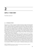
The brachial plexus
Commentary
An understanding of the anatomy of the brachial plexus is the key to successful
regional anaesthesia of the upper limb. The anatomy is detailed, but is not so com-
plex that it cannot be incorporated into a 7 or 8 min viva question. It is a clinically
important area of anatomy and it is asked frequently. It is worth learning a schematic
diagram of the plexus, because it makes it much easier to explain it to the examiners.
The viva
You will be asked about the formation of the brachial plexus.
●
The plexus forms in the neck from the anterior primary rami of C
5
,C
6
,C
7
,C
8
and T
1
.
●
These five roots merge in the posterior triangle of the neck to form three trunks.
●
C
5
and C
6
form the upper, and C
7
the middle trunk (above the subclavian artery).
C
8
and T
1
form the lower trunk (posterior to the subclavian artery).
●
At the lateral border of the first rib the three trunks each divide into anterior and
posterior divisions.
●
The three posterior divisions form the posterior cord (described according to its
relationship with the axillary artery), from which derives the radial nerve. (Also
the axillary, thoracodorsal and upper and lower subscapular nerves.)
●
The anterior divisions of upper and middle trunks form the lateral cord, from
which derive the median nerve (lateral head) and the musculocutaneous nerve.
(Also the lateral pectoral nerve.)
●
The anterior division of the lower trunk continues as the medial cord, from
which derive the ulnar nerve and the median nerve (medial head). (Also the
medial cutaneous nerves of arm and forearm and the medial pectoral nerve.)
Direction the viva may take
You will be asked about brachial plexus block. It is probable that you will be asked
to describe an approach of your choosing. Choose a block that you have actually
performed.
●
Interscalene block
— Interscalene local anaesthesia blocks the anterior primary rami of the
nerves of C
5
,C
6
,C
7
,C
8
and T
1
, before they merge in the posterior triangle to
form the trunks of the brachial plexus.
— The cervical nerves leave the intervertebral foramina, and pass caudad and
laterally between the scalenus anterior and the scalenus medius muscles.
The nerves are enclosed within a fascial compartment which comprises the
posterior fascia of the anterior scalene muscle, and the anterior fascia of the
middle scalene muscle.
— The patient should lie supine with the head turned slightly away from the
side of injection and with the arm by the side (gently pulled down if
necessary to depress the shoulder).
— After standard aseptic preparation, the interscalene groove between
scalenus anterior and medius should be identified at the level of the cricoid
cartilage (C
6
).
— If the awake patient is asked to lift the head off the pillow (which tenses the
sterno-cleidomastoid muscles) or to give a sniff, the groove becomes more
evident. In the anaesthetised patient identification is helped by the fact that
in more than 90% of subjects the external jugular vein overlies the gr
oove at
this level.
— The groove and the roots beyond are superficial and in most cases a
stimulating needle no longer than 30 mm is needed. The needle should be
CHAPTER
2
The anaesthesia science viva book
34
held perpendicular to the skin in all planes as it is directed medially,
posteriorly and caudally (inwards, backwards and downwards,
respectively) towards the transverse process of C
6
(Chaisssaignac’s
tubercle).
— Once muscle stimulation is apparent in the required distribution (usually
shoulder movements mediated by C
5
and C
6
) 30–40 ml of solution may be
injected after aspiration and with all due precautions. In common with
most plexus blocks into fascial compartments, large volumes of
appropriately dilute solutions may be needed to obtain adequate analgesia
of all the nerves involved.
— Interscalene block is particularly useful for shoulder surgery. It can be used
to provide analgesia for more distal structures in the upper limb, but it does
not provide reliable block of C
8
and T
1
and so ulnar sparing is frequent
(some reports quote 30–40%).
— It commonly blocks the phrenic nerve and so should be used cautiously in
those with respiratory disease. Bilateral blocks should not be performed.
— Complications: These include intravascular injection (particularly into the
vertebral artery), central spread via inadvertent dural puncture leading to a
total spinal, phrenic nerve palsy (which almost invariably accompanies an
effective block), Horner’s syndrome (cervical sympathetic block, which is
usually innocuous), vagal and recurrent laryngeal nerve block which
may cause hoarseness, but is usually benign, and pneumothorax. (There
are also the generic complications such as systemic toxicity and
neurapraxia.)
●
Supraclavicular block
— This block provides analgesia for most of the upper limb. The three trunks
are in close arrangement and the block is reliable. It can also be used for
shoulder surgery, although the interscalene approach is usually preferred.
— The three trunks lie on the first rib, between the insertion of the scalenus
anterior and scalenus medius muscles, and immediately posterior to the
subclavian artery (the pulsations of which can provide a landmark).
— The trunks cross the rib at about the mid-point of the clavicle.
— A number of approaches have been described: if you are familiar with one
of them then explain it. In essence the aim of the technique is to direct the
needle down onto the first rib, and to contact the brachial plexus where it
lies cephaloposterior to the subclavian artery.
— Once muscle stimulation is apparent in the appropriate distribution,
20–40 ml of appropriate local anaesthetic solution (such as
laevobupivacaine 0.25–0.5%) may be injected after aspiration and with the
usual precautions. If localisation is accurate, then the smaller volumes will
be effective.
— Complications: These include pneumothorax (the incidence may be 0.5–1.0%
even in experienced hands, and may takeupto24 h to develop),
intravascular injection or puncture (subclavian artery or vein), phrenic
nerve palsy (in 40–60%), Horner’s syndrome in 70–90% (cervical
sympathetic block) and neuritis (plus generic complications as above).
●
Subclavian perivascular or infraclavicular block (several variations have been
described).
— In effect this is an approach to the axillary sheath from a proximal direction,
although the blocks provides analgesia similar to that offered by the
supraclavicular approach. The subclavian perivascular block is actually
made through a needle inserted above the clavicle. Unlike the other
techniques these alone reliably block the intercostobrachial nerve. These
blocks are not widely used in the UK and unfamiliarity with their details
will not disadvantage you.
CHAPTER
2
Anatomy and its applications
35
●
Axillary block
— This has fewer complications than other approaches, is generally effective
and is a popular technique.
— The block provides good analgesia for surgery below the elbow. The
musculocutaneous nerve may leave the axillary sheath proximal to the site
of injection, in which event supplemental analgesia may be needed by
blocking the nerve between brachioradialis and the lateral epicondyle at the
elbow. This nerve innervates a substantial part of the radial side of the
forearm, and so local anaesthetic sparing of this area is not purely
academic.
— The arm is abducted to 90° (hyperabduction may abolish the arterial
pulsation). The advancing needle is directed at an angle of about 45° to the
skin as far proximally as possible. In practice this often means injecting at
the lateral border of pectoralis major.
— Once a twitch is elicited, the entire volume of local anaesthetic solution can
be injected (after aspiration). Ittakes just over 40 ml to fill the axillary
sheath as far as the coracoid process in adults, and in theory complete block
of all three cords will follow circumferential spread round the sheath. Some
anaesthetists prefer to identify the major nerves of the upper limb
separately, and block each one in turn.
— An alternative approach uses axillary arterial puncture as an end point.
Following transfixion of the vessel, the needle is either advanced or
withdrawn until aspiration is negative. The widespread use of nerve
stimulators has made this technique less respectable than once it was.
Further direction the viva could take
It is important that you understand the indications for these different approaches (for
instance, interscalene block for shoulder surgery; axillary block for a fasciectomy
involving the fifth finger) and that you are aware of their limitations and complica-
tions. You may be asked, therefore, to compare and contrast the blocks.
CHAPTER
2
The anaesthesia science viva book
36
The ulnar nerve
Commentary
This is a well-circumscribed area of anatomy, which is of interest not only because the
ulnar nerve can be blocked to provide surgical anaesthesia, but also because it is vul-
nerable to damage during general anaesthesia.
The viva
You will be asked about the anatomy of the ulnar nerve.
●
The ulnar nerve arises from the brachial plexus. (This is formed from the anterior
primary rami of C
5
,C
6
,C
7
,C
8
and T
1
. These roots merge in the posterior triangle
of the neck to form three trunks: C
5
and C
6
form the upper, C
7
the middle trunk,
and C
8
and T
1
form the lower trunk. At the lateral border of the first rib the three
trunks each divide into anterior and posterior divisions.)
●
The anterior division of the lower trunk continues as the medial cord, from
which derives the ulnar nerve. Its fibres originate mainly from C
8
and T
1
,
although it may also receive a contribution from C
7
.
●
It passes through the extensor compartment of the upper arm, lying medial to
the axillary and brachial arteries. It then continues medially on the anterior
aspect of the medial head of triceps to pass beneath the medial epicondyle of the
humerus, where it lies in the ulnar groove.
●
It enters the forearm between the two heads of flexor carpi ulnaris. In the upper
part of the forearm it lies deep to this muscle and separated from the ulnar
artery. In the distal forearm it lies lateral to flexor carpi ulnaris and near to the
medial side of the artery.
●
About 5 cm above the wrist it gives off a dorsal branch before continuing into the
hand lateral to the pisiform bone and above the flexor retinaculum.
●
The ulnar nerve pr
ovides the motor supply to flexor carpi ulnaris, to the medial
part of flexor digitorum pr
ofundus, and to the hypothenar muscles. It also
supplies all the small muscles of the hand apart fr
om the lateral two lumbricals
and the thr
ee muscles of the thenar eminence (abductor pollicis br
evis, opponens
pollicis and part of flexor pollicis br
evis). The deep head of flexor pollicis is
supplied by the ulnar nerve.
●
It supplies sensation to the elbow joint but gives off no branches in the upper
arm. It supplies the skin over the hypothenar eminence and over the fifth finger
as well as over the medial part of the fourth finger.
Direction the viva may take
You may be asked about the indications for, and techniques of, ulnar nerve blockade.
●
Indications for ulnar block follow from knowledge of its anatomy, and its main
use is to provide analgesia for procedures on the medial, ulnar side of the hand
and forearm. Digital nerve blocks are an easy and reliable method of providing
anaesthesia for finger surgery, and so ulnar block is reserved usually for more
proximal operations such as palmar fasciectomy. It is commonly performed
jointly with blocks of the other major nerves of the arm.
●
The nerve can be blocked at a number of sites:
— At the brachial plexus: See The brachial plexus, page 34.
— At mid-humeral level: A line is drawn between the upper border of pectoralis
major in the axilla and the mid-point of the flexor crease of the elbow.
A parallel line is drawn along the middle of the humerus about 1 cm medial
to it, and via a single injection point at this mid-point all three major nerves
of the forearm can be reached with a 50-mm stimulator needle.
— At the elbow: The nerve can be blocked with about 5 ml of solution injected
2–3 cm proximal to the ulnar groove. Injection into the actual fibrous sheath
CHAPTER
2
Anatomy and its applications
37
at the elbow is thought to be associated with a high incidence of residual
neuritis.
— At the wrist: The nerve lies beneath the tendon of flexor carpi ulnaris,
proximal to the pisiform bone, and medial and deep to the ulnar artery. An
approach from the ulnar side of the tendon (3–5 ml of solution injected at a
depth of around 1.5 cm) is less likely to encounter the artery
, and will also
block the cutaneous branches.
Further direction the viva could take
You may be asked about the potential for ulnar nerve damage and the clinical signs
of such damage.
●
Damage: Even when the arm is lying in the neutral position by the side of the
anaesthetised patient it is vulnerable to pressure, either from arm supports or
from the table itself. It has become routine practice to protect the elbow with
padding, and it has become equally routine to blame anaesthesia for any ulnar
nerve damage. This is despite the fact that ulnar nerve palsy has been reported
even when every precaution has been taken. The nerve is also vulnerable to
stretch and so the upper arm should not be displaced posteriorly, nor abducted
to greater than 90°.
●
Symptoms and signs of injury: Apart from the sensory loss and paraesthesia of
which the patient will complain, ulnar nerve injury is associated with the classic
main en griffe, or claw hand. This is because the extensors of the fingers and the
long flexors of the hand act unopposed. If the nerve is transected at the elbow
the clawing is less marked. This so-called ‘ulnar paradox’ occurs because flexor
digitorum profundus is also paralysed.
CHAPTER
2
The anaesthesia science viva book
38
The radial nerve
Commentary
The radial nerve is one of the three main nerves of the upper limb, and comprises
another well-defined area of anatomy. Upper limb surgery and trauma is common,
and radial nerve block is a reliable means of producing useful analgesia. The nerve
has a relatively large number of terminal branches whose detailed anatomy is
beyond the scope of this viva, but you will need to know the effects of blocking the
radial nerve proximal to its main divisions.
The viva
You will be asked about the anatomy of the radial nerve.
●
The radial nerve arises from the brachial plexus. (This is formed from the
anterior primary rami of C
5
,C
6
,C
7
,C
8
and T
1
. These roots merge in the posterior
triangle of the neck to form three trunks: C
5
and C
6
form the upper, C
7
the
middle trunk, and C
8
and T
1
form the lower trunk. At the lateral border of the
first rib the three trunks each divide into anterior and posterior divisions.)
●
The three posterior divisions form the posterior cord (described according to its
relationship with the axillary artery), from which derives the radial nerve. Its
fibres originate, therefore, from C
5
,C
6
,C
7
,C
8
and T
1
, and it is the largest branch
of the brachial plexus.
●
The radial nerve descends beneath the axillary artery and passes between the
long and medial heads of the triceps muscle into the posterior compartment of
the arm. It then passes obliquely behind the humerus where it lies in a shallow
spiral groove.
●
In the lower third of the humerus it enters the anterior compartment of the
upper arm, descending into the forearm between brachialis medially and
brachioradialis laterally. At the lateral epicondyle of the humerus it divides into
its terminal deep and superficial branches.
●
It is motor in the upper arm to triceps, in the lower arm to brachialis,
brachioradialis and to the extensor muscles of the wrist and hand.
●
The area of sensory innervation that is of particular anaesthetic relevance
includes much of the dorsum of the hand, and the radial side of the forearm.
(The ulnar nerve supplies the skin over the distal phalanges, the fifth finger and
medial side of the fourth finger and over the fifth and fourth metacarpals.) The
radial nerve also supplies cutaneous sensation to the posterior aspect of the
forearm and to the skin over the dorsal base of the thumb. (The musculo-
cutaneous nerve supplies much of the radial surface of the forearm.)
Direction the viva may take
You may be asked about the indications for, and techniques of, radial nerve blockade.
●
Its main use is in conjunction with other blocks to provide analgesia for
procedures on the lateral, radial side of the hand and forearm. Digital nerve
blocks provide reliable anaesthesia for finger surgery, but radial block can be
used for procedures on the base of the thumb and, in combination with
musculocutaneous block, to allow the cr
eation of forearm arterio-venous fistulae
for dialysis.
●
The nerve can be blocked at various sites:
— At the brachial plexus: See The brachial plexus, page 34.
— At mid-humeral level: A line is drawn between the upper border of pectoralis
major in the axilla and the mid-point of the flexor crease of the elbow.
A parallel line is drawn along the middle of the humerus about 1 cm medial
to it, and via a single injection point at this mid-point all three major nerves
of the forearm can be reached with a 50-mm stimulator needle.
CHAPTER
2
Anatomy and its applications
39
— At the elbow: The nerve can be blocked as it traverses the anterior aspect of
the lateral epicondyle of the humerus. The needle is inserted some 2 cm
lateral to the biceps tendon and directed towards the bone. Up to 10mlof
solution can be injected in a fanwise direction as the needle is withdrawn.
The musculocutaneous nerve can also be blocked at the elbow between the
biceps and brachioradialis muscles.
— At the wrist: Nerve block at the wrist is effectively a superficial field block of
the terminal sensory branches. Local anaesthetic solution can be injected
along the lateral border of the radial artery, extending dorsally to include
the area delineated by the extensor tendons of the thumb.
Further direction the viva could take
You may be asked about the potential for radial nerve damage and the clinical signs
of such damage.
●
Damage: The radial nerve is subject to various types of injury and may be
damaged by compression against the upper humerus, as in the so-called
‘Saturday night or crutch palsy’. The pressure exerted by an arterial tourniquet
can also damage the nerve by the same mechanism. Its close relation to the
humerus makes it vulnerable to damage in mid-humeral fractures, and the
posterior interosseous branch may be traumatised in injuries to the head of
the radius.
●
Symptoms and signs of injury: Overlap of innervation means that sensory loss
and paraesthesia may be confined to a relatively small area on the dorsum of the
hand. Otherwise radial nerve injury typically is associated with wrist drop due
to paralysis of the extensor muscles. If the damage to the nerve has occurred
below the elbow then the functional preservation of extensor carpi radialis
longus will minimise this effect.
CHAPTER
2
The anaesthesia science viva book
40
The median nerve
Commentary
This is the third of the main nerves of the upper limb, and comprises another well-
defined area of anatomy. As with the questions on the ulnar and radial nerves, you
will be expected to outline the anatomy and to discuss the relevant local anaesthetic
blocks.
The viva
You will be asked about the anatomy of the median nerve.
●
The median nerve arises from the brachial plexus. (This is formed from the
anterior primary rami of C
5
,C
6
,C
7
,C
8
and T
1
. These roots merge in the posterior
triangle of the neck to form three trunks: C
5
and C
6
form the upper, C
7
the
middle trunk, and C
8
and T
1
form the lower trunk. At the lateral border of the
first rib the three trunks each divide into anterior and posterior divisions.)
●
The anterior divisions of the upper and middle trunks form the lateral cord, from
which derive the lateral head of the median nerve.
●
The anterior division of the lower trunk continues as the medial cord, from
which derives the medial head of the median nerve. Its fibres originate,
therefore, from C
5
,C
6
,C
7
,C
8
and T
1
.
●
The nerve passes into the arm lying lateral to the brachial artery which it then
crosses to descend on its medial side to the antecubital fossa, where it is
protected by the bicipital aponeurosis.
●
It passes down into the forearm between the bellies of the deep and superficial
flexors of the fingers (flexor digitorum profundus and superficialis) and at the
wrist lies lateral to or just beneath the tendon of palmaris longus, and medial to
flexor carpi radialis.
●
It enters the hand beneath the flexor r
etinaculum befor
e dividing into a leash of
terminal branches.
●
It is motor in the forearm to several of the superficial flexors (excluding flexor
carpi ulnaris) and in the hand to muscles of the thenar emininence: abductor
pollicis brevis, part of flexor pollicis brevis and the opponens pollicis. Its anterior
interosseous branch also supplies flexor pollicis longus, pronator quadratus and
part of flexor digitorum profundus.
●
The cutaneous innervation extends to the radial aspect of the palm, and the
palmar surface of the radial 3½ digits, together with their dorsal tips as far as the
first interphalangeal joint.
Direction the viva may take
You may be asked about the indications for, and techniques of, median nerve blockade.
●
Its main use is the provision of analgesia for pr
ocedures on the radial palm. The
fingers and distal thumb can readily be anaesthetised using digital nerve blocks,
but median nerve block is useful for procedures such as carpal tunnel release
and palmar fasciectomy.
●
The nerve can be blocked at various sites:
— At the brachial plexus: See The brachial plexus, page 34.
— At mid-humeral level: A line is drawn between the upper border of pectoralis
major in the axilla and the mid-point of the flexor crease of the elbow.
A parallel line is drawn along the middle of the humerus about 1 cm medial
to it, and via a single injection point at this mid-point all thr
ee major nerves
of the forearm can be reached with a 50-mm stimulator needle.
— At the elbow: The nerve can be blocked immediately medial to the brachial
artery as it crosses the intercondylar line. The needle is directed
perpendicularly and should find the nerve within 1–2 cm.
CHAPTER
2
Anatomy and its applications
41
— At the wrist: The nerve lies in the midline on the radial border of the
palmaris longus tendon. The needle is directed perpendicularly some 2 cm
proximal to the distal flexor crease of the wrist. The nerve is superficial and
lies beneath the deep fascia at a depth of 1 cm or less.
Further direction the viva could take
You may be asked about the potential for median nerve damage and the clinical signs
of such injury.
●
Damage: The median nerve is most vulnerable to trauma at the wrist, although
it can be injured in supracondylar humeral fractures and following injury to the
distal radius. The most common lesion occurs as a result of compression of the
nerve in the carpal tunnel.
●
Symptoms and signs of injury: Trauma at the wrist will paralyse the thenar
muscles and cause significant sensory loss. More proximal injury leads to weak
wrist flexion, loss of pronation, and loss of flexion of the thumb, index and
middle fingers. Atrophic changes and wasting of the thenar eminence will flatten
the contours of the hand.
CHAPTER
2
The anaesthesia science viva book
42
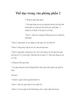
The antecubital fossa
Commentary
By analogy with the femoral triangle, the anatomy of the antecubital fossa is straight-
forward, and it too lends itself readily to simple diagrams. A transverse sketch is a
simple way of showing that you are aware of the important anatomical relations.
Alternatively you may find yourself automatically demonstrating on your own
arm: this can be an effective technique which may make the anatomy easier to learn.
Questioning may extend to practical clinical matters such as inadvertent intra-arterial
injection, nerve blocks at the elbow and the insertion of long lines. Non-medical staff
who undergo training in venepuncture and cannulation are required to learn the
detailed anatomy of this area, and so the FRCA examiners will expect no less.
The viva
You will be asked to describe the anatomy
.
●
The antecubital, or cubital fossa, is a triangular intermuscular depression on the
anterior surface of the elbow joint.
●
The base of the triangle is formed by the line which joins the medial and lateral
epicondyles of the humerus.
●
The lateral side of the triangle is formed by the medial edge of the
brachioradialis muscle, while the medial side is formed by the lateral border
of the pronator teres.
●
The floor consists of the brachialis and supinator muscles.
●
The roof (from above down) comprises skin, subcutaneous tissue, and the deep
fascia, which includes the bicipital aponeurosis.
●
Within the fossa lie the tendon of the biceps muscle and the terminal part of the
brachial artery, which lies in the centre of the fossa prior to its division into the
radial and ulnar arteries opposite the neck of the radius. It also contains the
associated veins and the median and radial nerves.
●
The anatomy of the superficial veins varies greatly, but that of a typical subject
can be described as follows.
— Cephalic vein: The cephalic vein drains the radial side of the forearm, and
ascends over the lateral side of the fossa to lie in a groove along the lateral
edge of the biceps. At the lower border of pectoralis major it moves deeper
to lie between pectoralis major and deltoid before penetrating the
clavipectoral fascia to join the axillary vein.
— Basilic vein: The basilic vein drains the ulnar side of the forearm and rises
along the medial border of biceps to pierce the deep fascia in the middle
upper arm before going on to form the axillary vein.
— Median cubital vein: The median cubital vein originates from the
cephalic vein distal to the lateral epicondyle, and then r
uns upwards
and medially across the antecubital fossa to join the basilic vein above
the elbow.
Direction the viva may take
You are likely to be asked about the clinical r
elevance of the anatomy.
●
The antecubital fossa is the most common site for venepuncture as well as being
a site for venous cannulation. One potential hazard is inadvertent puncture or
injection into the brachial artery. The danger of this happening is lessened by the
presence of the bicipital aponeurosis, which is an extension of the medial lower
border of the muscle and tendon of biceps. It passes downwar
ds and medially to
merge with the deep fascia at the origin of the forearm flexor muscles, separating
as it does so, the brachial artery from the median cubital vein. (This is the reason
why historically it was known as the ‘grâce à Dieu fascia’.)
CHAPTER
2
Anatomy and its applications
43
●
The lateral cutaneous nerve of the forearm crosses the fascia of the roof of the
fossa, and although it lies deep to the cephalic vein may still be vulnerable to
damage from a needle or a cannula.
●
Long lines can be inserted via the antecubital veins, which offer a safer route to
the central veins. Although cannulation at the elbow may be simple, the acute
curve at the clavipectoral fascia may pr
event a long venous catheter fr
om
gaining access to the central venous circulation.
Further direction the viva could take
You may be asked how you would recognise and manage inadvertent intra-arterial
injection, and about nerve blocks at this site.
●
This is detailed in Arterial supply of the hand, on page 45. An anomalous ulnar
artery which lies superficially just below the median cubital vein is present in 2%
of the population, and so it is not only accidental injection into the brachial
artery of which anaesthetists must be aware.
●
Nerve blocks at the elbow are described in The radial nerve, page 39,
The median nerve, page 41, and The ulnar nerve, page 37.
CHAPTER
2
The anaesthesia science viva book
44
Arterial supply of the hand
Commentary
This is a straightforward area of anatomy which in the modified Allen test has an
obvious clinical application, albeit one whose value is disputed. If you impart the
basic information too rapidly then you will find yourself discussing arterial pressure
waveforms and damping. That may suit you, but if you would prefer to stay with
the anatomy then it will be worth refreshing your memory of some of the relevant
muscles and tendons. This in any event will make your knowledge of anatomy
appear much more substantial.
The viva
You will be asked the basic anatomy of the arterial supply.
●
The hand is supplied by the radial and ulnar arteries.
●
Radial artery: In the distal forearm the radial artery lies between flexor carpi
radialis and brachioradialis. The tendons of these muscles comprise the
landmarks between which the artery is palpated at the wrist.
— Beyond the radial pulse the artery supplies a branch which contributes to
the superficial palmar arch.
— The main arterial branch continues over the scaphoid and beneath the
extensor and abductor tendons of the thumb (extensor pollicis longus and
brevis, and abductor pollicis longus), and passes between the first and
second metacarpal bones to contribute to the deep palmar arch.
●
Ulnar artery: In the distal forearm the ulnar artery lies superficially between the
tendons of flexor carpi ulnaris and flexor digitorum superficialis.
— It crosses beneath the flexor retinaculum to complete the superficial palmar
arch. The ulnar arterial component is much more significant than the radial.
— The deep branch enters the palm where it forms an anastomosis with the
radial artery to complete the deep palmar arch.
— The superficial palmar arch then gives off further branches including dorsal
metacarpal and dorsal digital arteries. The deep palmar arch similarly
branches to form palmar metacarpal and palmar digital arteries.
Direction the viva may take
You will probably be asked about the clinical relevance of this arterial supply.
●
Both arteries can be cannulated in order to allow direct intra-arterial
measurement of blood pressure, but anaesthetists prefer to be confident that the
circulation of the hand will not be jeopardised. The traditional method for
assessing the adequacy of radial or ulnar arterial flow is the modified Allen test.
After compression of both arteries at the wrist, the patient is asked to blanch the
palm by clenching and then opening the hand. On releasing the compression
of one or other of the arteries, depending on which is chosen as the site of
cannulation, the palm should reperfuse, demonstrating thereby the adequacy of
flow. Seven seconds or less is considered normal; longer than 15 s is abnormal.
Although the test continues to be used widely it has a poor predictive value.
Ischaemic complications have been reported following a normal Allen test and
vice versa.
Further direction the viva could take
This topic is not large, and so you may exhaust it fairly quickly. You will be assessed
mainly on the core information above, but there a number of directions the viva
could take. You may be asked about intra-arterial injection or about indications for,
and direct methods of measuring arterial pressure. There is unlikely to be enough
time to explore this latter subject in any depth.
CHAPTER
2
Anatomy and its applications
45
●
Intra-arterial injection: Accidental injection occurs when an artery is wrongly
identified as a vein, or when an intra-arterial catheter is mistaken for a venous
cannula. Drugs that have been so injected include anaesthetic induction agents,
phenytoin, benzodiazepines and antibiotics. In the awake patient severe pain in
the hand is a cardinal feature. In the anaesthetised or sedated patient there may
be ischaemic colour changes in the distal limb, which because of arterial spasm
may be pale, mottled or cyanosed. Thrombosis may follow. The degree of damage
depends on the substance injected. Thiopentone causes substantial damage
because at body pH it precipitates into crystals, which occlude small arterial
vessels and provoke intense vasospasm mediated via local noradrenaline
(norepinephrine) release. Propofol, in contrast, seems relatively innocuous. Any
such injection should be treated as for the worst-case scenario, because clinical
experience of intra-arterial injection of many drugs is limited.
●
Management: After the injection of 500–1000 heparin units to reduce thrombosis
risk, warm NaCl 0.9% can be given to dilute the substance.
Arterial spasm can be
treated with papaverine 40–80
mg, prostacyclin at rate of 1
g min
Ϫ1
, tolazoline
(which is a noradrenaline antagonist) and phenoxybenzamine (which is an
␣
1
-antagonist). Sound though the recommendation may be, these drugs may
well not be immediately available, and this advice may be impractical.
Dexamethasone 8 mg given immediately may reduce arterial oedema. Perfusion
can be enhanced by sympatholysis, either by a stellate ganglion block (which is
quick to perform) or via a brachial plexus block, using a catheter technique to
provide analgesia and a continuous block. Maintenance anticoagulation is
recommended for up to 14 days, and hyperbaric oxygen has also been suggested
as a means of minimising final ischaemic damage.
●
Intra-arterial monitoring: See Intra-arterial blood pressure measurement, page 263.
CHAPTER
2
The anaesthesia science viva book
46
Intercostal nerves
Commentary
This area of anatomy was of more direct relevance before thoracic epidural anaesthe-
sia, paravertebral injection and intrapleural catheterisation were more commonly
employed as analgesic techniques. Intercostal nerve blocks were used to provide
analgesia for subcostal surgical incisions and to treat the pain of fractured ribs. The
topic, however, continues to be asked, but because the list of indications for inter-
costal block is shrinking, the viva will concentrate on the anatomy and on the distri-
bution of injected drugs more than on clinical techniques of nerve blockade.
The viva
You will be asked the anatomy of an intercostal nerve.
●
The intercostal nerves are the ventral somatic rami of the spinal nerves from
T
1
to T
11
.T
12
is a subcostal nerve which is not closely associated with its
corresponding rib, and which in addition links with fibres from the first lumbar
nerve. T
1
,T
2
and occasionally T
3
, are also atypical, in that some of their fibres
join with fibres of the brachial plexus, as well as contributing to the formation of
the intercostobrachial nerve.
●
The typical intercostal nerve exits the intervertebral foramen to lie initially
between the posterior intercostal membrane and the pleura. Thereafter the nerve
lies between the internal and the innermost (intercostalis intimis) intercostal
muscles.
●
Each nerve lies in the neurovascular bundle comprising the artery, vein and
inferiorly the nerve, which runs in a groove beneath each rib. The overhanging
external edge of the rib protects this bundle from direct trauma. The groove is
also invested in the fascia of the external and internal intercostal muscles.
●
The groove is well defined until it reaches the mid-axillary line, at which point
the nerve divides.
●
Motor filaments supply the intercostal, the transversus thoracis and the serratus
posterior muscles. The lower intercostal nerves also supply motor fibres to the
abdominal muscles.
●
Sensory branches supply the overlying skin as well as supplying the parietal
pleura and the costal part of the diaphragm.
●
The first sensory branch arises as the posterior cutaneous branch, which
supplies the skin and muscles of the paravertebral area.
●
The second sensory branch arises as the lateral cutaneous branch after the
division of the nerve at around the mid-axillary line. The terminal fibres of this
branch supply the skin and subcutaneous tissue of much of the chest and
abdominal walls.
●
The third and final sensory branch arises as the anterior cutaneous branch which
is the continuation of the main intercostal nerve, and which supplies the skin
and subcutaneous tissue of the anterior chest and abdominal walls.
Direction the viva may take
You may be asked about indications for
, and the technique of, intercostal block. You
may never have seen this block performed, and it will come as no surprise to your
examiners if you admit as much. Your account, therefore, may be theoretical, but it
must be safe.
●
Intercostal nerve block can provide effective analgesia for upwards of 12h.
Historically it was used for analgesia following subcostal and loin incisions
(for gall bladder and renal surgery), after thoracotomy and to provide analgesia
for fractured ribs. Only the last indication now applies, and even here the
technique has been supplanted by intrapleural and epidural block. It has been
CHAPTER
2
Anatomy and its applications
47
used to alleviate the discomfort of herpes zoster. A block of T
10
,T
11
and T
12
provides effective analgesia following appendicectomy, but it is rarely utilised
for this purpose, possibly because in the UK relatively inexperienced trainees
give the majority of anaesthetics for this operation.
●
Technique of intercostal block
— The intercostal injection is made usually at the angle of the rib, before the
nerve divides.
— The skin of the back is tensed gently in a cranial direction before a needle
and syringe is advanced to encounter the lower surface of the appropriate
rib. The skin tension is then released. This helps the needle move to its
correct position.
— The needle is then carefully ‘walked off’ the inferior surface, before being
directed a further 2–3 mm inwards to pierce the fascia of the innermost
intercostal muscle (the posterior intercostal membrane) and enter the
subcostal groove.
— Following injection of 3 or 4
ml of solution, for example bupivacaine
0.25–0.5% with adr
enaline, the needle is withdrawn to r
est on the posterior
surface of the rib. The next space can then be located in the same way
without risking inadvertent injection in the same space. This can easily
happen in individuals even of modest size, and is common in the obese.
●
Complications include pneumothorax (incidence of less than 1%), respiratory
embarrassment in patients with any diaphragmatic impairment, and systemic
toxicity if a large number of nerves are blocked. The rich vascular supply of the
area means that systemic absorption following intercostal block exceeds that
from almost any other site.
Further direction the viva could take
You may be asked what happens to local anaesthetic once it has been injected.
●
Contrast studies have confirmed that local anaesthetic spreads not only along
the rib but also can track medially as far as the sympathetic chain. It also extends
to several dermatomes above and below the site of injection, probably via direct
sub-pleural spread. The intercostal, sub-pleural and paravertebral spaces are all
in anatomic continuity, and so it is not surprising that injection of sufficient
volume may lead to spread throughout all three.
CHAPTER
2
The anaesthesia science viva book
48
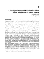
The diaphragm
Commentary
The diaphragm is an important anatomical area for anaesthetists although it acts
mainly as a radiographical marker for other disease processes. A raised hemi-
diaphragm, for example, may indicate pulmonary or abdominal pathology, while
gas under the diaphragm is pathognomonic of visceral perforation. So even though
primary diaphragmatic problems are rare, the examiners will expect you to demon-
strate knowledge of the anatomy that allows you to use it as an indicator for these
other conditions.
The viva
You will be asked to describe the basic anatomy of the structure.
●
Diaphragm: The diaphragm (from the Greek words for ‘across’ and ‘partition’) is
the dome-shaped muscular and fibrous partition which separates the abdominal
from the thoracic viscera.
●
Vertebral part: The vertebral part of the diaphragm originates from the right and
left crura, which arise from the front of the vertebral bodies of L
1
–L
3
and L
1
–L
2
,
respectively, and from the arcuate ligaments. The median ligament is a fibrous
band which links the crura; the medial ligament is a tendinous arch arising as a
thickening of the fascia of the psoas major muscle; the lateral ligament arises as
another thickening of fascia, in this case from the quadratus lumborum muscle.
●
Costal part: The costal part of the diaphragm arises from the six lowest ribs and
their costal cartilages.
●
Sternal part: The sternal part comprises two small attachments from the
xiphisternum.
●
Central tendon: The muscle fibres converge into the central tendon, which is a
tough aponeurosis near the centre of the dome of the diaphragm and which is
merged above with the connective tissue of the pericardium.
●
Foramina: There are three important openings in the diaphragm. Through a
foramen at the level of T
8
pass the inferior vena cava and right phrenic nerve.
Through an aperture at the level of T
10
pass the oesophagus and vagus nerves.
Through the final opening at the level of T
12
pass the aorta, the thoracic duct and
the azygos vein.
●
Motor supply: Motor innervation is supplied by the phrenic nerve (mainly C
4
but with contributions from C
3
and C
5
) and whose long thoracic course reflects
the descent of the diaphragm during fetal development.
●
Sensory supply: The central part of the diaphragm is innervated by the sensory
afferents of the phrenic nerve: hence the tendency for sub-diaphragmatic pain to
be referred to the shoulder tip, which shares the sensory innervation of C
5
. The
peripheral area of the diaphragm is innervated by the lower intercostal nerves.
Direction the viva may take
There are a number of miscellaneous areas of clinical relevance about which you
could be asked.
●
Position on chest X-ray: After forced expiration the right cupola (which is higher
than the left because of the upward pressure of the liver) is level anteriorly with
the fourth costal cartilage, and level posteriorly with the eighth rib. During quiet
respiration the diaphragm moves only about 1.5 cm but this excursion can
increase to 10 cm or more with deep inspiration.
●
The cardio-oesophageal sphincter: The fibres of the crura that surround the
cardio-oesophageal junction exert a pinchcock effect on the oesophagus which
contributes to the prevention of gastro-oesophageal reflux. Laxity of this
oesophageal hiatus is associated with hiatus hernia in which the lower
CHAPTER
2
Anatomy and its applications
49
oesophagus and stomach slide into the chest, causing symptoms of dyspepsia
and reflux. (This is a sliding hernia; the much less common rolling hernia occurs
when the fundus of the stomach rolls up through the hiatus in front of the
oesophagus which remains intra-abdominal. Patients have dyspepsia but no
reflux.) You should be prepared to detail your management of anaesthesia in a
patient with hiatus hernia. This commonly would involve a pr
ecise clinical
history seeking the symptoms and characteristics of oesophageal reflux, which if
positive would mandate rapid sequence induction following administration of
agents to reduce gastric acidity.
●
Phrenic nerve palsy: This may be caused by disease, or may be iatrogenic,
associated for example with a successful interscalene nerve block. On a plain
chest X-ray the affected hemidiaphragm will be raised (other causes include
pregnancy, ascites, obesity, intra-abdominal malignancy and lobar collapse)
while fluoroscopy will reveal paradoxical upward movement during inspiration.
During quiet breathing some 75% of respiratory function is diaphragmatic,
although when the minute volume is high, around 60% of the tidal volume is
provided by the accessory muscles. The phrenic nerve can be paced by stimuli
applied where it lies on the scalenus anterior muscle in the neck.
●
Spinal cord injury: Cord lesions at the level of C
2
and C
3
cause respiratory
tetraplegia. Injuries at C
4
and below permit some phrenic nerve function, but
vital capacity is reduced to about 25% of normal. Damage below C
6
allows full
diaphragmatic function.
●
Neuromuscular block: The diaphragm is among the muscles most resistant to
muscle relaxants. Post-operative respiration may therefore be adequate even
though the patient subjectively may feel profoundly weak.
●
Diaphragmatic hernia: These may be congenital, occurring in utero (the
incidence is 1 in 4000 live births) and preventing the proper development of the
lung, or traumatic. Surgical repair in the neonate requires tertiary paediatric
centre expertise, details of which you will not be expected to furnish. Traumatic
herniation may be associated with immediate symptoms, but equally there are
cases in which the abnormality has been diagnosed years after an injury from
which the patient has been asymptomatic.
CHAPTER
2
The anaesthesia science viva book
50
Innervation of the inguinal region
Commentary
This in essence is a straightforward question about field block for inguinal hernia
repair based on anatomical knowledge. If you provide reasonably comprehensive
anatomical details it will prevent the viva moving away from the core topic into more
vaguely related areas such as local anaesthetic toxicity.
The viva
You will be asked to describe the nerve supply to the inguinal region.
●
Supply: The skin over the lower abdomen is supplied by the first and second
nerves of the lumbar plexus, L
1
and L
2
, together with a contribution from the
subcostal nerve, T
12
.
●
Iliohypogastric nerve: This arises from L
1
, emerges from the lateral border of the
psoas muscle, and passes obliquely behind the kidney to perforate the posterior
part of the transversus abdominis muscle above the iliac crest. It lies then
between transversus and the internal oblique where it divides. Its anterior
cutaneous branch runs forward between those muscles before passing through
the internal oblique about 2 cm medial to the anterior superior iliac spine.
It pierces the aponeurosis of the external oblique muscle about 3 cm above the
external inguinal ring and supplies sensation to suprapubic skin.
●
Ilioinguinal nerve: This also arises from L
1
, emerging from the lateral border of
the psoas muscle and passing below the larger iliohypogastric nerve to perforate
the posterior part of the transversus abdominis muscle near the anterior iliac
crest. It lies below the internal oblique, before piercing it to traverse the inguinal
canal accompanied by the spermatic cord. It exits the external inguinal ring to
supply the skin of the upper thigh, the skin over the root of the penis or the
mons pubis, and the skin of the scrotum or labia.
●
Genitofemoral nerve: This arises from L
1
and L
2
, emerging on the abdominal
surface of the psoas muscle opposite the third or fourth lumbar vertebra. It runs
down on the body of the psoas muscle, retroperitoneally, and divides above the
inguinal ligament into genital and femoral branches. The genital branch enters
the inguinal canal via the deep inguinal ring to supply the cremaster muscle and
to send some fine terminal branches to innervate scrotal skin. (In the female it
accompanies the broad ligament and contributes to cutaneous sensation of the
mons and labia.) The femoral branch passes behind the inguinal ligament to
enter the femoral sheath, lateral to the artery, before perforating the sheath and
fascia lata anteriorly to supply the skin over the upper femoral triangle.
Direction the viva may take
You will be asked how you would perform a field block for inguinal herniorrhaphy.
There are various techniques described: choose the one with which you are most
familiar.
●
Reliable anaesthesia for inguinal hernia repair is not always easy to achieve, and
if the operation is done with the patient awake it is common for surgeons to
infiltrate considerable volumes of supplemental local anaesthetic. Field block is,
however, useful for post-operative analgesia.
●
All three nerves need to be blocked, and subsequent infiltration may also
be required over the skin incision itself, depending on its extent, and at the
internal ring.
●
A short bevelled needle is advanced via a point approximately 2 cm medial and
2 cm caudal to the anterior superior iliac spine. This blunter needle will better
appreciate the resistance offered by the external oblique aponeurosis, which is
penetrated often with a definite click. Injection of around 5 ml of local
CHAPTER
2
Anatomy and its applications
51
anaesthetic should be sufficient to block the iliohypogastric nerve at this point. If
the needle is then advanced through the internal oblique muscle for about
1–2 cm the same volume should block the ilioinguinal nerve which at this point
lies below the muscle. The genitofemoral nerve is approached via an injection
made from the pubic tubercle and extending fanwise from the midline to the
external inguinal ring.
●
Alternative techniques include the fanwise injection of large volume
low-concentration solutions in and between the oblique muscles (plus
genitofemoral nerve block as above), lumbar plexus and lumbar paravertebral
blocks. These latter two techniques are used infrequently.
Further direction the viva could take
You may be asked about the potential for local anaesthetic toxicity. See Local
anaesthetic toxicity, page 165.
CHAPTER
2
The anaesthesia science viva book
52