Báo cáo y học: "Nuclear import of Avian Sarcoma Virus integrase is facilitated by host cell factors" pdf
Bạn đang xem bản rút gọn của tài liệu. Xem và tải ngay bản đầy đủ của tài liệu tại đây (1.7 MB, 14 trang )
BioMed Central
Page 1 of 14
(page number not for citation purposes)
Retrovirology
Open Access
Research
Nuclear import of Avian Sarcoma Virus integrase is facilitated by
host cell factors
Mark D Andrake, Monica M Sauter, Kim Boland, Andrew D Goldstein,
Maryem Hussein and Anna Marie Skalka*
Address: Institute for Cancer Research, Fox Chase Cancer Center, Philadelphia, PA 19111, USA
Email: Mark D Andrake - ; Monica M Sauter - ; Kim Boland - ;
Andrew D Goldstein - ; Maryem Hussein - ;
Anna Marie Skalka* -
* Corresponding author
Abstract
Background: Integration of retroviral DNA into the host cell genome is an obligatory step in the
virus life cycle. In previous reports we identified a sequence (amino acids 201–236) in the linker
region between the catalytic core and C-terminal domains of the avian sarcoma virus (ASV)
integrase protein that functions as a transferable nuclear localization signal (NLS) in mammalian
cells. The sequence is distinct from all known NLSs but, like many, contains basic residues that are
essential for activity.
Results: Our present studies with digitonin-permeabilized HeLa cells show that nuclear import
mediated by the NLS of ASV integrase is an active, saturable, and ATP-dependent process. As
expected for transport through nuclear pore complexes, import is blocked by treatment of cells
with wheat germ agglutinin. We also show that import of ASV integrase requires soluble cellular
factors but does not depend on binding the classical adapter Importin-α. Results from competition
studies indicate that ASV integrase relies on one or more of the soluble components that mediate
transport of the linker histone H1.
Conclusion: These results are consistent with a role for ASV integrase and cytoplasmic cellular
factors in the nuclear import of its viral DNA substrate, and lay the foundation for identification of
host cell components that mediate this reaction.
Background
Integration of viral DNA into the genome of its host cell is
an essential step in the replication of all retroviruses. This
reaction is catalyzed by the retroviral integrase (IN), an
enzyme that, along with reverse transcriptase, enters the
cell within the infecting viral capsid. Reverse transcription
of the RNA genome to produce retroviral DNA is known
to take place in the cytoplasm, shortly after entry. How-
ever, the manner in which viral DNA and IN enter the
nucleus is not well understood and, indeed, may vary
among the different retroviruses. Nuclear import of the
human immunodeficiency virus type 1 (HIV-1) preinte-
gration complex, which includes viral DNA and IN, has
been the subject of intense investigation. As HIV and
other lentiviruses can infect non-dividing cells, in which
nuclei remain intact, some nuclear import mechanism
Published: 7 August 2008
Retrovirology 2008, 5:73 doi:10.1186/1742-4690-5-73
Received: 5 May 2008
Accepted: 7 August 2008
This article is available from: />© 2008 Andrake et al; licensee BioMed Central Ltd.
This is an Open Access article distributed under the terms of the Creative Commons Attribution License ( />),
which permits unrestricted use, distribution, and reproduction in any medium, provided the original work is properly cited.
Retrovirology 2008, 5:73 />Page 2 of 14
(page number not for citation purposes)
must exist for these viruses. In addition to IN, the HIV Gag
proteins, matrix (MA) and Vpr, as well as a unique central
DNA flap, have been proposed to contribute to this proc-
ess, although none of the latter three components appear
to be essential and details of the process remain contro-
versial and unresolved [1,2]. We and others have shown
that the avian sarcoma virus (ASV), an alpharetrovirus,
can infect cycle-arrested cells [3,4] and terminally-differ-
entiated neurons [5] quite efficiently. Furthermore, both
HIV and ASV can enter the nucleus in cycling cells during
interphase, before nuclear disassembly [6,7]. These find-
ings indicate that some mechanism for nuclear import
must also be available for ASV.
Nuclear import occurs through large, multi-protein pore
complexes that span the nuclear envelope of eukaryotic
cells. Passage through these pores is a multi-step process
facilitated by nuclear localization signals (NLSs) that are
embedded in import substrates called "cargos." Classical
NLSs are characterized by clusters of basic amino acids,
and can be grouped into two related categories [8]. The
monopartite NLSs, such as that in the SV40 large T antigen
(SV40 TAg) (Fig. 1C), contain a short, continuous stretch
of basic residues [9,10]. Bipartite NLSs, including the
nucleoplasmin NLS [11], contain two clusters of basic res-
idues separated by a spacer region of at least 10 amino
acids.
Much of our knowledge of the mechanism of nuclear
translocation comes from the study of these model NLSs
using an in vitro assay that employs digitonin-permeabi-
lized cells [12,13]. In this assay, nuclear import of pro-
teins containing classical NLSs requires a nucleoside
triphosphate, ATP or GTP, a functional NLS, and is
dependent on the addition of cytosolic extract or purified
cytosolic proteins [12]. Studies with this system have led
to the purification of two soluble proteins, Importin-α
(Impα) [14,15] and Importin-β (Impβ) [16,17], and oth-
ers [18,19] that participate in import [20] of these NLSs-
containing proteins. In the classical pathway, Impα acts as
an adaptor protein, binding both to the NLS on the cargo
protein and to a specific site on Impβ, which then medi-
ates transport through the nuclear pore complex. In other,
non-classical pathways, import is mediated by Impβ
alone, or by one or more of a number of other transport
receptors and NLSs [21].
Our previous investigations identified a nuclear localiza-
tion signal in a linker region between the catalytic core
and C-terminal domain of ASV IN (Fig. 1). This sequence,
comprising 30 amino acids (residues 206–235), is suffi-
cient to target a cytoplasmic protein to the nucleus of
mammalian cells in transient transfection assays [22]. We
have also observed that substitution of specific Lys or Arg
residues within this sequence had no effect on the activi-
ties of the purified ASV IN proteins in vitro, but prevented
nuclear accumulation of a Lac-fusion construct and
caused delayed replication kinetics when the correspond-
ing mutations were included in the viral genome [23].
Subsequent studies have shown that the IN domain of the
β subunit in the ASV heterodimeric reverse transcriptase
(RT) accounts for its nuclear accumulation when
expressed independently [24]. As integrase is a compo-
nent of the functional ASV pre-integration complex, we
have proposed that this protein may facilitate nuclear
transport of the viral DNA to which it is bound. Because
the NLS of ASV IN has only limited similarity to the
mono- or bi-partite classical NLSs [20], and no similarity
to several other known NLSs (Fig. 1C), it seemed possible
that this sequence represents a distinct class of karyophilic
signals. Here we describe studies of the nuclear import of
the ASV IN protein using in vitro assays with digitonin-per-
meabilized cells [12], and investigate whether such
import exploits the classical transport receptors.
Results
The NLS of ASV integrase mediates nuclear transport of a
cytoplasmic protein
To determine if the NLS of ASV IN can function in the in
vitro nuclear import assay we used HeLa cells [12], which
are known to support the early steps in replication of a
number of retroviruses, including ASV. A traceable import
substrate was prepared by crosslinking a peptide compris-
ing the 30 amino acid NLS to Texas red-labeled bovine
serum albumin (hereafter called ASV-BSA). As a positive
control, a peptide corresponding to the well-character-
ized, classical karyophilic signal of SV40 Large T antigen
[10] was also crosslinked to Texas red-labeled BSA (SV40-
BSA). HeLa cells were treated with digitonin to permeabi-
lize the plasma membrane to passage of macromolecules
while leaving the nuclear membrane intact, and import
assays were performed as described by Adam et al. [12]. A
HeLa cell cytosolic extract was added to provide any essen-
tial components that were lost during permeabilization.
Subsequent inspection of these cells by fluorescence
microscopy revealed that the ASV-BSA conjugate accumu-
lated in the nuclei (Fig. 2A; top, left panel), whereas there
was no nuclear accumulation in cells incubated in the
presence of Texas red-labeled BSA alone (TR-BSA) (Fig.
2A; top, middle panel). The latter result was expected, as a
molecule the size of BSA (68 kDa) is too large to enter the
nucleus by passive diffusion [25]. The SV40-BSA conju-
gate also accumulated in the nuclei of the permeabilized
cells, as was anticipated from previous reports [12] (Fig.
2A; top, right panel). To verify that the nuclear membrane
remained intact under our experimental conditions, the
cells were incubated in the presence of an antibody to the
cytosolic hnRNP protein A1 following digitonin treat-
ment. No nuclear staining of A1 was apparent (data not
Retrovirology 2008, 5:73 />Page 3 of 14
(page number not for citation purposes)
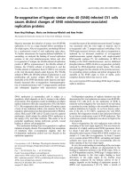
The ASV IN NLS and three well characterized NLSsFigure 1
The ASV IN NLS and three well characterized NLSs. A. Linear map of ASV IN showing the location of NLS sequence.
The 286 amino acid IN protein is composed of three domains. The N-terminal, Zn-binding (HHCC) domain (dark) and the
central catalytic core domain (red) with the locations of the active site residues (D, D, E) are indicated. The nuclear localization
signal, amino acids 206–235 (green), extends from a linker region and into the C-terminal domain (yellow). B. A 3-D structural
ribbon model of the catalytic core and C-terminal domains of ASV IN [58] with the with basic residues of the NLS shown in
space filling representation. Active site residues in the core domain are shown in ball and stick representation. C. Comparison
of the sequences of the ASV IN NLS with three well-characterized NLSs used in the studies reported herein. Residues under-
lined in the ASV IN NLS have been shown to be required for function.
A.
IN 'NLS'
206
235
N-TERMINAL
DDEHHCC
CATALYTIC
C-TERMINAL
286
1
B.
C.
Catalytic
Domain
C-terminal
Domain
Active
Site
Retrovirology 2008, 5:73 />Page 4 of 14
(page number not for citation purposes)
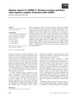
Figure 2
Nuclear import of ASV-BSA and SV40-BSA substrates; import of ASV-BSA does not require the Impα-Impβ
pathway. A. Digitonin-permeabilized HeLa cells were incubated in the presence of complete transport mixture containing the
ASV-BSA conjugate, the SV40-BSA conjugate, or Texas red-labeled BSA (TR-BSA). Top panels: Visualization of Texas red con-
jugates by fluorescence microscopy. Bottom panels: Differential interference contrast (DIC) microscopy of the same field to
show preservation of cell integrity. B. Digitonin permeabilized HeLa cells were untreated (no addition), treated with 50 μg/ml
wheat germ agglutinin (WGA), or 50 units/ml apyrase (Apyrase) prior to incubation with complete transport mixture contain-
ing either the ASV-BSA or the SV40-BSA import substrates. C. Free NLS peptides were added to the import reactions in
molar excess of the import substrates as indicated. "Self" signifies competition with the homologous peptides; "Cross" indicates
competition for ASV-BSA import by excess SV40TAg NLS peptide or competition for SV40-BSA import by excess ASV NLS
peptide. The left column panels show import in the absence of competitor peptides. D. Depletion of ASV-BSA import factor(s)
from cytosolic extracts. All assays included Texas-Red labeled ASV-BSA except that shown in the lower left hand corner (panel
4) which included Texas-Red labeled SV40-BSA. Cytosol was either not treated (1; no depletion) or pretreated with glutath-
ione-beads that bound GST alone (2) or fusion proteins of GST plus IN(1–207) which lacks the IN NLS (3), full-length IN(1–
286) (5), or a fragment of IN(201–236) that contains the IN NLS (panels 4 and 6).
Retrovirology 2008, 5:73 />Page 5 of 14
(page number not for citation purposes)
shown), confirming that the nuclear envelope was not
permeabilized by this treatment.
The lectin wheat germ agglutinin (WGA) binds specifi-
cally to O-linked N-acetylglucosamine residues, a modifi-
cation found on many nuclear pore complex proteins
[26]. Previous studies have demonstrated that import
through the nuclear pore is blocked by WGA both in vitro
and in vivo [27,28]. To determine if WGA inhibits nuclear
import of ASV-BSA, permeabilized cells were treated with
WGA for 20 min at 20°C prior to incubation in complete
transport mixture without added lectin. As shown in Fig.
2B (middle panels), nuclear import mediated by both the
ASV IN NLS and the SV40 T Ag NLS was inhibited by
WGA, providing evidence that the corresponding conju-
gates enter the nucleus through the nuclear pore com-
plexes.
To determine if import mediated by the ASV IN NLS
requires ATP, the digitonin-treated HeLa cells were pre-
treated with apyrase to deplete residual ATP. Cells were
then incubated in complete transport mixture supple-
mented with the same concentration of apyrase for 30
min at 30°C. As seen in Fig. 2B (right panels), apyrase
treatment reduced the nuclear accumulation of both the
ASV-BSA and SV40-BSA transport substrates. In addition,
no nuclear import was observed when the transport reac-
tions were performed at 4°C (data not shown). Collec-
tively, results from these experiments indicate that the
ASV IN protein contains an NLS that can mediate import
of a large cytoplasmic molecule through nuclear pore
complexes in a temperature-dependent manner, and that
this transport requires ATP or another nucleotide that is
dependent on ATP for regeneration [29,30].
Nuclear import of the ASV-BSA conjugate is saturable and
requires soluble cytosolic factor(s), but utilizes a pathway
distinct from that of SV40-T-Antigen
Protein import to the nucleus is a signal-mediated process
that exhibits saturation kinetics, which reflect the finite
amounts of transport receptors available for a given cargo
[31]. To determine if import of ASV-BSA can be saturated
in our in vitro assay, increasing amounts of free ASV IN
NLS peptide were added to the nuclear import reactions.
Results summarized in Fig. 2C (top, labeled Self) show
that addition of a 75-fold molar excess of the free peptide
was sufficient to completely inhibit nuclear accumulation
of ASV-BSA.
Although longer than the classical SV40TAg NLS, the ASV
NLS contains at least three basic amino acids that are crit-
ical for nuclear accumulation [[23], underlined in Fig.
1C]. To determine if the ASV IN NLS and the SV40 TAg
NLS interact with the same cytosolic NLS binding protein,
excess free SV40 TAg NLS peptide was added to the import
reactions. The results showed that although addition of
excess SV40 TAg NLS peptide blocked the SV40-BSA
import reaction (Fig. 2C bottom, Self), addition of an
equivalent or even higher (100-fold) molar excess of this
peptide had no effect on nuclear import of the ASV-BSA
conjugate (Fig. 2C top, labeled Cross). Furthermore,
equivalent or higher (150-fold) molar excess of the ASV
IN NLS peptide failed to block import of the SV40-BSA
conjugate (Fig. 2C bottom, Cross). These data strongly
suggest that Impα, the cytosolic adaptor known to bind
the NLS of SV40 TAg is not required for import of the ASV
IN NLS.
Importins are soluble transport receptors that bind to
NLS-containing cargo proteins in the cytoplasm [8]. How-
ever, some proteins do not require such receptors for
nuclear transport. In these cases, import many be medi-
ated through direct interactions with components of the
nuclear pore complex [32,33]. To determine if ASV IN
NLS import is dependent on a soluble factor(s) present in
the HeLa cytosolic extract, cellular proteins that bind to IN
were depleted from these extracts by treatment with
immobilized glutathione-S-transferase (GST)-fusion pro-
teins that contained all, or specific segments of IN. No
import of the ASV-BSA conjugate was detected after deple-
tion with the fusion protein that contains full length IN
(GST-IN (1–286)), or the isolated IN NLS (GST-IN(201–
236)) (Fig. 2D, panels 3 and 5). On the other hand, deple-
tion with the latter protein did not affect the ability of the
extract to support nuclear import of the SV40-BSA conju-
gate (Fig. 2D, panel 6). Depletion of the extract with GST-
beads alone or with GST-IN(1–207) that lacks the IN NLS,
had no effect on the nuclear import of ASV-BSA (Fig. 2D,
panels 2 and 4).
The results in Fig. 2 confirm that the ASV-BSA conjugate
cannot pass through the nuclear pore unassisted, but
rather that soluble cytosolic factor(s), necessary for
nuclear import, bind specifically to the ASV IN NLS to
facilitate its transport. The data also confirm that the
cytosolic component(s) that binds the ASV IN NLS to
facilitate nuclear transport is distinct from that which
binds SV40-BSA.
ASV IN does not compete for factors required for SV40 TAg
or U1A NLS-mediated import
The studies described above were designed to monitor the
activity of the isolated NLS of ASV IN in comparison to the
classical NLS of SV40 TAg. To compare the properties of
IN NLS-mediated import with those of other character-
ized but unusual classes of NLSs (Fig. 1C), we prepared
GST-fusion proteins that included the full length IN or
specific truncated versions of this protein, as well as fusion
proteins that included the following: the M9 NLS of
hnRNP-A1 protein, which binds the Impβ-related protein,
Retrovirology 2008, 5:73 />Page 6 of 14
(page number not for citation purposes)
Transportin (GST-M9) [34], the NLS of U1A protein,
which mediates import of U1 RNA (GST-U1A) [35,36]
and binds Impα, and the SV40 TAg NLS (GST-TAg) [9,10].
Use of a common fusion partner in this and subsequent
assays allowed uniform detection by immunofluores-
cence with a labeled antibody against GST. Results from
import assays with each of these purified GST-fusion pro-
teins are summarized in Fig. 3. They show that all of the
NLS-containing proteins were imported into HeLa nuclei
as expected, and that such import is dependent on the
addition of cytosolic extract. In contrast, the fusion pro-
tein GST-IN(1–207), which contains the first two
domains of IN but not the NLS, was excluded from the
nuclei.
To evaluate the significance of the findings in Fig. 2C and
2D, we next asked if import of the IN fusion proteins
shared any of the cytosolic components that are required
for import of GST-TAg or GST-U1A. For these studies, a
competitor thioredoxin fusion protein was prepared that
included the C-terminal domain of ASV IN (residues 195
to 270, which includes the NLS). As shown in Fig. 4A, the
presence of a 15-fold molar excess of this IN competitor
blocked nuclear accumulation of the full length IN pro-
tein (GST-IN(1–286)); only cytoplasmic staining was
observed. As expected, nuclear import of the fusion pro-
tein containing only the NLS peptide (GST-IN(201–236))
also was decreased upon addition of the competitor, and
there was no detectable effect of the competitor on the
nuclear accumulation of GST-TAg. Data tabulated in Fig.
4B were obtained by examining the localization of the
indicated fusion proteins in more than 100 cells in the
absence or presence of the competitor. The results of these
analyses indicate that ASV IN NLS-mediated import is dis-
tinct from that of both SV40-TAg and U1A NLSs.
As a final test of this hypothesis, a monoclonal antibody
(3E9) known to block classical import mediated by Impα/
Impβ heterodimer [37] was included in nuclear import
assays with the GST-IN proteins. As seen in Fig. 4C, addi-
tion of this reagent resulted in exclusion GST-TAg from
the nuclei. This result is expected, as import of the SV40
TAg is known to be dependent on formation of a complex
between Impα and Impβ. In contrast, the antibody had no
significant effect on nuclear accumulation of fusion pro-
teins that included full length IN, a C-terminal fragment
of IN containing the NLS or, as expected, GST-M9 (Fig.
4C; compare top and bottom rows). Quantitation of the
results of these experiments is summarized in Fig. 4D.
Nuclear import of ASV IN shares factors required for
import of linker histone H1
Impβ is known to play a role in the nuclear import of sev-
eral basic, nucleic-acid binding proteins such as histones
and ribosomal proteins, but does so using adapter
Importins other than Impα [38,39]. As ASV IN is also a
basic protein (pI of 9.8), it seemed possible that nuclear
import of ASV IN might involve other transport receptors
that mediate import of highly basic cellular proteins. To
examine this possibility, competition experiments were
performed with histone H1. The linker histone H1
appears to depend mainly on the action of an Impβ-Imp7
heterodimer, but other Impβ-like receptors can also medi-
ate its transport [38,39]. As illustrated in Fig. 5, nuclear
import of histone H1 is saturable in our assay; nuclear
accumulation of the labeled protein was competed by a
15-fold molar excess of unlabeled histone H1. Under
these same conditions, import of GST-IN(1–286) was also
inhibited by unlabeled histone H1. In contrast, import of
the GST-M9, which utilizes a distinct pathway, mediated
by Transportin, was unaffected by the competitor. This
result shows that the excess histone H1 is not simply
blocking all nuclear import, but is a specific competitor
for import of ASV IN. While the results with 3E9 antibody
in Fig. 4 rules out a role for the Impα/Impβ heterodimer
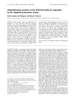
Nuclear import of GST-NLS substrates in digitonin-permea-bilized HeLa cellsFigure 3
Nuclear import of GST-NLS substrates in digitonin-
permeabilized HeLa cells. GST-NLS fusion proteins were
incubated in digitonin permeabilized HeLa cells for 30 min at
37°C prior to fixation with paraformaldehyde and staining
with fluorescent antibody against GST. Left column panels
are import without added cytosol and right column panels
with added HeLa cytosol extracts.
No Cyto + Cyto
GST-IN
(1-236)
GST-M9
GST-IN
(1-207)
GST-IN
(201-236)
GST-TAg
Retrovirology 2008, 5:73 />Page 7 of 14
(page number not for citation purposes)
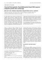
Figure 4
ASV IN NLS import does not compete for import factors required for SV40-TAg and U1A nuclear accumula-
tion. A. Digitonin permeabilized HeLa cells were either treated with buffer (PBS – top row), or with a molar excess of the
competitor protein trxIN(195–270) (bottom row). GST-IN(1–286) and GST-TAg had a 15-fold excess of competitor while
GST-IN-NLS(201–236) had a 30-fold molar excess. Import assays were performed as shown in Fig. 3 and staining was done
with fluorescent antibody against GST. B. Quantitative analysis of nuclear import of various GST fusion proteins with (+ comp)
and without (no comp) competitor. More than 100 cells were counted for each experimental condition and the percentage of
cells that had a mostly nuclear staining for the fusion protein was calculated. The percent decrease in the presence of the com-
petitor is shown in the column on the right. The lower value for import of GST-IN (201–236) compared to GST-IN (1–286)
reflects the fact that a larger percentage of cells had whole cell staining (in which nuclear import could not be assessed) or
nuclear exclusion. C. Digitonin permeabilized HeLa cells were either treated with buffer (PBS – top row), or with a 50 ug/ml
antibody 3E9 against Impβ (bottom row)during the import reaction. D. Quantitative analysis of nuclear import of various GST
fusion proteins with (+ Ab3E9) and without (no Ab) antibody 3E9. More than 100 cells were counted for each experimental
condition and the percentage of cells that had a mostly nuclear staining for the fusion protein was calculated. The percent
decrease in the presence of the antibody is shown in the column on the right.
A.
GST-IN
(1-286)
GST-IN
(201-236)
GST-TAg
B.
C.
D.
GST-M9
GST-IN
(201-236)
GST-TAg
GST-IN
(1-286)
Retrovirology 2008, 5:73 />Page 8 of 14
(page number not for citation purposes)
in ASV IN transport, it does not preclude Impβ cooperat-
ing with any of several other importins involved in his-
tone import. We conclude, therefore, that ASV IN NLS
import requires one or more of the transport receptors uti-
lized by histone H1.
Two characteristic import rates
During the course of our analyses, we observed variation
in the rates of nuclear accumulation with different GST-
fusion proteins. To examine these differences more sys-
tematically, we monitored nuclear uptake at specified
times subsequent to initiating the import reaction (Fig. 6).
We observed that these proteins fell into two categories.
Fusion proteins that contain full-length IN, C-terminally
truncated IN, or the UIA or SV40TAg NLSs, accumulated
in the nuclei slowly, and the proteins initially appeared to
be retained within the cytoplasmic compartment of the
permeabilized cells. Fusion protein containing the M9
NLS or the isolated IN NLS fragment were found only in
the nuclei even at the earliest time points, with nuclear
staining increasing over time. Control experiments veri-
fied that GST alone does not accumulate in nuclei or the
cytoplasm compartment. However, while the fusion pro-
tein containing IN that lacked the NLS (GST-IN(1–207))
was excluded from the nucleus as expected, it was retained
in the cytoplasmic compartment throughout the period
monitored in this assay. Similar phenomena are observed
in the absence of ASV IN NLS or SV40 Tag NLS-mediated
import in other data presented herein (see Figs. 2C, 3, 4A
&4C). From these results we conclude that determinants
in the N-terminal and/or catalytic core domains mediate
attachment of IN protein to cytoplasmic components of
the cell that remain after permeabilization.
Discussion
The studies reported here exploit an in vitro, permeabi-
lized cell assay to investigate the nuclear import of ASV
IN, mediated by an NLS initially identified in transient
transfection experiments [22,23]. This in vitro cell assay
makes it possible to monitor nuclear import directly, and
to delineate critical properties of the reaction. Use of a
large substrate comprising the NLS peptide crosslinked to
bovine serum albumin revealed that NLS-mediated
import can be blocked by wheat germ agglutinin and is,
therefore, dependent on transport through the nuclear
pore complex. Such transport was also shown to be satu-
rable, and to require soluble cellular factors. Sensitivity to
treatment with apyrase, which could be reversed by addi-
tion of ATP, was also observed.
The requirement for ATP could reflect a need for replen-
ishment of GTP. The GTP-bound form of the Ran GTPase
is concentrated in the nucleus, where it binds to importins
and causes release of their cargo. Depletion of ATP, with
concomitant decrease in Ran GTP, is known to decrease
the recycling of importins to the cytoplasm [40,41]. How-
ever, recycling of import receptors may not be required in
the permeabilized cell assay if an excess of the relevant
Importin is present in the cytosolic extract. Therefore, it is
also possible that the ASV IN NLS-mediated import is
Ran-GTP-independent and, as is the case for the transit of
some large proteins, ATP is required for transit through
the nuclear pore complex [42,43]. Further studies will be
required to distinguish between these two possibilities.
We have also used this permeabilized cell assay to analyze
the nuclear import of fusion proteins containing full
length ASV IN or specific segments of this protein. Our
results show that the ASV IN NLS is also active within the
context of the full protein or segments of the protein that
include the NLS. Constructs containing IN segments that
lacked the NLS were not imported to the nucleus, indicat-
ing determinants essential for nuclear import of IN are
contained within the identified NLS. These results are con-
sistent with our previous transfection studies, in which
nuclear accumulation of various Lac-IN fusion proteins
was monitored [23].
Although the ASV IN NLS comprises an apparently unique
sequence, it does bear some similarity to classical bipartite
NLSs such as nucleoplasmin, comprising clusters of basic
residues separated by a spacer. We therefore considered
the possibility that import of ASV IN might depend on the
same cellular factors that mediate import of the classical
NLSs, the adapter Impα and Impβ. This hypothesis was
tested in a variety of ways. Competition experiments with
ASV IN mediated import is inhibited by excess histone H1Figure 5
ASV IN mediated import is inhibited by excess his-
tone H1. The import of labeled histone H1, (GST-IN(1–
286), and the Impβ binding domain fused to GFP (IBB-GFP)
was examined in the absence (top) and presence (bottom) of
excess unlabeled histone H1. Incubations were for 30 min
and all exposure times were equivalent.
Histone H1 GST-IN(1-286) GST-M9
Retrovirology 2008, 5:73 />Page 9 of 14
(page number not for citation purposes)
Kinetics of ASV-NLS mediated importFigure 6
Kinetics of ASV-NLS mediated import. GST-NLS fusion proteins were incubated in digitonin permeabilized HeLa cells
for various times (labeled above each column) at 37°C prior to fixation with paraformaldehyde and staining with fluorescent
antibody against GST. The fusion protein used in each row is labeled at the right and the properties described in the text.
Fusion proteins that are imported with slower kinetics are grouped at the top (rows 1–4), and those with faster kinetics in the
middle (rows 5 and 6). Control fusion proteins that are not imported into the nucleus are in rows 7 and 8.
Minutes after initiation of import assay
GST-IN
(1-286)
GST-IN
(1-236)
GST-U1A
GST-TAg
GST-IN
(201-286)
GST-M9
GST
GST-IN
(1-207)
2102030
Retrovirology 2008, 5:73 />Page 10 of 14
(page number not for citation purposes)
the BSA conjugates showed that addition of excess
amounts of peptides corresponding to the classical SV40
TAg NLS or the IN NLS could block nuclear import medi-
ated by the corresponding NLS, but had no effect on the
activity of the other. We also found that excess IN NLS did
not compete for nuclear import mediated by the U1A
NLS, even though IN- or IN NLS-mediated import was
abolished. Lastly an antibody that blocks Impα/Impβ
mediated SV40 T-antigen import was not observed to
inhibit ASV IN import. All these experiments failed to sup-
port the hypothesis that transport of ASV IN requires this
classical pathway. We concluded from these results the
ASV IN NLS does not bind Impα nor utilize the Impα/
Impβ heterodimer.
Basic residues are also known to be critical for binding to
Impβ by various nonclassical NLS sequences that, like the
ASV IN NLS, are Impα-independent. For example, struc-
tural analyses of the parathyroid hormone-related protein
(PTHrP) NLS bound to Impβ reveal a requirement for a
cluster of basic amino acids followed by a twist in the pep-
tide and then an extended segment. This NLS binding is
stabilized by a combination of charge interactions with
the basic residues and hydrophobic interactions with the
extended peptide [44]. As several basic residues as well as
one proline are required for IN NLS function [23], both its
conformation and accessibility (see Fig. 1) are consistent
with this type of interaction, and it remains conceivable
that the soluble cellular factor(s) required for ASV IN
import is a β-like Importin [21] acting alone or in con-
junction with Impβ.
ASV IN is a highly basic protein (pI of 9.8), and excess his-
tone H1 competes for ASV IN import in our assay. While
H1 is best transported by the Impβ/Imp7 heterodimer it
has been shown to bind to Imp5, as well as Impβ or Imp7
alone. The core histones are even more promiscuous in
their usage of various importins [45,46], as are several
other proteins such as c-Jun [47], and other viral proteins
(Rev) [48]. As noted below, this also seems to be the case
for HIV IN, for which several import pathways have been
identified. An excess of histone H1 might then be
expected to sequester several other importins in addition
to the Impβ/Imp7 heterodimer. We speculate that ASV IN
may also have the capacity to utilize more than one
import receptor, for example, those that mediate the
nuclear import of other basic cellular proteins, such as
ribosomal proteins and core histones. Several of these are
reported to function as cytoplasmic chaperones that pre-
vent polyanion-mediated aggregation of these basic pro-
teins as well as mediators of nuclear import [39]. Our data
suggest that ASV IN takes advantage of one or more of the
transport pathways for such basic cellular proteins, which
are distinct from the classical NLS pathways, but essential
for cell metabolism.
In measuring the kinetics of nuclear import in the perme-
abilized cells, we observed very rapid accumulation
(within 2–10 min) with GST-fusion proteins that
included the isolated M9 or IN NLS sequences. A different
pattern was observed with fusions that included full
length IN or IN(1–236), which also contains the NLS. In
these cases we observed staining only in the cytoplasmic
compartment in the 2–10 min time period, and the fusion
proteins were largely excluded from the nuclei. Upon fur-
ther incubation, for 20–30 min, staining was no longer
seen in the cytoplasmic compartment, but the fusion pro-
teins with the IN NLS now localized to the nuclei. This dif-
ference could not be attributed to size of the cargo, as the
smaller fusion proteins containing only the SV40 TAg NLS
or the U1A NLS exhibited the same slow patterns
observed with the full length IN protein. Nor is this bind-
ing to cytosolic components likely to be due to aggrega-
tion; the IN fragment 1–207 is monomeric in solution at
high concentrations, and yet this protein exhibits promi-
nent cytoplasmic binding. The simplest explanation of
these results is that ASV IN protein and some of the iso-
lated NLSs can bind to cytoplasmic components. The bio-
logical significance of this observation is unclear, as
soluble components are lost from the permeabilized cells,
and cytoskeletal or other remaining components may be
exposed in some aberrant fashion. Comparison of the pat-
terns obtained with proteins containing the full length IN
or IN(1–236) with IN(201–286) suggest that interaction
with these cellular components may retard nuclear
uptake. When nuclear import cannot occur due to lack of
an NLS, as with GST-IN(1–207), cytoplasmic staining was
maintained throughout the course of the experiment. This
indicates that determinants responsible for interactions
with the cytoplasmic components are contained within
the N-terminal and catalytic core domains of the IN.
Investigations of the nuclear import of HIV-1 IN have
implicated the classical Impα-Impβ [49] and also Imp7 in
this process [50]. Using digitonin-permeabilized cells,
Fassati and coworkers [51] (supplementary data) reported
that Imp7 promotes nuclear transport of purified HIV-1
reverse transcription complexes (RTCs), and that siRNA-
knockdown of Imp7 inhibits HIV-1 infection. These find-
ings are consistent with a model in which the interaction
between Imp7 and HIV-1 IN facilitates Impβ nuclear
import of the preintegration complex. More recent exper-
iments with this same in vitro assay have provided evi-
dence that certain tRNAs may also promote RTC import
[52], and the role of another importin in HIV-1 infection,
Transportin 3, has been reported [53], further implicating
multiple pathways in this process.
As noted above, our results fail to support a role for Impα-
Impβ in nuclear transport of ASV IN. In preliminary exper-
iments, using transduction of a reporter gene as a readout
Retrovirology 2008, 5:73 />Page 11 of 14
(page number not for citation purposes)
for successful nuclear import, we observed that while
siRNA knockdown of Imp7 reduced transduction by an
HIV-1 vector, it had little effect on transduction by ASV.
Differences in import pathways for these two retroviruses
are not unexpected. The NLS of ASV IN is not conserved
among the retroviral genera, and although reports of the
location of NLS sequences in HIV-1 IN vary, residues that
bind Imp7 have been identified in the C-terminal, SH3-
like domain, distal to the location of the NLS in ASV IN
[50]. This suggests that some other member(s) of the
Importin superfamily or other karyophilic macromole-
cules promote import of ASV IN.
As with HIV [1,2], NLSs have been found in ASV Gag pro-
teins. Analysis of the function of these sequences suggest
that nuclear entry mediated by the basic NLS in the ASV
nucleocapsid (NC) protein requires the classical Impα-
Impβ, while import mediated by the more unusual NLS in
the matrix protein (MA) is facilitated by other members of
the Importin superfamily [54]. It has been proposed that
these signals may allow the ASV Gag polyprotein precur-
sor to enter the nucleus and capture viral RNA genomes
for virion assembly [55]. The possibility that the mature
Gag proteins could also contribute to nuclear import of
the preintegration complex has been noted, but the bio-
logical role for these Gag NLS sequences remain uncer-
tain. Further study, using the system described here and
purified transport receptors should make it possible to
identify the specific factors required for nuclear import of
ASV IN and to evaluate the role of this viral protein in
shepherding viral DNA through the nuclear pore.
Methods
Cell culture and antibodies and photomicroscopy
HeLa cells were obtained from the Fox Chase Cell Culture
Facility and passaged in DMEM with 10% FCS, 1 unit/ml
penicillin and 1 ug/ml streptomycin. Antibody against
hnRNP-A1 was a gift from Dr. Gideon Dreyfuss (Univer-
sity of PA). Monoclonal antibody 3E9 was provided by
Stephen Adam (Northwestern University). Immunofluo-
rescence microscopy was performed on an Olympus BK2
microscope. Color images were taken with Kodak Ekta-
chrome 400 film or Olympus MagnaFire digital camera,
maintaining equal exposure times within each experi-
ment. Glutathione-S-transferase (GST)-fusion proteins
used as import substrates were detected by direct immun-
ofluorescence with labeled antibody against GST (rabbit
IgG fraction, Alexa Fluor
®
488 conjugate – Molecular
Probes).
Preparation of labeled BSA import substrates and cytosolic
extract
The ASV IN NLS (NH
2
-cgggtKTPIQKHWRPTVLTEGP-
PVKIRIETGEWEK-COOH) and the SV40 TAg NLS (NH
2
-
cgggGPKKKRKVED-COOH) [10] peptides were synthe-
sized by Research Genetics (Huntsville, AL). The lower
case letters represent a linker containing three glycine res-
idues and an N-terminal cysteine for coupling to the BSA.
High purity bovine serum albumin (BSA) (Sigma) was
labeled with Texas red sulfonyl chloride (Pierce) follow-
ing published procedures [56]. Labeled BSA was activated
with the heterobifunctional cross-linker sulfosuccinimi-
dyl-4(maleimidomethyl) cyclohexane-1-carboxylate
(Sulfo-SMCC, Pierce) following the manufacturer's rec-
ommendation. A 50-fold molar excess of the ASV IN NLS
or the SV40 TAg NLS peptide was bound to the activated-
labeled BSA. The average number of peptides cross-linked
to the labeled BSA was determined by SDS-PAGE for each
import substrate.
HeLa cytosolic extract was prepared as described [12]
from pellets of exponentially growing HeLa S3 cells
obtained from the Cell Culture Center (Minneapolis,
MN) of the National Center for Research Resources. The
extract was concentrated to yield a final protein concentra-
tion of approximately 40 mg/ml, as determined by Bio-
Rad protein assay. Extracts were stored in aliquots at -
80°C, and diluted 1:1 for import assays.
Nuclear import assays
HeLa cells were grown on 8-chamber poly-lysine coated
culture slides (BD Biocoat) or coverslips coated with 0.2
mg/ml poly-D-lysine (Sigma). Cells were rinsed in cold
transport buffer (20 mM Hepes, pH 7.3, 110 mM potas-
sium acetate, 5 mM sodium acetate, 2 mM magnesium
acetate, 1 mM EGTA, 2 mM DTT, and 1 μg/ml each apro-
tinin, leupeptin, and pepstatin) and immersed in the
same buffer containing 30 μg/ml digitonin (Calbiochem)
for 5 min to permeabilize the plasma membrane. Cover-
slips were washed twice with cold transport buffer and
inverted over a drop of complete transport mixture for 30
min at 30°C. Cells were then washed in cold transport
buffer, fixed 10 min at room temperature with 2% para-
formaldehyde in PBS, and washed in PBS prior to mount-
ing with Citifluor (UKC Chem. Lab, Canterbury, UK). The
complete transport mixture contained 50% cytosolic
extract, approximately 10 μg of import substrate, 20 mM
Hepes, pH 7.3, 110 mM potassium acetate, 5 mM sodium
acetate, 2 mM magnesium acetate, 2 mM DTT, 1 mM
EGTA, 2 mM ATP, 2 mm GTP, 5 mM creatine phosphate
(Calbiochem), 20 U/ml creatine phosphokinase (Calbio-
chem), 10 μg/μl unlabeled BSA, and 1 μg/ml each apro-
tinin, leupeptin, and pepstatin. Using various
concentrations of digitonin and an anti-A1 antibody to
monitor nuclear breakdown, it was determined that 30
μg/ml digitonin gave optimal (30–50%) nuclear import
without significant nuclear destruction. Higher amounts
of digitonin produced detectable nuclear breakdown.
Retrovirology 2008, 5:73 />Page 12 of 14
(page number not for citation purposes)
For experiments with wheat germ agglutinin (WGA) inhi-
bition, cells were incubated with transport buffer contain-
ing 50 μg/ml WGA (Sigma) for 15 min at 20°C, prior to
incubation with complete transport mixture without
added lectin. For the apyrase experiments, coverslips were
pre-treated (10 min at 30°C) in transport buffer supple-
mented with 50 U/ml apyrase (Sigma), 1 mM CaCl
2
, and
10 μg/μl unlabeled BSA. Coverslips were then incubated
in the presence of complete transport mixture lacking
added ATP, creatine phosphatase or creatine phosphoki-
nase, but supplemented with 50 U/ml apyrase and 1 mM
CaCl
2
.
For the peptide competition experiments, SV40 TAg NLS
peptide or ASV IN NLS peptide preparations were solubi-
lized in a small amount of transport buffer and added
directly to the complete transport mixture at the stated
molar excess prior to the addition of the appropriate
import substrate. For experiments with thioredoxin-inte-
grase fusion protein (trx-IN(195–270)) as a competitor,
the fusion protein was added in either 15 or 30-fold molar
excess as indicated in the complete transport mixture,
prior to the addition of import substrate to be assayed. For
antibody inhibition experiments, digitonin permeabi-
lized HeLa cells were either treated with 50 ug/ml anti-
body 3E9 against Impβ during the import reaction. More
than 100 cells were counted for each experimental condi-
tion and the percentage of cells that had a mostly nuclear
staining for the fusion protein was calculated. These com-
petition and antibody experiments were repeated 3 times
and representative data shown in Fig. 4.
For the Histone H1 competition experiments, permeabi-
lized cell assays were done as above with 30 min incuba-
tion, but with the addition of a 15-fold molar excess of
unlabeled Histone H1 in the complete transport mixture
where indicated in Fig. 5. Histone H1 was labeled with
Alexa Fluor 488 according to manufacturer's instructions
(Molecular Probes) and its import assayed as with all
other substrates. This experiment was repeated 3 times
and representative data is shown.
Construction of GST-fusion expression plasmids,
preparation of GST-fusion proteins and cytosolic extract
depletion experiments
Construction of the GST-Integrase fusion proteins was
described previously [57]. A plasmid able to express the
NLS of the U1A protein fused to GST was constructed by
PCR of human cDNA with primers that amplified DNA
encoding amino acids 94 to 204 of the U1A protein [35].
This DNA fragment was digested with BamH1 and EcoR1
and ligated into the GST expression plasmid pGEX-2TK
(Amersham-Pharmacia). GST-M9 and GST-TAg were
kindly provided by Gideon Dreyfuss and Michael Malim,
respectively. All GST-fusion proteins were expressed and
purified by the same methodology as previously described
[57].
Cytosolic extract was depleted of factors which interact
with specific NLS sequences as follows. Purified fusion
proteins were mixed with glutathione-agarose beads
(Sigma) and the amount of bound fusion protein was
determined by SDS-PAGE. A standard volume (75 to 100
ul) of HeLa cytosolic extract was incubated with glutath-
ione agarose beads normalized for the amount of fusion
protein per bead volume (approximately 50–100 ug of
fusion protein was used in a typical binding reaction).
Extract and beads were incubated at 4°C for 1 hr with
rocking. Beads were then pelleted at low speeds in a
microfuge (4°C) and 25 ul of the resulting supernatant
was utilized in the in vitro nuclear import assays.
Conclusion
By use of an in vitro assay with digitonin-permeabilized
cells, we confirmed that nuclear import of ASV IN is medi-
ated by a previously identified NLS sequence. This import
is active, saturable, ATP-dependent, and relies on cytosolic
factors to transit through the nuclear pore complex. These
results are consistent with a role for ASV IN in the nuclear
import of the preintegration complex of this retrovirus.
Although the ASV NLS exhibits similarity to some classical
NLSs, we present a variety of evidence that make it
unlikely that the classical Impα/Impβ heterodimer is
required for its import. The results indicate that the ASV
IN NLS is recognized by other, perhaps Impβ-like soluble
karyophilic protein(s), which is also able to mediate
nuclear accumulation of the cellular linker histone H1.
The system we describe may be useful to identify the fac-
tor(s) and evaluate its role in ASV replication.
Abbreviations
IN: integrase; NLS: nuclear localization sequence; ASV:
avian sarcoma virus; HIV: human immunodeficiency
virus; BSA: bovine serum albumin; WGA: wheat germ
agglutinin; GST: glutathione S-transferase.
Competing interests
The authors declare that they have no competing interests.
Authors' contributions
MDA: participated in the design and coordination of the
study, performed or supervised all experiments with
fusion substrates, and helped to write the final manu-
script. MMS: participated in the design and execution of
assays with the BSA substrates, and the writing of an orig-
inal draft manuscript. AG: cloned and purified several
NLS fusion proteins, and participated in the ASV NLS
competition and mAb 3E9 inhibition experiments. KB:
efforts were essential for refinement of the permeabilized
Retrovirology 2008, 5:73 />Page 13 of 14
(page number not for citation purposes)
cell assay, and preparation of new substrates and reagents
tested. MH: purified NLS substrates and performed pre-
liminary experiments on siRNA-mediated knockdown of
Importins prior to infection experiments. AMS: partici-
pated in the design and coordination of this study, super-
vised its progress, and helped to write the final
manuscript. All all authors read and approved the final
manuscript.
Acknowledgements
This work was supported by National Institutes of Health grants
CA071515, AI040385, F32AI09924, Institutional grant CA006927 from the
National Institutes of Health, and also by an appropriation from the Com-
monwealth of Pennsylvania. We acknowledge use of the Fox Chase Cancer
Center DNA Sequencing Facility and the Cell Culture Facility. We thank
Marie Estes for help in preparing the manuscript. Dr. Michael Malim (Uni-
versity of Pennsylvania) was most generous with advice and provision of
important reagents. We thank Dr. D. Görlich (University of Heidelberg,
Germany) for his gracious hospitality to one of us (MDA) and providing
help in development expertise with the assay, as well as valuable reagents.
We are grateful to our colleague Dr. Richard Katz for advice and sugges-
tions during the course of this work and to Drs. Katz, Jonathan Chernoff,
and Glenn Rall for critical review of the manuscript. The work of MMS is
dedicated to the memory of Martin Z. Sauter. The contents of this manu-
script are solely the responsibility of the authors and do not necessarily
represent the official views of the National Cancer Institute, or any other
sponsoring organization.
References
1. Fassati A: HIV infection of non-dividing cells: a divisive prob-
lem. Retrovirology 2006, 3:74.
2. Suzuki Y, Craigie R: The road to chromatin - nuclear entry of
retroviruses. Nat Rev Microbiol 2007, 5(3):187-196.
3. Hatziioannou T, Goff SP: Infection of nondividing cells by Rous
sarcoma virus. J Virol 2001, 75(19):9526-9531.
4. Katz RA, Greger JG, Darby K, Boimel P, Rall GF, Skalka AM: Trans-
duction of interphase cells by avian sarcoma virus. J Virol 2002,
76(11):5422-5434.
5. Greger JG, Katz RA, Taganov K, Rall GF, Skalka AM: Transduction
of terminally differentiated neurons by avian sarcoma virus.
J Virol 2004, 78(9):4902-4906.
6. Katz RA, Greger JG, Boimel P, Skalka AM: Human immunodefi-
ciency virus type 1 DNA nuclear import and integration are
mitosis independent in cycling cells. J Virol 2003,
77(24):13412-13417.
7. Katz RA, Greger JG, Skalka AM: Effects of cell cycle status on
early events in retroviral replication. J Cell Biochem 2005,
94(5):880-889.
8. Dingwall C, Laskey R: The nuclear membrane. Science 1992,
258(5084):942-947.
9. Kalderon D, Roberts BL, Richardson WD, Smith AE: A short amino
acid sequence able to specify nuclear location. Cell 1984, 39(3
Pt 2):499-509.
10. Lanford RE, Butel JS: Construction and characterization of an
SV40 mutant defective in nuclear transport of T antigen. Cell
1984, 37(3):801-813.
11. Robbins J, Dilworth SM, Laskey RA, Dingwall C: Two interdepend-
ent basic domains in nucleoplasmin nuclear targeting
sequence: identification of a class of bipartite nuclear target-
ing sequence. Cell 1991, 64(3):615-623.
12. Adam SA, Marr RS, Gerace L: Nuclear protein import in perme-
abilized mammalian cells requires soluble cytoplasmic fac-
tors.
J Cell Biol 1990, 111(3):807-816.
13. Adam SA, Sterne-Marr R, Gerace L: In vitro nuclear protein
import using permeabilized mammalian cells. Methods Cell Biol
1991, 35:469-482.
14. Görlich D, Prehn S, Laskey RA, Hartmann E: Isolation of a protein
that is essential for the first step of nuclear protein import.
Cell 1994, 79(5):767-778.
15. Imamoto N, Shimamoto T, Takao T, Tachibana T, Kose S, Matsubae
M, Sekimoto T, Shimonishi Y, Yoneda Y: In vivo evidence for
involvement of a 58 kDa component of nuclear pore-target-
ing complex in nuclear protein import. EMBO J 1995,
14(15):3617-3626.
16. Radu A, Blobel G, Moore MS: Identification of a protein complex
that is required for nuclear protein import and mediates
docking of import substrate to distinct nucleoporins. Proc
Natl Acad Sci U S A 1995, 92(5):1769-1773.
17. Adam EJ, Adam SA: Identification of cytosolic factors required
for nuclear location sequence-mediated binding to the
nuclear envelope. J Cell Biol 1994, 125(3):547-555.
18. Moore MS, Blobel G: A G protein involved in nucleocytoplas-
mic transport: the role of Ran. Trends Biochem Sci 1994,
19(5):211-216.
19. Paschal BM, Gerace L: Identification of NTF2, a cytosolic factor
for nuclear import that interacts with nuclear pore complex
protein p62. J Cell Biol 1995, 129(4):925-937.
20. Görlich D, Mattaj IW: Nucleocytoplasmic transport. Science
1996, 271(5255):1513-1518.
21. Strom AC, Weis K: Importin-beta-like nuclear transport
receptors. Genome Biol 2001, 2(6):3008.
22. Kukolj G, Jones KS, Skalka AM: Subcellular localization of avian
sarcoma virus and human immunodeficiency virus type 1
integrases. J Virol 1997,
71(1):843-847.
23. Kukolj G, Katz RA, Skalka AM: Characterization of the nuclear
localization signal in the avian sarcoma virus integrase. Gene
1998, 223(1-2):157-163.
24. Werner S, Hardmarsh P, Napirei M, Vogel-Bachmayr K, Wöhrl BM:
Subcellular localization and integration activities of Rous
Sarcoma virus reverse transcriptase. J Virol 2002,
76:6205-6212.
25. Feldherr CM, Cohen RJ, Ogburn JA: Evidence for mediated pro-
tein uptake by amphibian oocyte nuclei. J Cell Biol 1983,
96(5):1486-1490.
26. Hanover JA, Cohen CK, Willingham MC, Park MK: O-linked N-
acetylglucosamine is attached to proteins of the nuclear
pore. Evidence for cytoplasmic and nucleoplasmic glycopro-
teins. J Biol Chem 1987, 262(20):9887-9894.
27. Dabauvalle MC, Schulz B, Scheer U, Peters R: Inhibition of nuclear
accumulation of karyophilic proteins in living cells by micro-
injection of the lectin wheat germ agglutinin. Exp Cell Res
1988, 174(1):291-296.
28. Finlay DR, Newmeyer DD, Price TM, Forbes DJ: Inhibition of in
vitro nuclear transport by a lectin that binds to nuclear
pores. J Cell Biol 1987, 104(2):189-200.
29. Newmeyer DD, Forbes DJ: Nuclear import can be separated
into distinct steps in vitro: nuclear pore binding and translo-
cation. Cell 1988, 52(5):641-653.
30. Richardson WD, Mills AD, Dilworth SM, Laskey RA, Dingwall C:
Nuclear protein migration involves two steps: rapid binding
at the nuclear envelope followed by slower translocation
through nuclear pores. Cell 1988, 52(5):655-664.
31. Goldfarb DS, Gariepy J, Schoolnik G, Kornberg RD: Synthetic pep-
tides as nuclear localization signals. Nature 1986,
322(6080):641-644.
32. Fouchier RA, Malim MH: Nuclear import of human immunode-
ficiency virus type-1 preintegration complexes. Adv Virus Res
1999, 52:275-299.
33. Jenkins Y, McEntee M, Weis K, Greene WC: Characterization of
HIV-1 vpr nuclear import: analysis of signals and pathways. J
Cell Biol 1998, 143(4):875-885.
34. Nakielny S, Siomi MC, Siomi H, Michael WM, Pollard V, Dreyfuss G:
Transportin: nuclear transport receptor of a novel nuclear
protein import pathway. Exp Cell Res 1996, 229(2):261-266.
35. Hetzer M, Mattaj IW: An ATP-dependent, Ran-independent
mechanism for nuclear import of the U1A and U2B" spliceo-
some proteins. J Cell Biol 2000, 148(2):293-303.
36. Hieda M, Tachibana T, Fukumoto M, Yoneda Y: Nuclear import of
the U1A splicesome protein is mediated by importin alpha /
beta and Ran in living mammalian cells. J Biol Chem 2001,
276(20):16824-16832.
Publish with BioMed Central and every
scientist can read your work free of charge
"BioMed Central will be the most significant development for
disseminating the results of biomedical research in our lifetime."
Sir Paul Nurse, Cancer Research UK
Your research papers will be:
available free of charge to the entire biomedical community
peer reviewed and published immediately upon acceptance
cited in PubMed and archived on PubMed Central
yours — you keep the copyright
Submit your manuscript here:
/>BioMedcentral
Retrovirology 2008, 5:73 />Page 14 of 14
(page number not for citation purposes)
37. Chi NC, Adam EJ, Adam SA: Sequence and characterization of
cytoplasmic nuclear protein import factor p97. J Cell Biol 1995,
130(2):265-274.
38. Bauerle M, Doenecke D, Albig W: The requirement of H1 his-
tones for a heterodimeric nuclear import receptor. J Biol
Chem 2002, 277(36):32480-32489.
39. Jakel S, Mingot JM, Schwarzmaier P, Hartmann E, Gorlich D: Import-
ins fulfil a dual function as nuclear import receptors and
cytoplasmic chaperones for exposed basic domains. The
EMBO journal 2002, 21(3):377-386.
40. Schwoebel ED, Talcott B, Cushman I, Moore MS: Ran-dependent
signal-mediated nuclear import does not require GTP
hydrolysis by Ran. J Biol Chem 1998, 273(52):35170-35175.
41. Kutay U, Izaurralde E, Bischoff FR, Mattaj IW, Gorlich D: Dominant-
negative mutants of importin-beta block multiple pathways
of import and export through the nuclear pore complex.
EMBO J 1997, 16(6):1153-1163.
42. Lyman SK, Guan T, Bednenko J, Wodrich H, Gerace L: Influence of
cargo size on Ran and energy requirements for nuclear pro-
tein import. J Cell Biol 2002, 159(1):55-67.
43. Schwoebel ED, Ho TH, Moore MS: The mechanism of inhibition
of Ran-dependent nuclear transport by cellular ATP deple-
tion. J Cell Biol 2002, 157(6):963-974.
44. Cingolani G, Bednenko J, Gillespie MT, Gerace L: Molecular basis
for the recognition of a nonclassical nuclear localization sig-
nal by importin beta. Mol Cell 2002, 10(6):1345-1353.
45. Muhlhausser P, Muller EC, Otto A, Kutay U: Multiple pathways
contribute to nuclear import of core histones. EMBO Rep
2001, 2(8):690-696.
46. Greiner M, Caesar S, Schlenstedt G: The histones H2A/H2B and
H3/H4 are imported into the yeast nucleus by different
mechanisms. European journal of cell biology 2004, 83(10):511-520.
47. Waldmann I, Walde S, Kehlenbach RH: Nuclear import of c-Jun is
mediated by multiple transport receptors. J Biol Chem 2007,
282(38):27685-27692.
48. Arnold M, Nath A, Hauber J, Kehlenbach RH: Multiple importins
function as nuclear transport receptors for the Rev protein
of human immunodeficiency virus type 1. J Biol Chem 2006,
281(30):20883-20890.
49. Hearps AC, Jans DA: HIV-1 integrase is capable of targeting
DNA to the nucleus via an importin alpha/beta-dependent
mechanism. Biochem J 2006, 398(3):475-484.
50. Ao Z, Huang G, Yao H, Xu Z, Labine M, Cochrane AW, Yao X: Inter-
action of human immunodeficiency virus type 1 integrase
with cellular nuclear import receptor importin 7 and its
impact on viral replication. J Biol Chem 2007,
282(18):13456-13467.
51. Fassati A, Gorlich D, Harrison I, Zaytseva L, Mingot JM: Nuclear
import of HIV-1 intracellular reverse transcription com-
plexes is mediated by importin 7. EMBO J 2003,
22(14):3675-3685.
52. Zaitseva L, Myers R, Fassati A: tRNAs Promote Nuclear Import
of HIV-1 Intracellular Reverse Transcription Complexes.
PLoS Biol 2006, 4(10):e332.
53. Brass AL, Dykxhoorn DM, Benita Y, Yan N, Engelman A, Xavier RJ,
Lieberman J, Elledge SJ: Identification of host proteins required
for HIV infection through a functional genomic screen. Sci-
ence 2008, 319(5865):921-926.
54. Butterfield-Gerson KL, Scheifele LZ, Ryan EP, Hopper AK, Parent LJ:
Importin-beta family members mediate alpharetrovirus gag
nuclear entry via interactions with matrix and nucleocapsid.
J Virol 2006, 80(4):1798-1806.
55. Scheifele LZ, Garbitt RA, Rhoads JD, Parent LJ: Nuclear entry and
CRM1-dependent nuclear export of the Rous sarcoma virus
Gag polyprotein. Proc Natl Acad Sci U S A 2002, 99(6):3944-3949.
56. Titus JA, Haugland R, Sharrow SO, Segal DM: Texas Red, a
hydrophilic, red-emitting fluorophore for use with fluores-
cein in dual parameter flow microfluorometric and fluores-
cence microscopic studies. J Immunol Methods 1982,
50(2):193-204.
57. Andrake MD, Skalka AM: Multimerization determinants reside
in both the catalytic core and C terminus of avian sarcoma
virus integrase. J Biol Chem 1995, 270(49):29299-29306.
58. Yang ZN, Mueser TC, Bushman FD, Hyde CC: Crystal structure of
an active two-domain derivative of Rous sarcoma virus inte-
grase. J Mol Biol 2000, 296(2):535-548.