Báo cáo y học: "Recovery of fitness of a live attenuated simian immunodeficiency virus through compensation in both the coding and non-coding regions of the viral genome" pot
Bạn đang xem bản rút gọn của tài liệu. Xem và tải ngay bản đầy đủ của tài liệu tại đây (906.85 KB, 10 trang )
BioMed Central
Page 1 of 10
(page number not for citation purposes)
Retrovirology
Open Access
Research
Recovery of fitness of a live attenuated simian immunodeficiency
virus through compensation in both the coding and non-coding
regions of the viral genome
James B Whitney
1,2,3
and Mark A Wainberg*
1,2
Address:
1
McGill University AIDS Centre, Lady Davis Institute-Jewish General Hospital, Montreal, Quebec, H3T 1E2, Canada,
2
Department of
Microbiology and Immunology, McGill University, Montreal, Quebec, H3A 2B4, Canada and
3
Division of Viral Pathogenesis, Beth Israel
Deaconess Medical Center, Harvard Medical School, Boston, MA 02115, USA
Email: James B Whitney - ; Mark A Wainberg* -
* Corresponding author
Abstract
We have analyzed a SIV deletion mutant that was compromised both in viral replication and RNA
packaging. Serial passage of this variant in two different T-cell lines resulted in compensatory
reversion and the generation of independent groups of point mutations within each cell line. Within
each group, single point mutations were shown to contribute to increased viral infectivity and the
rescue of wild-type replication kinetics. The complete recovery of viral fitness ultimately correlated
with the restoration of viral RNA packaging. Consistent with the latter finding was the rescue of
Pr
55
Gag processing, also restoring proper virus core morphology in mature virions. These
seemingly independently arising groups of compensatory mutations were functionally
interchangeable in regard to the recovery of wild type replication in rhesus PBMCs. These findings
indicate that viral reversion that overcomes a genetic bottleneck is not limited to a single pathway,
and illustrates the remarkable adaptability of lentiviruses.
Background
The packaging of full-length viral genomic RNA (vRNA)
into primate lentiviruses is regulated by a multipartite cis-
acting signal located within the 5' untranslated region
(UTR) or RNA-leader. In the leader of human immunode-
ficiency virus type-1 (HIV-1), the packaging signal or Psi
(Ψ) is distributed across multiple RNA domains that
include stem loop-1 (SL1), SL3 and SL4 [1-3]. There is
also evidence of vRNA packaging elements in other
regions, including those upstream of the primer-binding
site (PBS), as well as within downstream gag-coding
regions [4,5].
Comparative packaging studies of simian immunodefi-
ciency virus (SIV) by our group and of human immuno-
deficiency virus type-2 (HIV-2) by others, have assigned a
primary role in packaging to SL1, as compared to all other
regions within the SIV and HIV-2 genomes [6-10]. More-
over, SL1 sequences are also important in the formation of
5' linked vRNA duplexes or vRNA dimers [8,11-13]. RNA-
RNA interactions ultimately determine RNA tertiary con-
formation and have been shown to impact on both the
regulation and efficiency of vRNA packaging [14,15]. The
relationships among the packaging events of different len-
tiviruses have been extensively studied [16].
Published: 3 July 2007
Retrovirology 2007, 4:44 doi:10.1186/1742-4690-4-44
Received: 7 February 2007
Accepted: 3 July 2007
This article is available from: />© 2007 Whitney and Wainberg; licensee BioMed Central Ltd.
This is an Open Access article distributed under the terms of the Creative Commons Attribution License ( />),
which permits unrestricted use, distribution, and reproduction in any medium, provided the original work is properly cited.
Retrovirology 2007, 4:44 />Page 2 of 10
(page number not for citation purposes)
The foregoing implies the presence of multiple RNA-bind-
ing domains within Pr
55
Gag. In the context of Pr
55
Gag an
important trans-role has been ascribed to the viral nucle-
ocapsid (NC) protein [17,18], although several studies
have indicated that a functional separation of domains
within NC is present [17,19]. Other protein domains
within Gag have also been shown to be necessary for
vRNA packaging and dimerization, whereas the p2 region
has been shown to contribute to vRNA packaging specifi-
city [20,21].
Studies on the reversion of SL1 deleted virus in HIV-1
showed that compensatory point mutations in four dis-
tant Gag proteins, i.e. nucleocapsid (NC-T24I), matrix
(MA-V35I), capsid (CA-T24I) and the p2-spacer (p2-T21I)
were all involved in restoration of viral growth [22]. Grif-
fin et al have shown that there is a preferential use of co-
translation to impart packaging specificity for vRNA in
HIV-2 [23]. A similar process is thought to occur in SIV,
particularly in light of evidence that the 3' regions of the
leader possess an internal ribosome entry site (IRES) func-
tion [24].
Although Pr
55
Gag alone has been shown to be sufficient
for particle production, numerous host and viral proteins
are required for optimal viral assembly and budding [25].
Indeed, an appropriate conformation of packaged RNA is
critical, since mutations in viral RNA can severely impact
virus production and viability [26]. The late phase of len-
tiviral replication requires the assembly of virion compo-
nents at the cellular periphery, at which a series of
interrelated vRNA-protein interactions are required to
occur in a coordinated fashion; this positions vRNA in
precise relation to Pr
55
Gag during protease-mediated
cleavages that take place during assembly and at post-bud-
ding stages [27].
Previous work from our group described a mutant deleted
of 21 nucleotides within the 5' proximal stem of SL1 of
the infectious molecular clone of SIV
mac239
(Δnt +398 to
+418, termed-SD2) that resulted in a significant delay in
viral replication and reduced vRNA packaging. The serial
passage of this mutant virus in the CEMx174-T/B-hybrid
cell line or in C8166-T cells over protracted periods
resulted in the recovery of virus replication [7]. Our previ-
ous report showed that the original SD2 deletion had
been retained, but that each cell line specific isolate har-
boured three additional compensatory point mutations.
Briefly, virus passaged in C8166 cells, a single A-G com-
pensatory point mutation was identified within the viral
dimerization initiation site (DIS) at nucleotide position
+423 (A423G), while two other compensatory mutations
were found in the CA and p6 regions of gag, (i.e. K197R
and G49K, respectively) [22]. The forced evolution of the
SD2 variant in CEMx174 cells also selected the A423G
substitution. However, two distinct mutations were also
identified in NC, i.e. E18G and G31K (Fig. 1).
The present study was designed to elucidate mechanisms
whereby various compensatory mutations can restore
viral replicative fitness, and the role of different cellular
environments on the molecular evolution of SIV genomes
harboring deletions in leader sequences. We now show
that the recovery of Pr
55
Gag protein processing is com-
mensurate with the return of wild-type levels of packaged
vRNA. We also show that some mutations can facilitate
partial recovery of RNA dimerization, leading to restored
viral core morphology and placement. Thus, compensa-
tion may involve different viral gene products, leading to
restored infectivity and replicative fitness in primary
PBMCs.
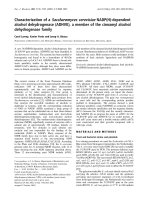
RNA secondary structure of SL1 and position of the SD2-nucleotide deletion in the SIV leaderFigure 1
RNA secondary structure of SL1 and position of the
SD2-nucleotide deletion in the SIV leader. Secondary
structure of the SIV
mac239
SL1 RNA element was predicted by
free energy minimization and adapted from published infor-
mation [6, 28, 48]. All nucleotide deletions are relative to the
transcriptional initiation site (1+) based on the sequence of
the wild type clone of SIV
mac239
. The DIS palindrome is
shown in bold, the A423G compensatory mutation is high-
lighted. Below is a diagram of the location of the various
compensatory mutations generated in different cell lines.
Asterisks denote substitutions selected in CEMx174 cells,
Bullets denote substitutions selected in C8166 cells.
CGGAG
U-A
C- G
C- G
U-A
C- G
G-C
U- G
G
G
C- G
G-C
C- G
G-U
A
A
A
A
U
G
G
G
C- G
U-A
G-C
G
G
G
G
U
U
A
A
A
C
C
C
C
GGA
+398
+419
CGGAG
A
G
G
A
G
C
U- G
G
G
C
G
U
G
C- G
A
C
G
G
G
G
U
U
A
A
A
C
C
C
C
GGA
G
G
* *
LTR
PBS DIS
MA CA NC p6
*/• •
•
Retrovirology 2007, 4:44 />Page 3 of 10
(page number not for citation purposes)
Results
The A423G point mutation plays an important role in the
restoration of viral RNA packaging
The SD2 variant (Δnt +398 to +418, Fig. 1) has been
shown to package diminished levels of viral RNA in com-
parison with wild type SIV
mac239
[6,7]. Forced evolution of
the SD2 variant through serial passage resulted in the res-
toration of wild-type replication kinetics. To further inves-
tigate the mechanism(s) involved, seven different SD2
derivates were analyzed that contained all possible com-
binations of the three point mutations that had been
identified in cell lines [7]. Viruses that reverted in C8166
cells contained either one, two or all three of the above
mentioned mutations, as follows: SD2-A423G, SD2-
K197R, SD2-G49L, SD2-A423G, K197R, SD2-A423G,
G49K, SD2-K197R, G49L, and SD2-A423G, K197R,
G49K. Similarly, viruses derived from reversions in
CEMx174 cells were termed SD2-A423G, SD2-E18G,
SD2-G31K, SD2-E18G, E31K, SD2-A423G, E18G, SD2-
A423G, E31K, and SD2-A423G, E18G, G31K (Fig. 1 and
Table 1).
Viral DNA of each of the two mutant groups (i.e. gener-
ated in either C8166 or CEMx174 cells) were transfected
into 293T cells. Mutant viral RNA was extracted from aliq-
uots of the supernatants of these transfections and nor-
malized on the basis of p27-CA. To assess relative
packaging efficiency, mutant viral RNAs were used as tem-
plate in an 18-cycle multiplex RT-PCR reaction run in par-
allel with multiple dilutions of wild type vRNA as a linear
range control, as described previously [6].
The results of RT-PCR (Fig. 2), were subjected to DNA
imaging analysis that showed that the SD2 deletion
mutant packaged viral RNA at levels that were approxi-
mately 40% of wild type; this is in agreement with previ-
ous studies. The compensatory A423G mutation within
the DIS-SL yielded the single largest increase in packaging
efficiency to about 80% of wild-type levels. In contrast,
the SD2-G49K and SD2-K197R variants packaged only
very low levels of vRNA (Fig 2A). Combinations of the
K197R and G49K mutations, i.e. SD2-K197R, G49K, or of
all three mutations, i.e., SD2-A423G, K197R, G49K,
showed increased packaging efficiency.
Next, we tested the ability of the two NC mutations to
restore vRNA incorporation. Fig 2B shows that the pres-
ence in SD2 of either E18G or G31K alone only margin-
ally affected levels of viral RNA packaging. In contrast, the
presence of both NC mutations resulted in moderately
increased RNA packaging. The addition of the A423G
mutation to the construct that contained both NC substi-
tutions completely compensated for the packaging deficit.
The SD2 variant (Δnt +398 to +418, Fig. 1) has also been
shown to be devoid of an RNA dimer [6-8]. To determine
the impact of multiple compensatory mutations on vRNA
Effects of untranslated-leader and gag-coding region muta-tions on viral RNA encapsidationFigure 2
Effects of untranslated-leader and gag-coding region
mutations on viral RNA encapsidation. Equivalent
amounts of virus derived from transfected 293T cells, based
on levels of p27-CAantigen, were used to prepare viral RNA
that was then used as template for quantitative RT-PCR to
detect full-length viral RNA genome in an 18-cycle PCR reac-
tion [6]. Relative amounts of a 114-bp DNA product were
quantified by molecular imaging, with wild-type values arbi-
trarily set at 1.0. Reactions run with RNA template, digested
by DNase-free RNase, served as a negative control for each
sample to exclude any potential DNA contamination. Rela-
tive amounts of viral RNA that were packaged were deter-
mined on the basis of four different experiments. A. RT-PCR
vRNA packaging results of SD2 variants harboring compensa-
tory mutations in the DIS (A423G), CA (K197R) and p6
(G49K) regions. B. RT-PCR vRNA packaging results of SD2
variants harboring mutations in the DIS (A423G), and NC
(E18G and G31K) regions.
0
20
40
60
80
100
120
WT
SD2
SD2-E18G
SD2-G31K
SD2-A423G-
E18G-G31K
SD2-E18G-G31K
123456
B.
0
20
40
60
80
100
120
WT
SD2
SD2-G49K
SD2-K197R
SD2-A423G
SD2-A423G-
G49K-K197R
SD2-G49K-K197R
2 345671
A.
Table 1: Impact of various coding and non-coding compensatory mutations on SD2 fitness.
Mutation Location Charge Processing Infectivity RNA incorporation
CEMx174 A420G DIS-SL n/a n/c +++ +++
E18G NC + +++ + +
G31K NC + +++ ++ ++
C8166 A420G DIS-SL n/a n/c +++ +++
K197R CA n/c n/c ++ +
G49K P6 + +++ ++ +
Legend: + = moderate recovery, +++ = near complete recovery, n/a = not applicable, n/c = no change
Retrovirology 2007, 4:44 />Page 4 of 10
(page number not for citation purposes)
dimerization, we analysed purifed vRNA on non-denatur-
ing Northern gels.
The results (Fig. 3A) show that RNA preparations recov-
ered from the SD2-mutants are compromised in regard to
vRNA dimerization compared to native wild-type RNA.
Figure 3B shows that the addition of the A423G mutation
to the SD2 backbone increased vRNA encapsidation levels
but appeared to have little impact on the amount of pack-
aged, mature vRNA dimer. Similarly, the amount of
mature dimer was not influenced by the addition of the
G49K or K197R mutations, i.e. SD2-G49K or SD2-K197R
(Fig. 3B). However, each of the abovementioned variants
did result in increased levels of high mobility RNA, inter-
preted to be dimeric RNA in an immature state, on non-
denaturing gels. This was also observed for combinations
of the K197R and G49K mutations, i.e. SD2-K197R,
G49K, or of all three compensatory mutations, i.e., SD2-
A423G, K197R, G49K.
Next, we tested the ability of the two NC mutations to res-
cue vRNA dimerization. Figure 3C shows that the SD2 var-
iant did have slightly increased levels of dimeric RNA in
the presence of either E18G or G31K. In contrast, the pres-
ence of both NC mutations resulted in a moderate
increase in levels of dimeric RNA. The addition of the
A423G and E18G mutations to the SD2 parental strain
also yielded an increase in RNA dimer levels. Finally, the
addition of G31K or of both NC substitutions to the SD2-
A423G variant increased levels of both packaged RNA
monomer and dimer.
To shed further light on the mechanisms involved, we per-
formed a thermodynamic RNA structural analysis of these
mutants by using M-Fold software [28]. RNA secondary
structure analysis suggests that the A423G point muta-
tion, that is located in the DIS-SL loop, cannot restore
native DIS-SL structure. However, our analysis indicated
that the A423G mutation altered the size of the DIS-loop
through nucleotide reorganization and loss of SL2 struc-
ture (not shown). Hence, the A423G point mutation plays
an important role in the compensation of the SD2 dele-
tion, but a full correction of packaging requires the pres-
ence of three mutations.
The G49K point mutation within p6 or, alternatively, the
E18G and G31K mutations within NC can restore Pr
55
Gag
processing in viruses that harbour the SD2 deletion
The SD2 deletion also resulted in delayed processing and
an altered processing pattern of Gag proteins. To study the
role of the aforementioned compensatory mutations in
this regard, Pr
55
Gag processing was evaluated by SDS-Page
analysis of viral proteins and Western blotting was per-
formed using monoclonal antibodies (MAbs) directed
against p27-CA as described previously [8]. Indeed, the
processing of each of three Gag proteins, i.e., the precursor
protein Pr55, the intermediate proteins p41, and p39,
were all impaired in the SD2 variant, but not in wild-type
virus. Interestingly, we found that all viruses that con-
tained the G49L mutation in p6, i.e SD2-G49K, SD2-
K197R, G49K, SD2-A423G, G49K, and SD2-A423G,
K197R, G49K, possessed similar proportions of these
products as wild-type virus. In contrast, the SD2-K197R,
SD2-A423G, and SD2-A423G, K197R viruses displayed
an accumulation of Pr55, p41, and p39 and diminished
levels of p27, similar to the parental SD2 virus (Fig. 4A).
The results of Fig. 4B show that either the E18G or G31K
substitution in NC was independently able to facilitate
complete Pr
55
Gag processing in the SD2 virus. In the pres-
ence of the A423G mutation, however, both NC muta-
tions i.e., SD2-A423G, E18G, G31K, were required to
restore processing of both the MA-CA (p41) and CA-NC
(p39) intermediate processing products, leading to a wild
type processing phenotype.
Thus, the A423G point mutation acts to rescue the deficit
in viral RNA packaging of the SD2 deletion, while the
G49K mutation in p6 or the E18G and G31K substitutions
in NC contribute to the restoration of Gag processing.
Native analysis of virion-associated RNAFigure 3
Native analysis of virion-associated RNA. Mutant or
wild-type virus was purified by sucrose gradient ultracentrifu-
gation. Virion RNA was then extracted from lysed particles
by protease K digestion followed by phenol chloroform
extraction. RNA was run under non-denaturing conditions at
room temperature. Membranes were analyzed with an SIV
specific probe as described in Materials and Methods. A.
Non-denaturing Northern analysis of the SD2 variant in con-
junction with compensatory mutants in the DIS, CA, and p6.
B. Non-denaturingNorthern analysis of the SD2 variant in
conjunction with compensatory mutants in the DIS and in
the NC protein.
M
D
WT
SD2
WT
SD2-E18G
SD2-G31K
SD2-E18G-G31K
M
D
SD2-A423G-E18G
SD2-A423G-G31K
SD2-A423G-E18G-G31K
A. C.
M
D
WT
SD2
SD2-K197R
SD2-A423G -G49K-K197R
SD2-G49K-K197R
SD2-A423G
SD2-A423G-G49K
SD2-A423G-K197R
SD2-G49K
B.
Retrovirology 2007, 4:44 />Page 5 of 10
(page number not for citation purposes)
Both sets of compensatory mutations are functionally
interchangeable in recovery of viral replication and
infectivity
In order to pursue the biological relevance of these com-
pensatory mutations, each mutant proviral construct was
transfected into 293T cells and viral supernatanst har-
vested after 48 hours. Viral stocks were titrated by p27-CA
ELISA and assayed for viral replication capacity in PHA-
stimulated rhesus PBMC. As shown in Figure 5A, the
mutations that had emerged in CEMx174 cells, i.e.
A423G, E18G, G31K, were also able to rescue the defec-
tive replication of the SD2-deleted viruses in these pri-
mary cells. Although each single mutation could
individually contribute to recovered viral growth, full res-
toration of replication capacity required the combination
of all three mutations, i.e. SD2-A423G, E18G, and G31K.
Similarly, the combination of A423G, K197R, and G49K
in the same SD2-backbone fully rescued SD2 replication
in rhesus PBMC (Fig. 5B).
The role of these various compensatory mutations in viral
replicative fitness was next assessed on the basis of viral
infectiousness in CEMx174 cells. For this purpose, relative
p27-CA concentrations in viral supernatants at the peak of
viral replication (as determined by RT assay and observed
cytopathicity in culture) were used to calculate TCID
50
per
ng p27-CA antigen (Fig. 5C). The results show that the
SD2 mutant was severely compromised, whereas each
compensatory mutation was independently capable of
restoring some degree of viral infectiousness, with the
largest increase attributable to A423G. However, recovery
to near wild-type replication levels required a full comple-
ment of either of the two groups of compensatory muta-
tions. The mutations identified in the C8166 cell line
restored infectiousness equally well when assayed in the
CEMx174 line and vice versa (not shown). Thus, both sets
of compensatory mutations seem to be functionally inter-
changeable in regard to restoration of viral replication,
independent of the cell line in which they were first
selected.
Replicative fitness of wild-type and mutated viruses in mon-key PBMCsFigure 5
Replicative fitness of wild-type and mutated viruses
in monkey PBMCs. Viral replication was assessed in PHA-
activated rhesus PBMCs using 10ng of viral inocula normal-
ized on the basis of p27-CA Ag. All replication experiments
were conducted in triplicate. Viral replication was monitored
by RT assay of culture supernatants at multiple time points.
All RT activity results are the average of duplicates. Mock
infection denotes exposure of cells to heat-inactivated wild-
type virus as a negative control. A. Growth curves of SD2-
variants harboring mutations in the DIS (A423G) and NC
(E18G and G31K) regions. B. Growth curves of variants har-
boring compensatory mutations in the DIS (A423G), CA
(K197R) and p6 (G49K) regions. C. Viral replication analysis
of mutated viruses by TCID
50
analysis of viral infectivity as
described in Materials and Methods. Results shown are rep-
resentative of three independent endpoint dilution assay
experiments. The scale of the ordinate is logarithmic. Mock
infection represents a negative control in which cells were
exposed to heat-inactivated wild-type virus.
0
50000
100000
150000
2.5
5
7.5
10
12.5
Days After Infection
MOCK
SD2-K197R-G49K
SD2-G49K
SD2-K197R
SD2-A423G-G49K
SD2-A423G-K197R
SD2-A423G-K197R-G49K
SD2-A423G
SD2
WT
0
50000
100000
150000
2.5
5
7.5
10
12.5
Days After Infection
MOCK
SD2-E18G-G31K
SD2-A423G-G31K
SD2-A423G-E18G
SD2-G31K
SD2-E18G
SD2-A423G
SD2-A423G-E18G-G31K
SD2
WT
0 100 200 300 400 500 600 700
0 100 200 300 400 500 600
700
WT
SD2
SD2-A423G
SD2-K197R
SD2-G49K
SD2-K197R-G49K
SD2-A423G-K197R
SD2-A423G-G49K
SD2-A423G-K197R-G49K
SD2-E18G
SD2-G31K
SD2-E18G-G31K
SD2-A423G-E18G
SD2-A423G-G31K
SD2-A432G- E18G-G31K
TCID50/ng p27
RT Activity (
cpm /ml)
RT Activity (
cpm /ml)
A.
B.
C.
Restoration of proteolytic Gag-processing by G49K, or by the E18G, G31K mutationsFigure 4
Restoration of proteolytic Gag-processing by G49K,
or by the E18G, G31K mutations. Viruses were purified
by ultracentrifugation of clarified culture supernatants over a
sucrose cushion at 48h after transfection. Western analysis
of viral Pr
55
Gag products were detected using MAb directed
against p27-CA antigen.
WT
SD2
SD2-K197R
SD2-A423G -G49K-K197R
SD2-G49K-K197R
SD2-A423G
SD2-A423G-G49K
SD2-A423G-K197R
A.
WT
SD2
SD2-E18G
SD2-G31K
SD2-A423G-E18G+G31K
SD2-E18G+G31K
SD2-A423G
SD2-A423G-E18G
SD2-A423G-G31K
B.
Pr55
MA-CA
p27-CA
CA-NC
SD2-G49K
Retrovirology 2007, 4:44 />Page 6 of 10
(page number not for citation purposes)
Forced evolution results in restoration of proper viral core
ultra-structure
We next hypothesized that the mutations selected through
serial passage might also correct morphological anoma-
lies in the viral core. Transmission electron microscopy
(TEM), of ultra-thin sections of transfected cell prepara-
tions showed that approximately 80% of wild-type virus
particles contained a fully condensed core, typical of
mature virus. In contrast, the SD2 mutant resulted in
diminished viral production, and about 70% of the SD2
particles observed possessed displaced and/or improperly
condensed cores and/or immature core structure (Fig. 6).
Both recombinant clones (i.e. SD2-A423G-E18G-G31K
and SD2-A423G-K197R-G49K) were also transfected in
parallel, and yielded comparable levels of particle produc-
tion as wild type, as measured by p27-CA levels in culture
supernatants (not shown). The results of the EM experi-
ments showed restoration of proper core morphology,
and levels of immature virus were comparable to the wild
type (Fig. 6).
Discussion
Here, we describe an SIV deletion mutant that was pas-
saged in two different T-cell lines and that employed two
different pathways to attain reversion. Retroviruses dis-
play genomic plasticity, and sequence diversification in
both HIV-1 and SIV can in some cases augment viral rep-
lication and pathogenesis [29-31]. The fitness of an RNA
virus population may be viewed as a continuum of
genomes of varying fitness. It is not surprising that these
viruses may be able to employ diverse routes to reach
higher fitness levels. However, such transitions may be
delimited by the tolerance of a particular gene for non-
synonomous mutation versus the maintenance of a native
function[32]. In the case of mutations that compensate
for deletion mutagenesis, a debilitated variant should
need to pass through a deterministic bottleneck to initiate
a new quasispecies distribution. Therefore, compensation
should be governed by selection for optimal viral fitness
and not by stochastic drift [33].
Our findings indicate that reversion is not limited to a sin-
gle trajectory. Compensatory mutations in both the
untranslated leader and the gag-coding region emerged
during long-term passage in different T-cell lines, and
these mutations were required for full restoration of viral
replication. Interestingly, the A423G substitution, located
within the DIS, was shown to be active in restoring effi-
cient levels of viral RNA packaging, while mutations in
either the nucleocapsid, G31K, E18G, or within the p6
protein of Gag, G49K, were essential for the proper
processing of Gag precursors. In each instance, the pres-
ence of three point mutations was functionally synergistic
in regard to rescue of both viral RNA packaging and Gag
processing. Moreover, the observed changes in regard to
impaired Gag processing could be corrected by either the
E18G, G31K or G49K mutations. We also showed that
RNA dimerization could be partially recovered due to
compensatory mutations in NC. Several studies have
shown the interplay that exists between viral RNA and
viral proteins that are involved in regulation of core struc-
ture, proteolytic processing, and maturation of RNA dim-
ers [34,35]. Interestingly, SD2 mutants harboring A423G
and various combinations of K197R and G49K did co-
package a high molecular weight RNA species reminiscent
of the "immature" dimer found in protease mutants of
MLV [11].
Numerous studies on HIV-1 have shown that NC is the
major protein domain within Gag that recognizes the
encapsidation signals present within leader sequences
Transmission election microscopy of wild-type and mutant viral particlesFigure 6
Transmission election microscopy of wild-type and
mutant viral particles. TEM of late (fixed 48 hr post-trans-
fection) wild-type and mutant particles were assessed and
scored from multiple sections. Panel A: the wild-type virus
displayed typical size and conical core morphology. Panel B:
the SD2 deletion mutant showed diminished production of
viral particles, with altered diameter and core morphology.
Panel C: the SD2-A423G-E18G-G31K mutant showed resto-
ration of proper core morphology. Panel D: the SD2-A423G-
K197R-G49K mutant also showed restoration of core place-
ment and morphology. Bar size is shown for each panel.
A.
B.
C.
D.
Retrovirology 2007, 4:44 />Page 7 of 10
(page number not for citation purposes)
[36]. The NC protein contains two zinc finger motifs that
contribute to its specific interactions with viral RNA,
including a well-described role in RNA dimer maturation
[19,37]. Deletions within SL1 of HIV-1 were shown to
impair viral replication, as well as to cause delayed
processing of Gag proteins and decreased levels of packag-
ing of viral RNA [38]. Forced evolution of SL1 deleted
virus in HIV-1 showed that compensatory reversion was a
result of substitutions in four disparate regions of Gag, i.e.
NC (T24I), MA (V35I), CA (T24I) and p2 (T21I) [22].
These substitutions all involve hydrophobic amino acids.
In contrast, we have shown that the deletions in leader
sequences of SIV
mac239
can be rescued by compensatory
point mutations elsewhere within the DIS and Gag. The
present work shows that restoration of SIV replication
involved two distinct sets of mutations, located in both
the DIS loop (A423G) and within different Gag proteins,
i.e. NC (E18G and G31K) or CA (K197R) and p6 (E49K);
these amino acid changes, with the exception of K197R,
result in a net increase in the number of positively charged
residues within Gag.
The finding that mutations within NC can rescue these
deficits further confirms the role of this protein in interac-
tions between Gag and RNA leader sequences of SIV,
which have been less intensively studied than for HIV. The
debilitated SD2 virus may be able to correct the deficit
caused by the deletion within the DIS stem by altering
both the leader sequence, as well as by reconfiguring Gag
proteins, presumably to facilitate both viral RNA-RNA
and RNA-protein interaction [39,40].
Our data also show that p6 plays an important role in the
incorporation of viral proteins into virions and the spe-
cific encapsidation of viral RNA [41]. We have also dem-
onstrated that a substitution within p6 resulted in
comparable levels of compensation as did mutations
within NC, i.e. E18G or G31K, in restoration of Gag
processing. This suggests that p6 may also be important at
core positioning and condensation during viral budding.
The multimerization of Pr
55
Gag has been shown to occur
on an RNA scaffold, and encapsidation of viral RNA likely
requires that leader RNA sequences exist within the con-
straints of proper tertiary structure, which are highly con-
served in both HIV-1 and SIV [40,42-44]. Deletions of
leader sequences may alter critical RNA-protein interac-
tions at early stages of viral assembly, thereby altering
morphogenesis. As a result, nascent particles may not be
able to undergo a "normal" intra-virion transition that
condenses the RNA genome and multiple viral proteins to
produce a "primed" infectious core [39].
These observations suggest the importance of functional
interactions between Gag-proteins and the RNA-leader in
both HIV-1 and SIV, but also imply that important differ-
ences may exist between SIV and HIV-1 in regard to such
interactions. We have also demonstrated that different cell
types can reproducibly select for different sets of compen-
satory mutations, but that both of these sets are function-
ally interchangeable in regard to their ability to restore
viral replication, regardless of the cell type in which the
virus is ultimately grown. Of course, it is conceivable that
either the same mutational spectrum or even different
ones may have been observed in either of the cell lines
tested had additional replication studies been performed.
It is not trivial that the mechanisms of compensation for
lentiviruses, grown under conditions of stress as demon-
strated here, are apparently not restricted to single path-
ways. The mechanisms behind viral escape from
antibodies, cytotoxic-T lymphocyte pressure and the gen-
eration of resistance to antiretroviral drugs are not mutu-
ally exclusive. Our results add to what is known about the
plasticity and adaptability of lentivirus genomes.
Methods
Construction of recombinant proviral SIV clones
A PCR-based mutagenesis method was applied together
with conventional cloning techniques using the full-
length infectious clone of SIV, termed SIV
mac239
wild type
as a template, to generate all the mutants described [6]. All
nucleotide designations are based on published
sequences; the transcription initiation site corresponds to
position +1 [45].
Viral RNA packaging analysis by RT-PCR
To study packaging of viral genomic RNA we used meth-
ods previously described [6-10]. Briefly, viral RNA was
isolated using the QIAamp viral RNA mini kit (QIAGEN)
from equivalent amounts of 293T cell-derived viral prep-
arations (normalized by SIV p27-CA antigen). RNA sam-
ples were treated with RNase-free DNase I at 37°C for 30
min to eliminate potential plasmid DNA contamination,
followed by inactivation by incubation at 75°C for 10
min. The viral RNA samples were quantified using the
Titan One Tube RT-PCR system (Boehringer Mannheim,
Montreal, Quebec, Canada). The primers sg1 and sg2 were
used to amplify a 114-bp fragment within the MA coding
region of gag representing full-length viral RNA. The
primer sg2 was radioactively labeled with δ-P
32
-ATP in
order to visualize PCR products. Equivalent RNA samples,
based on p27 antigen levels, were used as templates in an
18-cycle RT-PCR. The products were fractionated on 5%
polyacrylamide gels and exposed to X-ray film. Relative
amounts of products were quantified by molecular imag-
ing (BIO-RAD Imaging). RNA encapsidation was deter-
mined on the basis of four different reactions, and
calculated with wild type virus levels arbitrarily set at 1.0.
Retrovirology 2007, 4:44 />Page 8 of 10
(page number not for citation purposes)
Non-denaturing Northern analysis
Culture fluids from transfected 293T cells were collected
and clarified using a Beckman GS-6R bench centrifuge at
3,000 rpm for 30 min at 4°C. Viral particles were further
purified through a 20% sucrose cushion at 40,000 rpm for
1 hour at 4°C using a SW41 rotor in a Beckman L8-M
ultracentrifuge. Viral pellets were first dissolved in Tris-
EDTA (TE) buffer, then in lysis buffer containing protein-
ase K (100 μg/ml) and yeast tRNA (100 μg/ml). Samples
were incubated for 20 min at 37°C, in the presence of 50U
of DNAse I, followed by two extractions, first in phenol:
chloroform: isoamyl alcohol, then chloroform. Viral RNA
was then precipitated, washed in 70% ethanol and stored
at -80°C until required, at which time samples were resus-
pended in TE buffer at 4°C. RNA was then analysed by
non-denaturing electrophoresis on 0.9% agarose gels in
1× Tris-Borate-EDTA (TBE) running buffer for 4 hrs at
4°C. Products were subsequently denatured in 50 mM
NaOH and equilibrated in 200 mM Na-acetate. Following
electrophoresis, RNA was transferred to Hybond-N nylon
membranes by capillary blotting using a 20× concentra-
tion of SSPE buffer. Membranes were baked for 2 hrs at
80°C. Probes were prepared by digestion and purification
of the NdeI-BstE III fragment excised from the SIV
mac239
plasmid. These were recovered by gel purification and
labelled with δ-P
32
-ATP by nick-translation following
standard protocols (Roche, Indianapolis, IN, USA). The
denaturing Northern analysis of cellular RNA was also
conducted in parallel. RNA extraction was carried out in
similar fashion to that described for slot blotting above.
Cellular RNA from lysates was normalized on the basis of
p27-CA antigen present in cellular lysates. Total cellular
RNA preparations, i.e. equivalent volumes of RNA, were
also run on 1% ethidium bromide (EtBr) stained gels as
internal controls for total RNA and 28S and 18S ribos-
omal RNAs. Probes were prepared as described above.
Probes were labelled by nick-translation following stand-
ard manufacturer's protocols (Roche, Indianapolis, IN,
USA) and used in standard hybridization reactions.
Western analysis of viral protein
At 48 hrs post-transfection, virus-containing supernatants
recovered from transfected 293T cells were collected and
clarified at 3000 rpm for 30 min, at 4°C in a GS-6R Beck-
man centrifuge. Virus was further purified by pelleting
through a 20% sucrose cushion by ultracentrifugation at
35000 rpm in a Beckman ultracentrifuge for 1 hr at 4°C.
Cells were washed 2× in cold PBS and lysed by the addi-
tion of buffer containing 1% Nonidet P-40, 50 mM Tris-
CL (pH 7.4), 150 mM NaCl, 0.02% sodium azide, and a
cocktail of protease inhibitors (Roche, Laval, Quebec,
Canada). Virus was normalized on the basis of p27-CA
protein present in supernatants or cell lysates. Both pel-
leted virus and cellular lysates were subject to Western
blotting with monoclonal antibodies directed at SIV p27-
CA antigens (Fitzgerald industries, MA, USA) following
standard protocols [46].
Cell culture and preparation of virus stocks
293T cells were maintained in DMEM medium supple-
mented with 10% heat-inactivated fetal bovine serum,
penicillin, streptomycin and glutamine. CEMx174 or
C8166 cells were maintained in RPMI-1640 medium sup-
plemented with 10% heat-inactivated fetal bovine serum
and antibiotics. All media and sera were purchased from
Gibco inc. (Burlington, Ontario, Canada).
Monkey peripheral blood mononuclear cells (PBMCs)
were isolated from the blood of healthy rhesus macaques
(Macaca mulatta) housed at L.A.B. Pre-Clinical Research
International Inc., (Montreal, Quebec). All primates were
housed in accordance with accredited laboratory care
standards. All donor macaques were tested serologically
and were negative for simian type-D retrovirus-1 (SRV-1),
simian T-cell lymphotrophic virus type 1(STLV-1), and
simian foamy virus (SFV-1) at the initiation of the study.
PBMCs were purified on Ficoll cushions, washed in sup-
plemented RPMI-1640 media, and purified lymphocytes
were then phytohemagglutinin (PHA)-stimulated for 3
days, then maintained in supplemented RPMI-1640
medium containing 10% heat-inactivated fetal bovine
serum and 20 u/ml IL-2 at 37°C with 5% CO
2
overlay. All
recombinant viral constructs were purified using a maxi-
plasmid purification kit (Qiagen inc. Mississauga,
Ontario, Canada). For the production of infectious viral
stock, 293T cells were transfected using the above con-
structs together with Lipofectamine-Plus reagent (Gibco,
Burlington, Ontario, Canada). Virus-containing culture
supernatants were harvested at 48 hr post-transfection
and clarified by centrifugation for 30 min at 4°C at 3,000
rpm in a Beckman GS-6R centrifuge. Viral stocks were
passed through a 0.2 μm filter and stored in 1 ml aliquots
at -80°C. All wild type and mutant stocks were titered on
the basis of p27-CA antigen in culture supernatants using
a Coulter SIV core antigen ELISA assay (Immunotech inc.,
Westbrook, ME, U.S.A.).
Virus replication in macaque donor PBMCs
To initiate infection, viral stocks were thawed at room
temperature. Then, 100 U of Dnase I in the presence of 10
mm MgCl
2
were added at 37°C for 0.5 h to eliminate any
potential plasmid DNA contamination, prior to inocula-
tion of cells. Infection of rhesus PBMCs was performed by
incubating 4 × 10
6
PHA-activated cells with wild type or
mutant viral stocks containing 10 ng of p27-CA viral
equivalent at 37°C for 2 hours. Infected cells were then
washed three times with PBS to remove any remaining
virus. Finally, cells were resuspended in fresh supple-
mented RPMI-1640 medium. Cells were maintained in 3
Retrovirology 2007, 4:44 />Page 9 of 10
(page number not for citation purposes)
ml of culture medium as described above, and fresh stim-
ulated PBMCs were added to the cultures at weekly inter-
vals. Virus production in culture fluids was monitored by
both RT assay and SIV p27 antigen capture assay.
Virus infectivity (TCID
50
) was determined by infection of
CEMx174 cells as described previously. TCID
50
results
were calculated by the method of Reed and Muench [47].
Electron microscopic analysis of virion morphology
Viral ultra-structure for the described mutant viruses was
examined by transmission electron microscopy. Briefly,
COS-7 cells transfected with wild type or mutant SIV con-
structs were fixed at 48 hours post-tranfection in 2.5% glu-
taraldehyde/phosphate buffered saline followed by a
secondary fixation of lipids in 4% osmium tetroxide. Sam-
ples were routinely processed and serially dehydrated.
Samples were embedded in Epon under vacuum followed
by heat-induced polymerization. Thin-sectioned samples
were stained with lead citrate and uranyl acetate and visu-
alized at 80 Kev using a JEOL JEM-2000 FX transmission
electron microscope equipped with a Gatan 792 Bioscan
wide-angle 1024 × 1024 byte multi-scan CCD camera. At
least 100 viral particles were scored for each variant to
determine the relative percentage of particles with struc-
tural anomalies.
Acknowledgements
The following reagents were obtained through the AIDS Research and Ref-
erence Reagent Program, Division of AIDS, NIAID, NIH: the p239SpSp5'
and p239SpE3'plasmids contributed by R. Desrosiers. Research for this
study was supported by the Canadian Institutes for Health Research
(CIHR). We thank Maureen Olivera for conducting RT assays. We are also
grateful to Yonjun Guan for providing the many viral constructs. J. B. W.
was supported by both a pre-doctoral and post-doctoral fellowship from
The Canadian Institutes for Health Research (CIHR). We are also grateful
to Diane and Aldo Bensadoun for support of our work.
References
1. Lever A, Gottlinger H, Haseltine W, Sodroski J: Identification of a
sequence required for efficient packaging of human immun-
odeficiency virus type 1 RNA into virions. J Virol 1989,
63:4085-4087.
2. Li X, Liang C, Quan Y, Chandok R, Laughrea M, Parniak MA, Kleiman
L, Wainberg MA: Identification of sequences downstream of
the primer binding site that are important for efficient repli-
cation of human immunodeficiency virus type 1. J Virol 1997,
71:6003-6010.
3. Berkowitz R, Fisher J, Goff SP: RNA packaging. Curr Top Microbiol
Immunol 1996, 214:177-218.
4. Berkowitz RD, Hammarskjold ML, Helga-Maria C, Rekosh D, Goff SP:
5' regions of HIV-1 RNAs are not sufficient for encapsidation:
implications for the HIV-1 packaging signal. Virology 1995,
212:718-723.
5. Clever JL, Mirandar D Jr, Parslow TG: RNA structure and pack-
aging signals in the 5' leader region of the human immuno-
deficiency virus type 1 genome. J Virol 2002, 76:12381-12387.
6. Guan Y, Whitney JB, Diallo K, Wainberg MA: Leader sequences
downstream of the primer-binding site are important for
efficient replication of simian immunodeficiency virus. J Virol
2000, 74:8854-8860.
7. Guan Y, Diallo K, Detorio M, Whitney JB, Liang C, Wainberg MA:
Partial restoration of replication of simian immunodefi-
ciency virus by point mutations in either the dimerization
initiation site (DIS) or Gag region after deletion mutagenesis
within the DIS. J Virol 2001, 75:11920-11923.
8. Whitney JB, Wainberg MA: Impaired RNA incorporation and
dimerization in live attenuated leader-variants of
SIVmac239. Retrovirology 2006, 3:96.
9. Dirac AM, Huthoff H, Kjems J, Berkhout B: Regulated HIV-2 RNA
dimerization by means of alternative RNA conformations.
Nucleic Acids Res 2002, 30:2647-2655.
10. Dirac AM, Huthoff H, Kjems J, Berkhout B: The dimer initiation
site hairpin mediates dimerization of the human immunode-
ficiency virus, type 2 RNA genome. J Biol Chem 2001,
276:32345-32352.
11. Fu W, Rein A: Maturation of dimeric viral RNA of Moloney
murine leukemia virus. J Virol 1993, 67:5443-5449.
12. Rein A: Retroviral RNA packaging: a review. Arch Virol Suppl
1994, 9:513-522.
13. Berkhout B, van Wamel JL: Role of the DIS hairpin in replication
of human immunodeficiency virus type 1. J Virol 1996,
70:6723-6732.
14. Berkhout B, van Wamel JL: The leader of the HIV-1 RNA
genome forms a compactly folded tertiary structure. Rna
2000, 6:282-295.
15. Dirac AM, Huthoff H, Kjems J, Berkhout B: Requirements for RNA
heterodimerization of the human immunodeficiency virus
type 1 (HIV-1) and HIV-2 genomes. J Gen Virol 2002,
83:2533-2542.
16. Strappe PM, Hampton DW, Brown D, Cachon-Gonzalez B, Caldwell
M, Fawcett JW, Lever AM: Identification of unique reciprocal
and non reciprocal cross packaging relationships between
HIV-1, HIV-2 and SIV reveals an efficient SIV/HIV-2 lentiviral
vector system with highly favourable features for in vivo test-
ing and clinical usage. Retrovirology 2005, 2:55.
17. D'Souza V, Melamed J, Habib D, Pullen K, Wallace K, Summers MF:
Identification of a high affinity nucleocapsid protein binding
element within the Moloney murine leukemia virus Psi-RNA
packaging signal: implications for genome recognition. J Mol
Biol 2001, 314:217-232.
18. Darlix JL, Gabus C, Nugeyre MT, Clavel F, Barre-Sinoussi F: Cis ele-
ments and trans-acting factors involved in the RNA dimeri-
zation of the human immunodeficiency virus HIV-1. J Mol Biol
1990, 216:689-699.
19. Yovandich JL, Chertova EN, Kane BP, Gagliardi TD, Bess JW Jr, Sow-
der RC 2, Henderson LE, Gorelick RJ: Alteration of zinc-binding
residues of simian immunodeficiency virus p8(NC) results in
subtle differences in gag processing and virion maturation
associated with degradative loss of mutant NC. J Virol 2001,
75:115-124.
20. Shehu-Xhilaga M, Kraeusslich HG, Pettit S, Swanstrom R, Lee JY, Mar-
shall JA, Crowe SM, Mak J: Proteolytic processing of the p2/
nucleocapsid cleavage site is critical for human immunodefi-
ciency virus type 1 RNA dimer maturation. J Virol 2001,
75:9156-9164.
21. Russell RS, Roldan A, Detorio M, Hu J, Wainberg MA, Liang C:
Effects of a single amino acid substitution within the p2
region of human immunodeficiency virus type 1 on packag-
ing of spliced viral RNA. J Virol 2003, 77:12986-12995.
22. Liang C, Rong L, Quan Y, Laughrea M, Kleiman L, Wainberg MA:
Mutations within four distinct gag proteins are required to
restore replication of human immunodeficiency virus type 1
after deletion mutagenesis within the dimerization initiation
site. J Virol 1999, 73:7014-7020.
23. Griffin SD, Allen JF, Lever AM: The major human immunodefi-
ciency virus type 2 (HIV-2) packaging signal is present on all
HIV-2 RNA species: cotranslational RNA encapsidation and
limitation of Gag protein confer specificity. J Virol 2001,
75:12058-12069.
24. Ohlmann T, Lopez-Lastra M, Darlix JL: An internal ribosome
entry segment promotes translation of the simian immuno-
deficiency virus genomic RNA. J Biol Chem 2000,
275:11899-11906.
25. Gottlinger HG: The HIV-1 assembly machine. Aids 2001,
15(Suppl 5):S13-20.
26. Aldovini A, Young RA: Mutations of RNA and protein
sequences involved in human immunodeficiency virus type 1
Publish with BioMed Central and every
scientist can read your work free of charge
"BioMed Central will be the most significant development for
disseminating the results of biomedical researc h in our lifetime."
Sir Paul Nurse, Cancer Research UK
Your research papers will be:
available free of charge to the entire biomedical community
peer reviewed and published immediately upon acceptance
cited in PubMed and archived on PubMed Central
yours — you keep the copyright
Submit your manuscript here:
/>BioMedcentral
Retrovirology 2007, 4:44 />Page 10 of 10
(page number not for citation purposes)
packaging result in production of noninfectious virus. J Virol
1990, 64:1920-1926.
27. Cimarelli A, Darlix JL: Assembling the human immunodefi-
ciency virus type 1. Cell Mol Life Sci 2002, 59:1166-1184.
28. Zuker M: On finding all suboptimal foldings of an RNA mole-
cule. Science 1989, 244:48-52.
29. Nowak MA, Anderson RM, Boerlijst MC, Bonhoeffer S, May RM,
McMichael AJ: HIV-1 evolution and disease progression. Science
1996, 274:1008-1011.
30. Iwasa Y, Michor F, Nowak MA: Virus evolution within patients
increases pathogenicity. J Theor Biol 2005, 232:17-26.
31. Yeh WW, Cale EM, Jaru-Ampornpan P, Lord CI, Peyerl FW, Letvin
NL: Compensatory substitutions restore normal core assem-
bly in simian immunodeficiency virus isolates with Gag
epitope cytotoxic T-lymphocyte escape mutations. J Virol
2006, 80:8168-8177.
32. Yuste E, Borderia AV, Domingo E, Lopez-Galindez C: Few muta-
tions in the 5' leader region mediate fitness recovery of
debilitated human immunodeficiency type 1 viruses. J Virol
2005, 79:5421-5427.
33. Domingo E, Holland JJ: RNA virus mutations and fitness for sur-
vival. Annu Rev Microbiol 1997, 51:151-178.
34. Fu W, Dang Q, Nagashima K, Freed EO, Pathak VK, Hu WS: Effects
of Gag mutation and processing on retroviral dimeric RNA
maturation. J Virol 2006, 80:1242-1249.
35. Badorrek CS, Gherghe CM, Weeks KM: Structure of an RNA
switch that enforces stringent retroviral genomic RNA
dimerization. Proc Natl Acad Sci USA 2006, 103:13640-13645.
36. Feng YX, Copeland TD, Henderson LE, Gorelick RJ, Bosche WJ, Levin
JG, Rein A: HIV-1 nucleocapsid protein induces "maturation"
of dimeric retroviral RNA in vitro. Proc Natl Acad Sci USA 1996,
93:7577-7581.
37. Laughrea M, Shen N, Jette L, Darlix JL, Kleiman L, Wainberg MA: Role
of distal zinc finger of nucleocapsid protein in genomic RNA
dimerization of human immunodeficiency virus type 1; no
role for the palindrome crowning the R-U5 hairpin. Virology
2001, 281:109-116.
38. Liang C, Rong L, Laughrea M, Kleiman L, Wainberg MA: Compensa-
tory point mutations in the human immunodeficiency virus
type 1 Gag region that are distal from deletion mutations in
the dimerization initiation site can restore viral replication.
J Virol 1998, 72:6629-6636.
39. Tang S, Murakami T, Agresta BE, Campbell S, Freed EO, Levin JG:
Human immunodeficiency virus type 1 N-terminal capsid
mutants that exhibit aberrant core morphology and are
blocked in initiation of reverse transcription in infected cells.
J Virol 2001, 75:9357-9366.
40. Muriaux D, Costes S, Nagashima K, Mirro J, Cho E, Lockett S, Rein A:
Role of murine leukemia virus nucleocapsid protein in virus
assembly. J Virol 2004, 78:12378-12385.
41. von Schwedler UK, Stuchell M, Muller B, Ward DM, Chung HY,
Morita E, Wang HE, Davis T, He GP, Cimbora DM, et al.: The pro-
tein network of HIV budding. Cell 2003, 114:701-713.
42. Ganser-Pornillos BK, von Schwedler UK, Stray KM, Aiken C, Sun-
dquist WI: Assembly properties of the human immunodefi-
ciency virus type 1 CA protein. J Virol 2004, 78:2545-2552.
43. Muriaux D, Mirro J, Harvin D, Rein A: RNA is a structural ele-
ment in retrovirus particles. Proc Natl Acad Sci USA 2001,
98:5246-5251.
44. Muriaux D, Mirro J, Nagashima K, Harvin D, Rein A: Murine leuke-
mia virus nucleocapsid mutant particles lacking viral RNA
encapsidate ribosomes. J Virol 2002, 76:11405-11413.
45. Calef CMJ, O'Connor DH, Watkins DI, Korber BT: Numbering
Positions in SIV Relative to SIVMM239. In HIV Sequence Com-
pendium Edited by: Kuiken C, Foley B, Hahn B, Marx P, McCutchan F,
Mellors JW, Wolinsky S, Korber B. Theoretical Biology and Biophys-
ics Group, Los Alamos National Laboratory, Los Alamos, NM, LA-UR
02-2877. (Ed.); 2001:171-181.
46. Sambrook J, Fritsch EF, Maniatis T: Molecular cloning 2nd edition. New
York: Cold Spring Harbor Laboratory Press; 1989.
47. Reed LJ, Muench H: A simple method of estimating 50 per cent
end-points. Amer Jour Hygiene
1938, 27:493-497.
48. Zuker M: Mfold web server for nucleic acid folding and hybrid-
ization prediction. Nucleic Acids Res 2003, 31:3406-3415.