Báo cáo y học: " Differential resistance to cell entry by porcine endogenous retrovirus subgroup A in rodent species" potx
Bạn đang xem bản rút gọn của tài liệu. Xem và tải ngay bản đầy đủ của tài liệu tại đây (3.24 MB, 13 trang )
Retrovirology
BioMed Central
Open Access
Research
Differential resistance to cell entry by porcine endogenous
retrovirus subgroup A in rodent species
Giada Mattiuzzo, Magda Matouskova and Yasuhiro Takeuchi*
Address: Wohl Virion Centre, Division of Infection and Immunity, University College London, W1T 4JF, London, UK
Email: Giada Mattiuzzo - ; Magda Matouskova - ;
Yasuhiro Takeuchi* -
* Corresponding author
Published: 14 December 2007
Retrovirology 2007, 4:93
doi:10.1186/1742-4690-4-93
Received: 10 October 2007
Accepted: 14 December 2007
This article is available from: />© 2007 Mattiuzzo et al; licensee BioMed Central Ltd.
This is an Open Access article distributed under the terms of the Creative Commons Attribution License ( />which permits unrestricted use, distribution, and reproduction in any medium, provided the original work is properly cited.
Abstract
Background: The risk of zoonotic infection by porcine endogenous retroviruses (PERV) has been
highlighted in the context of pig-to-human xenotransplantation. The use of receptors for cell entry
often determines the host range of retroviruses. A human-tropic PERV subgroup, PERV-A, can
enter human cells through either of two homologous multitransmembrane proteins, huPAR-1 and
huPAR-2. Here, we characterised human PARs and their homologues in the PERV-A resistant
rodent species, mouse and rat (muPAR and ratPAR, respectively).
Results: Upon exogenous expression in PERV-A resistant cells, human and rat PARs, but not
muPAR, conferred PERV-A sensitivity. Exogenously expressed ratPAR binds PERV-A Env and
allows PERV-A infection with equivalent efficiency to that of huPAR-1. Endogenous ratPAR
expression in rat cell lines appeared to be too low for PERV-A infection. In contrast, the presence
of Pro at position 109 in muPAR was identified to be the determinant for PERV-A resistance.
Pro109. was shown to be located in the second extracellular loop (ECL2) and affected PERV-A Env
binding to PAR molecules.
Conclusion: The basis of resistance to PERV-A infection in two rodent species is different.
Identification of a single a.a. mutation in muPAR, which is responsible for mouse cell resistance to
PERV-A highlighted the importance of ECL-2 for the viral receptor function.
Background
Pig-to-human xenotransplantation presents potential
benefits for treatment of a range of diseases, such as diabetes, neurological disorders and for organ failures, and to
alleviate the shortage of human donor organs. Recent
advances in genetic engineering of animals, such as the
development of pigs devoid of α-galactosyltransferase
[1,2], help overcome immunological problems and bring
clinical xenotransplantation a step closer to reality. However, zoonotic pathogen transmission is a potential risk
and must be controlled (reviewed in [3] and [4]).
Although exogenous viruses can be removed from the
transplantation source by breeding pigs in specific pathogen-free environments, such techniques cannot eliminate
porcine endogenous retroviruses (PERV) present in the
pig germ line DNA. Furthermore, pig cells can produce
PERV capable of infecting human cells in vitro [5-7]. All
PERV known to be infectious belong to the gammaretrovirus genus and gammaretroviruses, such as gibbon ape
leukaemia virus (GALV) and murine leukaemia virus
(MLV), can cause cancer, leukaemia or neurodegeneration. If PERV cross the species barrier, adapt to new
Page 1 of 13
(page number not for citation purposes)
Retrovirology 2007, 4:93
human hosts and create epidemics, the risk will be not
only to the patient who receives the xenograft, but also to
the general public. The recent spread of koala endogenous
retrovirus in the koala population represents an example
of the hazards associated with gammaretroviral cross-species infection [8].
Three subgroups (A, B and C) of infectious PERV share
similar gag and pol genes, but differ substantially in the env
gene and therefore in their receptor usage and host range:
PERV-A and B, but not C, can infect human cells in vitro
[9]. All human-tropic PERV isolates derived from primary
porcine cells contain at least a part of PERV-A env and utilise PERV-A receptors for cell entry. As the greatest threat
comes from high-titre, human-tropic recombinant PERV
[10-13], such as PERV-A 14/220 isolate [12,13], PERV-A
receptors would be the major route for potential PERV
transmission to humans. Two PERV-A receptors (PAR) in
human cells, called huPAR-1 and huPAR-2, as well as their
murine homologue (named muPAR in this study) have
been cloned [14]. HuPAR-1 and huPAR-2 are paralogues
and their amino acid (a.a.) sequences share 86% homology. The muPAR genomic locus has been previously
described as syntenic to the huPAR-2 locus [14], whereas
complete sequencing of human and mouse genomes
shows that muPAR is syntenic to huPAR-1, not huPAR-2.
Our search for PAR homologues in the GenBank genomic
sequence database identified homologues syntenic to
huPAR-1 and muPAR in all complete genome sequences
(chimpanzee, rat, dog, rhesus macaque, cow and horse).
A pig cDNA coding for a PAR homologue is functional as
a PERV-A receptor [14]. Additional homologues were only
found in primate genomes, namely chimpanzee and rhesus macaque, and proved to be syntenic to huPAR-2,
while a baboon cDNA closely related to huPAR-2 has
been cloned [14]. It is likely that a duplication event gave
rise to PAR-2, since the extra copy of PAR appeared after
the separation of the primates from other mammalian
species. PAR expression has been shown in a wide variety
of human tissues by northern blot using a probe detecting
both huPAR-1 and huPAR-2 [14]. Our further investigation using EST Profile Viewer [15] has indicated ubiquitous expression of huPAR-1 in different human tissues,
whereas huPAR-2 expression appears to be low and limited to certain tissues including placenta, larynx and prostate. Function(s) of PAR other than that as a PERV-A
receptor are yet unknown.
The predicted multiple transmembrane structure of PAR
proteins and the ubiquitous expression of HuPAR-1 are
common characteristics among gammaretrovirus receptors. A number of them have physiological functions as
transporters of different substrates [16-20], suggesting
that PAR proteins are involved in the transport of unidentified substrates. The host range of retroviruses is often
/>
controlled at the cell entry level and fine structural differences in the receptor primary sequences generally determine species-sensitivity to gammaretroviral entry
(reviewed in [21]). However, alternative mechanisms to
block viral entry have also been described. N-linked glycosylation of the receptor or production of soluble factor(s) can inhibit the receptor function, while suboptimal
expression of the functional receptor may not support
infection [22-26].
Here we studied the resistance to PERV-A entry in cells of
two rodent species, mouse and rat, to better understand
the molecular mechanism of PERV-A entry. Implication
from our results in host-pathogen interaction is also discussed in the evolutionary context.
Results
Resistance of rodent cells against PERV-A infection
Mouse and rat cell lines have been shown to be resistant
to PERV-A infection [9,10]. The host range of gammaretroviruses are often determined by the functionality of
their receptor genes [21]. Transfection of cDNA for
human PAR receptors, huPAR-1 and 2, but not their
murine homologue, muPAR, conferred PERV-A infectivity
in otherwise resistant rabbit and murine cell lines [14].
Based on these results we hypothesised that the PERV-A
resistance of mouse and rat cells may be due to defective
mutations for PERV-A receptor function in muPAR and
the rat homologue, ratPAR, and that such mutations may
be shared in these two rodent species. We set out our initial experiments to test this hypothesis and first cloned a
cDNA for rat PAR from PERV-A resistant NRK cells. Its predicted amino acid sequence is almost identical (only 2 a.a.
difference in 450 a.a.) to that in the rat genome database
[GenBank: XM_343272] and differs from the muPAR
sequence by 9.6% (Table 1). MuPAR and ratPAR are similarly distant from huPAR-1 and -2, about 20% mismatch
and share 43 rodent-specific mutation (a.a. present in
mouse and rat but different from human) in 450 a.a..
Next, we tested the receptor function of rodent PARs in
comparison with human PARs. In this assay, all receptors
were expressed as C-terminal HA-tagged forms using an
MLV-based retroviral vector. This allowed stable PAR
expression in various target cells and quantification of
their surface expression by immunostaining with an antiHA antibody. Human 293T, murine MDTF, rat NRK and
quail QT6 cells were transduced to express various PARs,
Table 1: Amino acids identities
HuPAR-2
HuPAR-1
86.1%
MuPAR
81.1%
79.6%
RatPAR
90.4%
79.3%
79.0%
Page 2 of 13
(page number not for citation purposes)
Retrovirology 2007, 4:93
so that 50 to 70% of the cells expressed PAR on their surface (see Additional file 1 Fig. S1A). PERV-A infection of
cells with or without various PARs were tested using hightitre PERV-A containing an MLV vector genome encoding
EGFP [EGFP(PERV-A)] [13] (Fig 1A). The overexpression
of any PAR in human 293T cells did not increase the infection efficiency, suggesting that endogenous huPAR expression supports maximal PERV infection in these cells.
Despite no PERV-A infection being recorded in MDTF,
NRK and QT6 cells without exogenous PAR, these resistant cell lines became susceptible to PERV-A infection
upon expression of huPAR molecules (Fig 1A). This result
suggests that PERV-A infection is blocked at the entry level
and that expression of a functional receptor can overcome
this block. MuPAR, unlike huPARs, could not rescue PERV
infection when expressed in resistant cell lines (Fig 1A).
This result, consistent with the previous report [14], confirmed that muPAR expressed on the cell surface is defective in PERV-A receptor function.
RatPAR, like huPARs and unlike muPAR, allowed PERV-A
infection in all the resistant cell lines, including rat NRK
cells from which it was derived (Fig 1A). It was suspected
that the ratPAR expression level is critical for sensitivity to
PERV-A entry. Due to the unavailability of an anti-PAR
antibody, it was not possible to investigate endogenous
protein expression. Therefore, the amount of ratPAR
mRNA was measured by real time RT-PCR in three rat cell
lines, NRK, HSN, and XC, before and after exogenous
expression of ratPAR. PERV-A infectivity of these cultures
is plotted against the ratPAR mRNA level in Fig 1B. Rat
cells became PERV-A sensitive when the level of ratPAR
mRNA was increased 40–500 fold by exogenously
expressing ratPAR. The endogenous expression level of
ratPAR therefore appears to be too low to support PERV-A
infection, whereas exogenous ratPAR was overexpressed
to the level high enough to allow PERV-A entry in rat cells.
To demonstrate the dependence of PERV infection on ratPAR expression level, we produced QT6 cell clones with
various expression levels of C-terminal HA-tagged ratPAR.
PERV-A infection efficiency was dependent on the ratPAR
expression level as measured by anti-HA surface staining
(see additional file 2 Fig S2). Overall, the mechanism of
resistance to PERV-A entry differs between two rodent species, mouse and rat, and the molecular basis of muPAR
defect was further investigated.
Proline 109 in muPAR is responsible for PERV-A resistance
Few a.a. changes in gammaretrovirus receptors inactivate
the receptor function of their homologues in different
species resistant to viral infection [27-31]. To identify critical a.a. residues for PERV-A infection in PAR, humanmouse chimeric receptors were constructed. Their PERV-A
sensitivity was tested in non-permissive quail QT6 cells by
transduction of chimeric PAR in retroviral vectors fol-
/>
lowed by EGFP(PERV-A) infection (Fig 2). Similar results
were, however, obtained using murine MDTF cells (data
not shown). Figure 2 summarises infection assay results:
among the series of chimeric constructs between huPAR-2
and muPAR, H2M a-c, which contained Leu109 derived
from the huPAR-2 sequence, were as sensitive to PERV-A
infection as the wild-type huPAR-2. Conversely, H2M d-f,
possessing the murine Pro109, conferred either zero
(H2M f) or near background (H2M d and e) infection.
Similarly, no PERV-A infection was detected for huPAR-1
with a Leu-to-Pro change at position 109 (chimera H1M
g). These results demonstrated that single a.a. changes at
position 109 from Leu-to-Pro in both huPAR-1 and -2
inactivate their PERV-A receptor function and Pro-to-Leu
change in muPAR restores PERV-A sensitivity. The adjacent positions 108 and 110 also have different a.a.
between huPARs and muPAR. However, a.a. changes at
positions 108 and 110 did not affect PERV-A sensitivity of
chimeric constructs either in combination (compare H2M
b and c; H2M d and e in Fig 2) or separately (data not
shown). Pro109 is therefore solely responsible for the inability of muPAR to support PERV-A infection.
The critical amino acid at position 109 is located in the
second extracellular domain of PAR
A likely mechanism for how a.a. 109 affects the viral
receptor function of PAR is that this a.a. is located on the
cell surface and controls binding between PAR and PERVA Env. A previously proposed topology of the PAR molecule has 10 or 11 transmembrane domains (TM) [14].
Our updated transmembrane prediction analysis by
TMHMM server v.2.0 [32] suggested a topology with 11
TM, five extracellular loops (ECL), an intracellular N-terminus and extracellular C-terminus (Fig 3A). To validate
this topology, huPAR-2 was HA-tagged at its N- or C-terminus. Upon their expression, both the receptors are functional in supporting PERV-A infection in QT6 cells (data
not shown). The receptors were transfected into 293T cells
and their expression and localisation were studied by
immunostaining with or without cell permeabilisation.
Localisation of C-terminal HA-tagged huPAR-2 at the cell
membrane was visualised by staining both with and without permeabilisation, while N-terminal tagged molecules
were visualised only under permeabilised conditions (Fig
3B). Localisation of a fraction of GFP-tagged huPAR-2 to
the cell membrane has been previously shown with a
major signal also seen intracellularly [14]. In contrast, the
less bulky HA-tag used in this study demonstrated predominant membrane localisation of huPAR-2. These
stainings were consistently detected by FACS analysis,
whereas the staining of N-terminal-tagged molecules
without permeabilisation was negative (Fig 3C). Moreover, C-terminal HA tagged huPAR-2 staining was consistent with that obtained with an anti human transferrin
receptor (CD71), a protein expressed on the cell surface of
Page 3 of 13
(page number not for citation purposes)
Retrovirology 2007, 4:93
/>
A
B
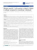
Figure receptor function of HuPARs and their rodent homologues
PERV-A 1
PERV-A receptor function of HuPARs and their rodent homologues. A. The different cell lines were transduced with
the same amount of retroviral vector encoding the HA-tagged receptor genes. Transduced cells were then infected with
EGFP(PERV-A). 48 hours post-infection cells were analysed by flow cytometry and the efficiency of infection was determined
as percentage of EGFP positive cells. The histograms represent the average ± SEM from three independent experiments. The
arrows indicate an infection below detectable levels. B. NRK, HSN and XC rat cells were transduced with a retroviral vector
encoding the ratPAR gene. Two independent transductions were performed on NRK and HSN cells. The RNA from transduced and untransduced rat cells were extracted. The amount of ratPAR was determined by real time RT-PCR and normalised
to equalised copies of 18S rRNA. The results were correlated with the efficiency of EGFP(PERV-A) infection. All the samples
were run in duplicate and the experiment repeated at least two times.
Page 4 of 13
(page number not for citation purposes)
Retrovirology 2007, 4:93
/>
(Fig 3A). As similar models were also obtained for huPAR1 and muPAR by transmembrane prediction, various PAR
molecules are likely to have the same topology and have
a.a.109 in the second ECL.
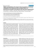
Figure 2
infection
Identification of critical amino acid residues for PERV-A
Identification of critical amino acid residues for
PERV-A infection. HA-tagged chimeric receptors (H2M af) between huPAR-2 (white bars) and muPAR (black bars) as
well as huPAR-1 (grey bar) and the mutant H1M g were
introduced into QT6 cells by MLV-based retroviral vectors.
50–70% of the QT6 cell population showed PAR expression
as confirmed by anti-HA staining. These cultures were
infected with EGFP(PERV-A). Cells were harvested 48 hours
later and PERV-A infection was measured by flow cytometry
as percentage of EGFP-positive cells. Arrows indicate infection below detectable levels. Results are expressed as average ± SEM from three independent experiments.
active proliferating cells. These results support the transmembrane prediction with 11 TM topology.
Further evidence to support the predicted topology was
obtained utilising a glycosylation study. Using NetNGlyc
1.0 software [33], one N-glycosylation site for huPAR-2 at
a.a. position 178 is postulated. This prediction agrees with
the proposed topology because Asp178 is located in the
third ECL (Fig 3A). To test this hypothesis, huPAR-2 harbouring the single a.a. mutation, Asp178 to Ala (N178A),
was generated. The construct expressed in QT6 cells supported PERV-A infection (data not shown). Cell lysates of
293T cells transfected with HA-tagged huPAR-2 wild type
or the mutant N178A were treated with PNGase F, an
enzyme which removes N-linked oligosaccharide chains.
The western blot analysis showed a shift of the signal in
the wild type huPAR-2 treated with PNGase F from 55
kDa to 48 kDa (Fig 3D). This shift indicated that huPAR2 carries N-linked oligosaccharide chains. In contrast, the
N178A mutant produced 48 kDa bands in both samples
with and without PNGaseF treatment (Fig 3D), suggesting
that Asp178 is indeed an N-glycosylation site and therefore located in an ECL. Together, these results strongly
support the predicted model for the huPAR-2 molecule
Pro109 abrogates binding of PERV-A Env to PAR
To further investigate the mechanism responsible for
abrogation of PERV-A infection by Pro109 in muPAR, we
analysed the binding properties of the receptors. Parental
and receptor-transduced QT6 cells, expressing similar levels of HA-tagged receptors (see Additional file 1 Fig. S1),
were incubated with soluble, c-myc-tagged PERV-A envelope protein (mycPERVEnv) and immunostained with an
anti-c-myc antibody. No difference was seen between
parental QT6 cells incubated in the presence or absence of
mycPERVEnv (Fig 4A, mock). However, expression of
huPAR-1, huPAR-2 and ratPAR, but not muPAR, produced a shift towards higher fluorescence intensity in the
FACS histogram profiles. These results indicate that
huPAR-1, huPAR-2 and ratPAR, but not muPAR, can bind
soluble PERV-A Env (Fig 4A). To verify whether Pro109
was responsible for the absence of binding of PERV-A Env
to muPAR, chimeric receptors huPAR-2 with murine
Pro109 (H2M d) and muPAR with human Leu109 (H2M
c) were tested in the binding assay. Pro109 completely
abrogated the binding of huPAR-2 with soluble PERV-A
Env, suggesting that the structure of the second ECL containing Pro109 does not support the interaction between
PERV-A Env and the receptor. However the exchange of
Pro109 to Leu in muPAR did not rescue the binding of
mycPERVEnv (Fig 4B), even if it supported PERV-A infection (Fig 2). This result suggests that other regions in the
muPAR molecule, probably involved in the kinetics or
affinity of the receptor-Env interaction, are important to
achieve a binding efficiency which can be detected in this
setting. Alternatively, the discrepancy between binding
and function of the mutant receptor H2M c may be caused
by a better binding to the trimeric Env form present on
viral particles than soluble surface unit monomers.
Unique structure of PAR ECL2 in murine species
PAR a.a. sequences of various species origin were compared. The alignment of ECL2 and the adjacent regions is
shown (Fig 5). Two additional murine species, Mus spretus
and Mus musculus castaneus, were also sequenced and in
this region, displayed the same a.a. sequence as Mus musculus and Mus dunni. Pro109 as well as the adjacent
Lys108 and Tyr110 are only found in muPAR. In contrast,
ratPAR from all 3 cell lines used in this study has the same
3 a.a. triplet, QLH, as huPAR-1 and -2 in the corresponding positions. This confirmed that the receptor function
defect is unique in muPAR and ratPAR does not share the
same defective mutation as muPAR. Gln108 and His110
are remarkably conserved among non-murine species
including rat, a mouse relative within the rodent lineage.
Page 5 of 13
(page number not for citation purposes)
Retrovirology 2007, 4:93
/>
A
B
Surface staining
Intracellular staining
N-glycosylation site
C-HA HuPAR-2
QLH
N-HA HuPAR-2
Anti-CD71
C
Surface staining
73.6%
D
Intracellular staining
59.6%
HuPAR-2
C-HA
Wild type C-HA wild type C-HA N178A
PGNase F
HuPAR-2
60
Anti-HA
56.9%
N-HA
HuPAR-2
84.0%
CELL COUNTS
8.7%
50
40
50
Anti-actin
67.1%
Anti-CD71
FLUORESCENCE INTENSITY
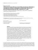
Figure topology
HuPAR 3
HuPAR topology. A. HuPAR-2 topology model derived by hydrophobicity algorithms and the experiments described in
panel B-D is depicted. B-C. HuPAR-2 bearing an N- or C-terminal HA-tag was transiently transfected into 293T cells. After 48
hours cells were treated with saponin (intracellular staining) or without (surface staining). Following immunostaining using an
anti-HA antibody and a FITC-conjugated secondary antibody, the samples were visualised either by confocal microscopy (B) or
processed by flow cytometry (C). Immunostaining of the cells with anti-human CD71 was used as cell surface protein control.
The cells nuclei were counter stained with propidium iodide. D. Cell lysates from 293T transiently transfected with an empty
pcDNA3 (-), HuPAR-2 (wild type), HA-tagged HuPAR-2 wild type (C-HA wild type) or glycosylation mutant (C-HA N178A)
were either treated (+) or untreated (-) with an enzyme removing N-linked oligosaccharide chains (PNGase F) and analysed by
western blotting.
Page 6 of 13
(page number not for citation purposes)
Retrovirology 2007, 4:93
/>
A
B
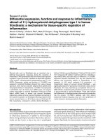
Figure 4
Envelope binding properties of PAR
Envelope binding properties of PAR. QT6 cells stably expressing HA-tagged receptors huPAR-1, huPAR-2, muPAR and
ratPAR (A) or the chimeric receptors huPAR-2 with L109P mutation and muPAR with P109L mutation (B) were incubated
with 1 ml of medium (grey filled) or with the supernatant of 293T containing N-terminal c-myc tagged soluble PERV-A 14/220
SU ENV (bold line). The cells were then immunostained with an anti- human c-myc antibody and a PE conjugated anti-mouse
IgG secondary antibody. The histograms show a representative result of at least three independent experiments.
Page 7 of 13
(page number not for citation purposes)
Retrovirology 2007, 4:93
/>
observed (data not shown). The mechanism which determines the threshold level of ratPAR expression for PERVA infection is currently unclear. However, our results suggest that other component(s) on the cell surface may be
responsible for a successful interaction between virus and
receptor, as has been previously proposed for other gammaretroviruses [34-36].
Figure 5
PAR amino acid sequences alignment
PAR amino acid sequences alignment. Amino acid
sequences retrieved from Entrez protein database [47] or
obtained by direct sequencing of PCR products on genomic
DNA were aligned using Clustal W software [48]. Mus spretus and Mus m. castaneus have the same identical a.a.
sequence in the ECL2 (boxed) of other murine species. RatPAR from different rat cell lines have identical a.a. sequences.
Considering a.a. 109 proved to be critical for muPAR
interaction with PERV-A, it would be expected to be well
conserved among the susceptible species. However, L109
is replaced in two PAR molecules that are functional as
PERV-A receptor: Leu-to-Val in porcine PAR [14] and Leuto-Ser in rhesus PAR-1 (GM and YT, unpublished data).
This observation suggests that the a.a. change to Pro109
may cause a substantial conformational change in the
ECL2, which results in the inactivation of PERV-A receptor
function.
Discussion
The mechanism of resistance to PERV-A cell entry is different between mouse and rat cells: the murine homologue
of PAR (muPAR) is defective in PERV-A receptor function,
whereas the rat cell encodes a fully functional PAR protein. RatPAR can rescue PERV-A infection in non-permissive cell lines, including the resistant rat cell lines from
which it has been cloned. The PERV-A infection of rat cells
upon overexpression of ratPAR is reminiscent of results
from a previous study which show that overexpression of
amphotropic MLV and GALV receptors from Chinese
hamster cells and FeLV-C receptor from MDTF cells, supports viral infection in the cell lines of their origin [26].
This type of resistance to viral infection can be explained
by subthreshold levels of receptor expression or stoichiometrically limited masking or interference mechanisms
[23-25]. We therefore explored the possibility that a Nglycosylation could mask the receptor and that an inhibitory factor is secreted from rat cells. However, no effect on
PERV-A infection by these possible mechanisms was
The defect in muPAR as a PERV-A receptor is due to the
presence of Pro at position 109. Our topology study indicated that a.a.. 109 is most likely to be located in the second extracellular loop (ECL2) and potentially accessible
for the direct binding by PERV-A Env. Our binding assay
consistently detected soluble PERV-A Env binding to cells
expressing 'functional' huPARs and ratPAR, but not
muPAR. Furthermore, a Leu-to-Pro mutation at a.a. 109 in
huPAR-2 abolished Env binding as well as PERV-A infection, further highlighting the important role of this a.a..
These results identified the ECL2 as the likely target for
PERV-A Env binding, leading to PERV-A entry. This,
together with recent studies on the determinants in PERV
Env for binding and entry [37,38] contribute to better
understanding of PERV-receptor interactions. These
advances may help develop reagents that block PERV
entry, such as neutralizing antibodies [39] and peptides
mimicking the receptor.
The amino acid sequence positions 108–110 of muPAR
ECL2, KPY instead of QLH, is intriguingly unique in
murine species. Since rats share QLH at the corresponding
positions with diverse non-murine species including primates, horse and dog, it is likely that murine species
acquired 3 mutations after separating from rats. Although
we cannot exclude the possibility that these changes are a
stochastic evolutionary outcome, it is more likely that certain selective pressure, at least partly, caused these
changes. It is tempting to speculate that severe epidemics
of PERV-A like viruses which target the ECL2-QLH structure may have selected 'PERV-A-resistant' murine species
with KPY. Our result showing that Leu-to-Pro 109 change
alone blocks PERV-A infection raises the question why
changes are also required at positions 108 and 110. It is
possible that all three a.a. changes were required to escape
viral attacks in the past. Alternatively the acquisition of
Lys108 and Tyr110 by murine species might be required
to maintain yet unknown physiological function of PAR
while escaping deadly viruses. To further gain insight into
this hypothesis, as well as to study involvement of PAR in
the possible PERV-A pathology, identification of physiological roles of PAR is warranted.
Conclusion
Different bases for PERV-A resistance between mice and
rats are shown. Expression of endogenous ratPAR in rat
cells appear to be under a threshold level to support
Page 8 of 13
(page number not for citation purposes)
Retrovirology 2007, 4:93
PERV-A infection. In mice, a single a.a. mutation in
muPAR in the ECL2 is responsible for the resistance to
PERV-A infection. ECL2 in muPAR has a unique sequence
with three a.a. changes compared with a wide range of
species. Possible selective pressure may have caused this
ECL2 diversion in mice.
/>
Table 2: Primers and probes used in this study
Name
Sequence (5' → 3')
G1
AGC TGG AGA TCTa GAG CAG AAA CTC ATC TCT
GAA GAG GAT CTGg CTT GTG ACC AGT CCG AAC
TCC CAT AAA CCC TTA TCT CTC ACC
ATG TTC TTA GCT AGCb CTA TTC ATC AAG GAT TGC
TTT TTC CGG
GAT TGA T GA ATT Cd AC CAC CAT GGiC AGC ACC
CAC G
GAT CTT GCG GCC GCeT CA A GCG TAT TCT GGA
ACA TCG TAT GGG TAh A AGC TTcG GGG CCA CAG
GGG TCT ACA CAG TCC TTT CTG CTT TG
GAA GGT AAG CTTc GGA GTC ACA GGG GTC
GAT TGA T GA ATT Cd AC CAC CAT GGiC AGC ACC
TCC G
GAA GGT AAG CTTc GAG GCC ACA CTG GTC
CGT GGC ATC TAG ATT AAG CTTc GGG GCC ACA
GGG GTC
TTG CAC TAG GGC TAG CAC ACA GG
CCT GTG TGC TAG CCC TAG TGC AA
TAG GAA GGC CAC AGA GTA CGG CTT CCC TGC
CAC TGG GGC
GCC CCA GTG GCA GGG AAG CCG TAC TCT GTG
GCC TTC CTA
TAG GAA GGC CAC AGA GTG GGG CTG CCC TGC
CAC TGG GGC
GCC CCA GTG GCA GGG CAG CCC CAC TCT GTG
GCC TTC CTA
TAG GAA GGC CAC TGA GTG GAG CTG TCC TGC
CAC TGG GGC
GCC CCA GTG GCA GGA CAG CTC CAC TCA GTG
GCC TTC CTA
TAG GAA GGC CAC CGA GTA GAG CTT TCC TGC
CAC TGG GGC
GCC CCA GTG GCA GGA AAG CTC TAC TCG GTG
GCC TTC CTA
AGA GGT GCC AGC GGT GGG CGC T
AGC GCC CAC CGC TGG CAC CTC T
TTA CAA GAA TTCd GCC ACC ATG GiTT TAC CCA
TAC GAT GTT CCA GAT TAC GCTh GCA GCA CCC
ACG CTG GGC CGT CTG GTG CTG A
GAT CTT AA G CGG CCG CeTC AGG GGC CAC AGG
GGT CTA
GCC AGA GGA GGT ACCf GCC ACC ATG GAT GCA
ATG AAG AGA G
GGG TAA GAT CTaG GCT CCT CTT CTG AAT CGG
GCA TGG ATT TCC TGG CTG GGC
GAT TGA T GA ATT Cd AC CAC CATG GiCA GCA CC
TGA CTG A GC GGC CGCe TCA AGG GCC ACA CTG
ATC CAC
GCA GGT AAG CTTc AGG GCC ACA CTG ATC
CTC ACT CCT TTA CAC TAC AC
CAA CCC ATT GGA TGA AGA TG
TCA AGG TGT CTC CCA TCA ATT TC
CGT CAA CAC CCA AAA GAA TGT G
TCG AGG CCC TGT AAT TGG AA
CCC TCC AAT GGA TCC TCG TT
TAC CTG GTT GAT CCT GCC AGT A
TTA CGA CTT TTA CTT CCT CTA GAT AG
CTG AGC GTT TCT CTG
AGT CCA CTT TAA ATC CTT
G2
Methods
Cell lines
Human embryonic kidney 293T cells were maintained in
Dulbecco's modified Eagle Medium (DMEM, Gibco) supplemented with 15% fetal bovine serum (FBS, BioSera).
Quail QT6 cell [ECACC: 93120831], murine MDTF (Mus
dunni tail fibroblast), rat NRK [ECACC: 86032002] HSN
cells [40] and XC [ECACC: 88120601] were grown in
DMEM supplemented with 10% FBS.
Plasmids and construction of chimeric receptors
The following plasmids have been previously described:
murine leukaemia virus (MLV)-based retroviral vectors
pCNCG carrying the eGFP gene [41], pCFCR with unique
EcoRI site [42], MLV gagpol expressor plasmid CMV [43],
G protein of vescicular stomatitis virus (VSV-G) expressor
plasmid pMDG [44]. Replication competent PERV-A 14/
220 plasmid has been previously described [13]. Oligonucleotide primers and probes are listed in Table 2. Soluble
surface unit of PERV-A 14/220 Envelope (PERVEnv) was
cloned into pCAGGS [45] using the restriction sites BglII
and NheI and a c-myc tag has been introduced at N-terminus of PERVEnv using primers G1 and G2 (mycPERVEnv).
The sequence of human tissue plasminogen activator
leader has been introduced in frame upstream to the cmyc tag by PCR of the construct PEE14 [46] using primers
G23 and G24 bearing the enzymatic restriction sites KpnI
and BglII, respectively. HuPAR-2 was tagged at the N-terminus with influenza virus HA-tag by PCR of the construct pcDNA3/huPAR-2 [14] using KOD HiFi polymerase
(Novagen) and the primers G21 and G22. C-terminal HAtagged HuPAR-2 was obtained by PCR using the primers
G3 and G4 and introduced into pcDNA3 using EcoRI and
NotI restriction sites. These primers introduced the Kozak
sequence at the ATG of the receptor and the HA-tag in the
C-terminus downstream of a HindIII restriction site.
Using EcoRI and NotI restriction sites, the HA-tagged
receptor was introduced again into pcDNA3. In this way
the resulting plasmid pcDNA3/huPAR-2HA contains two
HindIII restriction sites, one in pcDNA3 and the other
introduced in frame upstream of the HA-tag using the 3'
primer. HA-tagged huPAR-1 and muPAR genes were
obtained by PCR of constructs pcDNA3/huPAR-1 and
pcDNA3/muPAR [14] with the primer pairs G3;G5
(huPAR-1) and G6;G7 (muPAR). Using the HindIII
restriction site present in the reverse primers, huPAR-1
and muPAR were cloned into pcDNA3/huPAR-2HA
upstream of the HA-tag. All the HA-tagged receptors were
G3
G4
G5
G6
G7
G8
G9
G10
G11
G12
G13
G14
G15
G16
G17
G18
G19
G20
G21
G22
G23
G24
M1
M2
M3
M4
M5
Q1
Q2
Q3
Q4
ZF
ZR
PR
P18
a BglII, b NheI, c HindIII; d EcoRI; e NotI; f KpnI, g Human
c-myc tag; h
influenza virus HA tag, i Kozak sequence.
Page 9 of 13
(page number not for citation purposes)
Retrovirology 2007, 4:93
also subcloned into the retroviral vector pCFCR using
EcoRI and NotI restriction sites.
An NheI restriction site was introduced into huPAR-2 at
the site corresponding to that in muPAR [Genbank:
AK008081, nucleotide 805] by two-step PCR using primers G3;G9 and G10;G8, then G3;G8, where primers G9
and G10 contain the nucleotide change. Primer G8
includes a HindIII restriction site which allows the cloning of the mutant receptor into pcDNA3/huPAR-2HA.
Chimeric receptors H2M a and f were obtained by mixand-match cloning between huPAR-2 and muPAR using
the restriction sites EcoRI and NheI. The other huPAR-2derived chimeric receptors were produced in a similar way
using mutagenesis primers G11;G12 (H2M e) and
G13;G14 (H2M d) in association with the primers G3;G8.
Similarly, muPAR-derived chimeric receptors were produced using primers G15;G16 (H2M b) and G17;G18
(H2M c) in combination with primers G6;G7.
The N178A mutation in huPAR-2 was introduced by PCRmutagenesis using the primers G19;G20 in association
with the primers G3;G8 and the mutant huPAR-2 was
cloned into a partially digested pcDNA3/huPAR2HA
using EcoRI and HindIII restriction sites.
The mutant huPAR-1 carrying a proline at position 109
(H1M g) was generated by PCR-mutagenesis using the
primers G13;G14 in combination with the primers
G3;G5.
All the PCRs described above were performed using KOD
HiFi polymerase in accordance with manufacturer's
instructions. Chimeric receptors were verified by sequencing based on a modification of the Sanger method and
analysed using the CEQ 8000 DNA Sequencer (Beckman
Coulter).
Cloning of rat PERV-A receptor
Total RNA from NRK cells was extracted using the RNeasy
kit (Qiagen) and incubated with 5 U of RNase-free DNase
(Promega) for 30 min at 37°C. First strand cDNA was produced by incubation of 2 μg of DNase-treated RNA with
200 U of Moloney MLV Reverse Transcriptase (Promega),
1 μg of random primers (Promega), 20 U of RNasin Ribonuclease Inhibitor (Promega), 1 mM dNTPs (Qiagen) in a
final volume of 20 μl for 10 min at 25°C, 1 hr at 42°C and
an inactivation step of 10 min at 70°C. The ratPAR coding
sequence was then amplified using HotStart polymerase
(Qiagen) and primers M1;M2 with PCR conditions: 95°C
30 sec, 52°C 30 sec, 72°C 90 sec. Primers M1 and M2
were designed to anneal to the rat homologue of huPAR1 [Genbank: XM_343272]. The M1 primer introduced the
Kozak sequence in front of the ATG of the receptor. The
PCR product was cloned into pcDNA3 using EcoRI and
/>
NotI restriction sites present in the primers. HA-tagged Cterminal ratPAR was obtained by PCR using KOD HiFi
polymerase and the primers M1;M3 which contain the
HindIII restriction site, and introduced into pcDNA/
huPAR-2HA. This product was then subcloned into
pCFCR.
Transfection, virus production and infection
Transfection of huPAR-2 (N- or C- terminal HA-tagged or
N178A mutant) was performed on confluent 293T in a 6well plate using 4 μl of FuGene-6 reagent (Roche) and 1
μg of plasmid.
Viral particles carrying the receptor genes were produced
by co-transfection of 3.5 μg of three plasmids, CMVi for
MLV Gag-Pol, MDG for VSV-G and MLV vector genome
pCFCR carrying the receptor gene (ratio 1:1:1.5) on confluent 293T cells in 100 mm-dish using 18 μl of FuGene6 reagent (Roche). Cells were washed 24 hours later and
at 48 and 72 hours the supernatant containing viral particles were harvested and passed through a 0.45 μm filter
(Millipore). A replication-competent PERV-A 14/220
expressing the reporter gene EGFP, EGFP(PERV-A), was
produced as follows. A similar three plasmid transfection
on 293T cells was performed using pCNCG instead of
pCFCR in order to produce MLV/EGFP particles. The
virus-containing supernatant was used to transduce 293T
cells. The stable EGFP-expressing 293T cells were then
transfected using FuGene-6 with the replication competent PERV-A 14/220 plasmid. The titer of EGFP(PERV-A)
viral particles was assessed by infection of 1 × 10⁵ 293T
seeded in a 6-well plate using serial dilutions of the supernatant. After two months the titer was stable at 2 × 10⁵
EGFP 293T transducing units/mL.
The receptor transduction and EGFP(PERV-A) infection
were performed as follows: 5 × 10⁴ target cells were seeded
in a 12-well plate and the day after, 500 μl of virus-containing supernatant was added. Receptor or EGFP expression was verified 48 hours post transduction/infection by
flow cytometry analysis.
Flow Cytometry analysis
Cells transfected or transduced with HA-tagged PAR were
detached with PBS-5 mM EDTA and blocked by incubation for 30 min in PBS-10% FBS on ice. The cells were
washed twice in PBS, resuspended in PBS-2% FBS containing 1:100 dilution of mouse monoclonal antibody
HA.11 (Covance) or 1 μg of mouse monoclonal antihuman CD71 antibody (Santa Cruz) and incubated for 1
hour at 4°C. After two washes with PBS-2% FBS, the cells
were incubated with 1:200 dilution of the secondary antibody anti- mouse IgG fluorescein isothiocyanate (FITC)conjugate (Jackson Immunoresearch) in PBS-2% FBS for
45 min at 4°C. Cells were washed three times and resus-
Page 10 of 13
(page number not for citation purposes)
Retrovirology 2007, 4:93
pended in PBS. To assess EGFP(PERV-A) infection efficiency, 48 hours post-infection cells were harvested and
resuspended in PBS. All the samples were processed on a
FACScan cytometer (Becton-Dickinson) and analysed
using CellQuest software.
Immunofluorescence microscopy
One day post transfection, 293T expressing HA-tagged
huPAR-2 were seeded on cover slides and incubated for
further 48 hours. The cells were fixed by incubation with
4% paraformaldehyde (Sigma) in PBS for 20 min at room
temperature. The permeabilized samples were obtained
by incubation with PBS-0.1% saponin (Fluka) for 10 min
at room temperature. For the permeabilized samples,
0.1% saponin was added during all antibody incubations.
All slides were washed in PBS and placed on a 30 μl drop
of PBS-1% FBS containing antibody HA.11 (dilution
1:100) or anti-human CD71 antibody (dilution 1:50) for
1 hr at 37°C in a humidified chamber. Cells were then
washed three times with PBS and the slides placed in a 30
μl drop of PBS-1% FBS containing the secondary antibody
FITC-conjugated anti-mouse IgG (dilution 1:100) for 45
min at 37°C in a humidified chamber. After three washes,
the cover slides were mounted in Vecta Shield mounting
medium containing propidium iodide (Vector Laboratories). Images were collected using a DM IRE2 confocal
microscope (Leica).
Glycosylation assay
293T cells transfected with wild type huPAR-2 or N178A
mutant were harvested, washed and incubated in RIPA
lysis buffer (50 mM TRIS-HCl pH 7.5, 150 mM NaCl, 1%
Igepal CA-630, 0.5% deoxycholic acid, 0.1% SDS, 1% Triton ×-100) in the presence of protease inhibitors (Complete mini, Roche) for 30 min on ice. The cell lysates were
then digested with 1500 U of N-glycosidase F enzyme
(PGNase F, New England Biolabs) at 37°C for 2 hrs. Proteins from digested and undigested samples were separated by SDS polyacrylamide (BioRad) electrophoresis
(SDS-PAGE) and transferred to PVDF membrane (Amersham Biosciences) by using a semi-dry blotting system
(Amersham Biosciences). The membrane was blocked in
PBS-5% non-fat skimmed milk powder (Oxoid) and then
probed for 1 hr at room temperature with the HA.11 monoclonal antibody diluted 1:1000 in PBS-2% milk, followed by incubation with an anti-mouse IgG conjugated
with horseradish peroxidase (Dako, dilution 1:10,000 in
PBS-2% milk) for 30 min at room temperature. Signals
were detected by incubation with ECL chemiluminescence reagent (Amersham Biosciences) and exposure to xray film (Hyperfilm, Amersham Biosciences). To control
for protein loading, the same blots were incubated with
mouse anti-human β-actin (Sigma, 1:1000 in PBS-2%
milk).
/>
Soluble Envelope Binding Assay
C-myc tagged PERV-A 14/220 Env was produced by transient transfection of 293T in a 100 mm dish using 18 μl of
Fugene-6 (Roche) and 3 μg of myc14/220ENV plasmid.
One day post-transfection, medium was replaced with
DMEM supplemented with 10% FBS. The supernatant
was then harvested at 48 and 72 hours and passed
through a 0.45 μm filter. Target cells for binding assay
were detached using PBS-5 mM EDTA, washed twice and
1 × 10⁶ cells for each sample were resuspended in 1 mL of
293T supernatant containing soluble 14/220ENV. After 1
hr incubation at 37°C, the cells were washed twice with
PBS-2%FBS and incubated with 100 μl of anti c-myc antibody 9E10 (Santa Cruz Biotechnology, Inc) diluted 1:100
in PBS-2%FBS for 1 hr on ice. The cells were washed twice
and incubated for 30 min on ice with a 1:200 dilution of
phycoerythrin (PE)-conjugated secondary antibody antimouse IgG (Jackson Immunoresearch) in PBS-2%FBS.
After two washes with PBS-2%FBS, the cells were resuspended in PBS and analysed by flow cytometry (FACScan,
Becton Dickinson).
Quantitative RT-PCR
Total RNA from cells was extracted using an RNeasy kit
(Qiagen) and cleaned using 5 U of RNase-free DNase
(Promega) according to the manufacturer's instructions.
The RNA was quantified and 2 μg of total RNA was subjected to reverse transcription (RT) as described for the ratPAR cloning. 2.5 μl of the RT reaction were used in the
Real-Time PCR using Quantitect Probe PCR Mix (Qiagen)
0.4 μM of each primers (Q1;Q2), 0.2 μM of Fam-Tamra
labelled probes (PR) (Sigma). The amount of RNA
between each samples was normalized using the housekeeping gene 18S rRNA, primers Q3;Q4 and probe P18.
The assay was performed in duplicate using the ABI
PRISM 7000. Thermocycling conditions were: 50°C, 2
min; 95°C, 15 min; 40 cycles of 95°C, 15 sec and 60°C,
1 min. The number of copies of each products were calculated from standard curves obtained by serial dilution of
the plasmid pCFCR/ratPAR. Part of the 18S mRNA gene
were amplified using primers ZF;ZR from human total
RNA and cloned into TOPO BLUNT 2 (Invitrogen) following the manufacturer's instruction.
Genomic PAR sequence analysis
Genomic DNA was extracted from murine MDTF and rat
XC, HSN cell cultures using DNeasy Tissue kit (Qiagen).
Genomic DNA from Mus m. castaneus and Mus spretus is a
kind gift from Dr. Jiri Hejnar (Academy of Sciences of the
Czech Republic, Prague, Czech Republic). Genomic
sequences of rodent PAR were amplified by PCR using
high fidelity DNA polymerase KOD HiFi according to the
manufacturer's instructions and the primers M4;M5
(muPAR) and M1;M2 (ratPAR). The PCR products were
directly sequenced.
Page 11 of 13
(page number not for citation purposes)
Retrovirology 2007, 4:93
Amino acid sequence accession number
The amino acid sequences used in this study are: huPAR1 [RefSeq: NP_078807] and huPAR-2 [RefSeq:
NP_060456],
chimpanzee
PAR-1
[RefSeq:
XP_001156784] and PAR-2 [RefSeq: XP_001164395],
Rhesus macaque PAR-1 [RefSeq: XP_001091189] and
PAR-2 [RefSeq: XP_001099620], baboon PAR-2 [Swissprot: Q863Y8], dog PAR [RefSeq: XP_532355], horse PAR
[RefSeq: XP_001505049], pig PAR [Swissprot: Q863Y7],
cow PAR [RefSeq: NP_001069369], muPAR [RefSeq:
NP_083919] and ratPAR [RefSeq: NP_001103140].
/>
3.
4.
5.
6.
7.
Competing interests
The author(s) declare that they have no competing interests.
8.
9.
Authors' contributions
YT conceived the study. GM and YT designed the experiments and wrote the manuscript. GM carried out the
experiments. MM contributed to cloning and initial characterization of ratPAR. All the authors read and approved
the final manuscript.
Additional material
10.
11.
12.
13.
Additional File 1
PERV-A receptors cell surface expression. Expression of C-terminal HAtagged PAR constructs in QT6 cells was demonstrated by flow cytometry
analysis following surface immunostaining with an anti-HA antibody.
Click here for file
[ />
14.
15.
16.
Additional File 2
RatPAR function as PERV-A receptor depends on its expression on cell surface. PERV-A infection was measured in quail QT6 cell clones expressing
different levels of ratPAR.
Click here for file
[ />
17.
18.
19.
Acknowledgements
20.
This work was supported by UK Medical Research Council and European
Commission funded project LSHB-CT-2006-037377.
We thank Benjamin LJ Webb for critical reading of the manuscript.
References
1.
2.
Phelps CJ, Koike C, Vaught TD, Boone J, Wells KD, Chen SH, Ball S,
Specht SM, Polejaeva IA, Monahan JA, Jobst PM, Sharma SB, Lamborn
AE, Garst AS, Moore M, Demetris AJ, Rudert WA, Bottino R, Bertera
S, Trucco M, Starzl TE, Dai Y, Ayares DL: Production of alpha 1,3galactosyltransferase-deficient
pigs.
Science
2003,
299(5605):411-414.
Kolber-Simonds D, Lai L, Watt SR, Denaro M, Arn S, Augenstein ML,
Betthauser J, Carter DB, Greenstein JL, Hao Y, Im GS, Liu Z, Mell GD,
Murphy CN, Park KW, Rieke A, Ryan DJ, Sachs DH, Forsberg EJ,
Prather RS, Hawley RJ: Production of alpha-1,3-galactosyltrans-
21.
22.
23.
24.
ferase null pigs by means of nuclear transfer with fibroblasts
bearing loss of heterozygosity mutations. Proc Natl Acad Sci U
S A 2004, 101(19):7335-7340.
Takeuchi Y, Magre S, Patience C: The potential hazards of
xenotransplantation: an overview.
Rev Sci Tech 2005,
24(1):323-334.
Fishman JA, Patience C: Xenotransplantation: infectious risk
revisited. Am J Transplant 2004, 4(9):1383-1390.
Martin U, Winkler ME, Id M, Radeke H, Arseniev L, Takeuchi Y, Simon
AR, Patience C, Haverich A, Steinhoff G: Productive infection of
primary human endothelial cells by pig endogenous retrovirus (PERV). Xenotransplantation 2000, 7(2):138-142.
Patience C, Takeuchi Y, Weiss RA: Infection of human cells by an
endogenous retrovirus of pigs. Nat Med 1997, 3(3):282-286.
Wilson CA, Wong S, Muller J, Davidson CE, Rose TM, Burd P: Type
C retrovirus released from porcine primary peripheral blood
mononuclear cells infects human cells.
J Virol 1998,
72(4):3082-3087.
Tarlinton RE, Meers J, Young PR: Retroviral invasion of the koala
genome. Nature 2006, 442(7098):79-81.
Takeuchi Y, Patience C, Magre S, Weiss RA, Banerjee PT, Le Tissier
P, Stoye JP: Host range and interference studies of three
classes of pig endogenous retrovirus.
J Virol 1998,
72(12):9986-9991.
Wilson CA, Wong S, VanBrocklin M, Federspiel MJ: Extended analysis of the in vitro tropism of porcine endogenous retrovirus.
J Virol 2000, 74(1):49-56.
Oldmixon BA, Wood JC, Ericsson TA, Wilson CA, White-Scharf ME,
Andersson G, Greenstein JL, Schuurman HJ, Patience C: Porcine
endogenous retrovirus transmission characteristics of an
inbred herd of miniature swine. J Virol 2002, 76(6):3045-3048.
Harrison I, Takeuchi Y, Bartosch B, Stoye JP: Determinants of high
titer in recombinant porcine endogenous retroviruses. J Virol
2004, 78(24):13871-13879.
Bartosch B, Stefanidis D, Myers R, Weiss R, Patience C, Takeuchi Y:
Evidence and consequence of porcine endogenous retrovirus
recombination. J Virol 2004, 78(24):13880-13890.
Ericsson TA, Takeuchi Y, Templin C, Quinn G, Farhadian SF, Wood
JC, Oldmixon BA, Suling KM, Ishii JK, Kitagawa Y, Miyazawa T, Salomon DR, Weiss RA, Patience C: Identification of receptors for
pig endogenous retrovirus. Proc Natl Acad Sci U S A 2003,
100(11):6759-6764.
EST Profile Viewer [ />Tailor CS, Nouri A, Zhao Y, Takeuchi Y, Kabat D: A sodiumdependent neutral-amino-acid transporter mediates infections of feline and baboon endogenous retroviruses and simian type D retroviruses. J Virol 1999, 73(5):4470-4474.
Rasko JE, Battini JL, Gottschalk RJ, Mazo I, Miller AD: The RD114/
simian type D retrovirus receptor is a neutral amino acid
transporter. Proc Natl Acad Sci U S A 1999, 96(5):2129-2134.
Olah Z, Lehel C, Anderson WB, Eiden MV, Wilson CA: The cellular
receptor for gibbon ape leukemia virus is a novel high affinity
sodium-dependent phosphate transporter. J Biol Chem 1994,
269(41):25426-25431.
Mendoza R, Anderson MM, Overbaugh J: A putative thiamine
transport protein is a receptor for feline leukemia virus subgroup A. J Virol 2006, 80(7):3378-3385.
Kavanaugh MP, Miller DG, Zhang W, Law W, Kozak SL, Kabat D,
Miller AD: Cell-surface receptors for gibbon ape leukemia
virus and amphotropic murine retrovirus are inducible
sodium-dependent phosphate symporters. Proc Natl Acad Sci U
S A 1994, 91(15):7071-7075.
Tailor CS, Lavillette D, Marin M, Kabat D: Cell surface receptors
for gammaretroviruses. Curr Top Microbiol Immunol 2003,
281:29-106.
Marin M, Tailor CS, Nouri A, Kabat D: Sodium-dependent neutral amino acid transporter type 1 is an auxiliary receptor for
baboon endogenous retrovirus. J Virol 2000, 74(17):8085-8093.
Eiden MV, Farrell K, Wilson CA: Glycosylation-dependent inactivation of the ecotropic murine leukemia virus receptor. J
Virol 1994, 68(2):626-631.
Miller DG, Miller AD: Inhibitors of retrovirus infection are
secreted by several hamster cell lines and are also present in
hamster sera. J Virol 1993, 67(9):5346-5352.
Page 12 of 13
(page number not for citation purposes)
Retrovirology 2007, 4:93
25.
26.
27.
28.
29.
30.
31.
32.
33.
34.
35.
36.
37.
38.
39.
40.
41.
42.
43.
44.
45.
Miller DG, Miller AD: Tunicamycin treatment of CHO cells
abrogates multiple blocks to retrovirus infection, one of
which is due to a secreted inhibitor. J Virol 1992, 66(1):78-84.
Tailor CS, Nouri A, Kabat D: Cellular and species resistance to
murine amphotropic, gibbon ape, and feline subgroup C
leukemia viruses is strongly influenced by receptor expression levels and by receptor masking mechanisms. J Virol 2000,
74(20):9797-9801.
Johann SV, van Zeijl M, Cekleniak J, O'Hara B: Definition of a
domain of GLVR1 which is necessary for infection by gibbon
ape leukemia virus and which is highly polymorphic between
species. J Virol 1993, 67(11):6733-6736.
Eiden MV, Farrell KB, Wilson CA: Substitution of a single amino
acid residue is sufficient to allow the human amphotropic
murine leukemia virus receptor to also function as a gibbon
ape leukemia virus receptor. J Virol 1996, 70(2):1080-1085.
Marin M, Tailor CS, Nouri A, Kozak SL, Kabat D: Polymorphisms
of the cell surface receptor control mouse susceptibilities to
xenotropic and polytropic leukemia viruses. J Virol 1999,
73(11):9362-9368.
Yoshimoto T, Yoshimoto E, Meruelo D: Identification of amino
acid residues critical for infection with ecotropic murine
leukemia retrovirus. J Virol 1993, 67(3):1310-1314.
Tailor CS, Takeuchi Y, O'Hara B, Johann SV, Weiss RA, Collins MK:
Mutation of amino acids within the gibbon ape leukemia
virus (GALV) receptor differentially affects feline leukemia
virus subgroup B, simian sarcoma-associated virus, and
GALV infections. J Virol 1993, 67(11):6737-6741.
TMHMM server v.2.0 [ />NetNGlyc 1.0 software [ />lyc]
Pizzato M, Marlow SA, Blair ED, Takeuchi Y: Initial binding of
murine leukemia virus particles to cells does not require specific Env-receptor interaction. J Virol 1999, 73(10):8599-8611.
Chung M, Kizhatil K, Albritton LM, Gaulton GN: Induction of syncytia by neuropathogenic murine leukemia viruses depends
on receptor density, host cell determinants, and the intrinsic
fusion potential of envelope protein.
J Virol 1999,
73(11):9377-9385.
Wang H, Kavanaugh MP, North RA, Kabat D: Cell-surface receptor for ecotropic murine retroviruses is a basic amino-acid
transporter. Nature 1991, 352(6337):729-731.
Gemeniano M, Mpanju O, Salomon DR, Eiden MV, Wilson CA: The
infectivity and host range of the ecotropic porcine endogenous retrovirus, PERV-C, is modulated by residues in the Cterminal region of its surface envelope protein. Virology 2006,
346(1):108-117.
Watanabe R, Miyazawa T, Matsuura Y: Cell-binding properties of
the envelope proteins of porcine endogenous retroviruses.
Microbes Infect 2005, 7(4):658-665.
Chiang CY, Pan YR, Chou LF, Fang CY, Wang SR, Yang CY, Chang
HY: Functional epitopes on porcine endogenous retrovirus
envelope protein interacting with neutralizing antibody
combining sites. Virology 2007, 361(2):364-371.
Currie GA, Gage JO: Influence of tumour growth on the evolution of cytotoxic lymphoid cells in rats bearing a spontaneously metastasizing syngeneic fibrosarcoma. Br J Cancer 1973,
28(2):136-146.
Neil S, Martin F, Ikeda Y, Collins M: Postentry restriction to
human immunodeficiency virus-based vector transduction in
human monocytes. J Virol 2001, 75(12):5448-5456.
Ylinen LM, Keckesova Z, Wilson SJ, Ranasinghe S, Towers GJ: Differential restriction of human immunodeficiency virus type 2
and simian immunodeficiency virus SIVmac by TRIM5alpha
alleles. J Virol 2005, 79(18):11580-11587.
Towers G, Bock M, Martin S, Takeuchi Y, Stoye JP, Danos O: A conserved mechanism of retrovirus restriction in mammals.
Proc Natl Acad Sci U S A 2000, 97(22):12295-12299.
Naldini L, Blomer U, Gage FH, Trono D, Verma IM: Efficient transfer, integration, and sustained long-term expression of the
transgene in adult rat brains injected with a lentiviral vector.
Proc Natl Acad Sci U S A 1996, 93(21):11382-11388.
Niwa H, Yamamura K, Miyazaki J: Efficient selection for highexpression transfectants with a novel eukaryotic vector.
Gene 1991, 108(2):193-199.
/>
46.
47.
48.
Jeffs SA, McKeating J, Lewis S, Craft H, Biram D, Stephens PE, Brady
RL: Antigenicity of truncated forms of the human immunodeficiency virus type 1 envelope glycoprotein. J Gen Virol 1996,
77 ( Pt 7):1403-1410.
Entrez protein database [ />Thompson JD, Higgins DG, Gibson TJ: CLUSTAL W: improving
the sensitivity of progressive multiple sequence alignment
through sequence weighting, position-specific gap penalties
and weight matrix choice.
Nucleic Acids Res 1994,
22(22):4673-4680.
Publish with Bio Med Central and every
scientist can read your work free of charge
"BioMed Central will be the most significant development for
disseminating the results of biomedical researc h in our lifetime."
Sir Paul Nurse, Cancer Research UK
Your research papers will be:
available free of charge to the entire biomedical community
peer reviewed and published immediately upon acceptance
cited in PubMed and archived on PubMed Central
yours — you keep the copyright
BioMedcentral
Submit your manuscript here:
/>
Page 13 of 13
(page number not for citation purposes)