Báo cáo y học: " Characterization of a new 5'''' splice site within the caprine arthritis encephalitis virus genome: evidence for a novel auxiliary protein" pot
Bạn đang xem bản rút gọn của tài liệu. Xem và tải ngay bản đầy đủ của tài liệu tại đây (563.25 KB, 17 trang )
BioMed Central
Page 1 of 17
(page number not for citation purposes)
Retrovirology
Open Access
Research
Characterization of a new 5' splice site within the caprine arthritis
encephalitis virus genome: evidence for a novel auxiliary protein
Stephen Valas*
1
, Morgane Rolland
1,2,4
, Cécile Perrin
1
, Gérard Perrin
1
and
Robert Z Mamoun
2,3
Address:
1
AFSSA-Niort, Laboratoire d'Etudes et de Recherches Caprines, 79012 Niort, France,
2
INSERM U577, Université Victor Segalen Bordeaux
2, 146 rue Léo Saignat, 33076 Bordeaux, France,
3
CNRS, UMR 5235 DIMNP UMII, UMI, Université Montpellier II, CC 107, place E. Bataillon,
34095 Montpellier cedex 5, France and
4
Department of Microbiology, University of Washington, Seattle, WA 98195-8070, USA
Email: Stephen Valas* - ; Morgane Rolland - ; Cécile Perrin - ;
Gérard Perrin - ; Robert Z Mamoun -
* Corresponding author
Abstract
Background: Lentiviral genomes encode multiple structural and regulatory proteins. Expression
of the full complement of viral proteins is accomplished in part by alternative splicing of the genomic
RNA. Caprine arthritis encephalitis virus (CAEV) and maedi-visna virus (MVV) are two highly
related small-ruminant lentiviruses (SRLVs) that infect goats and sheep. Their genome seems to be
less complex than those of primate lentiviruses since SRLVs encode only three auxiliary proteins,
namely, Tat, Rev, and Vif, in addition to the products of gag, pol, and env genes common to all
retroviruses. Here, we investigated the central part of the SRLV genome to identify new splice
elements and their relevance in viral mRNA and protein expression.
Results: We demonstrated the existence of a new 5' splice (SD) site located within the central
part of CAEV genome, 17 nucleotides downstream from the SD site used for the rev mRNA
synthesis, and perfectly conserved among SRLV strains. This new SD site was found to be functional
in both transfected and infected cells, leading to the production of a transcript containing an open
reading frame generated by the splice junction with the 3' splice site used for the rev mRNA
synthesis. This open reading frame encodes two major protein isoforms of 18- and 17-kDa, named
Rtm, in which the N-terminal domain shared by the Env precursor and Rev proteins is fused to the
entire cytoplasmic tail of the transmembrane glycoprotein. Immunoprecipitations using
monospecific antibodies provided evidence for the expression of the Rtm isoforms in infected cells.
The Rtm protein interacts specifically with the cytoplasmic domain of the transmembrane
glycoprotein in vitro, and its expression impairs the fusion activity of the Env protein.
Conclusion: The characterization of a novel CAEV protein, named Rtm, which is produced by an
additional multiply-spliced mRNA, indicated that the splicing pattern of CAEV genome is more
complex than previously reported, generating greater protein diversity. The high conservation of
the SD site used for the rtm mRNA synthesis among CAEV and MVV strains strongly suggests that
the Rtm protein plays a role in SRLV propagation in vivo, likely by competing with Env protein
functions.
Published: 29 February 2008
Retrovirology 2008, 5:22 doi:10.1186/1742-4690-5-22
Received: 9 October 2007
Accepted: 29 February 2008
This article is available from: />© 2008 Valas et al; licensee BioMed Central Ltd.
This is an Open Access article distributed under the terms of the Creative Commons Attribution License ( />),
which permits unrestricted use, distribution, and reproduction in any medium, provided the original work is properly cited.
Retrovirology 2008, 5:22 />Page 2 of 17
(page number not for citation purposes)
Background
Caprine arthritis encephalitis virus (CAEV) and ovine
maedi-visna virus (MVV) are small-ruminant lentiviruses
(SRLVs) that cause slow and persistent inflammatory dis-
eases primarily in the joints, lungs, central nervous sys-
tem, and mammary glands of sheep and goats [1]. In vivo,
the predominant target cells of SRLV infection are of the
monocyte/macrophage lineage [2,3]. Several lines of evi-
dence suggest that SRLVs have evolved complex strategies
to escape the host immune control. Virus exposure to the
host immune response is limited because infected circu-
lating monocytes do not express a threshold level of viral
mRNA necessary to allow virus production [4], and only
differentiated tissue macrophages are permissive to SRLV
infection [4,5]. A large fraction of infectious particles
accumulates in intracellular vesicles of SRLV-infected cells
[3,4,6-9], sequestering virus from host defense mecha-
nisms. Together, the nonproductive infection of circulat-
ing monocytes and the assembly of viral structural
products in specific intracellular compartments, presuma-
bly promote efficient dissemination and persistence of
virus into the host. However, cellular and viral factors
involved in the control of SRLV expression are still largely
unknown.
The genomic organization of SRLVs appears to be less
complex than those of primate lentiviruses. In addition to
the gag, pol, and env genes coding for the structural pro-
teins and enzymes common to all retroviruses, SRLVs
encode three auxiliary proteins, namely, Tat, Rev, and Vif.
The SRLV Tat protein was initially described as a trans-
activator protein which weakly enhances the transcription
initiation from the viral promoter [10,11]. Recent studies
demonstrating the incorporation of this protein into viral
particles and its ability to mediate cell cycle arrest in the
G2/M phase led to the conclusion that the SRLV Tat pro-
tein would better be considered as an accessory protein
similar to the Vpr protein of the primate lentiviruses [12].
The Rev protein allows the cytoplasmic expression of the
incompletely spliced SRLV mRNAs that encode the struc-
tural proteins [13,14]. Thus, Rev is required for virus gene
expression and replication. The Vif protein acts at the late
stage of virus formation and/or release [15], and is
required for viral replication in vivo [16,17].
The expression of the various SRLV gene products is com-
plex and temporally regulated [18-20]. The production of
the full panel of the different spliced messages is achieved
by alternative splicing using many splice sites, most of
them being located in the pol/env intermediate region of
the SRLV genome. The fine tuning of each viral mRNA
level regulates the ratio of the different SRLV proteins. Ini-
tially, the multiply-spliced transcripts that encode the Tat
and Rev regulatory proteins are predominant. Then, a Rev-
mediated transition occurs to permit the cytoplasmic
accumulation of singly-spliced and full-length RNA spe-
cies encoding the viral structural and enzymatic proteins.
In CAEV-infected cells, Vif and Env are expressed from dif-
ferent singly-spliced mRNAs, Tat and Rev are each
encoded by at least two alternatively multiply-spliced
mRNAs [18,21,22].
Here, we report the identification of a novel 5' splice (SD)
site highly conserved in all SRLV genomes sequenced to
date. The sequence of this SD site matches perfectly the
canonical SD site. In CAEV-infected cells, the use of this
SD site leads to an alternatively spliced mRNA that
encodes two major protein isoforms of 18- and 17-kDa,
designated Rtm. These proteins are expressed in infected
cells and contain the N-terminal part of Env/Rev fused to
the entire cytoplasmic domain of the transmembrane
glycoprotein (TM). The Rtm proteins interact specifically
with the cytoplasmic domain of TM in vitro, and modulate
the fusion activity of viral envelope glycoproteins.
Results
In an attempt to identify cis-acting viral element that
would be the signature of new SRLV auxiliary proteins, we
looked for sequences within the pol/env intermediate
region of the CAEV Cork genome. We found, immediately
downstream from the previously described SD site
(SD
6123
) used for the rev mRNA synthesis [23,24], a
sequence AGGTAAGT which was a perfect repeat of the
SD
6123
sequence (Fig. 1). Interestingly, the SD
6123
site and
this putative SD
6140
site were 17 nt distant from each
other, and were consequently in different frames.
The SD
6140
site is competent for splicing activity
To test whether the putative SD
6140
site corresponded to a
bona fide SD site, we first analyzed the functionality of this
element in a heterologous context (Fig. 2A). The original
SD site of the rabbit β-globin intron in the parental
pKCR3 plasmid was substituted by the viral sequence (nt
6117–6369) encompassing both the SD
6123
and SD
6140
sites (plasmid pKR12). In the plasmid pKRm, the
upstream SD
6123
site was disrupted by a G
6124
→C muta-
tion. For functional assay of the SD
6140
site, cytoplasmic
RNAs were extracted from either pKRm or pKR12 trans-
fected 293T cells and amplified by RT-PCR. As shown in
Fig. 2B, the presence of the SD
6140
site alone induced effi-
cient splicing of the rabbit β-globin intron (lane 2). As
expected, the control pKR12 plasmid led to a shorter
product (lane 3) originating from a splicing at the SD
6123
site. Similar result was obtained with plasmid pKRmB1,
generated from the pKRm plasmid, in which the 3' splice
(SA) site of the rabbit β-globin intron was substituted by
3' end of Cork proviral genome (nt 8813–9251) harbor-
ing the well described SA
8514
site used with the SD
6123
site
to produce the rev-specific mRNAs (Fig. 2A). Indeed, a 660
nt signal corresponding to the expected SD
6140
/SA
8514
Retrovirology 2008, 5:22 />Page 3 of 17
(page number not for citation purposes)
splicing product was detected from pKRmB1 transfected
cells (Fig. 2B, lane 4).
Sequence analysis of the 660 nt PCR product confirmed
the junction between the SD
6140
and SA
8514
sites (data not
shown), demonstrating that the CAEV genome contains
an additional SD site at position 6140, leading to a new
splicing event within the Env coding region.
Analysis of RT-PCR fragments from cells transfected with
plasmid pKR12 containing the native viral sequence
revealed a spliced product shorter than that obtained with
plasmid pKRm in which the SD
6123
was disrupted (Fig. 2B,
compare lines 2 and 3), suggesting that no or few splicing
occured at the SD
6140
site in the presence of the upstream
SD
6123
site. To determine whether splicing activity at the
SD
6140
site occurred or not in the presence of a functional
SD
6123
site, Southern blot analysis was performed on RT-
PCR products produced from cells transfected with either
pKRB1 or pKRmB1 plasmids containing native or
mutated SD
6123
site, respectively. Two radiolabeled probes
were designed to specifically detect RNAs spliced at the
SD
6140
site (Fig. 2A, bottom). The probe MarN2 was tar-
geted against the sequence located between the SD
6123
and
SD
6140
sites, while the probe MarS overlapped the splice
junction between the SD
6140
and SA
8514
sites. As shown in
Fig. 2C, the SD
6140
site promoted splicing of the SRLV env
sequence even in the presence of the functional SD
6123
site
(lanes 2). As expected, the splicing activity at the SD
6140
site greatly increased in the absence of the upstream com-
petitive SD
6123
site (lanes 1). These results demonstrated
the functionality of the SD
6140
site in the context of a wild-
type viral sequence, and reinforced the potential complex-
ity of the CAEV mRNA pool.
Characterization of the rtm ORF
The splice junction between the SD
6140
and SA
8514
sites
predicted the existence of a novel ORF in which the N-
and C-terminal parts of the Env precursor were merged
together (Fig. 3A). Depending of the env initiation codon
used (positions 6012, 6033, or 6072), the encoded pro-
teins would contain either the first 43, 36 or 23 amino
acids of the Env precursor fused to the entire 110-amino
acid cytoplasmic domain of TM. These novel chimeric
proteins, that we termed Rtm (for Rev-TM), would exhibit
molecular masses of 17.8-kDa, 17-kDa and 15.5-kDa,
respectively. Since the synthesis of the SRLV Rev protein is
also initiated at the env initiation codon, the Env precur-
sor, Rev and Rtm proteins would share a common N-ter-
minal sequence. To test the coding ability of the rtm ORF,
immunoprecipitation experiments were performed from
293T cells transfected with a Rtm expression plasmid. This
expression vector (pKcRtm) was derived from the
pKRmB1 plasmid in which the 5' end of the rtm ORF was
reconstructed by inserting of the viral sequence contain-
ing the env initiation codon (Fig. 3B). Since rev and rtm
ORFs predicted that both proteins had very similar sizes,
the SD
6123
site was disrupted (G
6124
→C mutation) in the
Rtm expression plasmid in order to improve the specifi-
city of the detection of the protein. A Rev expression plas-
mid (pKcRev) was constructed as a control by using
similar strategy, except that this plasmid contained a wild-
type SD
6123
site and a mutated (G
6141
→C mutation)
SD
6140
site (Fig. 3B). In order to identify the Env-derived
domains within the Rtm protein, immunoprecipitations
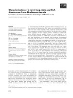
Schematic representation of the SRLV ORFsFigure 1
Schematic representation of the SRLV ORFs. The env sequence of the prototype CAEV (Cork) strain carrying the SD
site used for the rev mRNA synthesis (SD
rev
) is enlarged. The nucleotide motifs corresponding to the canonical SD sequence
are boxed, with splice points designated by bent arrows.
JDJ
HQY
SRO
YLI
WDW
UHY
777&$&7*&***$&$*&$$**7$$*7$7&$$&&&&$**7$$*7$$*&$$$7$***$$&$*$$$7$&7$$&
6'
6'
QW
UHY
Retrovirology 2008, 5:22 />Page 4 of 17
(page number not for citation purposes)
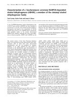
Splicing activity assays of SD sites within the CAEV env geneFigure 2
Splicing activity assays of SD sites within the CAEV env gene. A, Schematic representation of constructs used for splic-
ing activity assays. Reporter constructs were based on the vector pKCR3 which contained the β-globin intron flanked by its
splicing sequences inserted between the early promoter and poly-A site of SV40. CAEV sequences are included in open boxes.
In all constructs, the β-globin SD site was replaced by CAEV sequences containing the SD
6123
(grey box) and SD
6140
(hatched
box) sites. In plasmids pKRmB1 and pKRB1, the β-globin SA site was substituted by the 3' end viral genome containing the
SA
8514
site. The positions of the primers used for PCR amplification of cDNA are indicated (horizontal arrows). The positions
of probes MarN2 and MarS used in southern blot analysis are indicated. The MarN PCR primer used in experiment reported in
Fig. 4 is indicated. B, RT-PCR analysis of RNAs extracted from transfected 293T cells. cDNAs were PCR amplified using primer
pairs PK5 and PK3, or PK5 and M3b, as indicated. PCR products were resolved on an agarose gel and visualized by ethidium
bromide staining. Lane M, DNA size markers. C, Southern blot analysis of transcripts from cells transfected with pKRmB1 and
pKRB1 plasmids. PCR-amplified cDNAs were fractionated through a 2.5% agarose gel, blotted to nylon, and hybridized to
probes MarN2 (left panel) and MarS (right panel).
0DU1 0DU6
6'
6'
&$*&$$**7$$*7$7&$$&&&&$**7$$*7$*$7$7$&$*$$&
0DU1
0DU1
0DU6
/75
6$
S.5%
S.5P%
6'
369 3RO\$
6$JORELQ
S.&5
6'JORELQ
0E
$
% &
S
.
5
%
S
.
5
P
%
S
.
5
%
S
.
5
P
%
S.5P
3. 3.
00
ES
ES
S
.
5
P
%
S
.5
P
S
.5
S
.
&
5
0
R
F
N
3ULPHUV
3.3. 3.0E
S.5
6'
Retrovirology 2008, 5:22 />Page 5 of 17
(page number not for citation purposes)
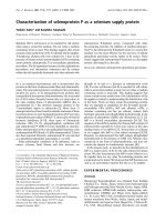
rtm ORF codes for two 18- and 17-kDa protein isoforms related to envelope precursor and TM proteinsFigure 3
rtm ORF codes for two 18- and 17-kDa protein isoforms related to envelope precursor and TM proteins. A, Rela-
tionships between domains shared by Env precursor, Rev and Rtm proteins. Splicing events within the Env coding region lead-
ing to rev and rtm ORFs are shown. Env precursor and Rev derived domains are represented by open and shaded boxes,
respectively. B, Schematic representation of Rev and Rtm expression constructs. Plasmids pKcRev and pKcRtm are predicted
to express singly-spliced mRNAs encoding the Rev and Rtm proteins, respectively. The pKRtm expression vector contains the
rtm cDNA generated by RT-PCR from cells transfected with pKcRtm. The approximate positions of PCR primers are indicated
(horizontal arrows). C, Coding capacity of the rtm ORF. Transfected 293T cells were radiolabeled 5 h with [
35
S]-methionine 48
h after transfection, and protein extracts were subjected to immunoprecipitation analysis using rabbit affinity-purified antibod-
ies raised against either the first 38 amino acids of Env precursor (anti-NH
2
Env), the 110-amino acid cytoplasmic domain of TM
(anti-CD™), or the 98-amino acid carboxy terminus of Rev (anti-Rev). Immunoprecipitated proteins were resolved by electro-
phoresis through a SDS-15% polyacrylamide gel and visualized by autoradiography. D, Analysis of in vitro translation products of
rtm cDNA. [
35
S]-methionine labeled polypeptides were synthesized in an in vitro coupled transcription-translation reaction with
pGEM-1 (lanes 1 and 2) or rtm cDNA (lanes 3 and 4). Crude products (lanes 1 and 3) and proteins immunoprecipitated with
affinity-purified anti-CD™ antibodies (lanes 2 and 4) were analyzed as described above.
& '
N'D
N'D
N'D
N'D
N'D
N'D
$QWL&'
70
$QWL1+
(QY
$QWL5HY
S
.
F
5
H
Y
S
.
F
5
W
P
S
.
5
W
P
S
.
&5
$
/75
6$
S.F5HY
6'
369
S.F5WP
6'
3RO\$
S.5WP
6$JORELQ6'JORELQ
0H
0E
UHY
UWP
HQY
%
6'
1
&
&
6'
6$
$7*
$7*
$7*
1
6$
Retrovirology 2008, 5:22 />Page 6 of 17
(page number not for citation purposes)
of
35
S-labeled proteins expressed from transfected cells
were performed by using three distinct antibodies devel-
oped by immunization of rabbits with GST fused pro-
teins.
The specificities of these antibodies were as follows: i)
anti-NH
2
Env antibodies recognizing the 38 N-terminal
amino acids of the Env precursor; ii) anti-CD™ antibodies
recognizing the cytoplasmic domain of TM; iii) monospe-
cific anti-Rev antibodies recognizing the 98 C-terminal
amino acids of Rev. As shown in Fig. 3C, two major pro-
tein species of apparent molecular weights of 18- and 17-
kDa expressed from cells transfected with the Rtm expres-
sion vector (lane 3) were immunoprecipitated with either
anti-NH
2
Env or anti-CD™ antibodies. A minor protein
species with a size slightly smaller than 18-kDa was also
immunoprecipitated with the anti-NH
2
Env antibodies.
None of these proteins were immunoprecipitated with
monospecific anti-Rev antibodies. Two proteins exhibit-
ing slightly different mobilities were immunoprecipitated
from cells transfected with the Rev expression vector (lane
2) by using either anti-NH
2
Env or anti-Rev antibodies,
but not with anti-CD™ antibodies. No corresponding pro-
tein was immunoprecipitated from cells transfected with
the empty parental plasmid pKCR3 (lane 1). These results
demonstrated that the rtm ORF encoded two major pro-
tein isoforms carrying antigenic determinants derived
from both the N- and C-termini of the Env precursor. As
expected, these proteins did not share any antigenicity
with the C-terminus of Rev. These two major protein iso-
forms of apparent molecular weights of 18- and 17-kDa
corresponded likely to the expected proteins of 17.8- and
15.5-kDa, the minor band corresponding to the expected
protein of 17-kDa. The fact that Rtm proteins were recog-
nized by antibodies directed against both NH2 and
COOH termini of the Env precursor indicated that they
were not degradation products of the Env precursor.
Expression of two isoforms from cells transfected with the
CAEV rev cDNA has been previously reported [13]. It has
been suggested that they resulted from initiation at the
first methionine codon (position 6012) and from leaky
scanning and initiation at one of the two downstream in
frame initiation codons (positions 6033 and 6072)
within the env gene (Fig. 3A), leading to a protein of 15.3-
kDa and to an isoform of either 14.5- or 13-kDa, respec-
tively. Since rev and rtm ORFs shared the same 5' coding
region, it was likely that the Rtm-related isoforms resulted
from a similar leaky translational mechanism. Alterna-
tively, they could originate from an alternative splicing
removing part of the rtm ORF. To discriminate between
these two hypotheses, immunoprecipitations were per-
formed from cells transfected with the plasmid pKRtm
carrying the fully spliced rtm cDNA (Fig. 3B), which was
obtained by RT-PCR from cells transfected with plasmid
pKcRtm. Two major proteins with similar mobilities and
antigenic properties were produced from cells transfected
with pKcRtm and pKRtm plasmids (Fig. 3C, compare
lanes 3 and 4), indicating that these protein isoforms did
not result from alternative splicing of the rtm transcript.
To rule out any post-translational modifications or pro-
tein degradations, the rtm cDNA was used as a template in
an in vitro transcription-translation reaction. As shown in
Fig. 3D, analysis of the cell-free radiolabeled translated
proteins also revealed the two Rtm proteins (lane 3),
which were specifically immunoprecipitated by anti-CD™
antibodies (lane 4), whereas no product was detected in
mock experiments (lanes 1 and 2). Interestingly, the fact
that the in vitro 18-kDa:17-kDa ratio was inversely related
to that observed in vivo was in favor of a leaky scanning
origin of the 17-kDa protein. Altogether, these results
strongly suggested that the two isoforms of 18- and 17-
kDa encoded by the rtm ORF resulted from translational
initiation at different in frame start codons, as previously
reported for Rev protein synthesis.
Splicing activity at the SD
6140
site occurs in CAEV-infected
cells, leading to the production of the rtm ORF
To determine whether splicing activity at the SD
6140
site
occurred in an infectious context, cDNAs from CAEV-
infected GSM cells were amplified by RT/PCR, and then
analyzed by Southern blot hybridization using probes
MarN2 and MarS (Fig. 2A). The primers used in PCR were
first Mar52 and M3b, located in the CAEV leader non-cod-
ing exon and the U3 region, respectively (Fig. 4A), and
then MarN and M3b, allowing amplification of cDNAs
corresponding to mRNAs generated by splicing at the
SD
6140
site (Fig. 2A and 4A). As a control, cDNAs from
293T cells transfected with plasmids pKRB1 and pKRmB1
were obtained similarly, except that the forward Mar52
primer was substituted by the PK5 primer in the first
round PCR (Fig. 2A). These controls led to a 617-bp
amplified product specifically detected by both MarN2
and MarS probes (Fig. 4B, lanes 1 and 2), a size expected
in view of the sequence of the plasmid used. The CAEV-
infected GSM cells led to a slightly smaller product (desig-
nated as ~617-bp) revealed with both probes (Fig. 4B,
lanes 3), whereas no product was detected from mock-
infected GSM cells (Fig. 4B, lanes 4). The signals corre-
sponding to vif, tat, and env singly-spliced transcripts were
not observed, but such long fragments were not expected
to be efficiently amplified using our experimental condi-
tions. To find out the origin of the unexpected slight dif-
ference in size between products from infected and
transfected cells, the ~617-bp cDNA amplified from
CAEV-infected cells was cloned and sequenced (Fig. 5).
Nucleotide sequence analysis revealed (i) the splice junc-
tion between the SD
6140
and SA
8514
sites, (ii) both synon-
ymous (nt 8606) and nonsynonymous (nt 8838 to 8840)
substitutions, and (iii) a 37 nt deletion (nt 8920 to 8957)
Retrovirology 2008, 5:22 />Page 7 of 17
(page number not for citation purposes)
Splicing junction between SD
6140
and SA
8514
sites occurs in CAEV-infected cellsFigure 4
Splicing junction between SD
6140
and SA
8514
sites occurs in CAEV-infected cells. A, Proviral organization and splicing
pattern of CAEV genome. The nucleotide numbers of SD sites (open triangles) and SA sites (solid triangles) are shown. All
splice sites were identified by cDNA sequencing. Exons are represented by solid lines. Alternative exons which are present in
only some of the mRNAs are shown in parenthesis. The putative structure of rtm transcript generated by splicing between
SD
6140
and SA
8514
sites is shown. The arrows represent PCR primers used for cDNA amplification. B, Southern blot analysis of
cDNAs from either transfected or infected cells. Cytoplasmic RNAs extracted from either 293T cells transfected with plas-
mids pKRB1 (lane 1) and pKRmB1 (lane 2) or CAEV-infected (lane 3) and non-infected (lane 4) GSM cells were submitted to
RT/PCR. Primer pairs PK5/M3b and Mar52/M3b were used to amplify in a first-round PCR the cDNAs from transfected and
infected cells, respectively. Primer pair MarN/M3b was used in the second-round PCR. PCR-amplified cDNA fragments were
electrophoresed through an 2.5% agarose gel, blotted to nylon, and hybridized to either probe MarN2 (left panel) or probe
MarS (right panel). Size of PCR-amplified fragments corresponding to the splice junction 6140–8514 is indicated.
$
ES
0DU6
%
YLI
WDW
HQY
UHY
UHY
"
JHQRPLF
0DU 0DU1 0E
UWP
UHY
HQY
/75
WDW
SRO
JDJ
YLI
/75
0DU1
*607
S.5%
&$(9
0RFN
S.5P%
*607
S.5%
&$(9
0RFN
S.5P%
Retrovirology 2008, 5:22 />Page 8 of 17
(page number not for citation purposes)
Identification of a novel CAEV ORFFigure 5
Identification of a novel CAEV ORF. The 617-bp cDNA amplified by nested-PCR from CAEV-infected GSM cells (see Fig.
4B) was cloned and sequenced. The region sequenced (uppercase letters) is bound by primers MarN and M3b (overlined) used
in the second-round PCR. The region in lowercase letters is from the previously published CAEV nucleotide sequence. Num-
bers in brackets indicate the nucleotide positions of the CAEV genomic sequence (22). The predicted translation product
(named Rtm) is shown below the sequence. The amino acids shared by the Rev and Rtm proteins are boxed. The nucleotide
and amino acid substitutions of the cDNA compared to the previously published CAEV-Cork sequence are underlined. Dele-
tion is represented by an open triangle. Stop codon is designated as asterisk.
DWJJDWJFWJJJJFFDJDWDFDWJFJFWWDDFWJJJDDJJDD
0'$*$5<05/7*.(
DDFWJJJWWJDDJWDDFFDWJJDFJJDJDJDDJJDDDJJDDD
1:9(970'*(.(5.
DJDJDDJJWWWFDFWJFJJJD&$*&$$**7$$*7$7&$$&&&
5(*)7$*44*.<43
&$*$7$7$&$*$$&7&7$$*7$&&&&*$&$7$&&$$&***7&
4,<57/6737<459
$&$*7&$7&$7**$$$&$$*$*&$*$&*7&*&$**$*$$$$7
79,0(75$'9$*(1
&$**$7777**$
*$7**&77$*$**$$7&$*$&$$&$*&*$$
4')*'*/((6'16(
$&$$*&*$$$*$*7*$&$*7$&$*$$$*&77**$*&&*7*&&
76(59794.$:65$
7***$*&777**&$*$$&7&$&&&7**$$**$*&&$7**$$$
:(/:4163:.(3:.
$****&&7*&7*$**&7*&7&*7&&77&&*&7*$&*$7***$
5*//5//9/3/70*
$7&7**$7$$$7**$7**&77**$*$$&$&&$&$$$$$7$$$
,:,1*:/*(++.1.
$$$$*$$$***7*$&7*7*$*$&$7***&7$$$$*7
*$&7$$
.5.*'&(7:$.6
'
7$$&$$*&7$**&&$$$77&&7*7$$$7&$&77******77$
7$$*$$$$*&$$*77&$&7$7*$&$$$*&$$7$7$$*$$$$*
&$$*77&$&7$7*$&$$$*&$$$$7*7$$&&*&$$*7*&7*$
&$*$7*7$$&$*&7*$&$7$7&$*&7*$7*&77*&7&$7*&7
*$&$&7*7$*&7&7*$*&7*7$7$7$$**$*$$*&77*&7*&
77*&
>@
0E
>@
8
0DU1
>@
>@
Retrovirology 2008, 5:22 />Page 9 of 17
(page number not for citation purposes)
within one copy of a duplicated 70-bp motif located in
the U3 region downstream from the rtm, rev and env ORFs.
All these features were confirmed by an independent
experiment. This confirmed that the 37 nt deletion
accounted for the size difference of RT/PCR products
obtained from infected and transfected cells. Such dele-
tion was previously found in some CAEV genomes (25).
These results demonstrated that the splicing activity of the
SD
6140
site also occurred in CAEV-infected cells, leading to
the synthesis of the rtm ORF.
Rtm is expressed in CAEV-infected cells
To investigate whether Rtm protein was expressed in
CAEV-infected cells, we needed to rule out numerous
drawbacks including common antigenic determinants
shared by the Rtm, Env and Rev proteins, and similar
molecular weights of Rtm and Rev proteins. Considering
that a Rtm-specific epitope might be encoded by the
nucleotide sequence overlapping the splice junction spe-
cific to the rtm mRNA, we generated a rabbit antiserum
against the synthetic peptide (KYQPQIYRT) correspond-
ing to the translated product of this specific Rtm coding
region. First of all, the specificity of these anti-Rtm anti-
bodies was tested by immunoprecipitation of [
35
S]-radi-
olabeled proteins produced from transfected cells. As
shown in Fig. 6A, the anti-Rtm antibodies immunoprecip-
itated neither the Env precursor nor the mature SU and
TM glycoproteins produced from cells transfected with an
Env expression vector (lane 1), while these Env products
were recovered by immunoprecipitation using either
CAEV-infected goat serum or anti-CD™ rabbit serum
(lanes 2 and 3, respectively). Similarly, no protein was
immunoprecipitated by the anti-Rtm antibodies from
mock-transfected cells or cells transfected with the Rev
expression vector (Fig. 6B, lanes 4 and 5, respectively). In
contrast, the anti-Rtm antibodies recognized both Rtm
isoforms produced from cells transfected with the Rtm
expression vectors (Fig. 6B, lanes 6 and 7), demonstrating
that this rabbit antiserum was specific to the Rtm protein.
Next, we determined whether Rtm protein was expressed
in CAEV-infected cells. A lysate from GSM cells infected by
the CAEV-Cork strain was immunoprecipitated with
either anti-Rtm or anti-CD™ antibodies. The two proteins
of 18- and 17-kDa were clearly detected by both types of
antibodies in infected cells whereas no product was
detected in uninfected cells (Fig. 6C, compare lanes 2 and
4 with lanes 1 and 3). The signal using the anti-Rtm anti-
bodies was faint compared with that obtained with the
anti-CD™ antibodies, indicating probably a low peptide-
antibody affinity. We concluded that the Rtm protein was
expressed in CAEV-infected GSM cells.
To know whether the Rtm protein was expressed in
infected animals we looked for humoral immune
response against it. Considering that most antibodies that
would recognize the Rtm protein might in fact result from
an immune response against the Rev and/or Env proteins,
we tested for the presence of antibodies recognizing the
KYQPQIYRT Rtm-specific epitope. For this purpose, fifty
milliliters of pooled sera from three seropositive goats
experimentally infected with the CAEV-Cork strain were
loaded onto a resin matrix covalently bound with the Rtm
peptide. After extensive washing, bound antibodies were
eluted and tested by ELISA using either the Rtm peptide or
the GST-CD™ protein as antigens and by Western blot
using the GST-CD™ protein. None of these assays pro-
vided positive results. We concluded that if the Rtm was
expressed in infected animals the KYQPQIYRT epitope
was not enough immunogenic to give rise to the produc-
tion of antibodies or that these antibodies did not recog-
nize the synthetic peptide.
Rtm protein interacts with the cytoplasmic domain of TM
Considering that the major part of the Rtm sequence cor-
responded to the cytoplasmic domain of TM and that the
homologue domain of HIV TM was reported to self-
assemble as an oligomer [26], we looked for an interac-
tion between the Rtm and the cytoplasmic domain of TM.
In this attempt, a GST pull-down assay was performed to
identify potential interaction of Rtm with Env protein. In
vitro-translated, radiolabeled Rtm protein was incubated
with either a GST fusion protein containing the entire
cytoplasmic domain of TM (GST-CD™) or with GST alone
coupled to glutathione-Sepharose beads. Equal amounts
of protein were used in all binding experiments, as veri-
fied by SDS-PAGE and Coomassie blue staining (data not
shown). After extensive washing of the bead-bound com-
plexes in different stringent conditions, the bound pro-
teins were analysed by SDS-PAGE and autoradiography.
As shown in Fig. 7, the Rtm protein interacted with the
GST-CD™, and this interaction was resistant to high ionic
strength washes. In contrast, no significant interaction
was observed in association with the GST protein alone.
These results clearly indicated that Rtm protein strongly
and specifically interacts with the cytoplasmic domain of
TM in vitro.
The SD
6140
site is strictly conserved throughout the SRLV
phylum
To assess the biological importance of the SD
6140
site and
of the Rtm protein for SRLVs, we assumed that it should
be conserved in all SRLV genomes, as the rev SD
6123
site is.
To look for the conservation of the SD
6140
site among
SRLV strains, previously described env sequences repre-
sentative of highly divergent phylogenetic clusters were
aligned (Fig. 8). This alignment confirmed that the 5'
region of the SRLV env gene was extremely variable, except
two quasi perfect repeat sequences (GGTAAG) corre-
Retrovirology 2008, 5:22 />Page 10 of 17
(page number not for citation purposes)
Detection of Rtm expression in transfected 293T cells and infected GSM cells using a Rtm-specific peptide antiserumFigure 6
Detection of Rtm expression in transfected 293T cells and infected GSM cells using a Rtm-specific peptide
antiserum. A and B, Specificities of rabbit anti-Rtm peptide antibodies. 293T cells were transfected with Env (pKEnv) plasmid
(A), or with either parental (pKCR3), Rev (pKcRev), or Rtm (pKcRtm and pKRtm) plasmids, as indicated (B). Cells were radi-
olabeled 5 h with [
35
S]-methionine 48 h after transfection. Lysates were subjected to immunoprecipitation and fractionated on
an SDS-10% polyacrylamide gel. Immunoprecipitations were performed using either anti-CD™ antibodies, a serum from
CAEV-infected goat (anti-CAEV), or anti-Rtm peptide antibodies (anti-Rtm). C, Immunoprecipitation of the Rtm protein from
infected cells. GSM cells were either mock infected (-) or infected with CAEV-Cork strain (+). When cytopathic effects
appeared in infected cell culture, cells were radiolabeled 5 h with [
35
S]-methionine, and lysates were immunoprecipitated with
either anti-Rtm or anti-CD™ antibodies, as indicated. Immunoprecipitated proteins were resolved on a SDS-15% polyacryla-
mide gel.
$
&
$QWL &'
70
5WP &$(9
N'D
N'D
S.
F
5
H
Y
S
.
F
5
W
P
S
.
5
W
P
S
.
&
5
$QWL5WP
70
%
7FHOOV
3U(QY
68
N'D
7FHOOV
N'D
N'D
$QWL&'
70
$QWL5WP
*60FHOOV
Retrovirology 2008, 5:22 />Page 11 of 17
(page number not for citation purposes)
sponding to the SD
6123
and SD
6140
sites of Cork genome.
Remarkably, the 17 nt distance between the two SD sites
was also perfectly conserved among all SRLV sequences.
Moreover, the downstream SD site was even better con-
served than the SD site used for the rev mRNA synthesis.
The high conservation of all these genetic features
(sequence, frame, and nt distance) within a highly varia-
ble region strongly suggested that the Rtm protein, like the
rev protein, is very important for SRLVs.
Rtm protein affects the fusion activity of Env protein
Given the importance of the cytoplasmic domain of TM in
several Env protein functions such as Env sorting and
membrane fusion activity and the specific interaction
between this domain and Rtm protein in our in vitro bind-
ing assay, we looked for the effects of Rtm expression on
the Env fusion activity. For this purpose, 293T cells were
transfected with pKEnv plasmid or cotransfected with
pKEnv and pKRtm plasmids, then fusion activity was
assessed by coculture with GSM target cells. In all transfec-
tions, the plasmid amounts were normalized by addition
of empty plasmid pKCR3 in order to have the same copy
number of SV40 promotor. As shown in Table 1, the
expression of Rtm reduced by 79% the syncytium number
with respect to that obtained with Env protein expressed
alone.
Discussion
A novel CAEV protein, designated Rtm, was characterized
on the basis of its immunological cross-reactivity with
antibodies directed against either the N-terminus of the
Env precursor or the C-terminus of TM, and by cDNA
sequence analysis which established that rtm ORF results
from a novel splicing junction linking the 5' and 3' part
ends of the env gene. Immunoprecipitation experiments
using monospecific antibodies raised against the peptide
encoded by the sequence straddling this splice junction
confirmed the existence of the rtm ORF. Like Rev, the Rtm
expression was initiated at the env ORF start codons and
led to the production of two major protein isoforms of 18-
and 17-kDa. Previous studies together with this work indi-
cated that both Rev and Rtm protein doublets may be
attributed to a leaky scanning at the first AUG initiation
codon and initiation at one of the two downstream in
frame env initiation codons [13,23,24]. The ratio between
the 18- and 17-kDa isoforms depends on the context and/
or the level of the Rtm expression, the 18-kDa being the
major product in transfected 293T cells whereas the 17-
kDa is the major product in infected GSM cells and in the
in vitro transcription/translation reaction.
The rtm ORF was generated by splicing between the SD
6140
site identified in this study and the well described SA
8514
site. Since the rev ORF is produced by splicing between the
SD
6123
and SA
8514
sites, the Rev and Rtm proteins are
expressed from distinct but structurally closely related
multiply-spliced mRNAs. The SD
6140
site matched per-
fectly the canonical SD sequence and was highly con-
served among all SRLV genomes. Moreover, the distance
between the SD
6123
and SD
6140
sites is also strictly con-
served among SRLV sequences, despite the variability of
the surrounding region. Altogether, these results strongly
suggested that the Rtm protein is an additional auxiliary
factor required for successful SRLV propagation in vivo.
The Rtm protein is a chimeric protein in which the N-ter-
minal part of the envelope precursor is joined to the com-
plete cytoplasmic domain of TM. An expression of this
domain in a viral envelope-free context has been also
reported for MVV in a previous study showing that a 16.5-
kDa protein corresponding to the C-terminal domain of
TM is produced in an in vitro transcription/translation
reaction from the rev transcript by leaky scanning and ini-
tiation at a downstream initiation codon [23]. For SRLVs,
equine infectious anemia virus (EIAV), bovine and simian
(SIV) immunodeficiency viruses, the 5' terminal part of
the env gene also constitutes the first coding exon of Rev.
Thus, Rtm could exhibit some functions of its related pro-
teins, Rev and/or TM. Several studies demonstrated that
motifs involved in CAEV Rev function are exclusively
localized in the C-terminal domain of the protein [13,27].
Moreover, a truncated Rev protein devoid of its N-termi-
nal domain is still functional [21], although this domain
has been shown to be responsible for optimal binding to
the Rev responsive element [28]. Our attempts to identify
a Rev activity in Rtm were unsuccessful (data not shown).
Altogether, these results strongly indicate that Rtm does
not harbor any Rev activity. More likely, the biological
functions of Rtm would be harbored by its C-terminus
which corresponds precisely to the entire cytoplasmic
domain of TM.
Rtm binds to the cytoplasmic domain of Env proteinFigure 7
Rtm binds to the cytoplasmic domain of Env protein.
Purified recombinant GST-CD™ or GST alone were incu-
bated with in vitro-translated Rtm protein labeled with
[
35
S]methionine. The beads were washed six times in the
presence of different ionic strengths of KCl, as indicated, and
the bound proteins were subjected to 15% SDS-PAGE analy-
sis and autoradiography.
.&O P0
N'D
N'D
*67&'
70
*67
Retrovirology 2008, 5:22 />Page 12 of 17
(page number not for citation purposes)
The cytoplasmic domain of the TM protein is considerably
longer for lentiviruses than for other retroviruses [29], and
has been reported to be involved in multiple crucial steps
of virus infection, such as particle assembly, infectivity,
replication, and pathogenesis. For HIV and SIV, the cyto-
plasmic domain of TM modulates Env fusogenicity,
expression on the cell surface, and incorporation into viral
particles as well as the biochemical and immunologic
properties of the Env ectodomain [30-39]. The underlying
mechanisms responsible for the regulation of Env protein
function involve association of the cytoplasmic domain of
TM with cellular and viral components [40-50]. These
interactions require canonical tyrosine-based and di-leu-
cine motifs as well as small domains called lentivirus lytic
peptide. Since most of these signals were found in the
cytoplasmic domain of CAEV TM (Fig. 5), it was likely that
Genetic homologies of the internal exon sequence carrying the SD
rev
and SD
rtm
sites among SRLV strainsFigure 8
Genetic homologies of the internal exon sequence carrying the SD
rev
and SD
rtm
sites among SRLV strains. All
available sequences belonging to the three phylogenetic groups (A, B and C) are aligned. Dashes indicate deletions. A consen-
sus sequence is presented. The conserved SD motifs are boxed.
.$&$$&$***$&7**7$7&$**7$$*$$$$$$7&$7***7$$*7$77*$&&7&&7****$&$*$$**$$$$
296**$$&&$*&**&* &$
32/9&$7*$*$&$$$**&$&$ 7&$**
&*$&77$$&*$&**&7* &**&
.0**$** $**
(9$**7$**&*$$$*&$7*7
6:*$7$&**&7* $*&$7**
6$2099$$$$77$*7 &$&$*
&RUN777&$&7*&***$&$*&$$**7$$*7$7&$$&&&&$**7$$*7$$*&$$$7$***$$&$*$$$7$&7$$&
* $*&$7$7*
&*$*$$&&$*$**
&*$$$$&&$*$**
&&$*$$*$7&$7$**
&*7$$**$&*&$&7& *$*
&KL&&$$$*$&$&&$7$$*$*
*$$$$**$*&&$&7$$7&&$***7$$*7$7$$$$$$&$**7$$*7$*$$7$$&7$7$*77$7$77$&7$$&
.(**$7&$$*&7&$*7 $&&$ $*
.(**$7&$$*&7&$*7 $&&$ $*
**$$$7*7$&77**&&7$7& *$7
**&$$7***&*&$7777$7& && *&7
**&$$7*$*&*&$77*&7$7& 7& *&7
/0&$$7***&$&7&$*****7& 7$& *7
**7$***7$$*7
$
*
%
6'
UHY
6'
QW
$
&
Table 1: Effects of Rtm on the fusion activity of Env protein.
Plasmid
a
No. of syncytia/well (293T/GSM cocultivation)
b
% of syncytia
c
pKCR3 0 0
pKEnv 954 ± 105 100
pKEnv + pKRtm 200 ± 46 21
a
The plasmid amounts were normalized by addition of empty plasmid pKCR3 in order to have the same copy number of SV40 promotor in all
transfections.
b
Numbers of syncytia in 30 randomly selected fields per well. Averages of three wells are shown.
c
Number of syncytia divided by the number obtained with the positive control (pKEnv + pKCR3 plasmids) × 100.
Retrovirology 2008, 5:22 />Page 13 of 17
(page number not for citation purposes)
similar mechanisms have been developed by SRLVs to reg-
ulate the Env protein function. Consequently, expression
of the cytoplasmic domain of TM in a viral glycoprotein-
free context like Rtm could potentially interfere with Env
protein function. We showed here that the Env-mediated
syncytium induction is dramatically reduced in the pres-
ence of Rtm protein. Using a GST pull-down assay, we
also found that Rtm protein interacts specifically with the
cytoplasmic domain of TM. Altogether, these findings sug-
gest that the Rtm protein acts in the infectious cycle of
SRLV by modulating the Env protein function through
direct interaction with the cytoplasmic domain of TM
and/or through competitive interaction with cellular fac-
tors.
Assuming that Rtm acts as a down regulator of Env protein
function, it is likely that Rtm is not expressed during the
late phase of the viral cycle when Env proteins must be
highly expressed, correctly sorted and incorporated in the
virions. This is sustained by the fact that despite Rtm inter-
acts strongly with the cytoplasmic domain of the TM, all
our attempts to detect Rtm in virions failed (data not
shown). Moreover, Rtm is translated from a multiply-
spliced transcript, arguing in favor of its expression early
during the viral replication cycle since previous studies
have revealed a temporal shift from multiply-spliced to
mono- and unspliced SRLV transcripts [20,22]. Since the
Rev protein is absolutely required for the expression of the
viral structural proteins, we can hypothesize that expres-
sions of Rev and Rtm proteins are temporally dissociated
from each other. This assumption can accommodate with
the fact that SRLV expression is restricted in monocytes,
and upregulated during maturation of monocytes into
macrophages [4,5]. In latently infected monocytes, virus
replication is blocked at a post-transcriptional stage [2,4],
which could result from the downregulation effects of
Rtm. Differentiation of monocytes into macrophages is
also required for EIAV expression [51]. Interestingly, a
very similar mRNA encoding a Rtm-like protein has been
described in an acutely infected horse [52]. This hybrid
protein, designated Ttm, contains the domain encoded by
the first coding exon of Tat fused to the complete cytoplas-
mic tail of TM. Similarly to the Rev-derived sequence in
the Rtm protein, the Tat-derived sequence in the Ttm pro-
tein does not contain functional domains of the native
protein. Thus, the role of the cytoplasmic domain of TM
expressed in these two similar hybrid proteins would be
interesting to examine in the context of the high variation
of viral expression of these two macrophage-tropic lentivi-
ruses in their hosts.
Conclusion
In the present study we have identified a new competent
SD site at position 6140 in the CAEV genome that
presents two remarkable features. First, despite its location
within a highly variable region, the sequence of this SD
site is perfectly conserved among CAEV and MVV strains,
the only other conserved stretch of nucleotides in this
region corresponding to the SD site used for the rev mRNA
synthesis. Second, the 17 nt distance between the two SD
sites is also strictly conserved in the genome of all SRLV
isolates. Splicing at the SD
6140
site was demonstrated in
both transfected and CAEV-infected cells, leading to the
production of an ORF encoding two major protein iso-
forms of 18- and 17-kDa, named Rtm. These proteins are
generated by differential translation initiation, contain
the N-terminal part of the Env precursor fused to the com-
plete 110-amino acid cytoplasmic domain of TM, and are
expressed in CAEV-infected cells. The exceptional degree
of conservation of the SD
6140
site sequence among CAEV
and MVV isolates and the fact that rtm and rev transcripts
were structurally closely related strongly suggest that the
Rtm protein is a fourth important auxiliary factor of
SRLVs. We showed that this protein interacts specifically
with the cytoplasmic domain of TM and dramatically
affects the fusion activity of Env protein. Altogether, these
findings support a model of regulation of SRLV expression
by a new viral factor, which can accommodate confirmed
observations regarding SRLV infection in vivo. In this
aspect, the functional activity of the cytoplasmic domain
of the TM expressed in a viral glycoprotein-free context
should be considered to better understand the parameters
of SRLV propagation in vivo.
Methods
Cells and sera
Primary foetal goat synovial membrane (GSM) cells
infected with CAEV-Cork strain were maintained in cul-
ture in Eagle's minimal essential medium supplemented
with 1% glutamine, 200 U/ml penicillin, 200 μg/ml strep-
tomycin, 5 μg/ml fungizone, and 10% foetal bovine
serum (FBS). The human 293T cell line was propagated in
Dulbecco's modified Eagle's medium (DMEM) supple-
mented with 1% glutamine, 25 μg/ml gentamycin, and
5% FBS. CAEV-specific sera were collected from goats
experimentally infected with CAEV-Cork strain. Rabbit
polyclonal antisera against the cytoplasmic domain of TM
(anti-CD™), the N-terminus of Env precursor (anti-NH
2
Env), and the C-terminus of Rev (anti-Rev) were gener-
ated by using GST fusion proteins as immunogens. Puri-
fied fusion proteins were used in association with Freund
adjuvant to immunize New Zealand White rabbits. Rabbit
antisera were affinity purified on GST fusion protein/affi-
gel 10 columns according to the manufacturer's instruc-
tions (BIO-RAD).
A synthetic peptide derived from the predicted Rtm open
reading frame (ORF) product (amino acids 39 to 47) was
designed for the production of monospecific anti-Rtm
antibodies. This Rtm-specific peptide (KYQPQIYRT) was
Retrovirology 2008, 5:22 />Page 14 of 17
(page number not for citation purposes)
synthesized using a 9-fluorenylmethyloxycarbonyl chem-
ical strategy (Sigma-Genosys) and rabbit peptide-specific
antiserum was generated by immunization with the pep-
tide cross-linked to the keyhole limpet hemocyanin car-
rier.
Plasmid constructions
All eukaryotic expression plasmids were derived from the
pKCR3 vector [53] in which the rabbit β-globin intron 2
flanked by its splice sites was inserted between the early
promoter and polyA signal site of the simian virus 40
(SV40). The viral sequences were derived from both the 9-
kbp and 0.5-kbp HindIII clones of CAEV-Cork strain
[22,54]. The nucleotides (nt) are numbered according to
the Cork sequence [22]. To generate constructs used for
splicing activity assays, a fragment (nt 6117 to 6674)
spanning the well-described SD
6123
site and the putative
SD
6140
site was PCR-amplified from the 9-kbp HindIII
clone, and subcloned into the SmaI site of pGEM-1 plas-
mid. Disruption of the SD
6123
site (G to C substitution at
nt 6124) was achieved with primer 5'-TCTAAAGGATC-
CCCCAGCAAGC
TAAGTATCAACCCCAG-3' (non CAEV
sequences are shown in italic) by using the CLONTECH
Transformer site-directed mutagenesis kit (mutated nt are
underlined in the nucleotide sequences). A 261-bp frag-
ment containing either the wild-type or mutated viral
sequence was double digested with BamHI (in the multi-
ple cloning site [MCS] of pGEM-1) and HincII (position
6369 in Cork sequence), and cloned in place of the origi-
nal β-globin SD site in the pKCR3 plasmid, to generate
pKR12 and pKRm plasmids, respectively. The plasmids
pKRB1 and pKRmB1 were generated by inserting the ApaI-
BamHI fragment of the 0.5-kbp HindIII clone (nt 8113 to
9251) into the ApaI-BglII-digested pKR12 and pKRm plas-
mids, respectively.
To generate eukaryotic expression plasmids coding for
Env, Rev and Rtm proteins, the SpeI-BamHI fragment of
the 9-kbp HindIII clone, containing the env initiation
codon (nt 6012) and both the SD
6123
and SD
6140
sites, was
cloned into the XbaI/BamHI-digested pGEM-1 plasmid.
To facilitate cloning, a BamHI site was introduced by site-
directed mutagenesis at position 5979, upstream the env
initiation codon, using the primer 5'-TGCAAATAAAT-
GG
ATCCAACAAGTAGCAAAAGT-3' (nt 5968 to 6000).
Mutagenic primers 5'-GGGACAGCAAGC
TAAGTATCAA-
3' (nt 6113 to 6134) and 5'-TATCAACCCCAGC
TAAG-
TAAGCAA-3' (nt 6129 to 6152) were used to disrupt the
SD
6123
and SD
6140
sites, respectively. The resulting 231-bp
BamHI-EcoRV fragments (nt 5980 to 6211) containing
either the mutated SD
6123
site or the mutated SD
6140
site
were cloned into the similarly-digested pKRmB1 to pro-
duce pKcRtm and pKcRev plasmids, respectively. The
pKEnv plasmid was generated by cloning the EcoRI-BstXI
fragment (nt 6348 to 8368) of the 9-kbp HindIII clone
into the similarly-digested pKcRev plasmid. The Plasmids
encoding the Rtm protein from the cDNA were generated
as follows. Two PCR primers were used to amplify cDNA
from 293T cells transfected with pKcRtm. Forward primer
M5e (5'-GGAATTCATGGATGCTGGGGCCAGATAC-3'; nt
6012 to 6032) and reverse primer M3b (5'-CGGGATCCG-
CAAGCAGCAAGCTTCTCCTTATATA-3'; nt 9098 to 9073)
contained EcoRI and BamHI sites at their 5' end, respec-
tively. The EcoRI-BamHI digested PCR product was cloned
into pKCR3 digested with EcoRI and BglII to produce plas-
mid pKRtm. The same digested PCR product was cloned
under the T7 promoter control between the EcoRI/BamHI
sites of pGEM-1, generating the pGRtm plasmid used as
template in cell-free transcription/translation reaction. All
clones were verified by sequencing.
RNA isolation and RT-PCR
RNAs were extracted from either transfected 293T cells or
infected GSM cells with the RNA Now extraction kit (Bio-
gentex) according to the manufacturer's protocol. Five
micrograms of Dnase-treated RNAs were used for reverse
transcription in a final volume of 20 μl containing 200 U
of Moloney murine leukemia virus reverse transcriptase
(Promega), 1 mM of each deoxynucleoside triphosphate
(dNTPs), 1 mM DTT, 500 ng of oligo(dT)
12–18
and 26 U of
RNase inhibitor (Amersham). Reverse transcription (RT)
was carried out for 15 min at 37°C and then for 45 min at
42°C. PCR amplifications were carried out in a final vol-
ume of 50 μl of 1× PCR buffer (Perkin-Elmer), with 200
μM (each) dNTPs, 2 mM MgCl
2
, 100 ng of each primer,
0.5 U AmpliTaq DNA polymerase (Perkin-Elmer), and 10
μl of cDNA, using the following conditions: 3 min dena-
turation at 94°C, followed by 35 amplification cycles of
40 sec at 94°C, 50 sec at 53°C, 50 sec at 72°C, followed
by a final 4 min extension step at 72°C. Nested-PCR reac-
tions were performed under similar conditions using one-
tenth of the RT-PCR products as templates. PCR products
were separated by 2.5% agarose electrophoresis and visu-
alized by ethidium bromide staining. After blotting onto
a nylon membrane (Hybond-N
+
, Amersham), the mem-
brane was prehybridized for 3 h at 46°C in 5× SSC (1×
SSC is 0.15 M NaCl plus 0.15 M sodium citrate), 0.5%
sodium dodecyl sulfate (SDS), 100 μg/ml salmon sperm
DNA. The prehybridization solution was then replaced by
the hybridization solution containing the
32
P-labeled oli-
gonucleotide probe. After overnight incubation at 46°C,
the membrane was washed four times with 2× SSC – 0.1%
SDS at room temperature for 15 min and then once with
1× SSC – 0.1% SDS at 46°C for 15 min before being
exposed to X-ray film (Kodak) at -70°C. The oligonucle-
otide used to detect messages spliced at the SD
6140
site was
MarN2 (5'-AGGTAAGTATCAACCCCAG-3'; nt 6122 to
6140). The oligonucleotide used to detect Rtm-specific
spliced message was MarS (5'-AGTATCAACCCCAGATAT-
ACAGAAC-3'; nt 6127 to 6140 and nt 8514 to 8524),
Retrovirology 2008, 5:22 />Page 15 of 17
(page number not for citation purposes)
which overlapped the splice junction between the SD
6123
and SA
8514
sites.
Two primer pairs were alternatively used in a first round
PCR reaction to amplify cDNAs from transfected cells:
PK5 (5'-TAGTGAGGAGGCTTTTTTGGAG-3'; forward
primer) and PK3 (5'-GAAGATCTCAGTGGTATTTGT-
GAGCCA-3'; reverse primer), both located in pKCR3
sequence, or PK5 and M3b. The primer pair used in a first
round PCR reaction to amplify cDNAs from infected GSM
cells were Mar52 (5'-TAATCTGTGCAATACCAGAGCG-
GCT-3'; nt 131 to 155; forward primer) and M3b. Primer
pair MarN (5'-CAGCAAGGTAAGTATCAACCCCAG-3'; nt
6117 to 6140; forward primer) and M3b was used in a sec-
ond round PCR reaction.
Construction of GST fusion proteins
Plasmids pGST-NH
2
Env, pGST-Rev, and pGST-CD™
encode the gluthatione S-transferase (GST) fused C-termi-
nally to the N-terminus of Env precursor (amino acids 1
to 38), the C-terminus of Rev (amino acids 36 to 133),
and the cytoplasmic domain of TM (amino acids 203 to
312), respectively. To generate pGST-NH
2
Env, a PCR
product was amplified from pGRtm by using primers M5e
and 5'-CGCGGATCCTTGCTGTCCCGCAGTGAAACCT-3',
digested with EcoRI-BamHI, and then cloned into the
EcoRI/BglII sites of pGEX-A (Pharmacia). To generate the
GST-Rev fusion protein, cDNA from 293T cells transfected
with pKRB1 was used as template to amplify by PCR the
Rev C-terminus coding region by using primers PK5 and
M3x (5'-TGTCTAGAGCAAGCAGCAAGCTTCTCCT-
TATATA-3'; nt 9098 to 9073). The PCR product was
digested with EcoRI-XbaI and then cloned into the corre-
sponding sites of pGEM-1. Then, a BamHI/HincII frag-
ment encoding the C-terminal domain of Rev was excised
and subcloned into the BamHI/SmaI sites of pGEX-3X
(Pharmacia). Similar cloning strategy was applied to
cDNA from 293T cells transfected with pKRmB1 to gener-
ate pGST-CD™. GST fusion proteins were produced in
Escherichia coli strain DH5α and purified by using Glu-
tathione-Sepharose 4B beads (Pharmacia Biotech) by
standard procedures [55].
Metabolic labeling and immunoprecipitation
The 293T cells were transfected by the calcium phosphate
coprecipitation technique [56]. At 48 h post-transfection,
293T cells were incubated for 30 min in methionine/
cysteine-free DMEM supplemented with 5% dialyzed FBS
and then metabolically labeled for 5 h with 100 μCi of
[
35
S] methionine/cysteine mixture (Promix; Amersham)
per ml. Cells were then lysed in 1× RIPA buffer (10 mM
Tris-HCl [pH 7.8], 1 mM Na
2
HPO
4
, 1 mM EDTA, 0.5%
NP-40, 0.5% sodium deoxycholate, 0.2 mM PMSF), and
cell lysates were cleared by centrifugation at 15,000 × g for
15 min at 4°C. Immunoprecipitation reactions were per-
formed by incubating cell lysates with protein A-Sepha-
rose beads and either 300 μl of affinity-purified rabbit
antibodies or 5 μl of serum from CAEV-Cork infected goat
preadsorbed on normal 293T cell lysate, overnight at 4°C
with rocking. The bead-antibody-protein complexes were
washed five times with RIPA buffer, boiled in sample
loading buffer (60 mM Tris-HCl [pH 6.8], 1% SDS, 1% β-
mercaptoethanol, 10% glycerol, 0.01% bromophenol
blue), and recovered proteins were subjected to SDS-15%
polyacrylamide gel electrophoresis (SDS-PAGE) analysis.
The gel was fixed, soaked in Amplify (Amersham) for 30
min, dried and autoradiographied.
Cell fusion activity
The 293T cells were plated (2 × 10
5
cells per well) in trip-
licate in six-well plates in complete DMEM the day prior
transfection. The cells were transfected with 1.5 μg of
pKEnv plasmid or cotransfected with 1.5 μg of pKEnv
plasmid and 1.5 μg of pKRtm plasmid. The copy number
of the SV40 promotor was normalized in each transfec-
tion by adding appropriate amounts of empty plasmid
(pKCR3). On the next day, primary GSM cells (2.5 × 10
5
cells per well) were added to transfected cells. One day
post-coculture, cells were fixed and stained to visualize
nuclei. Fusion activity was assessed by counting syncytia
in 30 randomly selected fields at 100× magnification of
each well.
In vitro transcription and translation
One microgram of linearized plasmids pGRtm or pGEM-
1 were transcribed and translated in a coupled rabbit retic-
ulocyte lysate system (Promega) according to the manu-
facturer's instructions with T7 RNA polymerase and 0.8
μCi/μl of [
35
S] methionine/cysteine mixture (Amersham)
in a final volume of 50 μl. The translated products were
analyzed on SDS-PAGE and detected by autoradiography.
Immunoprecipitations of translation products with affin-
ity purified anti-CD™ antibodies were carried out as
described above.
GST pull-down assays
Equal amounts, as judged by Coomassie blue staining, of
the different GST fusion proteins complexed to glutath-
ione-Sepharose beads were incubated with 10 μl of [
35
S]-
labeled Rtm protein produced from in vitro transcription/
translation reaction, in binding buffer (10 mM Tris [pH
7.4], 10% glycerol, 0.2 mM EDTA, 0.5 mM DTT, 0.25%
Triton X-100) supplemented with 80 mM KCl and 100
μg/ml of bovine serum albumin in a final volume of 300
μl overnight at 4°C with constant agitation. The beads
were washed six times in binding buffer containing differ-
ent concentrations of KCl (150 mM, 300 mM, or 500
mM), boiled in sample loading buffer, and the proteins
were subjected to 15% SDS-PAGE analysis and autoradi-
ography as described above.
Retrovirology 2008, 5:22 />Page 16 of 17
(page number not for citation purposes)
Competing interests
The author(s) declare that they have no competing inter-
ests.
Authors' contributions
SV performed most of the laboratory work, MR contrib-
uted to the Western blot analyses, and CP contributed to
the preparation of serum samples. GP and RZM conceived
the strategies and designed the experiments. SV and RZM
wrote the manuscript. All authors read and approved the
final manuscript.
Acknowledgements
This work was supported by grants from AFSSA and from the Etablisse-
ments Publics Régionaux de Poitou-Charentes et d'Aquitaine.
References
1. Narayan O, Clements JE: Biology and pathogenesis of lentivi-
ruses. J Gen Virol 1989, 70:1617-1639.
2. Gendelman HE, Narayan O, Molineaux S, Clements JE, Ghotbi Z:
Slow, persistent replication of lentiviruses: role of tissue
macrophages and macrophage precursors in bone marrow.
Proc Natl Acad Sci USA 1985, 82:7086-7090.
3. Narayan O, Wolinsky JS, Clements JE, Strandberg JD, Griffin DE,
Cork LC: Slow virus replication: the role of macrophages in
the persistence and expression of visna viruses of sheep and
goats. J Gen Virol 1982, 59:345-356.
4. Gendelman HE, Narayan O, Kennedy-Stoskopf S, Kennedy PG,
Ghotbi Z, Clements JE, Stanley J, Pezeshkpour G: Tropism of sheep
lentiviruses for monocytes: susceptibility to infection and
virus gene expression increase during maturation of mono-
cytes to macrophages. J Virol 1986, 58:67-74.
5. Narayan O, Kennedy-Stoskopf S, Sheffer D, Griffin DE, Clements JE:
Activation of caprine arthritis-encephalitis virus expression
during maturation of monocytes to macrophages. Infect
Immun 1983, 41:67-73.
6. Filippi P, Vigne R, Quérat G, Jouanny C, Sauze N: Intracellular ribo-
nucleoprotein complexes of visna virus are infectious. J Virol
1982, 42:1057-1066.
7. Haziza B, Chauvin JP, Gluschankof P, Suzan M: Caprine arthritis
encephalitis virus: evidence for a B/D-type assembly pathway
in a C-type lentivirus replication. Virology 2001, 286:434-445.
8. Lairmore MD, Akita GY, Russell HI, DeMartini JC: Replication and
cytopathic effects of ovine lentivirus strains in alveolar mac-
rophages correlate with in vivo pathogenicity. J Virol 1987,
61:4038-4042.
9. Lee WC, McConnell I, Blacklaws BA: Electron microscope stud-
ies of the replication of a British isolate of maedi visna virus
in macrophages and skin cell lines. Vet Microbiol 1996,
49:93-104.
10. Davis JL, Clements JE: Characterization of a cDNA clone encod-
ing the visna virus trans-acting protein. Proc Natl Acad Sci USA
1989, 86:414-418.
11. Hess JL, Pyper JM, Clements JE: Nucleotide sequence and tran-
scriptional activity of the caprine arthritis-encephalitis virus
long terminal repeat. J Virol 1986, 60:385-393.
12. Villet S, Bouzar BA, Morin T, Verdier G, Legras C, Chebloune Y:
Maedi-visna virus and caprine arthritis encephalitis virus
genomes encode a Vpr-like but no Tat protein. J Virol 2003,
77:9632-9638.
13. Schoborg RV, Saltarelli MJ, Clements JE: A rev protein is
expressed in caprine arthritis encephalitis virus (CAEV)-
infected cells and is required for efficient viral replication.
Virology 1994, 202:1-15.
14. Tiley LS, Brown PH, Le SY, Maizel JV, Clements JE, Cullen BR: Visna
virus encodes a post-transcriptional regulator of viral struc-
tural gene expression. Proc Natl Acad Sci USA 1990, 87:7497-7501.
15. Harmache A, Bouyac M, Audoly G, Hieblot C, Peveri P, Vigne R, Suzan
M: The vif gene is essential for efficient replication of caprine
arthritis encephalitis virus in goat synovial membrane cells
and affects the late steps of the virus replication cycle. J Virol
1995, 69:3247-3257.
16. Harmache A, Russo P, Guiguen F, Vitu C, Vignoni M, Bouyac M, Hie-
blot C, Pepin M, Vigne R, Suzan M: Requirement of caprine
arthritis encephalitis virus vif gene for in vivo replication.
Virology 1996, 224:246-255.
17. Kristbjornsdottir HB, Andresdottir V, Svansson V, Torsteinsdottir S,
Matthiasdottir S, Andresson OS: The vif gene of maedi-visna
virus is essential for infectivity in vivo and in vitro. Virology
2004, 318:350-359.
18. Kalinski H, Yaniv A, Mashiah P, Miki Y, Tronick SR, Gazit A: Rev-like
transcripts of caprine arthritis encephalitis virus. Virology
1991, 183:786-792.
19. Schoborg RV: Analysis of caprine arthritis encephalitis virus
(CAEV) temporal gene expression in infected cells. Virus Res
2002, 90:37-46.
20. Vigne R, Barban V, Querat G, Mazarin V, Gourdou I, Sauze N: Tran-
scription of visna virus during its lytic cycle: evidence for a
sequential early and late gene expression. Virology 1987,
161:218-227.
21. Gazit A, Mashiah P, Kalinski H, Gast A, Rosin-Abersfeld R, Tronick SR,
Yaniv A:
Two species of rev proteins, with distinct N termini,
are expressed by caprine arthritis encephalitis virus. J Virol
1996, 70:2674-2677.
22. Saltarelli M, Querat G, Konings DA, Vigne R, Clements JE: Nucle-
otide sequence and transcriptional analysis of molecular
clones of CAEV which generate infectious virus. Virology 1990,
179:347-364.
23. Mazarin V, Gourdou I, Querat G, Sauze N, Vigne R: Genetic struc-
ture and function of an early transcript of visna virus. J Virol
1988, 62:4813-4818.
24. Saltarelli MJ, Schoborg R, Pavlakis GN, Clements JE: Identification
of the caprine arthritis encephalitis virus rev protein and its
cis-acting rev-response element. Virology 1994, 199:47-55.
25. Sherman L, Gazit A, Yaniv A, Dahlberg JE: Nucleotide sequence
analysis of the long terminal repeat of integrated caprine
arthritis encephalitis virus. Virus Res 1986, 5:145-155.
26. Lee SF, Wang CT, Liang JY-P, Hong SL, Huang CC, Chen SS-L: Mul-
timerization potential of the cytoplasmic domain of the
human immunodeficiency virus type 1 transmembrane glyc-
oprotein gp41. J Biol Chem 2000, 275:15809-15819.
27. Schoborg RV, Clements JE: Definition of the RRE binding and
activation domains of the caprine arthritis-encephalitis virus
rev protein. Virology 1996, 226:113-121.
28. Abelson ML, Schoborg RV: Characterization of the caprine
arthritis encephalitis virus (CAEV) rev N-terminal elements
required for efficient interaction with the RRE. Virus Res 2003,
92:23-35.
29. Hunter E, Swanstrom R: Retrovirus envelope glycoproteins.
Curr Top Microbiol Immunol 1990, 157:187-253.
30. Day JR, Munk C, Guatelli JC: The membrane-proximal tyrosine-
based sorting signal of human immunodeficiency virus type
1 gp41 is required for optimal viral infectivity. J Virol 2004,
78:1069-1079.
31. Dubay JW, Roberts SJ, Hahn BH, Hunter E: Truncation of the
human immunodeficiency virus type 1 transmembrane glyc-
oprotein cytoplasmic domain blocks virus infectivity. J Virol
1992, 66:6616-6625.
32. Edwards TG, Wyss S, Reeves JD, Zolla-Pazner S, Hoxie JA, Doms
RW, Baribaud F: Truncation of the cytoplasmic domain
induces exposure of conserved regions in the ectodomain of
human immunodeficiency virus type 1 envelope protein. J
Virol 2002, 76:2683-2691.
33. Kalia V, Sarkar S, Gupta P, Montelaro RC: Antibody neutralization
escape mediated by point mutations in the intracytoplasmic
tail of human immunodeficiency virus type 1 gp41. J Virol
2005, 79:2097-2107.
34. Piller SC, Dubay JW, Derdeyn CA, Hunter E: Mutational analysis
of conserved domains within the cytoplasmic tail of gp41
from human immunodeficiency virus type 1: effects on glyc-
oprotein incorporation and infectivity. J Virol 2000,
74:11717-11723.
35. Sauter MM, Pelchen-Matthews A, Bron R, Marsh M, LaBranche CC,
Vance PJ, Romano J, Haggarty BS, Hart TK, Lee WMF, Hoxie JA: An
internalization signal in the simian immunodeficiency virus
transmembrane protein cytoplasmic domain modulates
Publish with BioMed Central and every
scientist can read your work free of charge
"BioMed Central will be the most significant development for
disseminating the results of biomedical research in our lifetime."
Sir Paul Nurse, Cancer Research UK
Your research papers will be:
available free of charge to the entire biomedical community
peer reviewed and published immediately upon acceptance
cited in PubMed and archived on PubMed Central
yours — you keep the copyright
Submit your manuscript here:
/>BioMedcentral
Retrovirology 2008, 5:22 />Page 17 of 17
(page number not for citation purposes)
expression of envelope glycoproteins on the cell surface.
Journal of Cell Biology 1996, 132:795-811.
36. Spies CP, Ritter GD, Mulligan MJ, Compans RW: Truncation of the
cytoplasmic domain of the simian immunodeficiency virus
envelope glycoprotein alters the conformation of the exter-
nal domain. J Virol 1994, 68:585-591.
37. Wyss S, Dimitrov AS, Baribaud F, Edwards TG, Blumenthal R, Hoxie
JA: Regulation of human immunodeficiency virus type 1
envelope glycoprotein fusion by a membrane-interactive
domain in the gp41 cytoplasmic tail. J Virol 2005,
79:12231-12241.
38. Ye L, Bu Z, Vzorov A, Taylor D, Compans RW, Yang C: Surface sta-
bility and immunogenicity of the human immunodeficiency
virus envelope glycoprotein: role of the cytoplasmic domain.
J Virol 2004, 78:13409-13419.
39. Yu X, Yuan X, McLane MF, Lee T-H, Essex M: Mutations in the
domain of human immunodeficiency virus type 1 transmem-
brane protein impair the incorporation of env proteins into
mature virions. J Virol 1993, 67:213-221.
40. Berlioz-Torrent C, Shacklett BL, Erdtmann L, Delamarre L, Bouchaert
I, Sonigo P, Dokhelar MC, Benarous R: Interactions of the cyto-
plasmic domains of human and simian retroviral transmem-
brane proteins with components of the clathrin adapter
complexes modulate intracellular and cell surface expres-
sion of envelope glycoproteins. J Virol 1999, 73:1350-1361.
41. Blot G, Janvier K, Le Panse S, Benarous R, Berlioz-torrent C: Target-
ing of the human immunodeficiency virus type 1 envelope to
the trans-Golgi network through binding to TIP47 is
required for env incorporation into virions and infectivity. J
Virol 2003, 77:6931-6945.
42. Boge M, Wyss S, Bonifacino JS, Thali M: A membrane-proximal
tyrosine-based signal mediates internalization of the HIV-1
envelope glycoprotein via interaction with the AP-2 clathrin
adaptor. J Biol Chem 1998, 273:15773-15778.
43. Bowers K, Matthews A, Honing S, Vance PJ, Creary L, Haggarty BS,
Romano J, Ballensiefen W, Hoxie JA, Marsh M: The simian immu-
nodeficiency virus envelope glycoprotein contains multiple
signals that regulate its cell surface expression and endocy-
tosis. Traffic 2000, 1:661-674.
44. Bultmann A, Muranyi W, Seed B, Haas J: Identification of two
sequences in the cytoplasmic tail of the human immunodefi-
ciency virus type 1 envelope glycoprotein that inhibit cell
surface expression. J Virol 2001, 75:5263-5276.
45. Chan WE, Chen SS-L: Downregulation of human immunodefi-
ciency virus type 1 gag expression by a gp41 cytoplasmic
domain fusion protein. Virology 2006, 348:418-429.
46. Cosson P: Direct interaction between the envelope and
matrix proteins of HIV-1. EMBO J 1996, 15:5783-5788.
47. Kalia V, Sarkar S, Gupta P, Montelaro RC: Rational site-directed
mutations of the LLP-1 and LLP-2 lentivirus lytic peptide
domains in the intracytoplasmic tail of human immunodefi-
ciency virus type 1 gp41 indicate common functions in cell-
cell fusion but distinct roles in virion envelope incorporation.
J Virol 2003, 7:3634-3646.
48. Ohno H, Aguilar RC, Fournier MC, Hennecke S, Cosson P, Bonifacino
JS: Interaction of endocytosis signals from the HIV-1 enve-
lope glycoprotein complex with members of the adapter
medium chain family. Virology 1997, 238:305-315.
49. Vincent MJ, Melsen LR, Martin AS, Compans RW: Intracellular
interaction of simian immunodeficiency virus gag and env
proteins. J Virol 1999, 73:8138-8144.
50. Wyss S, Berlioz-Torrent C, Boge M, Blot G, Honing S, Benarous R,
Thali M: The highly conserved C-terminal dileucine motif in
the cytosolic domain of the human immunodeficiency virus
type 1 envelope glycoprotein is critical for its association
with the AP-1 clathrin adaptor. J Virol 2001, 75:2982-2992.
51. Sellon DC, Walker KM, Russell KE, Perry ST, Covington P, Fuller FJ:
Equine infectious anemia virus replication is upregulated
during differentiation of blood monocytes from acutely
infected horses. J Virol 1996, 70:590-594.
52. Beisel CE, Edwards JF, Dunn LL, Rice NR: Analysis of multiple
mRNAs from pathogenic equine infectious anemia virus
(EIAV) in an acutely infected horse reveals a novel protein,
Ttm, derived from the carboxy terminus of the EIAV trans-
membrane protein. J Virol 1993, 67:832-842.
53. Landais D, Waltzinger C, Beck BN, Staub A, McKean DJ, Benoist C,
Mathis D: Functional sites on Ia molecules : a molecular dis-
section of A alpha immunogenicity. Cell 1986, 47:173-181.
54. Pyper JM, Clements JE, Gonda MA, Narayan O: Sequence homol-
ogy between cloned caprine arthritis encephalitis virus and
visna virus, two neurotropic lentiviruses. J Virol 1986,
58:665-670.
55. Smith DB, Corcoran LM: Current protocols in molecular biology Volume
2. Edited by: Ausubel FM, Brent R, Kingston RE, Moore DD, Seidman
JD, Smith JA, Struhl K. New York: Wiley and Sons; 1994.
56. Graham FJ, van der Eb AJ: A new technique for the assay of infec-
tivity of human adenovirus 5 DNA. Virology 1973, 52:456-460.