Kaplan anatomy coloring book - part 6 potx
Bạn đang xem bản rút gọn của tài liệu. Xem và tải ngay bản đầy đủ của tài liệu tại đây (746.06 KB, 21 trang )
BRAIN
ARTERIES
The brain is nourished by two main arterial conduits. The first of these is
the flow from the
internal
carotid
arteries. Blood from the internal
carotid arteries comes from the neck and enters a circular pathway
known as the
arterial
circle (circle
of
Willis). The
other
conduit comes
from the vertebra and these are the
vertebral
arteries. These arteries
connect at a vessel called the
basilar
artery
and it leads to the arterial
Anterior
a. _
b.
c.
d
_
e.
Posterior
d. _
Chapter
Eight
I UPLANd'· I 211
Cardiovascular System me lea
circle. The arterial circle consists of the
anterior
communicating
arteries
and the
posterior
communicating
arteries. From this circle
blood then moves into one of manyarteries that feed the brain. The
cerebrum is fed by the
anterior,
middle
and
posterior
cerebralarteries.
The cerebellum is fed by the cerebellar arteries. If there is a blockage in
any of these vesselsthen blood does
not
reach the affected part of the
brain and this produces a stroke. Color the arteries red and label the
illustration. Arteries are abbreviated
aa.
!\:·{:-'::\.i:l
= Cranial nerves
:.:::.:
:.
f
_
~ ,,;'=:::O=~l
b.
Answer
Key: a. Anterior cerebral a., b. Middle cerebral a., c. Internal carotid a., d. Posterior cerebral a., e. Basilar a.,
f.
Anterior communicating a., g. Arterial circle,
h. Posterior communicating a., i. Cerebellar aa.,
J.
Vertebral a.
UPPER
LIMB
ARTERIES
The arteries of the upper limb
receive blood from the
subclavian
artery
which takes blood to the
axillary artery. Blood in the axillary
artery travels to the anterior scapula
by the
subscapular
artery.
to the
external chest wall by the
lateral
thoracic
artery,
to the
upper
humeral region by the
posterior
circumflex
humeral
artery,
and to
the distal regions of the arm by the
brachial
artery.
The brachial artery
is the major artery of the arm
and
it
divides distally to form the
radial
and
ulnar
arteries. The radial artery
is frequently palpated at the wrist to
determine the pulse rate. The radial
and ulnar arteries rejoin (called
collateral circulation) in the
hand
as
the superficial and
deep
palmar
arch
arteries. These arteries take
blood to the lingers as digital
arteries. Label these blood vessels
and color
them
red.
Answer Key:a.
Subclavian
a.,b.
Axillary
a.,c.
Posterior
circumflexhumeral a.,
d.
Brachial
a.,
e.
Radial
a, f.
Lateral
thoracica, g
Subscapular
a., h. Ulnar
a.,
i. Deep palmar
arch,
j.
Superficial
palmar
arch,
k. Digitala
Chapter
Eight
I
mKAPeLA(lN'
."cal
213
Cardiovascular System
a. _
b. _
c.
Teres
major
muscle
d. _
g
e. _
1. _
LOWER
LI
MB
ARTERI
ES
Blood in the lower limb comes from
the branches of the iliac arteries.
Blood in the
common
iliac
artery
flows into the
internal
iliac
artery
and into the
external
iliac
artery.
Once it passes by the inguinal
ligament (a connective tissue
band
that stretches from the ilium to the
pubis) the external iliac artery
becomes the
femoral
artery. The
femoral artery takes blood down the
anterior
thigh
but
there is a
branch
called the deep
femoral
artery
that
takes blood closer to the bone. The
femoral artery moves posteriorly to
become the
popliteal
artery
and
branches of the popliteal artery
become the
anterior
and
posterior
tibial
arteries
and the
peroneal
(fibular)
artery.
The tibial arteries
take blood to the
dorsal
arcuate
artery,
the
dorsalis
pedis
artery,
and the
dorsal
metatarsal
arteries
which take blood to the digital
arteries. Label the lower limb
arteries and color
them
in red.
Answer
Key:
a. Common
iliac
a.,
b.
Internal
iliac
a., c.
External
iliac
a.,
d.
Femoral
a., e. Deep femoral a.,
I.
Popliteal
a.,g.
Anterior
tibial
a.,
h. Posterior
tibial
a, i Peroneal a.,
J.
Dorsalis
pedis a.,
k.
Arcuatea.,
I.
Dorsal
metatarsal a.
a.
b.
~
c
_
d
e.
f. _
g
h
1
_
J
k. _
1
_
Chapter
Eight
I UPLANd'· I
215
CardiovascularSystem me lea
ABDOMINAL/THORACIC
ARTERIES
The aorta starts at the
ascending
aorta
and curves via the
aortic
arch.
The
thoracic
aorta
is a
portion
of
the descending aorta. It has several
branches
that
take blood to
most
of
the ribs
and
intercostal muscles.
These are the
posterior
intercostal
arteries.
Below the
diaphragm
the
descending aorta is
known
as the
abdominal
aorta
and
it has several
branches. The first
of
these is the
celiac
trunk
and it branches to take
blood to the stomach, spleen
and
liver. The next
branch
is the
superior
mesenteric
artery.
Below this are
the
renal
arteries
that
take blood to
the kidneys. The
gonadal
arteries
are
found
inferior to the renal
arteries and they take
blood
to the
testes in males or the ovaries in
females. A single
inferior
mesenteric
artery
is
found
below
the gonadal arteries. The aorta
terminates as it divides into the
common
iliac arteries. Label these
vessels and color
them
in red.
Answer
Key:
a.
Aortic
arch,
b. Ascendingaorta,
c.Thoracicaorta,
d.
Posteriorintercostalarteries,e. Celiac
trunk,f.Superior mesenteric
artery,
g.
Renal
artery,
h.Abdominalaorta,
I. Gonadal
artery,
J
Inferior
mesenteric
artery,
k. Common
iliac
artery
Chapter
Eight I
KAPLAlf
d
- I
217
Cardiovascular System me lea
a.
b. _
c. _
d.
, ,-:'"" :-""'-'='-7-c =~~,__+_
Vena caval foramen
Esophageal hiatus
UL<f;;:)~-=:~~~:+f-~
Aortic hiatus
e.
f.
g.
h.
1.
J.
k.
ARTERIES
OF
DIGESTIVE
SYSTEM
The celiac
trunk
splits
into
three
branches,
the
common
hepatic
artery,
the left
gastric
artery
and
the
splenic
artery.
There
are
other
branches to the
stomach
which have collateral circulation (two or
more
arteries taking blood to one area).
One
of
these is the
right
gastroepi-
ploic
artery
and
another
is the left
gastroepiploic
artery.
Below the
celiac
trunk
is the
superior
mesenteric
artery
which takes
blood
to the
Chapter
Eight
I KAPLANd'. I
219
Cardiovascular System me lea
small intestine
and
to several
of
the colic arteries
that
supply blood to
the proximal
portion
of the large intestine. These are the
middle
colic
artery,
the
intestinal
branches,
the
right
colic
artery
and the ileocolic
artery.
The
inferior
mesenteric
artery
takes
blood
to the distal
portion
of the large intestine via the left colic
artery,
sigmoid
artery
and
the
rectal
artery.
Gall bladder
e
g
Branches
of
g:
h
L
J.
k.
"
','
.v
"
b.
Rectum
a.
c
Spleen
Stomach
1
Branches
of
1:
m
n.
o.
Answer Key:a. Celiactrunk, b. Common hepatic a., c. Leftgastrica., d. Splenic a, e.
Right
gastroepiploica.,
f.
Left
gastroepiploic a., g. Superior mesenteric a, h.
Middlecolica., i.Intestinalbranches,
j.
Right
colica.,
k.
Ileocolic
a, LInferiormesenteric a.,m.
Left
cohca, n. Sigmoida., o. Superior rectala.
Chapter
Eight
I
KAPLA~.
I 221
CardiovascularSystem meulCa
Uterus
(dotted)
Female
a.+-
_
d. _
b.
-~k___
e. _
c.
~~
The
common
iliac
artery
takes
blood to the
external
iliac
artery
and
the
internal
iliac
artery
that
takes blood to the pelvis. In females,
branches
of
the internal iliac
artery
take blood to the
inner
pelvis.
The
vesical
arteries
takes
blood
to the
bladder, the
uterine
arteries
take
blood to the uterus, the
vaginal
arteries
feed
the
vagina, the
rectal
arteries
feed the rectum,
and
the
sacral arteries go to the
sacrum.
The
pudendal
artery
takes
blood
to the
external regions where it supplies
blood
to the pelvic floor, the labia
majora
and
minora
and
the clitoris.
In males the internal iliac
artery
takes
blood to the bladder, rectum,
sacrum, the prostate,
and
seminal
vesicles on the inside.
The
pudendal
artery takes blood to the scrotum,
penis
and
external pelvic floor. In
both
sexes the
obturator
artery
takes
blood
from
the internal iliac
artery
to
the medial thigh while the
gluteal
arteries
take blood to the muscles
posterior to the pelvic cavity.
MALE
AND
FEMALE
PELVIC
ARTERI
ES
g._
Answer Key:a. Common
iliac
a.,
b. Internal
iliac
a., c.
External
iliac
a.,
d. Obturator a.,e. Superior vesical aa.,
f.
Lateral
sacral a., g. Gluteal aa.,
h. Superior gluteal a., i.
Inferior
gluteal
a.,
j. Uterine a.,
k.
Pudendal a.,
I.Middlerectal a., rn.
Vaginal
a,
n.
Inferior
vesical a.
h. _
a.
1
b.
+
C.
+
Urinary bladder
(dotted)
Male
VEINS
Veins are blood vessels that return
blood to the heart. They are charac-
teristically colored in blue on illus-
trations. The deep veins typically
take the name of the artery next to
them or the
name
of the organ
that
provides
them
with blood.
Therefore the femoral vein runs
next to the femoral artery
and
the
splenic vein receives blood from the
spleen. Some veins have names
unique to
them
and
these are typi-
cally the superficial veins. Use the
following list
and
label the major
veins of the
body
and color
them
blue.
Cephalic vein
Basilic vein
Radial veins
Ulnar
veins
Brachial vein
Axillary vein
Subclavian vein
Brachiocephalic vein
Superior
vena
cava
Vertebral vein
Internal
jugular
vein
External
jugular
vein
Femoral
vein
Great
saphenous
vein
Small
saphenous
vein
External
iliac vein
Internal
iliac vein
Common
iliac vein
Inferior
vena
cava
Renal veins
Gonadal
veins
Answer Key: a. Internaljugularvein,
b. Brachiocephalic vein,
c. Superior
vena
cava,
d.
Brachial
veins,
e. Ulnar
veins,
f.
Radial
veins,g. Internaliliac
vein, h.
External
iliacvein, i.
Femoral
vein,
J.
Vertebral
vein, k.
External
jugular
vein,
I.
Subclavian
vein, m.
Axillary
vein,
n.
Cephalic
vein,
o.
Basilic
vein,
p. Inferiorvena
cava,
q.
Renal
vein,
r.
Gonadalvein, s.Common iliacvein,
1. Greatsaphenousvein, u. Small
saphenousvein
a.
b
c.
Deep veins:
d
e.
f.
g.
h
1.
/ )
'&;~
Chapter
Eight
I IAPLAN
d
·· I
223
Cardiovascular System me lea
1.
m
Superficial veins:
n
0
Superficial veins:
HEAD/NECK
VEINS
Superior
Vena CavaVeins
The drainage
of
the head occurs by the
jugular
veins
or the
vertebral
veins. Some
of
the
blood
coming
from the
brain
travels
down
the
superior
sagittal
sinus
and
through
the large
internal
jugular
veins.
These veins take
blood
down
both
sides
of
the neck
and
enter
the
a.
b.
c. _
d
_
e.
_
f. _
Chapter
Eight
I UPLANd'· I
225
Cardiovascular System me lea
brachiocephalic
veins. The external
portion
of
the head is
drained
by
several veins. The facial
vein
and
the
maxillary
vein
take blood to the
internal jugular vein while the
superficial
temporal
vein
and
the
posterior
auricular
vein
take
blood
to the
external
jugular
vein which
then
flows into the subclavian vein before reaching the brachiocephalic
vein.
Answer Key:a.
Sagittal
sinus, b. Superficial temporal V., c. PosteriorauricularV., d.
External
jugular
v.,
e.
Vertebral
v. (plexus), f.Subclavianv, g.
Maxillary
v.,
h.
Facial
v.,
i.InternalJugular
v.,
j. Brachiocephalic
v.,
k.
Superior vena cava
UPPER
LIMB
VEINS
The veins of the upper limb are
somewhat variable and have many
cross connections between them but
they can be divided into the deep
veins and the superficial veins. The
deep veins of the upper limb fre-
quently form a meshwork
around
the arteries (venae comitantes)
which allows for a great
amount
of
heat transfer. Cool blood from the
extremities is warmed by the arterial
blood flowing in a counter current.
Blood in the fingers returns to the
forearm by the digital veins and
then the superficial and deep pal-
mar
arch veins. The deep veins of
the upper limb are the radial veins,
the
ulnar
veins, and the
brachial
veins. The brachial veins lead to the
axillaryvein which takes blood to
the subclavian vein. The superficial
veins of the upper limb are the
basilic vein, found on the medial
aspect of the forearm and arm, the
median
antebrachial vein, on the
anterior aspect of the forearm, the
cephalic vein, found on the lateral
aspect of the forearm and arm and a
small vein that connects the basilic
vein with the cephalic vein called
the
median
cubitalvein. This vein
is used frequently to withdraw
blood. Label the veins of the
upper
limb and color them in blue.
Answer Key:a.
Subclavian
v.,
b.
Cephalic
v, c.
Axillary
v.,
d.
Radial
vv.,
e. Median
antebrachial
v.,
f. Deep
palmar
arch,
g.
Digital
vv.,
h.
BasillC
v.,
i.
Brachial
vv., j.Mediancubital
v.,
k.Ulnarvv., I.
Superficial
palmararchv.
Chapter
Eight
I IAPLAlf
d
-
I
227
Cardiovascular System me lea
a. _
b. _
c. _
d. _
e. _
:,:::::::::
Superficial veins
cr::::::=
Deep veins
1.
_
g._
c.
e.
LOWER
LIMB
VEINS
Blood in the toes returns by the
digitalveins. These veins take blood
to the
dorsal
metatarsal
veins
and
the
dorsal
venous
arch
veins.
On
the
underside
of
the foot are the
plantar
veins. Blood moves up the leg by the
posterior
and
anterior
tibial
veins
and the
great
and
small
saphenous
veins. The
anterior
and
posterior
tibial
veins join together to form the
popliteal
vein posterior to the knee.
The small saphenous vein joins the
popliteal
vein taking blood to the
femoral
vein. The
great
saphenous
vein begins
around
the medial
malleolus and runs the entire length
of
the medial lower limb
when
it
enters into the femoral veins. Once
the femoral vein crosses the inguinal
ligament it becomes the
external
iliac vein.
Answer
Key:
a.
External
iliac
v.,
b.
Femoral
v.,
c.
Deep
femoral
v.,
d.
Anterior
tibial
v.,
e.
Dorsal
venous
arch,
f.
Dorsal
metatarsal
v.,
g.
Digital
v.,
h. Great saphenous
v.,
i.
Popliteal
v.,
j
Small
saphenous
v.,
k.
Posterior
tibial
v.,
I.Plantarv.
a. _
b.
Anterior
d
~';J
f· ~I~\J
g.
JllfJJ
Chapter Eight I
IAPLA~.
I
229
Cardiovascular System meulCa
1.
J
Posterior
1.
HEPATIC
PORTAL
VEINS,
TRUNK
VEINS
Most
of
the blood
of
the
body
returns to the heart by capillaries
flowing into venules and finally into
veins before reaching the heart. In a
portal
system blood moves from
one capillary system to another
capillary system before reaching the
heart. The
hepatic
portal
system
takes blood from the capillary beds
of many of the abdominal organs
and carries it
to the liver where
metabolic processing takes place.
The
hepatic
portal
vein receives
blood from various veins including
the splenicvein, the gastroepiploic
vein, the left gastricvein and the
colic veins which take blood to the
superior
mesenteric
and
inferior
mesenteric
veins. Once the blood is
processed in the liver it enters the
systemic circulation by the
hepatic
veins.
The return of blood from
other
parts
of
the pelvic and abdominal cavities
does not go through the hepatic
portal system
but
enters the inferior
vena cava. The
renal
veins take
blood from the kidneys to the
inferior vena cava. The
gonadal
veins take blood from the testes or
the ovaries. The left
gonadal
vein
enters
the
left renal vein while the
right
gonadal
vein enters the
inferior
vena cava. The
intercostal
veins take blood to the hemiazygos
and the azygos veins.
Answer
Key:
a.
Azygos
V., b.
Inferior
vena cava,c. Hepaticvv, d.
Renal
v.,
e. Posterior intercostal vv.,
f.
Hemiazygos
v.,
g.Gonadal
v.,
h. Hepaticportal
v.,
i.Superior
mesenteric
v.,
J.
Right
colic
v.,
k.
Gastric
v.,
I.
Splenic
v.,
m.
Gastroepiploic
v.,
n.
Inferior
mesenteric
v.
a
b. _
c. _
d.
h
1. _
J.
Chapter
Eight
I
KAPLAlf
d
-
I 231
Cardiovascular System me lea
c.
FETAL
CIRCULATION
The significant difference in fetal circulation from adult circulation lies
in the fact that the lungs are non-functional in the fetus. The source of
oxygen for the fetus is the placenta where maternal blood carries oxygen
and nutrients to the fetus. Blood from the placenta travels to the fetus by
the umbilical vein.
It
is called a vein because it carries blood to the fetal
heart. The blood flowing in the umbilical vein is oxygenated blood
which is not typical of most blood that occurs in veins. From the
umbilical vein the blood passes through a small shunt vesselknown as
the
ductus
venosus and enters the
inferior
vena cava where it mixes
with blood returning from the lower extremities. The fetus receives a
mixture of oxygenated and deoxygenated blood.
a.
b.
d.
Chapter
Eight
I
Km·Peu~·lcal
233
Cardiovascular System U
This mixed blood reaches the fetal heart and begins the first of two
bypass routes. Since the lungs do not oxygenate blood in the fetus they
do not require the entire blood volume to pass through them. The first
bypass route is through the foramen ovale, a hole between the right and
left atria of the heart. Another bypass route occurs as the blood enters
the
pulmonary
trunk.
Blood moves from the pulmonary
trunk
through
the
ductus
arteriosus and into the aortic arch.
Blood traveling back to the fetus is not fully deoxygenated
but
isa
mixture of oxygenated and deoxygenated blood. This blood flows from
the
internal
iliac arteries of the fetus and into the umbilical arteries.
From the umbilical arteries the blood flows into the placenta.
f
1
J
1.
Answer Key: a. Superiorvena
cava,
b. Rightatrium, c. Foramenovale,d.
Placenta,
e. Ductus
arteriosus,
f. Rightventricle,g.Ductus
venosus,
h. Inferiorvena
cava,
i. Umbilical
v.,
J.
Abdominal aorta,k. Internal iliaca.,I.Umbilical aa.
Chapter
Nine:
The
Lymph
System
235
OVERVIEW
OF
THE
LYMPH
SYSTEM
The lymph system is composed of
lymphatics or
lymph
vessels and
glands and is a system with many
functions. Fluid that bathes the cells
(interstitial fluid) is returned to the
cardiovascular system, in part, by the
lymph system. This fluid, called
lymph, passes through
lymph
nodes
where impurities and foreign
microbes are removed. Other parts
of the lymph system include lymph
organs such as the spleen. These
organs produce cells that protect the
body from foreign compounds, and
have other immune functions such
as cleansing the body of cellular
debris and removing old blood cells
from circulation.
The main exchange of fluid from the
cardiovascular system occurs at the
capillary level.Arterioles carry
blood to the capillary bed and the
venules return blood from the
capillaries. About ninetypercent of
the fluid that flows from the blood
capillaries to the interstices around
the cells is reabsorbed by the
capillaries. The remaining ten
percent of the interstitial fluid enters
the lymph system by
lymph
capillaries and travels through
lymphatics. These lymph capillaries
have one-way valvesthat allow the
fluid to enter the lymphatics and not
return to the cells.Once the fluid
enters the lymphatic system it is
called lymph. The lymph travels
through the lymphatics and some of
these merge into a large vessel in the
abdomen called the cisterna chyli.
This vessel,in turn, takes lymph to
the thoracic
duct
that returns the
lymph to the cardiovascular system.
Label the structures of the lymph
system and color them in.
e. _
c.
d.
g._
Answer Key: a.
Thoracic
duct,
b.
Spleen,
c.
Cisterna
chili,
d.
Lymphatics,
e. Lymph nodes,
f.
Venule,
g.
Arteriole,
h. Lymph
capillaries
h. _
Chapter
l\Jine
I
KAPLAlf
d
- I
237
Lymph
System
me lea
~j
a. _
1. _
b. _
c. _
One of the functions of the lymph
system is to return tissue fluid to the
cardiovascular system.
The
right
lymphatic
duct
returns
blood to the
right
internal
jugular
vein. This occurs at the junction
where the
right
subclavianvein and
the right internal jugular vein reach
the right brachiocephalic vein. The
thoracic
duct
enters the
cardiovascular system at the point
where the left
internal
jugular
vein
and the left subclavianveinenter
the left brachiocephalic vein. Lymph
nodes
occur along the path and
cleanse the lymph. The
thymus
is a
lymph organ that occurs near these
drainage areas. The thoracic duct
receiveslymph from most of the
body while the right lymphatic duct
receives lymph from the right side of
the head, the right pectoral region,
shoulder and right upper extremity.
Label and color in the veins of the
neck and upper thorax and label the
lymphatic vessels that return fluid to
the cardiovascular system.
RETURN
DRAINAGE
,"
;'
'.'
,"
Answer Key: a.Rightinternaljugular
vein,b.
Right
lymphaticduct,c.
Right
subclavian
vein, d. Lymph nodes,e. Left
internaljugularvein,
f.
Thoracic
duct,
g. Left
subclavian
vein, h.
Thymus,
i.
Right
drainage
area,
j. Left
drainage
area
TONSILS
The tonsils are lymph organs that
provide protection against microbes
entering the
mouth
and nose. Tonsils
are regions of mucous membrane
with lymph tissue. The pharyngeal
tonsils are located in the naso-
pharynx (a region posterior to the
nasal cavityand superior to the oral
cavity) and they provide some
protection from inhaled material.
The lingual tonsils are on the
posterior part of the
tongue
and,
along with the palatinetonsilson
the side of the oral cavity,they
provide protection from material
that enters the body by mouth. These
tonsils cluster to form a tonsillar
(Waldeyer's)
ring
that protects the
body from microbial invasion. Label
the tonsils and associated structures
and color them in.
Answer Key:a.
Pharyngeal
tonsil,
b.
Tongue,
c.
Palatine
tonsil, d.
Lingual
tonsil,e.
Tonsillar
(Waldeyer's) ring
e. _
Chapter Nine I 1
m
A
PeLA
!I cal
239
Lymph
System U
c. _
d. _
(behind
soft
palate)
Uvula
c. _
SPLEEN
The spleen is on the left side of the
body and is close to the pancreas.
The splenic
artery
takes blood to the
spleen and the splenicvein takes
blood from the spleen. The spleen is
important in removing aging red
blood cells from circulation and
recycling them. The spleen has both
red
pulp
and
white
pulp.
The red
pulp isinvolved in red blood cell
removal and the white pulp
produces lymphocytes. The spleen
has splenic
cords
that have
lymphocytes along their length.
Labelthe parts of the spleen and
associated structures and color them
in. Select red for the red pulp and
leavethe white pulp white.
Chapter Nine I
KAPLAlf
d
"
I 241
Lymph System
me
lea
~.
Ninth
rib
~
~
a. _
b. _
c. _
d. _
e. _
Answer Key:a,
Spleen,
b. Splenic
artery,
c. Splenicvein, d.
Red
pulp,
e.
Arteriole,
f.
Sinuses,
g.Whitepulp
g._
f. _
(filled with red blood cells)
Chapter Nine I UPLANd'· I
243
Lymph
System me lea
LYMPH
NODES
Lymph
nodes
are found typically in clusters along the route that
lymphatics
take as lymph is returned to the cardiovascular system.
Afferent
lymphatics
bring lymph to the node
and
efferent
lymphatics
receive lymph from the node.
Lymph nodes consist of an outer cortexand an inner medulla. The cortex
produces lymphocytes and the medulla has
medullary
cords that have
clusters
oflymph
cellsthat cleanse the lymph passing through the nodes.
Label the lymphatics and parts
of
the lymph node and color them in.
f.
c.
e.
d.
/J?
./
b.
a.
Answer
Key:
a. Efferentlymphatics, b.
Capsule,
c.
Cortex,
d. Medullawith medullary
cords.
e. Afferentlymphatics, f. Blood
vessels
LACTEALS
The lymph system has a special
function in digestion. Not only are
there lymph nodes along parts of the
digestive tract that protect the body
from possible invasion from ingested
microbes, but fatty acids from
digestion are absorbed by special
vesselscalled lacteals. Lacteals are
found in the small
intestine
in
finger-like structures called villi.
These villi also contain capillaries
which absorb sugars and amino
acids. The lacteals absorb the fatty
acids, products from the digestion of
lipids in the diet. They travel
through the
lymphatic
vessels to the
cardiovascular system. Label the
villi, capillaries, and lacteals and
color them in.
a. _
0)
I
_~~_//\
. .
Chapter Nine I
UPLA~.
I
245
Lymph System meulca
Answer Key: a. Smallintestine,
b. Lymphatic
vessel,
c.
Villi,
d.
Capillaries,
e.
Lacteal
Chapter Nine I KAPLAlf
d
- I
247
Lymph
System
me lea
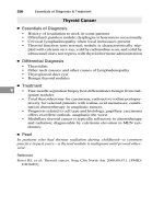
TWO
TYPES
OF
IMMUNITY
The body can control against foreign particles either by cell-mediated
immunity
or
antibody
mediated immunity. In antibody mediated
immunity, foreign particles called antigens (typically proteins or
carbohydrates on the surface of invading cells) stimulate Bcells to
become plasma cells and
memory
Bcells. The plasma cells produce
antibodies and these react with the antigens stimulating their
destruction.
In cell-mediated immunity, the reacting cells are called
helperTcells
and they cause the activation of and the differentiation of otherT cells
into
memory
T cells and effector or cytotoxicT cells. The cytotoxic T
cellscan recognize foreign cellsand destroy them. The steps in immune
reactions are much more complex than this but this description provides
a general understanding of the process. Fill in the illustration using the
terms provided. Color the different cells and antibodies using one type
of color (various types of orange for Bcells) and another for T cells.
Cell-mediated
immunity
~
~
Activates
g._
• e
h
- -
I
~)(
~
Antigen-bearing cell
1. _
b.
/
c. _
a
_
Antibody-mediated
immunity
Destroyed cell
Answer Key:a.
Antigens,
b. B cell,c. Memory B cell, d. Antibodies,e.
Plasma
cell,f.HelperTcell,g.
Activated
T cell, h. Effector
(CytotOXIC)
T cell,i.Memory T cell
OVERVIEW
OF THE
RESPIRATORY
SYSTEM
The respiratory system consists
of
the nose, nasal cavity, pharynx,
larynx, trachea, lungs,
the linings of
the lungs
(pleura)
and
the
respiratory muscles, such as the
diaphragm and intercostal muscles.
Label the respiratory figure
and
color in the major parts of the
system.
Answer
Key: a.
Pharynx,
b.
Trachea,
e. Rightlung,d.
Pleura,
e.
Nasal
cavity,
f.
Larynx,
g. Left lung,h.
Diaphragm
Chapter
Ten:
Respiratory
System
I
249
a
b
c
d
LARYNX,
TRACHEA,
AND
LUNGS
OVERVIEW
Two main cartilages of the larynx can be seen from an anterior view.The
thyroid
cartilage is superior to the cricoid cartilage. Below the larynx is
the
trachea
which divides into the
right
and left
primary
bronchi. The
right primary bronchus leads to the
right
lung
and the left primary
b. _
c.
_
Chapter
Ten
I
IAPLA~.
I 251
Respiratory System meulca
bronchus leads to the left lung. Label the parts of the respiratory system
illustrated. Color the two visible cartilages of the larynx different colors
and the trachea another color. Color the bronchi in first with a darker
color and then color the lungs in with a lighter color.
g._
Answer Key:a.
Trachea,
b.
Right
primary
bronchus,
c.
Right
lung,d.
Thyroid
cartilage,
e.
Cricoid
cartilage,
f. Leftprimary
bronchus,
g. Leftlung