Pocket Atlas of Human Anatomy 4th edition - part 3 ppsx
Bạn đang xem bản rút gọn của tài liệu. Xem và tải ngay bản đầy đủ của tài liệu tại đây (1.52 MB, 51 trang )
94
1
2
3
4
5
6
7
8
9
10
11
12
13
14
15
16
17
18
19
20
21
22
23
24
25
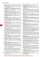
1 MUSCLES OF LOWER LIMB. Musculi membri
inferioris.
2 M. iliopsoas. Comprised of two muscles, the
psoas major and iliacus. o: Lesser trochanter. A:
Most important flexor and pre-elevator muscle
of the legs; me dial and lateral rotation of thigh
at the hip joint. B C D
3
M. iliacus. o: Iliac fossa. i: Lesser trochanter. A:
Flexion, medial and lateral rotation of thigh at
the hip joint. I: Femoral nerve and lumbar
plexus. C
4
M. psoas major. o: Bodies and transverse
processes of L1−4. i: Lesser trochanter. A: Flex-
ion, medial and lateral rotation of thigh at the
hip joint. I: Lumbar plexus. C
5 [M. psoas minor]. o: Bodies of T12 and L1. i:
Iliac fascia. I: Lumbar plexus. C
6 M. gluteus maximus. o: Posterior, external sur-
face of ilium, sacrum, coccyx, sacrotuberous
ligament. i: Iliotibial tract, gluteal tuberosity,
lateral intermuscular septum, linea aspera. A:
Extension, lateral rotation, abduction and ad-
duction of thigh at the hip joint. I: Inferior
gluteal nerve. A D E
7 M. gluteus medius. o: External surface of
ilium. i: Greater trochanter. A: Abduction, me-
dial and lateral rotation, flexion and extension
of thigh at the hip joint. I: Superior gluteal
nerve. A D E
8 M. gluteus minimus. o: External surface of
ilium between anterior and inferior gluteal
lines. i: Greater trochanter. A: Abduction, me-
dial and lateral rotation, flexion and extension
of the thigh at the hip joint. I: Superior gluteal
nerve. A D E
8a Gluteal aponeurosis. Aponeurosis glutealis.
Deep, sheet-like tendon of origin of the gluteus
maximus lying on the gluteus medius.
9 M. tensor fasciae latae. o: Near the anterior su-
perior iliac spine. i: Above the iliotibial tract
lateral to the tibial tuberosity. A: Flexion, ab-
duction and medial rotation of thigh at the hip
joint. Flexion, extension and final rotation at
the knee joint. I: Superior gluteal nerve. C E
10 M. piriformis. o: Anterior surface of sacrum. i:
Greater trochanter, inner side of apex. A: Ab-
duction, extension and lateral rotation of thigh
at the hip joint. I. Sacral plexus. A D
11 M. obturator internus. o: Inner surface of ob-
turator membrane and environment. i: Tro-
chanteric fossa. A: Lateral rotation, abduction
and adduction of thigh. I: Sacral plexus. A D
12 M. gemellus superior. o: Ischial spine. i: Ten-
don of obturator internus and trochanteric
fossa. A: Lateral rotation, adduction and abduc-
tion of thigh. I: Sacral plexus. A D E
13 M. gemellus inferior. o: Ischial tuberosity. i:
Tendon of obturator internus, trochanteric
fossa. A: Lateral rotation, adduction and abduc-
tion of thigh. I: Sacral plexus. A D E
14 M. quadratus femoris. o: Ischial tuberosity. i:
Intertrochanteric crest. A: Lateral rotation and
adduction of thigh. I: Sacral plexus. A D E
15 M. sartorius. o: Anterior superior iliac spine. i:
Medial to tibial tuberosity. A: Flexion, abduc-
tion, lateral rotation of thigh at the hip joint,
flexion and medial rotation of leg at the knee
joint. I: Femoral nerve. C E
16 M. quadriceps femoris. The muscle group
comprising the three vasti muscles and the rec-
tus femoris. I: Femoral nerve.
17
M. rectus femoris. o: Anterior inferior iliac
spine = straight head and upper margin of
acetabulum. = reflected head. i: Tibial tuberos-
ity. A: Flexion of thigh at the hip joint, exten-
sion of leg at the knee joint. B C E
18
M. vastus lateralis. o: Greater trochanter,
lateral lip of linea aspera. i: Quadriceps tendon.
A: Extension of leg at the knee joint. B C D
19
M. vastus intermedius. o: Anterior surface of
femur. i: Quadriceps tendon. A: Extension of leg
at the knee joint. B D
20
M. vastus medialis. o: Distal to intertrochan-
teric line, medial lip of linea aspera. i: Quadri-
ceps tendon. A: Extension of leg at the knee
joint. C D
21 M. articularis genus. o: Anterior surface of
femur. i: Knee joint capsule. A: Tenses capsule.
I: Femoral nerve. D
22 M. pectineus. o: Pecten pubis. i: Pectineal line
below the lesser trochanter. A: Flexion, adduc-
tion and lateral rotation of thigh at the hip joint.
I: Femoral and obturator nerves. B C D E
23 M. adductor longus. o: Near the symphysis. i:
Medial lip of linea aspera. A: Adduction and
flexion of thigh at the hip joint. I: Obturator
nerve. B C D E
24 M. adductor brevis. o: Inferior ramus of pubis.
i: Medial lip of linea aspera. A: Adduction, flex-
ion, extension and lateral rotation of thigh at
the hip joint. I: Obturator nerve. B D E
25 M. adductor magnus. o: Ischial tuberosity,
ischial ramus. i: Medial lip of linea aspera and
with a long tendon to the medial epicondyle. A:
Adduction and extension of thigh at the hip
joint. I: Obturator and sciatic nerves. B C D E
25 a M. adductor minimus. Uppermost part of the
adductor magnus muscle. It arises from a more
anterior part of the pelvis.
26 M. gracilis. o: Inferior ramus of pubis medial to
the adductor magnus muscle. i: Medial to tibial
tuberosity. A: Adduction, flexion and extension
of thigh at the hip joint. Flexion and medial ro-
tation of the knee joint. I: Obturator nerve. A C E
Muscles
Feneis, Pocket Atlas of Human Anatomy © 2000 Thieme
All rights reserved. Usage subject to terms and conditions of license.
95
1
2
3
4
5
6
7
8
9
10
11
12
13
14
15
16
17
18
19
20
21
22
23
24
25
a
a
a
6
7
8
9
15
17
22
23
24
26
14
13
12
25
11; 12 ; 13
10
7
14
6
18
23
20
25
24
22
2
25
21
19
20
2
18
8
10
11; 12 ; 13
4
5
4
3
9
2
22
23
15
25
26
20
18
17
98.14
2
22
96.1
22
18
19
98.18
17
25
23
24
7
8
7
10
12
11
13
14
26
98.14
96.6
6
96.5
96.3
25
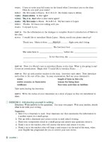
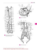
Deep muscles of hip,
posterior view
A
Thigh, anterior view
B
Thigh,
anterior view
C
Femur, posterior and anterior viewsD Hip bone, lateral viewE
Muscles
Feneis, Pocket Atlas of Human Anatomy © 2000 Thieme
All rights reserved. Usage subject to terms and conditions of license.
96
1
2
3
4
5
6
7
8
9
10
11
12
13
14
15
16
17
18
19
20
21
22
23
24
25
1 M. obturator externus. o: External surface of
obturator membrane and environment. i: Tro-
chanteric fossa. A: Lateral rotation and adduc-
tion of thighatthehipjoint. I: Obturatornerve.A
2 M. biceps femoris. o: Arises from the pelvis
and femur via two heads. i: Head of fibula. I:
Sciatic nerve, tibial part. A B E F
3
Long head of biceps femoris. Caput longum.
o: Ischial tuberosity. i: Head of fibula. A: Exten-
sion, adduction and lateral rotation of thigh at
the hip joint; flexion and lateral rotation of the
knee joint. I: Tibial nerve. A B
4
Short head of biceps femoris. Caput breve. o:
Lateral lip of linea aspera. i: Head of fibula. A:
Flexion and lateral rotation of the knee joint. I:
Common peroneal nerve. A B
5 M. semitendinosus. o: Ischial tuberosity. i: Me-
dial to tibial tuberosity [pes anserinus]. A: Ex-
tension, medial rotation and adduction at the
hip joint; flexion and medial rotation at the
knee joint. I: Tibial nerve. A D E
6 M. semimembranosus. o: Ischial tuberosity. i:
Medial condyle of tibia and oblique popliteal
ligament. It is partially covered by the semiten-
dinosus muscle. A: Extension, adduction and
medial rotation of thigh at the hip joint; flexion
and medial rotation of the knee joint. Tenses
knee joint capsule. I: Tibial nerve. A B F
7 M. tibialis anterior. o: Lateral surface of tibia,
interosseous membrane, fascia of leg (crural
fascia). i: Medial aspect of medial cuneiform
bone and 1
st
metatarsal. A: Dorsiflexion and
supination of foot. I: Deep fibular nerve. D E
8 M. extensor digitorum longus. o: Lateral tibial
condyle, interosseous membrane, fibula. i: Dor-
sal aponeurosis of toes 2−5. A: Dorsiflexion and
pronation of foot, extension of toes. I. Deep
fibular nerve. D E
9 M. peroneus tertius (m. fibularis tertius).
Muscle split off from the extensor digitorum
longus and inserting into the base of the 5
th
metacarpal. A: Dorsiflexion and pronation of
foot. I: Deep fibular nerve. D
10 M. extensor hallicus longus. o: Interosseous
membrane and fibula. i: Distal phalanx of big
toe. A: Dorsiflexion of foot, extension of big toe.
I: Deep fibular nerve. D E
11 M. peroneus longus [[m. fibularis longus]]. o:
Fibula and crural fascia. i: Medial cuneiform
bone and 1
st
metatarsal after an oblique course
below the dorsum of the foot. A: Plantar flexion
anf pronation of foot. I: Superficial fibular
nerve. C D E F
12 M. peroneus brevis [[m. fibularis brevis]]. o:
Distal 2/3 of fibula. i: Tuberosity of 5
th
metatar-
sal. A: Plantar flexion and pronation. I: Superfi-
cial fibular nerve. C D E F
13 M. triceps surae. Muscle group consisting of
the gastrocnemius and soleus; it forms the
Achilles tendon (tendo calcaneus). I: Tibial
nerve.
14
M. gastrocnemius. The superficial calf muscle
with two heads (lateral, medial). A: Flexes the
knee joint, plantar flexes and supinates the
ankle joint. A B C D
15
Lateral head of gastrocnemius. Caput laterale. o:
Proximal to the lateral femoral condyle. i:
Achilles tendon. A B C
16
Medial head of gastrocnemius. Caput laterale. o:
Proximal to the medial femoral condyle. i:
Achilles tendon. A B C D
17
M. soleus. o: Proximal ends of fibula and tibia.
i: Achilles tendon. A: Plantar flexes and supi-
nates the foot. B F
18
Tendinous arch of soleus muscle. Arcus tendineus
musculi solei. Tendinous arch above the inter-
osseous membrane. Passageway for the tibial
nerve and posterior tibial artery and vein. B
19
Tendo calcaneus [[Achilles tendon]]. The ten-
don of the triceps surae at the tuber of the cal-
caneus. B C
20 M. plantaris. o: Above the lateral femoral con-
dyle. i: Achilles tendon or tuberosity of the cal-
caneus. I: Tibial nerve. B C
21 M. popliteus. o: Lateral femoral condyle. i:
Posterior surface of tibia. A: Flexion of knee
joint and medial rotation of leg. B C F
22 M. tibialis posterior. o: Tibia, fibula, interos-
seous membrane. i: Navicular, cuneiforms,
cuboid and metatarsals 2−4. One bundle of
fibers extends backward to the sustentaculum
tali of the calcaneus. A: Plantar flexor and supi-
nator. I: Tibial nerve. C F
23 M. flexor digitorum longus. o: Tibia. i: Distal
phalanges of toes 2−5. A: Plantar flexion and
supination of foot, flexion of toes. I: Tibial
nerve. C F
24 M. flexor hallucis longus. o: Fibula. i: Distal
phalanx of big toe. A: Plantar flexion and supi-
nation of foot, flexion of big toe. I: Tibial nerve.
CF
25 M. extensor hallucis brevis. o: Dorsal surface
of calcaneus. i: Proximal phalanx of big toe. A:
Extends the big toe. I: see p. 26. D
26 M. extensor digitorum brevis. o: Dorsal sur-
face of calcaneus. i: Dorsal aponeuroses of toes
2−4. A: Extends toes. I: Deep fibular nerve. D
Muscles
Feneis, Pocket Atlas of Human Anatomy © 2000 Thieme
All rights reserved. Usage subject to terms and conditions of license.
97
1
2
3
4
5
6
7
8
9
10
11
12
13
14
15
16
17
18
19
20
21
22
23
24
25
a
a
a
211
24 12
17
21
6
22 23
57
82 11 8 10 12
5
16
7
11
12
8
10
8
26
8
25
9
20
15
21
22
11
23
24
12
19
12
11
24
23
22
16
16
19
15
17
18
20
15
4
3
2
16
6
21
94.10
94.12
94.11
94.13
94.25
94.18
2
14
16
15
6
2
5
3
4
1
94.14
Thigh,
posterior view
A Lower leg,
posterior view
B Deep muscles of
lower leg, posterior view
C Lower leg,
anterior view
D
Tibia and fibula,
anterior view
E
Tibia and fibula,
posterior view
F
Muscles
Feneis, Pocket Atlas of Human Anatomy © 2000 Thieme
All rights reserved. Usage subject to terms and conditions of license.
98
1
2
3
4
5
6
7
8
9
10
11
12
13
14
15
16
17
18
19
20
21
22
23
24
25
1 M. abductor hallucis. Abductor muscle of great
toe. o: Medial process of tuber calcanei. i: Me-
dial sesamoid and proximal phalanx of big toe.
A: Me dial abduction, supports the longitudinal
arch. I. See 2. A B
2 M. flexor hallucis brevis. Short flexor muscle of
the great toe. Origin: Cuneiform I, long plantar
ligament, tendon of the posterior tibial m. and
plantar aponeurosis. Forms a groove for trans-
mission of the flexor hallucis longus m., stabi-
lizes the longitudinal arch. A B
2a Medial head. Caput mediale. o: Tendon of the
abductor m. of the great toe, sesamoid bone
and proximal phalanx.
2b Lateral head. Caput laterale. o: Tendon of the
adductor m. of the great toe, lateral sesamoid
bone and proximal phalanx of the great toe.
3 M. adductor hallucis. Important muscle for the
transverse arch of the foot consisting of the fol-
lowing two heads.
4
Oblique head. Caput obliquum. o: Metatarsals
2−4, lateral cuneiform and cuboid bones. i:
Lateral sesamoid bone and proximal phalanx of
big toe together with the transverse head. A:
Important for stabilization of transverse and
longitudinal arches. B
5
Transverse head. Caput transversum. o: Cap-
sules of metatarsophalangeal joints 3−5. i:
Lateral sesamoid bone. A: Primary function is
to support the transverse arch of the foot. A B
6 M. abductor digiti minimi. o: Calcaneus and
plantar aponeurosis. i: Laterally on proximal
phalanx of 5
th
toe. A: Plantar flexion and abduc-
tion of the 5
th
toe. I: Lateral plantar nerve. A B
7 M. flexor digiti minimi brevis. o: Base of 5
th
metatarsal, long plantar ligament. i: Proximal
phalanx of little toe. A: Flexion and abduction
of little toe. I: Lateral plantar nerve. A B
7a [M. opponens digiti minimi]. Muscle occasion-
ally split off from the flexor digiti minimi
brevis. o: Distal half of 5
th
metatarsal.
8 M. flexor digitorum brevis. o: Tuber calcanei
and plantar aponeurosis. i: Middle phalanges of
toes 2−5 via divided tendons. A: Flexes toes and
supports the longitudinal arch of the foot. I:
Medial plantar nerve. A B
9 M. quadratus plantae (m. flexor accessorius).
o: Calcaneus. i: Lateral border of tendon of flexor
digitorum longus. A: Flexes toes and supports
longitudinal arch of foot. I: Lateral plantar nerve.
B
10 Mm. lumbricales pedis. Lumbrical muscles of
the foot. o: Tendonsofflexordigitorum longus. i:
Bases of proximal phalanges 2−5. A: Flexion at
the metatarsophalangeal joint. Brings toes
closer to the big toe. I: Medial and lateral plantar
nerves. A B
11 Mm. interossei dorsales pedis. o: Arises by two
heads from adjacent metatarsal bones. i: Base of
the proximal phalanx, plantar ligament. A: Ab-
duction and flexion of toes at the metatar-
sophalangeal joints and extension at the inter-
phalangeal joints. I: Lateral plantar nerve. C
12 Mm. interossei plantares. o: Single-headed
from metatarsal bones 3−5. i: Base of proximal
phalanges. A: Adduction and flexion of toes at
the metatarsophalangeal joints. I: cf. p. 11. C
13 Fascia lata. Fascia of thigh which envelops the
entire thigh musculature. D
14
Iliotibial tract. Tractus iliotibialis. Vertical
thick band of fascia lata that extends from the
anterior segment of the iliac crest to the lateral
tibial condyle and into which radiate the tensor
fasciae latae and gluteus maximus. D
15 Lateral intermuscular septum of thigh. Sep-
tum intermusculare femoris laterale. Firm con-
nective tissue layer extending from the fascia
lata to the lateral lip of the linea aspera between
the biceps femoris and vastus lateralis muscles.
16 Medial intermuscular septum of thigh. Sep-
tum intermusculare femoris mediale. Stout con-
nective tissue layer extending from the fascia
lata to the medial lip of the linea aspera between
the vastus medialis, sartorius and adductor
muscles.
17 Adductor canal. Canalis adductorius. Channel
between adductors, vastus medialis and [vasto-
adductor membrane]. It ends with the hiatus
tendineus within the adductor magnus. D
18 Hiatus tendineus (adductorius). Opening near
the attachment of the adductor magnus at the
level of the inferior margin of the adductor lon-
gus.
19 Iliac fascia. Fascia iliaca. Fascia over the iliac and
inferior portion of the psoas muscles. It attaches
totheiliaccrest and arcuatelineaswell as the in-
guinal ligament. D
20 Muscular lacuna. Lacuna musculorum. Com-
partment forpassageoftheiliopsoas muscle and
the femoral and lateral femoral cutaneous
nerves between the ilium,inguinalligamentand
iliopectineal arch. E
21 Iliopectineal arch. Arcus iliopectineus. Portion
of the iliac fascia between the inguinal ligament
and the iliopubic [iliopectineal] eminence. It
separates the vascular and muscular lacunae. E
22 Vascular lacuna. Lacuna vasorum. Compart-
ment between the pubis, inguinal ligament and
iliopectineal arch for passage of the femoral
artery and the femoral branch of the geni-
tofemoral nerve. E
23 Femoral triangle. Trigonum femorale. Triangle
between the sartorius and adductor longus
muscles and the inguinal ligament. D
24 Femoral canal. Canalis femoralis. Passage
within the medial segment of the vascular
lacuna that extends from the inguinal ligament
to the saphenous opening. E
Muscles
Feneis, Pocket Atlas of Human Anatomy © 2000 Thieme
All rights reserved. Usage subject to terms and conditions of license.
99
1
2
3
4
5
6
7
8
9
10
11
12
13
14
15
16
17
18
19
20
21
22
23
24
25
a
a
a
20
21
22
86.11
24
14
13
13
19
23
17
12
11
5
10
7
6
9
8
96.23
1
4
2
8
5
6
8
100.17
96.24
2
10
7
1
Superficial plantar musclesA Deep plantar muscles
B Interosseous muscles
C
Thigh, lateral and anterior medial view
D Vascular lacuna
E
Muscles
Feneis, Pocket Atlas of Human Anatomy © 2000 Thieme
All rights reserved. Usage subject to terms and conditions of license.
100
1
2
3
4
5
6
7
8
9
10
11
12
13
14
15
16
17
18
19
20
21
22
23
24
25
1 Femoral ring. Anulus femoralis. Entrance into
the femoral canal bordered by the femoral vein,
inguinal ligament, falx inguinalis and pectineal
ligament. A
2 Femoral septum. Septum femorale. Fibrous
membrane that closes the entrance of the
femoral canal. A
3 Saphenous opening. Hiatus saphenus. Large
opening in the fascia lata directly below the in-
guinal ligament for passage of the great
saphenous vein. B
4
Falciform margin. Margo falciformis. Curved,
principal lateral margin of the saphenous open-
ing. B
5
Superior horn. Cornu superius. Upper, curved
portion of the falciform margin. B
6
Inferior horn. Cornu inferius. Lower, curved por-
tion of the falciform magin. B
7
Cribriform fascia. Fascia cribrosa. Loose, per-
forated connective tissue lamina covering the
saphenous opening. B
8 Fascia of the leg (crural fascia). Fascia cruris.
Superficial investing fascia of the leg which
serves partially for muscle attachment and is
fused to the free bony margins of the tibia. C D F
9 Anterior intermuscular septum of leg. Septum
intermusculare cruris anterius. Connective
tissue septum between the peroneal and exten-
sor compartments. F
10 Posterior intermuscular septum of leg. Sep-
tum intermusculare cruris posterius. Connec-
tive tissue septum between the peroneal and
flexor compartments. F
11 Superior extensor retinaculum. Retinaculum
mm. extensorum superius. Transverse
thickened (about two finger’s breadth) of the
crural fascia that hold the extensor tendons in
place. C D
12 Flexor retinaculum. Retinaculum mm. flex-
orum. Fibrous band on the long flexor tendons
that extends from the medial malleolus to the
calcaneus. It forms an osteofibrous compart-
ment for the posterior tibial m., then divides
into two parts. The lower portion forms com-
partments for the flexor digitorum longus and
flexor hallucis longus muscles. The tibial nerve
and posterior tibial artery and vein lie between
the two membranous parts. D
13 Inferior extensor retinaculum. Retinaculum
mm. extensorum inferius. Usually cruciate
band that supports the extensor tendons, ex-
tending from both malleoli to the foot margins
of the opposite side, primarily to the calcaneus.
CD
14 Superior peroneal (fibular) retinaculum. Reti-
naculum mm. peroneorum (fibularium) su-
perius. Upper band that holds peroneal tendons
in place; it extends from the lateral malleolus
to the calcaneus. C
15 Inferior (fibular) peroneal retinaculum. Reti-
naculum mm. peroneorum (fibularium) in-
ferius. Lower band that holds the peroneal ten-
dons in place. It passes from the extensor reti-
naculum to the outer surface of the calcaneus.
A fibrous tract goes to the peroneal trochlea
and separates the upper lying peroneus brevis
from the peroneus longus muscle. C
16 Fascia dorsalis pedis. Thin fascia on the dor-
sum of the foot connected above with the infe-
rior extensor retinaculum. C D
17 Plantar aponeurosis. Aponeurosis plantaris.
Tough, tendinous sheet on the sole of the foot
extending from the tub er calcanei to as far as
the middle phalanges. It braces the longitudinal
arch of the foot. E
18
Transverse fasciculi. Fasciculi transversi.
Transverse fibrous sheets in the distal plantar
aponeurosis. E
19 Superficial transverse metatarsal ligament.
Lig. metatarsale transversum superficiale.
Transverse fibrous tract in the vicinity of the
distal transverse fibers of the plantar
aponeurosis. E G
19 a Synovial bursae (sacs) and sheaths. Bursae et
vaginae synoviales.
20 Synovial sheaths of the digits of the foot.
Vaginae synoviales tendinum digitorum pedis.
Synovial portion of the tendon sheaths for the
flexors of the toes. G
21
Vincula tendinum. Connective tissue tract
passing obliquely through the tendon sheaths
bearing blood vessels. G
22 Fibrous sheaths of the digits of the foot.
Vaginae fibrosae tendinum digitorum pedis.
Tough fibrous sheath that reinforces the tendon
sheaths on the flexor side of the toes. G
23
Annular par t of f ibrous sheath. Pars anularis
vaginae fibrosae. Circular tracts in the f ibrous
sheaths between the joints. G
24
Cruciate part of fibrous sheath. Pars cruci-
formis vaginae fibrosae. Crucitate connective
tissue tracts in the fibrous sheaths over the
joints. G
Muscles, synovial bursae and sheaths
Feneis, Pocket Atlas of Human Anatomy © 2000 Thieme
All rights reserved. Usage subject to terms and conditions of license.
101
1
2
3
4
5
6
7
8
9
10
11
12
13
14
15
16
17
18
19
20
21
22
23
24
25
a
a
a
21
20 19 19
24
22
20
23
24
8
8
9
10
8
19
17
18
11
13
16
8
12
8
11
13
14
15
16
7
4
6
3
5
86.20
86.18
1
2
86.21
Foot, medial viewD
Toes, plantar view
GCross-section of lower legFPlantar surface of footE
Foot, lateral viewC
Fascia lata in inguinal regionB
Vascular lacuna from behind
A
Muscles, synovial bursae and sheaths
Feneis, Pocket Atlas of Human Anatomy © 2000 Thieme
All rights reserved. Usage subject to terms and conditions of license.
102
1
2
3
4
5
6
7
8
9
10
11
12
13
14
15
16
17
18
19
20
21
22
23
24
25
1 Tendon sheath of superior oblique muscle.
Vagina tendinis m. obliqui superioris. Synovial
sheath of the superior oblique m. of the eyeball,
situated at the site where its tendon passes
through the trochlea. See p. 364. 12
2 Synovial bursa of tensor veli palatini. Bursa m.
tensoris veli palatini. Synovial bursa between
the pterygoid hamulus and the tendon of the
tensor veli palatini muscle. See pp. 116.20, 117.
C
3 Subcutaneous bursa of the laryngeal promi-
nence. B. subcutanea prominentiae laryngealis.
Synovial bursa between the skin and the laryn-
geal prominence of the thyroid cartilage. A
4 Retrohyoid bursa. B. retrohyoidea. Synovial
bursa between the body of the hyoid bone and
the median thyrohyoid ligament. A
5 Infrahyoid bursa. B. infrahyoidea. Synovial
bursa between the upper end of the sternohy-
oid muscle and the thyrohyoid membrane. A B
5a Synovial bursae of upper limb. Bursae membri
superioirs.
6 Subtendinous bursa of trapezius. B. subten-
dinea m. trapezii. Synovial bursa between the
trapezius muscle (ascending part) and the
spine of the scapula. C
7 [B. subcutanea acromialis]. Synovial bursa be-
tween acromion and the skin. D
8 Subacromial bursa. B. subacromialis. Synovial
bursa between the acromion, coracoacromial
ligament and supraspinous tendon. It and its
tendons lie on the joint capsule. D E
9 Subdeltoid bursa. B. subdeltoidea. Synovial
bursa between the deltoid muscle and the
greater tubercle of the humerus. It often com-
municates with the subacromial bursa. D
10 Coracobrachial bursa. [b. m. coracobrachialis].
Synovial bursa between the tendons of the sub-
scapularis and coracobrachialis muscles below
the apex of the coracoid process. D
11 Subtendinous bursa of infraspinatus muscle.
B. subtendinea m. infraspinati. Synovial bursa
between the tendon of the infraspinatus and
the capsule of the shoulder joint. E
12 Subtendinous bursa of subscapularis muscle.
B. subtendinea m. subscapularis. Synovial
bursa between the tendon of the subscapularis
and the capsule of the shoulder joint. It com-
municates with the joint cavity. D
13 Subtendinous bursa of teres major muscle. B.
subtendinea m. tertis majoris. Synovial bursa
between the tendon of the teres major and the
humerus. D
14 Subtendinous bursa of latissimus dorsi
muscle. B. subtendinea m. latissimi dorsi. Syn-
ovial bursa between the tendons of the teres
major and latissimus dorsi. D
15 Subcutaneous bursa of olecranon. B. subcu-
tanea olecrani. Synovial bursa between the
olecranon and the skin. F
16 Intratendinous bursa of olecranon. [B. in-
tratendinea olecrani]. Synovial bursa within
the triceps tendon near the olecranon. F
17 Subtendinous bursa of triceps brachii. B. sub-
tendinea m. tricipitis brachii. Synovial bursa
between the triceps tendon and the olecranon.
F
18 Bicipitoradial bursa. B. bicipitoradialis. Syn-
ovial bursa between the biceps tendon and the
anterior part of the radial tuberosity. F
19 [B. cubitalis interossea]. Synovial bursa be-
tween the biceps tendon and the ulna or ob-
lique cord. F
20 Tendon sheath of abductor pollicis longus and
extensor pollicis brevis muscles. Vag. ten-
dinum mm. abductoris longi et extensoris
brevis pollicis. Common tendon sheath forming
the first tendon compartment on the dorsum of
the hand. G
21 Tendon sheath of extensor carpi radialis lon-
gus and brevis muscles. Vag. tendinum mm.
extensorum carpi radialium. Common tendon
sheath forming the second tendon compart-
ment on the dorsum of the hand. G
22 Tendon sheath of extensor pollicis longus
muscle. Vag. tendinis m. extensoris pollicis
longi. Forms the third tendon compartment. G
23 Tendon sheath of extensor digitorum and ex-
tensor indicis muscles. Vag. tendinum mm. ex-
tensoris digitorum et extensoris indicis. Tendon
sheath forming the fourth tendon compart-
ment on the dorsum of the hand. G
24 Tendon sheath of extensor digiti minimi
muscle. Vag. tendinis m. extensoris digiti min-
imi. Forms the fifth tendon compartment on
the dorsum of the hand. G
25 Tendon sheath of extensor carpi ulnaris
muscle. Vag. tendinis m. extensoris carpi ul-
naris. Forms the sixth tendon compartment on
the dorsum of the hand. G
26 Sheath of extensor carpi radialis brevis
muscle. Vag. m. extensoris carpi radialis brevis.
Synovial bursa at the attachment between the
tendon and base of the 3
rd
metacarpal.
Synovial bursae and sheaths
Feneis, Pocket Atlas of Human Anatomy © 2000 Thieme
All rights reserved. Usage subject to terms and conditions of license.
103
1
2
3
4
5
6
7
8
9
10
11
12
13
14
15
16
17
18
19
20
21
22
23
24
25
a
a
a
22
20
21
23
24
25
17
16
15
18
19
8
11
7
8
10
12
14
13
9
6
5
4
5
3
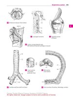
Sagittal section
of larynx
A Larynx,
lateral view
B Right shoulder,
posterior view
C
Shoulder joint, anterior view
D Shoulder joint, posterior view
E
Section of elbow joint sawed open
F Wrist and hand, dorsal viewG
Synovial bursae and sheaths
Feneis, Pocket Atlas of Human Anatomy © 2000 Thieme
All rights reserved. Usage subject to terms and conditions of license.
104
1
2
3
4
5
6
7
8
9
10
11
12
13
14
15
16
17
18
19
20
21
22
23
24
25
1 Tendon sheath of flexor carpi radialis muscle.
Vag. tendinis m. flexoris carpi radialis. In-
dividual tendon sheath for the flexor carpi
radialis at the insertion of the tendon to the
base of the 2
nd
metacarpal bone. A
2 Common sheath of flexor muscles. Vag. com-
munis mm. flexorum. Common tendon sheath
for the two long flexors of the fingers. A
3 Tendon sheath of flexor pollicis longus
muscle. Vag. tendinis m. flexoris pollicis longi.
Separate synovial sheath for the long flexor of
the thumb. A
4 Tendon sheaths for flexors in region of fin-
gers. Vag. tendinum digitorum manus. A
4a Synovial bursae of lower limb. Bursae membri
inferioris.
5 Subcutaneous trochanteric bursa. Bursa sub-
cutanea trochanterica. Synovial bursa on the
tendon of the gluteus maximus between the
skin and greater trochanter. B
6 Trochanteric bursa of gluteus maximus. B.
trochanterica m. glutei maximi. Synovial bursa
between the tendon of the gluteus maximus
and the greater trochanter. B
7 Trochanteric bursae of gluteus medius. Bb.
trochantericae m. glutei medii. This designa-
tion comprises two synovial bursae, an anterior
one between the tendon of insertion of the glu-
teus medius and the greater trochanter and a
posterior one between this tendon and the piri-
formis muscle. B C
8 Trochanteric bursa of gluteus minimus. B. tro-
chantericae m. glutei minimi. Synovial bursa
between the tendon of insertion of the gluteus
minimus and the greater trochanter. B C
9 Bursa of piriformis muscle. B. m. piriformis.
Synovial bursa between the piriformis tendon,
femur and superior gemellus muscle. B
10 Ischial bursa of obturator internus muscle. B.
ischiadica (sciatica) m. obturatoris interni. Syn-
ovial bursa between the cartilage-covered sur-
face of the lesser sciatic notch and the tendon
of the obturator internus. B
11 Subtendinous bursa of obturator internus
muscle. B. subtendinea m. obturatoris interni.
Synovial bursa below the insertion of the obtu-
rator internus. B
12 Intermuscular bursae of gluteal muscles. Bb.
intermusculares mm. gluteorum. 2−3 synovial
bursae that extend inferiorly from the gluteus
maximus to the linea aspera. B
13 Ischial bursa of gluteus maximus muscle. B.
ischiadica (sciatica) m. glutei maximi. Synovial
bursa between the ischial tuberosity and the
inferior surface of the gluteus maximus. B
14 Iliopectineal bursa. [B. iliopectinea]. Synovial
bursa between the iliopsoas muscle and the
pelvic bone. It lies above and often communi-
cates with the hip joint. C
15 Subtendinous iliac bursa. B. subtendinea iliaca.
Synovial bursa between the lesser trochanter
and the iliopsoas tendon. C
16 Superior bursa of biceps femoris muscle. B. m.
bicipitis femoris superior. Synovial bursa be-
tween the origins of the biceps femoris and
semimembranosus muscles. B
17 Subcutaneous prepatellar bursa. B. subcu-
tanea prepatellaris. Synovial bursa directly be-
tween the skin and the fascia in front of the
knee. D
18 Subfascial prepatellar bursa. [B. subfascialis
prepatellaris]. Synovial bursa between the in-
vesting fascia of the knee and the tendon of the
quadratus femoris muscle. D
19 Subtendinous prepatellar bursa. [B. subten-
dinea prepatellaris]. Synovial bursa directly on
the knee joint below the tendon of the quadra-
tus femoris. D
20 Suprapatellar bursa. B. suprapatellaris. Syn-
ovial bursa b etween the quadriceps tendon and
the femur. It almost always communicates with
the joint cavity. D
21 Subcutaneous infrapatellar bursa. B. subcu-
tanea infrapatellaris. Synovial bursa between
the ligamentum patellae and the skin. D
22 Deep infrapatellar bursa. B. infrapatellaris pro-
funda. Synovial bursa between the ligamentum
patellae and the tibia. D
23 Subcutaneous bursa of tibial tuberosity. B.
subcutanea tuberositas tibiae. Synovial bursa
between the tibial tuberosity and the skin. It is
mostly involved in kneeling. D
24 Subtendinous bursae of sartorius muscle. Bb.
subtendineae m. sartorii. Synovial bursae be-
tween the sartorius tendon and the tendons of
the gracilis and semitendinosus situated below
it. E
Synovial bursae and sheaths
Feneis, Pocket Atlas of Human Anatomy © 2000 Thieme
All rights reserved. Usage subject to terms and conditions of license.
105
1
2
3
4
5
6
7
8
9
10
11
12
13
14
15
16
17
18
19
20
21
22
23
24
25
a
a
a
24
20
18
17
19
22
21
23
14
7
15
8
7
5
12
16
13
10
11
6
8
9
4
3
2
1
3
2
4
Palmar view of handA
Deep hip region,
dorsal view
B
Hip joint, anterior view
C
Knee, sagittal section sawed open
D Knee, anterior viewE
Synovial bursae and sheaths
Feneis, Pocket Atlas of Human Anatomy © 2000 Thieme
All rights reserved. Usage subject to terms and conditions of license.
106
1
2
3
4
5
6
7
8
9
10
11
12
13
14
15
16
17
18
19
20
21
22
23
24
25
1 Anserine bursa. B. anserina. Synovial bursa on
the tibial collateral ligament below the tendons
of the semitendinosus, gracilis and sartorius
muscles. It occasionally communicates with
the subtendinous bursa of the sartorius. A
2 Inferior subtendinous bursa of biceps femoris
muscle. B. subtendinea m. bicipitis femoris in-
ferior. Synovial bursa located partially on the
fibular collateral ligament below the tendon of
insertion of the biceps femoris. B
3 Subpopliteal recess. Recessus subpopliteus
[bursa m. poplitei]. Synovial bursa on the
lateral femoral condyle below the tendon of
origin of the popliteal muscle. It always com-
municates with the knee joint cavity, more
rarely with the tibiofibular joint. B
4 Lateral subtendinous bursa of gastrocnemius
muscle. B. subtendinea m. gastrocnemii later-
alis. Synovial bursa between the lateral condyle
of the femur and the lateral gastrocnemius ten-
don. B
5 Medial subtendinous bursa of gastrocnemius
muscle. B. subtendinea m. gastrocnemii medi-
alis. Synovial bursa between the medial con-
dyle of the femur and the medial gastrocne-
mius tendon. A B
6 Bursa of semimembranosus muscle. B. m.
semimembranosi. Synovial bursa between the
semimembranosus tendon and the upper mar-
gin of the tibia. A
7 Subcutaneous bursa of lateral malleolus. B.
subcutanea malleoli lateralis. Synovial bursa
between the skin and the lateral malleolus. C
8 Subcutaneous bursa of medial malleolus. B.
subcutanea malleoli medialis. Synovial bursa
between the skin and the medial malleolus. D
9 Tendon sheath of tibialis anterior muscle. Vag.
tendinis m. tibialis anterioris. It begins just
below the extensor retinaculum. D
10 Tendon sheath of extensor hallucis longus
muscle. Vag. tendinis m. extensoris hallucis
longi. Sheath extending below the extensor ret-
inaculum and further distal. C D
11 Tendon sheath of extensor digitorum longus
muscle. Vag. tendinum m. extensoris digitorum
pedis longi. Sheath extending below the exten-
sor retinaculum and further distal. C
12 Tendon sheath of flexor digitorum longus
muscle. Vag. tendinum m. flexoris digitorum
pedis longi. It lies behind and below the medial
malleolus covered by the flexor retinaculum. D
13 Tendon sheath of tibialis posterior muscle.
Vag. tendinis m. tibialis posterioris. It resides
below the flexor retinaculum and begins at the
point where it is crossed over by the flexor digi-
torum longus. D
14 Tendon sheath of flexor hallucis longus
muscle. Vag. tendinis m. flexoris hallucis longi.
It extends up to the proximal end of the sole,
where it crosses under the tendon of the flexor
digitorum longus. D
15 Common tendon sheath for peroneal muscles.
Vag. tendinum mm. peroneorum (fibularium)
communis. It lies below the peroneal reti-
naculum and extends to the cuboid bone. C
16 Subtendinous bursa of tibialis anterior
muscle. B. subtendinea m. tibialis anterioris.
Synovial bursa between the tibialis anterior
tendon and the medial cuneiform bone. D
17 Subcutaneous calcaneal bursa. B. sucutanea
calcanea. Synovial bursa between the skin and
the posterior surface of the calcaneus. D
18 Bursa of calcaneal [[Achilles]] tendon. B. ten-
dinis calcanei [Achilles]. Synovial bursa be-
tween the calcaneus and the Achilles tendon. D
19 Tendon sheath of peroneus longus muscle at
the sole of the foot. Vag. tendinis m. peronei
(fibularis) longi plantaris. D
20 Tendon sheaths for the flexors of the toes.
Vagg. tendinum digitorum pedis. D
Synovial bursae and sheaths
Feneis, Pocket Atlas of Human Anatomy © 2000 Thieme
All rights reserved. Usage subject to terms and conditions of license.
107
1
2
3
4
5
6
7
8
9
10
11
12
13
14
15
16
17
18
19
20
21
22
23
24
25
a
a
a
10
9
13
12
14
18
17
14121916
20
8
10
7
15
10
11
5
4
3
2
5
1
6
Right knee joint, posterior viewA
Right knee joint, posterior view
B
Foot, lateral view
C
Foot, medial view
D
Synovial bursae and sheaths
Feneis, Pocket Atlas of Human Anatomy © 2000 Thieme
All rights reserved. Usage subject to terms and conditions of license.
108
1
2
3
4
5
6
7
8
9
10
11
12
13
14
15
16
17
18
19
20
21
22
23
24
25
1 DIGESTIVE SYSTEM. Apparatus digestorius
(systema alimentarium).
2 ORAL CAVITY. Cavitas oris.
3 Vestibule of mouth. Vestibulum oris. Space be-
tween the rows of teeth and the lips or cheeks.
BC
4 Oral fissure. Rima oris. Mouth opening be-
tween the lips. A
5 Lips. Labia oris.
6
Upper lip. Labium superius. A B C
7
Philtrum. Groove extending from nasal septum
to upper lip. A
8
Tuberculum. Small eminence on upper lip mark-
ing end of philtrum. A
9
Lower lip. Labium inferius. A B C
10 Commissure of lips. Commissura labiorum.
Transition of upper lip into lower lip at the
angle of the mouth. A B
11 Angle of mouth. Angulus oris. A
12 Cheek. Bucca. Lateral wall of vestibule of
mouth. A
13
Buccal fat pad. Corpus adiposum buccae. [[Bi-
chat]]. Encapsulated body of fat between the
buccinator and masseter muscles. A
14 Oral cavity proper. Cavitas oris propria. True
oral cavity enclosed anteriorly and laterally by
the teeth and extending as far as the isthmus of
fauces (oropharyngeal isthmus). C
15 Palate. Palatum. Partition between oral and
nasal cavities.
16
Hard palate. Palatum durum. Hard, bony part
of the palate. C D
17
Soft palate. Palatum molle (velum palatinum).
Soft, posterior part of the palate. C D
18 Palatine raphe. Raphe palati. Median mucosal
ridge at the junction of the right and left bony
palatal processes. D
19 Oral mucosa. Tunica mucosa oris. Mucous
membrane of oral cavity consisting of
stratified, nonkeratinized squamous
epithelium throughout and underlying mixed
glands.
20 Frenulum of upper lip. Frenulum labii super-
ioris. Median mucosal fold between the gums
and upper lip. B
21 Frenulum of lower lip. Frenulum labii inferi-
oris. Median mucosal fold between the gums
and lower lip. B
22 Gums. Gingivae. Mucous membrane united
firmly with the teeth and jaw bones. B D
23
Gingival (gum) margin. Margo gingivalis. B D
24
Gingival (interdental) papilla. Papilla ging-
ivalis (interdentalis). B D
25
Gingival sulcus. Sulcus gingivalis. Shallow fur-
row between the gum margin and the tooth. Its
deepening leads to cavity formation. See p. 113.
A
26 Sublingual papilla. Caruncula sublingualis. A
small mucosal eminence on either side of the
frenulum. It receives the opening of the sub-
mandibular duct and the major sublingual
duct. B
27 Sublingual fold. Plica sublingualis. Mucosal
fold overlying the sublingual gland and extend-
ing posterolaterally from the sublingual papilla.
B
28 Parotid papilla. Papilla ductus parotidei. Small
mucosal elevation at the opening of the parotid
duct lateral to the second upper molar tooth. B
29 Transverse palatine folds. Plicae palatinae
transversae. Mucosal folds running trans-
versely on the anterior part of the hard palate.
D
30 Incisive papilla. Papilla incisiva. Small mucosal
elevation over the incisive foramen at the ante-
rior end of the palatine raphe. D
30 a GLANDULAE ORIS. The glands of the mouth.
31 Small glands of the oral cavity. Glandulae
salivariae minores.
32 Labial glands. Gll. labiales. Small salivary
glands at the inner aspect of the lips. B
33 Buccal glands. Gll. buccales. Small mucous
salivary glands at the inner aspect of the
cheeks. B
34 Molar glands. Gll. molares. Salivary glands cor-
responding to the buccal glands situated
beneath the mucosal at the level of the molar
teeth. B
35 Palatine glands. Gll. palatinae. Salivary glands
situated beneath the mucosa of the palate.
(Two large groups right and left of the midline.)
D
36 Lingual glands. Gll. linguales. Numerous
mucous, serous and mixed glands primarily in
the lateral and posterior areas of the tongue. B
37
Anterior lingual glands. Gl. lingualis anterior
[[gl. apicis linguae, Nuhn’s glands]]. Mixed
glands near the apex of the tongue providing
several drainage ducts on the undersurface of
the tongue. B
Digestive system
Feneis, Pocket Atlas of Human Anatomy © 2000 Thieme
All rights reserved. Usage subject to terms and conditions of license.
109
1
2
3
4
5
6
7
8
9
10
11
12
13
14
15
16
17
18
19
20
21
22
23
24
25
a
a
a
Digestive system
24
30
23
18
16
29
16
22
1717
35
16
14
17
6
3
9
20 22
6
33
34
37
36
28
10
26
9
3
21 22
24
23
27
32
7
1013
6
8
411
12
9
Face, anterior viewA
Mouth with tongue elevated
B
Sagittal section of oral cavity
C Palate, inferior viewD
Feneis, Pocket Atlas of Human Anatomy © 2000 Thieme
All rights reserved. Usage subject to terms and conditions of license.
110
1
2
3
4
5
6
7
8
9
10
11
12
13
14
15
16
17
18
19
20
21
22
23
24
25
1 Major salivary glands. Glandulae salivariae
majores.
2 Sublingual gland. Glandula sublingualis. Pre-
dominantly mucous gland situated on the my-
lohyoid muscle diaphragma oris. It contains
several drainage ducts. D
3
Major sublingual duct. Ductus sublingulis
major. Main drainage duct of the sublingual
gland. It opens at the sublingual caruncle next
to the submandibular duct. D
4
Minor sublingual ducts. Ductus sublinguales
minores. About 40 small ducts that drain the
sublingual gland and open along the sublingual
fold and the sublingual caruncle. D
5 Submandibular gland. Glandula submandibu-
laris. Predominantly serous salivary gland lo-
cated almost entirely below the mylohyoid. D F
6
Submandibular duct. Ductus submandibu-
laris. Duct that drains the submandibular
gland. It loops around the posterior margin of
the mylohyoid accompanied by glandular
tissue and opens at the sublingual caruncle. D
7 Parotid gland. Glandula parotidea. Salivary
gland located behind and on the mandibular
ramus. F
8
Superf icial part. Pars superficialis. The parts
of the parotid gland located superficial to the
facial nerve. F
9
Deep par t. Pars produnda. Parts of the parotid
gland located deep with respect to the facial
nerve. F
10
Accessory parotid gland. Glandula parotidea
accessoria. Portion of the parotid gland located
on the masseter muscle near the parotid ex-
cretory duct. F
11
Parotid duct. Ductus parotideus. Excretory
duct of the parotid gland. It passes around the
anterior margin of the masseter and opens near
the second upper molar tooth. F
12 TEETH. Dentes. A B C D E G
13 Crown of tooth. Corona dentis. Portion of the
tooth covered by enamel. E
14
Cusp of tooth. Cuspis dentis [[tuberculum]]. 1−
5 protuberances on the occlusal surface of
tooth (with the exception of the incisors). E
15
Apex of cusp. Apex cuspidis. E
16 Dental tubercle. Tuberculum dentis. Distinct
eminence on the side of the crown, especially
in canine and incisor teeth. A
17
Transverse ridge. Crista transversalis. Trans-
verse connecting ridge between adjacent
cusps. B
18
Triangular ridge. Crista triangularis. Triangu-
lar connecting ridge between the cusps of the
molars. B
19 Clinical crown. Corona clinica. Portion of the
tooth projecting above the gum. C
20 Neck of tooth. Cervix [[collum]] dentis. Portion
of the tooth at the enamel-cementum border. E
21 Root of tooth. Radix dentis. Portion of the
tooth covered by cementum. E
22
Apexofrootoftooth.Apex radicis dentis. E
23 Clinical root. Radix clinica. Portion of the tooth
situated below the gum. C
24 Occlusal (masticator y) surface. Facies oc-
clusalis (masticatoria) dentis. B E
25 Vestibular (facial) surface. Facies vestibularis
(facilis) dentis. Tooth surface facing the vesti-
bule. D G
25 a
Buccal sur face. Facies buccalis dentis. Tooth
surface facing the cheek.
25 b
Labial surface. Facies labialis dentis. Tooth
surface facing the lips.
26 Lingual and palatine surfaces. Facies lingualis/
palatalis dentis. Tooth surfaces facing the
tongue and palate, respectively. A G
27 Approximal surface. Facies approximalis.
Tooth surface facing the adjacent tooth. G
28
Mesial surface. Facies mesialis. Vertical con-
tact surface of a tooth turned away from the last
molar. G
29
Distal surface. Facies distalis. Vertical contact
surface of a tooth facing away from the first in-
cisor. G
29 a
Contingent area. Area contingens. Direct contact
surface of adjacent teeth.
30 Cingulum. Ridge near the neck of a tooth con-
necting both marginal crests at the lingual sur-
face of incisor and canine teeth. A
31 Marginal crest. Crista marginalis. Lateral
marginal ridge on the lingual surface of the in-
cisor and canine teeth which goes over into the
cingulum at the neck region. A
32 Incisal margin. Margo incisalis. Occlusal edge
of incisor and canine teeth. A
Digestive system
Feneis, Pocket Atlas of Human Anatomy © 2000 Thieme
All rights reserved. Usage subject to terms and conditions of license.
111
1
2
3
4
5
6
7
8
9
10
11
12
13
14
15
16
17
18
19
20
21
22
23
24
25
a
a
a
Digestive system
25
28
28
26
28 29
27
29
29
11
10
8
9
7
5
14 15
24
20
13
21
22
First lower molarE
56
3
25
4
2
19
23
17
18
24
First and second molars,
occlusal surface
B
32
2626
30
16
31
Incisor tooth and canine tooth,
lingual surface
A
Oral cavity,
medial view
D
Salivary glands, lateral viewF Teeth of lower jawG
Incisor, sagittal
section
C
Feneis, Pocket Atlas of Human Anatomy © 2000 Thieme
All rights reserved. Usage subject to terms and conditions of license.
112
1
2
3
4
5
6
7
8
9
10
11
12
13
14
15
16
17
18
19
20
21
22
23
24
25
1 Pulp cavity of tooth. Cavitas dentis (pulparis).
Cavity in the dentin. Towards the root, it be-
comes continuous with the root canal. A
2
Pulp chamber in the crown. Cavitas coronae.
Crown portion of the pulp cavity. A
3
Root canal of tooth. Canalis radicis dentis.
Canal between the pulp cavity and the apical
foramen. A
4
Apical foramen of root of tooth. Foramen apicis
radicis dentalis. The opening of the root canal at
the apex of the root. A
5 Pulp of tooth. Pulpa dentis. Contents of the
pulp cavity consisting of loose, finely fibered
connective tissue, blood vessels and nerves.
6
Crown pulp. Pulpa coronalis. Pulp within the
crown portion of the pulp cavity.
7
Root pulp. Pulpa radicularis. Pulp within the
root canal portion of the pulp cavity.
8 Dental papilla. Papilla dentis. A mass of
mesenchyme present in the bell stage of tooth
development. B
9 Dentine. Dentinum [[substantia eburnea]]. Pre-
dominant mass of a tooth consisting of inor-
ganic and organic material (especially col-
lagenous fibers). A C
10 Enamel. Enamelum [[substantia adamantina]].
The extremely hard substance surrounding the
crown of the tooth like a mantle. A C
11 GOMPHOSIS. Type of fibrous joint in which a
conical process, e. g., a tooth, is inserted into a
socket, e. g., alveolus of the jaw (dentoalveolar
articulation).
12 Periodontium. Tissues that invest and support
the tooth within the alveolus. It consists of the
following parts: A
13
Periodontium protectoris [[gingiva]]. The
outer part of the periodontium with the exter-
nal border epithelium. A
14
Periodontium insertionis. Portion of the peri-
odonium touching the tooth. It consists of the
inner border epithelium and the periodontal
ligament. A
15
Periodontal ligament. Desmodontium. All con-
nective tissue fibers which are anchored in the
cementum and, with their vessels and nerves,
extend partly into the gum and partly into the
alveolar wall. A
16
Cementum. Substance similar to bone. It sur-
rounds the tooth from the enamel border to the
apex of the root and receives the fibers of the
periodontal ligament. A
17
Alveolar bone. Os alveolare. Bony wall of the
alveolus. A
18 Superior dental arch. Arcus dentalis superior.
Curved row of teeth of the maxilla.
19 Inferior dental arch. Arcus dentalis inferior.
Curved row of teeth of the mandible.
20 Incisor teeth. Dentes incisivi. Cutting teeth lo-
cated on both sides of the midline at the 1
st
and
2
nd
positions of the dental arch. D
21 Canine teeth. Dentes canini. Teeth located at
the 3
rd
position of the dental arch. D
22 Premolar teeth. Dentes premolares. Teeth oc-
cupying the 4
th
and 5
th
positions of the dental
arch. D
23 Molar teeth. Dentes molares. Teeth located at
the 6
th
,7
th
and 8
th
positions of the dental arch. D
24
Wisdom tooth. Dens serotinus [molaris ter-
tius]. Tooth located at the 8
th
position of the
dental arch. D
25 Deciduous (milk) teeth. Dentes decidui.
26 Permanent teeth. Dentes permanentes. Teeth
that develop after the deciduous teeth.
27
Diastema. Space between adjacent teeth.
28 TONGUE. Lingua. D E
29 Body of tongue. Corpus linguae. Portion of the
tongue situated between the apex and root. E
30 Root of tongue. Radix linguae. Anchoring re-
gion of the tongue at the mandible and hyoid
bone. Also the posterior, vertical segment of the
tongue. E
31 Dorsum of tongue. Dorsum linguae. E
32
Anterior, oral portion of tongue. Pars pre-
sulcalis (anterior). The part of the dorsum of
the tongue situated anterior to the sulcus ter-
minalis. 115 B
33
Posterior, pharyngeal par t of tongue. Pars
postsulcalis (posterior). The vertical portion of
the dorsum of the tongue between the sulcus
terminalis and the epiglottis. 115 B
34 Inferior sur face of tongue. Facies inferior lin-
guae. D E
35
Fimbriated fold. Plica fimbriata. Serrated fold
lateral to the frenulum. It is a remnant of the in-
ferior tongue. D
36 Lateral margin of tongue bordering the teeth.
Margo linguae. D
37 Apex of tongue. Apex linguae. D E
Digestive system
Feneis, Pocket Atlas of Human Anatomy © 2000 Thieme
All rights reserved. Usage subject to terms and conditions of license.
113
1
2
3
4
5
6
7
8
9
10
11
12
13
14
15
16
17
18
19
20
21
22
23
24
25
a
a
a
Digestive system
29
31
30
34
37
22
20
21
24
21
20
21
23
36
35
37
34
114.2
910
8
1
10
108.25
14
13
17
16
2
15
4
12
3
9
Longitudinal section of toothA
Tooth development
B
Enamel-dentin border
C
Mouth with tongue elevatedD Tongue, sagittal sectionE
Feneis, Pocket Atlas of Human Anatomy © 2000 Thieme
All rights reserved. Usage subject to terms and conditions of license.
114
1
2
3
4
5
6
7
8
9
10
11
12
13
14
15
16
17
18
19
20
21
22
23
24
25
1 Mucous membrane of tongue. Tunica mucosa
linguae. C
2 Frenulum of tongue. Frenulum linguae. Mu-
cosal fold extending from the floor of the
mouth to the inferior side of the tongue. D; see
also p. 113. D
3 Lingual papillae. Papillae linguales. Collective
term for the following five different types of
mucosal formations: A B
4
Filiform papillae. Papillae filiformes. Fine, al-
most threadlike epithelial elevations on a con-
nective tissue core. Their tips that are of ten
cleft. A
5
Conical papillae. Papillae conicae. Special
form of filiform papilla. They are somewhat
larger, longer and exhibit conical apices which
bend backwards. A
6
Fungiform papillae. Papillae fungiformes.
Mushroom-like papillae which are not pointed
at the tip, but terminate with a small plateau. A
B
7
Vallate (circumvallate) papillae. Papillae val-
latae. 7−12 large papillae located in front of the
sulcus terminalis. They are circular in cross-
section and are surrounded by a moat, the wall
of which contains taste buds. A B
8
Lenticular papillae. Papilla lentiformes. Short
fungiform papillae. A
9
Foliate papilla. Papillae foliatae. Several paral-
lel folds containing taste buds at the post-
erolateral margin of the tongue. B D
10 Median groove of tongue. Sulcus medianus
linguae. Shallow, median longitudinal groove
situated above the lingual septum. B C
11 Sulcus terminalis [[”V” linguae]]. Bilateral
groove passing obliquely forward from the
foramen caecum. It lies behind the row of val-
late papillae which run parallel to it. B
12 Foramen caecum linguae. Pit situated at the
apex of the sulcus terminalis. It is the embryo-
logical remains of the thyroglossal duct. B
13
Thyroglossal duct. Ductus thyroglossalis.
Embryological connection between thyroid
gland and tongue. At the site of the future fora-
men caecum, it extends downward from the
base of the tongue as an epithelial cone.
14 Lingual tonsil. Tonsilla lingualis. Accumulation
of lymphatic tissue (lingual follicles) which is
irregularly distributed over the root of the
tongue. B D
15
Lingual follicles. Folliculi linguales. Dome-
shaped protrusions of the mucosa, 1−5 mm in
diameter, caused by masses of lymphatic tissue
beneath them. Each has a central crypt. A
16 Lingual septum. Septum lingualis. Connective
tissue plate with a special fibrous architecture
located in the midsagittal plane. C
17 Lingual aponeurosis. Aponeurosis lingualis.
Stout connective tissue framework of the
tongue between the muscles and the mucosa. C
18 TONGUE MUSCULATURE. Musculi linguae (lin-
guales). The following eight tongue muscles are
innervated by the hypoglossal nerve (XII).
19 M. genioglossus. o: Mental spine of mandible.
i: Fan-shaped distribution within the tongue
from the apex to the base. A: It pulls the tongue
forward or towards the chin. I: Hypoglossal
nerve. C D
20 M. hyoglossus. o: Body and greater horn of
hyoid bone. i: Coming from below, it radiates
into the lateral parts of the tongue and pene-
trates up to the mucosa. A: It draws the base of
the tongue backward and downward. I: Hypo-
glossal nerve. D
21 M. chondroglossus. o: Lesser horn of hyoid. i:
Same as hyoglossus. I: Hypoglossal nerve. D
22 M. styloglossus. o: Styloid process. i: Coming
from behind and above, it radiates to the lateral
parts of the tongue and interweaves with the
hyoglossus. A: It draws the tongue backward
and upward. I: Hypoglossal nerve. D
23 Superior longitudinal muscle of tongue. M.
longitudinalis superior. Longitudinal bundles of
muscle just below the mucosa that extend from
the apex of tongue to the region of the hyoid
bone. I: Hypoglossal nerve. C
24 Inferior longitudinal muscle of tongue. M.
longitudinalis inferior. Longitudinal fibrous
system situated close to the inferior surface of
the tongue. It passes from the base to the apex
of the tongue. I: Hypoglossal nerve. C
25 Transverse muscle of tongue. M. transversus
linguae. Transversely oriented muscle fibers
extending between the longitudinal system of
fibers. o: Lingual septum. i: Mucous membrane
along lateral margins of tongue. A: Extension of
tongue together with the vertical muscle of the
tongue. I: Hypoglossal nerve. C
26 Vertical muscle of tongue. M. verticalis lin-
guae. Vertical muscle fibers coursing from the
back of the tongue to the inferior surface. I: Hy-
poglossal nerve. C
Digestive system
Feneis, Pocket Atlas of Human Anatomy © 2000 Thieme
All rights reserved. Usage subject to terms and conditions of license.
115
1
2
3
4
5
6
7
8
9
10
11
12
13
14
15
16
17
18
19
20
21
22
23
24
25
a
a
a
Digestive system
2
9
19
82.12
14
20
21
118.27
118.22
118.29
116.22
22
123101725
24
196.5
19
16
26
10
112.32
6
9
7
11
12
14
112.33
15
15
15
8
6
5
4
6
7
Surface of tongue, enlargedA
Dorsum of tongue, overview
B
Cross-section of tongue
C
Tongue muscles
D
Feneis, Pocket Atlas of Human Anatomy © 2000 Thieme
All rights reserved. Usage subject to terms and conditions of license.
116
1
2
3
4
5
6
7
8
9
10
11
12
13
14
15
16
17
18
19
20
21
22
23
24
25
1 Pharynx. Passageway for air and food. 14−
16 cm long, it extends from the fornix to the
beginning of the esophagus in front of the 6
th
cervical vertebra. E
2 FAUCES. Space between soft palate and base of
tongue. E
3 Isthmus of fauces. Isthmus faucium. Space be-
tween right and left palatoglossal and pala-
topharyngeal arches.
4 Soft palate. Palatum molle (velum palatinum).
The dorsal portion projects downward in front
of the posterior pharyngeal wall and assists in
swallowing by closing off like a valve the na-
sopharyngeal space from the oral cavity. A D E
5
Uvula. Uvula palatina. Conical process project-
ing downward from the posterior margin of the
soft palate. A D E
6
Palatoglossal arch. Arcus palatoglossus. Mu-
cosal fold overlying the palatoglossal muscle
and extending from the palate to the tongue in
front of the tonsillar fossa. A
7
Palatopharyngeal arch. Arcus palatopharyn-
geus. Mucosal fold overlying the palatopharyn-
geal muscle and extending between the palate
and pharyngeal wall behind the tonsillar fossa.
A
8 Salpingopalatine fold. Plica salpingopalatina
[[plica palatotubalis]]. Fold extending from the
anterior lip of the auditory tube to the soft pa-
late in front of the tubal elevation. A
9 Palatine tonsil. Tonsilla palatina. Tonsil sit-
uated between the palatoglossal and pala-
topharyngeal arches. A
10
Tonsillar pits. Fossulae tonsillae. Pit-like open-
ings of the tonsillar crypts visible on the sur-
face. B
11
Tonsillar crypts. Cryptae tonsillares. Epithelial
invaginations extending into the tonsil from
the tonsillar pits. B
12
Capsule of tonsil. Capsula tonsillaris. Fibrous
capsule covering the organ.
13 Triangular fold. Plica triangularis. Triangular
fold emanating from the palatoglossal arch in
front of the tonsil. A
14 Semilunar fold. Plica semilunaris. Arched fold
between the palatoglossal and palatopharyn-
geal arches. It forms the upper boundary of the
tonsillar fossa. A
15 Tonsillar fossa. Fossa tonsillaris. Recess for the
tonsil bordered by the palatoglossal and pala-
topharyngeal arches as well as by the triangular
and semilunar folds. D
16 Supratonsillar fossa. Fossa supratonsillaris. Su-
perior portion of tonsillar fossa not occupied by
the tonsil. A
17 Muscles of palate and fauces. Musculi palati et
faucium.
18 Palatine aponeurosis. Aponeurosis palatina. It
is formed primarily by the tendon of the tensor
veli palatini muscle. C
19 M. levator veli palatini. o: Petrous portion of
temporal bone in front of the lower opening of
the carotid canal. i: Palatine aponeurosis. It
passes through the pharyngeal wall above the
superior constrictor muscle and moves the soft
palate backward and upward, thereby taking
along the dorsomedial part of the auditory tube
cartilage below the pharyngeal opening of the
auditory tube. I: Vagus (X) nerve. C
20 M. tensor veli palatini. o: Spine of sphenoid,
scaphoid fossa and anterior (lateral) lip of car-
tilaginous auditory tube. i: After looping
around the pterygoid hamulus, it radiates into
the palatine aponeurosis, stiffens the anterior
(lateral) membranous wall of the auditory tube
and tenses the soft palate. I: Mandibular nerve.
C
21 M. uvulae. o: Palatine aponeurosis. i: Connec-
tive tissue of uvulae. I: Vagus nerve. C
22 M. palatoglossus. o: Transversus linguae
muscle. i: Palatine aponeurosis. A: Elevates the
base of the tongue, depresses the palate and
narrows the isthmus of fauces. I: Vagus nerve. D
23 M. palatopharyngeus [[m. pharyngopalat-
inus]]. o: Palatine aponeurosis, pterygoid
hamulus and medial plate of pterygoid process.
i: Lateral wall of pharynx and thyroid cartilage.
A: It lowers the palate and constricts the isth-
mus of fauces. I: Vagus nerve. D
24 PHARYNGEAL CAVITY. Cavitas phar yngis. Space
enclosed by the pharyngeal walls.
25 Pharyngeal fornix. Fornix pharyngis. Roof of
the pharyngeal cavity beneath the sphenoid
bone. E
26 Nasopharynx. Pars nasalis pharyngis. The por-
tion of the pharyngeal cavity located behind
the choanae. E
27 Nasopharyngeal tonsil (adenoids). Tonsilla
pharyngealis (adenoidea). It lies at the pharyn-
geal fornix. E
28
Tonsillar pits. Fossulae tonsillares. Openings
of crypts visible on surface of tonsil. See also
p. 116.10. B
29
Tonsillar crypts. Cryptae tonsillares. Epithelial
invaginations emanating from the tonsillar
pits. See also p. 116.11. B
Digestive system
Feneis, Pocket Atlas of Human Anatomy © 2000 Thieme
All rights reserved. Usage subject to terms and conditions of license.
117
1
2
3
4
5
6
7
8
9
10
11
12
13
14
15
16
17
18
19
20
21
22
23
24
25
a
a
a
Digestive system
25
26
27
4
5
118.8
2
11 8 .1 2
1
22
4
5
23
15
19
20
102.2
20 21
10; (28)
11; (29)
8
4
14
5
16
9
7
6
13
Tonsillar fossa
and soft palate
A
Palatine tonsil, microscopic viewB
Nasal cavity from behind and
muscles of soft palate
C
Muscles of tonsillar fossa
D
Head, sagittal section
E
Feneis, Pocket Atlas of Human Anatomy © 2000 Thieme
All rights reserved. Usage subject to terms and conditions of license.
118
1
2
3
4
5
6
7
8
9
10
11
12
13
14
15
16
17
18
19
20
21
22
23
24
25
1 Pharyngeal bursa. [Bursa pharyngealis]. Blind
pouch in the roof of the pharynx; it is more
frequently present in children, less often in
adults. A
2 Pharyngeal opening of auditory tube. Ostium
pharyngeum tubae auditivae (auditoriae). Open-
ing found in the nasopharynx. A
3
Torus tubarius. Elevation produced by the dor-
somedial cartilage of the auditory tube posterior
to the tube opening. A
4
Salpingopharyngeal fold. Plica salpingopharyngea.
Mucosal fold overlying the salpingopharyngeal
muscle and extending obliquely downward from
the dorsomedial lip of the auditory tube cartilage.
A
5
Torus levatorius. Elevation situated in front of
the dorsomedial lip of the cartilage of the audi-
tory tube and below the tube opening. It overlies
the levator veli palatini muscle. A
6
Tubal tonsil. Tonsilla tubaria. Submucosal lym-
phatic tissue near the opening of the auditory
tube.
7
Pharyngeal recess. Recessus pharyngeus
[[Rosenmüller’s]]. Lateral recess of the na-
sopharyngeal space behind the auditory tube. A
8 Oropharynx. Pars oralis pharyngis. The portion
of the pharyngeal cavity located behind the oral
cavity. See p. 117. E
9 Vallecula epiglottica. Fossa between the median
and lateral glossoepiglottic folds. B
10
Median glossoepiglottic fold. Plica glos-
soepiglottica mediana. Unpaired mucosal fold lo-
cated in the median plane between the base of
the tongue and the epiglottis. B
11
Lateral glossoepiglottic fold. Plica glos-
soepiglottica lateralis. Bilateral mucosal fold be-
tween the base of the tongue and the epiglottis. B
12 Laryngopharynx. Pars laryngea pharyngis. The
portion of the pharyngeal cavity situated behind
the larynx. See p. 117. E
13 Piriform recess. Recessus piriformis. Channel be-
tween the aryepiglottic fold and the thyrohyoid
membrane or thyroid cartilage. B
14
Plica nervi laryngei. Mucosal fold in the pir-
iform recess produced by the underlying internal
branch of the superior laryngeal nerve and the
superior laryngeal artery. B
15 Pharyngobasilar fascia. Fascia phar yn-
gobasilaris. Membranous wall of the uppermost
muscle free portion of the pharynx. It corre-
sponds to the thickened tela submucosa. C D E
16 Submucosa. Tela submucosa. Connective tissue
layer between the mucosa and muscularis. A
17 Mucosa. Tunica mucosa. Pharyngeal mucous
membrane lined by stratified squamous or pseu-
dostratified ciliated columnar (nasopharynx)
epithelium.
18 Pharyngeal glands. Gll. pharyngis. Small sub-
epithelial mixed salivary glands.
19 Muscularis of pharynx. Tunica muscularis
pharyngis. Muscular layer of pharyngeal wall. A
20 Pharyngeal raphe. Raphe pharyngis. Connective
tissue band between the right and left pharyn-
geal muscles, extending posteriorly in the mid-
line. C
21 Pterygomandibular raphe. Raphe pterygoman-
dibularis. Tendinous seam between the buccina-
tor muscle and the superior pharyngeal constric-
tor. It extends from the pterygoid hamulus to the
mandible. D
22 Superior pharyngeal constrictor muscle. M.
constrictor pharyngis superior. Uppermost con-
strictor of the pharynx consisting of the four
parts described below, which attach to the
pharyngeal raphe. I: Pharyngeal plexus. C D
23
Pterygopharyngeal part. Pars pterygophary-
nea. o: Medial plate of pterygoid process and
pterygoid hamulus. D
24
Buccopharyngeal part. Pars buccopharyngea. o:
Pterygomandibular raphe. D
25
Mylopharyngeal part. Pars mylopharyngea. o:
Posterior end of mylohyoid line of mandible. D
26
Glossopharyngeal par t. Pars glossopharyngea.
o: Intrinsic muscles of tongue. D
27 M. stylopharyngeus. o: Styloid process. i: Be-
tween superior and middle pharyngeal constric-
tors radiating into the pharyngeal wall, thyroid
cartilage and epiglottis. I: Glossopharyngeal (IX)
nerve. C
28 M. salpingopharyngeus. o: Dorsomedial lip of
auditory tube cartilage and part of longitudinal
musculature of pharyngeal wall. i: Lateral wall of
pharynx. A: Prevents backward displacement of
the levator veli palatini muscle. I: Pharyngeal
plexus. A
29 Middle pharyngeal constrictor muscle. M. con-
strictor pharyngis me dius. More centrally located
pharyngeal constrictor arising from the hyoid
bone. o: Pharyngeal raphe. I: Pharyngeal plexus. C
30
Chondropharyngeal par t. Pars chondropharyn-
gea. o: Lesser horn of hyoid. D
31
Ceratopharyngeal part. Pars ceratopharyngea.
o: Greater horn of hyoid. D
32 Inferior pharyngeal constrictor muscle. M. con-
strictor pharyngis inferior. o: Larynx. I: Pharyn-
geal plexus. C D
33
Thyropharyngeal par t. Pars thyropharyngea. o:
Oblique line of thyroid cartilage. D
34
Cricopharyngeal par t. Pars circopharyngea. o:
Cricoid cartilage. D
34 a Buccopharyngeal fascia. Fascia buccopharyn-
gealis. Continuation of the buccopharyngeal fas-
cia into the loose tissue connecting the pharynx
to the deep cervical fascia.
35 Peripharyngeal space. Spatium peripharyn-
geum. Connective tissue space associated with
the pharynx.
36
Retropharyngeal space. Spatium retropharyn-
geum. The portion of the peripharyngeal space
between the pharynx and the prevertebral layer
of the cervical fascia. A
37
Lateropharyngeal space. Spatium latero-
pharyngeum. Portion of the peripharyngeal space
lateral to the pharynx.
Digestive system
Feneis, Pocket Atlas of Human Anatomy © 2000 Thieme
All rights reserved. Usage subject to terms and conditions of license.