Pocket Atlas of Human Anatomy 4th edition - part 5 doc
Bạn đang xem bản rút gọn của tài liệu. Xem và tải ngay bản đầy đủ của tài liệu tại đây (1.34 MB, 51 trang )
196
1
2
3
4
5
6
7
8
9
10
11
12
13
14
15
16
17
18
19
20
21
22
23
24
25
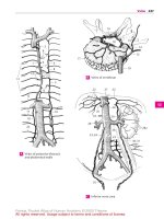
1 Lingual artery. A. lingualis. Second anterior
branch of external carotid artery. It is covered
by the hyoglossus muscle and passes laterally
into the tongue. A B C
2 Suprahyoid branch of lingual artery. Ramus
suprahyoideus. It anastomoses at the hyoid
bone with the infrahyoid branch and the
branch from the opposite side. B
3 Sublingual artery. A. sublingualis. Arising at
the anterior margin of the hyoglossus, it passes
between the mylohyoid and the sublingual
gland and extends up to the gingiva. B
4 Rami dorsales linguae. Dorsal lingual branches
of lingual artery that supply the base of the
tongue. B
5 Deep lingual artery. A. profunda linguae. As
the main branch of the lingual artery, it passes
between the genioglossus and inferior longi-
tudinal muscles of the tongue to the apex of the
tongue and anastomoses with the artery from
the opposite side. B
6 Linguofacial trunk. [Truncus linguofacialis].
Occasionally present common trunk of lingual
and facial arteries. A
7 Facial artery. A. facialis. Third anterior branch
of external carotid artery. It runs below the sty-
loyoid muscle, first upward, then laterad, and
crosses the mandible at the anterior margin of
the masseter. A B C
8 Ascending palatine artery. A. palatina ascen-
dens. Arising from the proximal portion of the
facial artery, it passes medial to the styloglos-
sus muscle at the lateral wall of the pharynx to
supply the palatal arches and adjacent muscu-
lature, often also the tonsils from above. It and
the ascending pharyngeal artery can replace
each other. C
9 Tonsillar branch. Ramus tonsillaris. Branch
frequently arising from the ascending palatine
artery and supplying the palatine tonsils. C
10 Submental artery. A. submentalis. It lies
caudal to the mylohyoid muscle and supplies
mainly this muscle and the submandibular
gland. It anastomoses with the sublingual
artery. C
11 Glandular branches. Rami glandulares. Direct
branches for the submandibular gland. C
12 Inferior labial artery. A. labialis inferior. Artery
for the lower lip situated between the muscle
and the mucosa. It anastomoses with the sub-
mental and mental arteries as well as the
artery of the opposite side. C
13 Superior labial artery. A. labialis superior.
Artery for the upper lip situated between the
muscle and mucosa. It anastomoses with the
transverse facial and infra-orbital arteries as
well as the artery of the opposite side. C
13 a
Nasal septal branch. Ramus septi nasi. It con-
nects with the cavernous body of the septum
(Kiesselbach’s area). C
13 b Lateral nasal branch. Ramus lateralis nasi. It
supplies the base of the nasal ala. C
14 Angular artery. A. angularis. Terminal branch
of facial artery. It anastomoses with the oph-
thalmic artery. C
15 Occipital artery. A. occipitalis. Second dorsal
branch of external carotid artery. It passes me-
dial to the mastoid process at the occiput and
anastomoses with the superficial temporal,
vertebral, deep cervical and posterior auricular
arteries. C D
16 Mastoid branch of occipital artery. Ramus
mastoideus. It passes through the mastoid
foramen to the diploë and dura. It also supplies
mastoid cells. C
17 Auricular branch of occipital artery. Ramus
auricularis. It passes beneath the sternoclei-
domastoid muscle and runs obliquely behind
the pinna. C
18 Sternocleidomastoid branch. Ramus sterno-
cleidomastoidei. Small branches of occipital
artery that supply the sternocleidomastoid
muscle. C
19 Meningeal branch. [Ramus meningeus]. In-
constant branch of the occipital artery that oc-
casionally passes through the parietal foramen
and supplies the dura mater. C
20 Occipital branches. Rami occipitales. Usually
very tortuous branches of occipital artery that
penetratethe trapeziusandsupplytheocciput. C
21 Descending branch of occipital artery. Ramus
descendens. It passes beneath the splenius
capitis to supply the muscles there. C
22 Posterior auricular artery. A. auricularis poste-
rior. Third dorsal branch of external carotid
artery. It lies under the parotid gland on the sty-
loid process between the mastoid process and
the ear. C D
23 Stylomastoid artery. A. stylomastoidea. Slen-
der companion artery of facial nerve. It courses
with it through the stylomastoid foramen to the
hiatus of the canal for the greater petrosal nerve,
and then into the middle and inner ear. D
24 Posterior tympanic artery.A.tympanica poste-
rior. It passes with the chorda tympani from the
facial canal to the tympanic membrane. D
25
Mastoid branches. Rami mastoidei. Branches
of posterior tympanic artery that supply the
mastoid cells. D
26
Stapedial branch.[Ramusstapedialis].Slender
branch that supplies the stapedial muscle.
27 Auricular branch. Ramus auricularis. It supplies
the posterior side of the pinna with perforating
branches as well as the anterior side and the
small auricular muscles. D
28 Occipital branch.Ramus occipitalis.Branchthat
courses above the mastoid process and anasto-
moses with the occipital artery. D
28 a Parotid branch. Ramus parotideus. It supplies
the parotid gland. D
Arteries
Feneis, Pocket Atlas of Human Anatomy © 2000 Thieme
All rights reserved. Usage subject to terms and conditions of license.
197
1
2
3
4
5
6
7
8
9
10
11
12
13
14
15
16
17
18
19
20
21
22
23
24
25
a
a
a
28a
15
28
27
23
22
24
25
19
21
8
7
12
10
7
15
18
16
14
20
17
22
9
1
11
13a
13b
13
4
53
2
1
7
194.17
1
194.9
6
194.10
7
200.10
194.6
Linguofacial trunkA Branches of external carotid arteryB
Branches of external
carotid artery
C
Branches of external
carotid artery
D
Arteries
Feneis, Pocket Atlas of Human Anatomy © 2000 Thieme
All rights reserved. Usage subject to terms and conditions of license.
198
1
2
3
4
5
6
7
8
9
10
11
12
13
14
15
16
17
18
19
20
21
22
23
24
25
1 Superficial temporal artery. A. temporalis su-
perficialis. One of two terminal branches of the
external carotid artery. It passes upward in front
of the pinna accompanied by the auriculotem-
poral nerve. A B
2 Parotid branch. Ramus parotideus. It supplies
the parotid gland. A
3 Transverse facial artery. A. transversa faciei
(facialis). Branch that is covered by the parotid
gland and passes below the zygomatic arch to
the cheek. A
4 Anterior auricular branches. Rami auriculares
anteriores. Several small branches to the pinna
and the external acoustic meatus. A
5 Zygomatico-orbital artery. A. zygomatico-
orbitalis. It passes above the zygomatic arch to
the lateral margin of the orbit. A
6 Middle temporal artery. A.temporalismedia. It
passes above the zygomatic arch beneath the
temporalis muscle. A
7 Frontal branch. Ramus frontalis. Anterior
branch of superficial temporal artery. It anasto-
moses with its counterpart from the opposite
side as well as with the supra-orbital and su-
pratrochlear arteries from the internal carotid.
A
8 Parietal branch. Ramus parietalis. Posterior
branch of superficial temporal artery. It anasto-
moses with its counterpart from the opposite
side as well as with the posterior auricular and
occipital arteries. A
9 Maxillary artery. A. maxillaris. Larger terminal
branch of external carotid artery. It arises
beneath the temporomandibular joint, passes
lateral or medial to the lateral pterygoid muscle
and ramifies in the pterygopalatine fossa. A B
10 Deep auricular artery. A. auricularis profunda.
It passes backward and upward to the temporo-
mandibular joint, external acoustic meatus and
tympanic membrane. B
11 Anterior tympanic artery. A. tympanica ante-
rior. Accompanied by the chorda tympani, it
passes through the petrotympanic fissure into
the tympanic cavity. B
12 Inferior alveolar artery. A. alveolaris inferior. It
passes between the medial pterygoid muscle
and mandibular ramus into the mandibular
canal up to the mental foramen. B
13
Dental rami. Rami dentales. Branches to roots
of the teeth. B
13 a
Peridental branches. Rami peridentales.
14
Mylohyoid branch. Ramus mylohyoideus.
Branch that exits in front of the mandibular
canal and accompanies the mylohyoid nerve in
the mylohyoid groove; it runs anteriorly
beneath the mylohyoid muscle and anasto-
moses with the submental artery. B
15
Mental branch. Ramus mentalis. Terminal
branch of inferior alveolar artery. It supplies the
chin. B
16 Middle meningeal artery. A. meningea media.
It passes medial to the lateral pterygoid muscle
and through the foramen spinosum into the
middle cranial fossa where it ramifies. B C
17
Accessory ramus. Ramus accessorius. Acces-
sory branch from the middle meningeal artery
or from the maxillary artery that extends to the
adjacent muscles, the auditory tube and
through the foramen ovale to the dura up to the
trigeminal (semilunar) ganglion. B
18
Petrosal branch. Ramus petrosus. Small
branch that arises directly after the entrance of
the middle meningeal artery into the cranial
cavity and anastomoses with the stylomastoid
artery via the hiatus of the canal for the greater
petrosal nerve. C
19
Superior tympanic artery. A. tympanica su-
perior. It arises close to the petrosal branch and
passes into the tympanic cavity with the lesser
petrosal nerve. C
20
Frontal branch. Ramus frontalis. Anterior,
large terminal branch in the cranium. It lies in a
bony groove often closed to form a canal. C
21
Parietal branch. Ramus parietalis. Terminal
branch passing to the posterior half of the
cranium. C
22
Orbital branch. Ramus orbitalis. It runs
through the superior orbital fissure in the
direction of the lacrimal gland. C
23
Anastomotic branch connecting the orbital
branch and the lacrimal ar tery.
Ramus anas-
tomoticus [[cum a. lacrimalis]]. C
23 a Pterygomeningeal artery. A. pterygo-
meningea. Artery that supplies the pterygoid
muscles, the tensor veli palatini, and the audi-
tory tube; it emerges from the maxillary and
middle meningeal arteries and passes through
the foramen ovale to the trigeminal ganglion
and the dura mater.
24 Masseteric artery. A. masseterica. Artery that
supplies the masseter muscle passing laterally
through the mandibular notch. B
25 Anterior deep temporal artery. A. temporalis
profunda anterior. Artery passing upwards into
the temporalis muscle. B
25 a Posterior temporal artery. A. temporalis post-
erior.
26
Pterygoid branches. Rami pterygoidei.
Branches that supply the pterygoid muscles. B
27 Buccal artery. A. buccalis. Artery that passes
downward and forward onto the buccinator
muscle to supply the cheek and gingiva. B
28 Posterior superior alveolar artery. A. alve-
olaris superior posterior. It passes posteriorly
into the maxilla and the maxillary sinus and
supplies the upper molar teeth and their ging-
iva. B
29
Dental branches. Rami dentales. They supply
the maxillary molars. B
29 a
Peridental branches. Rami peridentales.
Arteries
Feneis, Pocket Atlas of Human Anatomy © 2000 Thieme
All rights reserved. Usage subject to terms and conditions of license.
199
1
2
3
4
5
6
7
8
9
10
11
12
13
14
15
16
17
18
19
20
21
22
23
24
25
a
a
a
20
22
21
23
19
18
16
1
9
16 1711
25
28
29
24
26
27
13 15
1214
10
8
7
6
5
32
91
4
Superficial temporal arteryA Maxillary artery
B
Middle meningeal artery
C
Arteries
Feneis, Pocket Atlas of Human Anatomy © 2000 Thieme
All rights reserved. Usage subject to terms and conditions of license.
200
1
2
3
4
5
6
7
8
9
10
11
12
13
14
15
16
17
18
19
20
21
22
23
24
25
1 Infraorbital artery. A. infraorbitalis. Terminal
branch of maxillary artery. It passes to the face
via the inferior orbital fissure, groove and canal.
A
2
Anterior superior alveolar ar teries. Aa. alve-
olares superiores anteriores. They leave the in-
fraorbital artery in the infraorbital canal and
pass through bone to the anterior teeth. A
3
Dental branches. Rami dentales. Terminal
branches passing to the teeth. A
3a
Peridental branches. Rami peridentales.
4 Artery of pterygoid canal. A . canalis ptery-
goidei. It traverses the pterygoid canal and
passes posteriorly to the auditory tube and its
environment. A B
4a
Pharyngeal branch. Ramus pharyngeus.
Branch to the pharyngeal mucosa.
5 Descending palatine artery. A. palatina de-
scendens. It descends through the greater
palatine canal. A B
6
Greater palatine artery. A. palatina major. It
passes through the greater palatine foramen to
the anterior palate and the neighboring ging-
iva. B
7
Lesser palatine arteries. Aa. palatinae
minores. They leave the greater palatine artery
and canal and pass through the lesser palatine
foramina to the soft palate. B
7a
Pharyngeal branch. Ramus pharyngeus.
Branch that supplies the pharyngeal mucosa up
to the level of the tonsil and gingiva.
8 Sphenopalatine artery. A. sphenopalatina. It
passes through the sphenopalatine foramen
into the nasal cavity. B
9
Lateral posterior nasal branches. Aa. nasales
posteriores laterales. As the terminal branches
of the sphenopalatine artery, they supply the
nasal cavity laterally and posteriorly. B
9a
Posterior septal branches. Rami septales
posteriores. Branches of the sphenopalatine
artery that supply the posteroinferior part of
the nasal septum. B
10 INTERNAL CAROTID ARTERY (ICA). Arteria
carotis interna. It extends branchless from the
bifurcation of the common carotid to the base
of the skull where it extends through the
carotid canal up to its terminal division into the
anterior and middle cerebral arteries. B C
11 Cervical part of internal carotid artery. Pars
cervicalis. Branchless segment that extends up
to the site where it enters the carotid canal in
the petrous part of the temporal bone. B C
12 Carotid sinus. Sinus caroticus. It is occasionally
displaced from the end of the common carotid
artery (p. 194.8) to the beginning of the inter-
nal carotid. Site of baroreceptors. B
13 Petrous part of internal carotid artery. Pars
petrosa. Segment coursing through the carotid
canal in the petrous part of the temporal bone.
C
14
Caroticotympanic arteries. Aa. caroticotym-
panicae. Slender arteries extending from the
carotid canal to the tympanic cavity. C
15
Pterygoid branch. Ramus pterygoideus.
Branch that accompanies the nerve of the pter-
ygoid canal in the lateral wall of the sphenoid
sinus. C
16 Cavernous part of internal carotid. Pars caver-
nosa. Segment in the cavernous sinus; it ex-
tends up to the vicinity of the optic canal. C
17
Basal tentorial branch. Ramus basalis ten-
torii. Branch of ICA that extends across the
petrosal ridge to the tentorium. C
18
Marginal tentorial branch. Ramus marginalis
tentorii. Branch of ICA located near the ten-
torial notch. C
19
Meningeal branch. Ramus meningeus. Branch
of ICA that supplies the dura mater of the
middle cranial fossa. C
20
Branch to trigeminal ganglion. Ramus gan-
glionis trigeminalis. C
21
Nerve branches. Rami nervorum.
22
Cavernous sinus branch. Ramus sinus caver-
nosi. Twig from the cavernous part of the ICA. C
23 Inferior hypophysial artery. A. hypophysialis
inferior. It supplies the posterior lobe of the hy-
pophysis. C
24 Cerebral part of internal carotid artery. Pars
cerebralis. As the terminal intradural segment,
it extends from the exit of the ophthalmic
artery at the last bend of the carotid up to the
terminal branches that form the anterior and
middle cerebral arteries. C
25 Superior hypophysial artery. A. hypophysialis
superior. It supplies the hypophysial stalk, in-
fundibulum and part of the lower hy-
pothalamus. C
26
Clival branch. Ramus clivi. Branch that sup-
plies the clivus. C
Arteries
Feneis, Pocket Atlas of Human Anatomy © 2000 Thieme
All rights reserved. Usage subject to terms and conditions of license.
201
1
2
3
4
5
6
7
8
9
10
11
12
13
14
15
16
17
18
19
20
21
22
23
24
25
a
a
a
24
25
17
18
14
13
11
15
16
16
20
26
2322
21
19
10
4
7
6
5
202.16b
9
9a
9
8
12
11
4
5
1
3
1
2
Infraorbital arteryA
Lateral nasal arteryB
Branches of internal
carotid artery
C
Arteries
Feneis, Pocket Atlas of Human Anatomy © 2000 Thieme
All rights reserved. Usage subject to terms and conditions of license.
202
1
2
3
4
5
6
7
8
9
10
11
12
13
14
15
16
17
18
19
20
21
22
23
24
25
1 Ophthalmic artery. A. ophthalmica. It passes
from the anterior convex arch of the internal
carotid artery through the optic canal into the
orbit accompanied by the optic nerve. A
2 Central retinal artery. A. centralis retinae. It
passes into the optic nerve from below about
1 mm behind the eyeball. Distribution: internal
layers of the retina. A
3 Lacrimal artery. A. lacrimalis. Exits lateral to the
ophthalmic artery and passes to the lacrimal
gland along the upper margin of the lateral rectus
muscle. A
4 Anastomotic branch [with middle meningeal
artery]. Ramus anastomoticus [cum a. meningea
media]. Branch that anastomoses with the orbital
branch of the middle meningeal artery. It oc-
casionally replaces the ophthalmic artery. A
5
Lateral palpebral arteries. Aa. palpebrales
laterales. They arises from the lacrimal artery and
pass lateral to the eyelids. A B
5a
Recurrent meningeal branch. Ramus mening-
eus recurrens. Branch of the lacrimal artery that
runs through the superior orbital fissure into the
cranial cavity. It anastomoses with the anasto-
motic branch.
6 Short posterior ciliary arteries. Aa. ciliares post-
eriores breves. 10−15 arteries which penetrate
the sclera circumferentially around the optic
nerve and supply the choroid. A
7 Long posterior ciliary arteries. Aa. ciliares poste-
riores longae. Lateral and medial arteries which
pass between the sclera and choroid to the ciliary
body. A B
8 Muscular arteries. Branches that supply the eye
muscles.
9 Anterior ciliary arteries. Aa. ciliares anteriores.
They arise from the lacrimal artery or muscular
branches, penetrate the sclera and supply the
choroid and ciliary body. A B
10 Anterior conjunctival arteries. Aa. conjuncti-
vales anteriores. Branches arising from the ante-
rior ciliary arteries and supplying the conjunc-
tivae. B
11 Posterior conjunctival arteries. Aa. conjuncti-
vales posteriores. They arise from the lacrimal
and supraorbital arteries. A
12 Episcleral arteries. Aa. episclerales. Twigs of the
anterior ciliary arteries situated on the sclera. B
13 Supraorbital artery. A. supraorbitalis [[a.
frontalis lat.]]. It passes below the roof of the orbit
and through the supraorbital notch to the fore-
head. A B
13 a
Diploic branch. Ramus diploicus. Branch to
bone.
14 Posterior ethmoidal artery. A. ethmoidalis post-
erior. It passes below the superior oblique muscle
and through the posterior ethmoidal foramen to
the posterior ethmoidal air cells and the posterior
part of the nasal cavity. A
15 Anterior ethmoidal artery. A. ethmoidalis ante-
rior. It passes upward through the anterior eth-
moidal foramen into the anterior cranial fossa
and through the cribriform plate into the nasal
cavity and frontal sinus as well as the anterior
ethmoidal cells. A
16
Anterior meningeal branch. Ramus meningeus
anterior. Branch from the part of the anterior eth-
moidal artery lying in the cranial fossa. It supplies
the dura. A
16 a
Anterior septal branches. Rami septales anteri-
ores. Branches that extend from the anterior eth-
moidal artery to the upper portion of the nasal
septum.
16 b
Lateral anterior nasal branches. Rami nasales
anteriores lateralis. Branches of the anterior eth-
moidal artery that extend to the upper lateral
wall of the nasal septum and the anterior upper
ethmoidal air cells. See p. 201 B
17 Medial palpebral arteries. Aa. palpebrales medi-
ales. Paired arteries for each eyelid, upper and
lower. They arise from the ophthalmic artery and
anastomose with the lateral palpebral artery via
the superior and inferior palpebral arches. A B
18
Superior palpebral arch. Arcus palpebralis su-
perior. Connects the medial and lateral palpebral
arteries superiorly on the tarsal plate. B
19
Inferior palpebral arch. Arcus palpebralis infe-
rior. Connects the medial and lateral palpebral ar-
teries inferiorly on the tarsal plate. B
20 Supratrochlear artery. A. supratrochlearis [[a.
frontalis med.]]. Terminal branch of the ophthal-
mic artery which passes through the frontal
notch to the forehead. Anastomoses with the
contralateral, supraorbital and superficial tem-
poral arteries. A B
21 Dorsal nasal artery. A. dorsalis nasi (a. nasi ex-
terna). It penetrates the orbicularis oculi and
passes downward to the bridge of the nose. Anas-
tomoses with the facial artery. A B
22 Anterior choroidal artery [[AChA]]. Arteria
choroidea anterior. Usually arises from the inter-
nal carotid artery. It follows the optic tract and
enters the choroid plexus of the inferior horn of
the lateral ventricle, where it occasionally passes
up to the interventricular foramen. C D
23 Choroidal branches of lateral ventricle. Rami
choroidei ventriculi lateralis. Branches that
supply the choroid plexus of the lateral ventricle.
CD
24 Choroidal branches to third ventricle. Rami
choroidei ventriculi tertii. Branches that supply
the choroid plexus of the third ventricle. C
25 Branches to anterior perforated substance.
Rami substantiae perforatae anterioris. AChA
branches that supply internal capsule. D
26 Branches of optic tract. Rami tractus optici.
Branches to the AchA that supply the optic tract.
D
27 Branches to lateral geniculate body. Rami cor-
poris geniculati lateralis. AChA branches that
supply the lateral genticulate body. D
28 Branches to internal capsule. Rami capsulae in-
ternae. AChA branches that supply the posterior
part of the internal capsule.
29 Branches to globus pallidus. Rami globi pallidi.
AChA branches that supply the globus pallidus.
30 Branches to tail of caudate nucleus. Rami
caudae nuclei caudati. AChA branches that supply
the tail of the caudate nucleus.
31 Branches to tuber cinereum. Rami tuberis cin-
erei. AChA branches that supply the tuber cin-
ereum. D
Arteries
Feneis, Pocket Atlas of Human Anatomy © 2000 Thieme
All rights reserved. Usage subject to terms and conditions of license.
203
1
2
3
4
5
6
7
8
9
10
11
12
13
14
15
16
17
18
19
20
21
22
23
24
25
a
a
a
34
33
31
26
25
27
23
22
22
35
23
24
17
21
13
20
18
5
19
7
12
910
1
5
21
16
7
11
13
20
15
14
17
9
3
6
2
4
Ophthalmic arteryA Facial branches of ophthalmic arteryB
Anterior choroidal artery
from above
C Anterior choroidal artery
from below
D
32 Branches to hypothalamic nuclei. Rami nu-
cleorum hypothalamicorum. AChA branches that
supply the hypothalamic nuclei.
33 Branches to substantia nigra. Rami substantiae
nigrae. AChA branches that pass through the crus
cerebri and supply the substantia nigra. D
34 Branches to red nucleus. Rami nuclei rubri. AChA
branches that pass through the crus cerebri and
supply the red nucleus. D
35 Branches to amygdaloid body. Rami corporis
amygdaloidei. Branches of the AchA that supply
the medial amygdaloid nucleus. C
Arteries
Feneis, Pocket Atlas of Human Anatomy © 2000 Thieme
All rights reserved. Usage subject to terms and conditions of license.
204
1
2
3
4
5
6
7
8
9
10
11
12
13
14
15
16
17
18
19
20
21
22
23
24
25
1 Anterior cerebral artery. A. cerebri anterior.
One of the two terminal arteries of the internal
carotid artery. It runs posteriorly above the cor-
pus callosum and supplies the greater part of
the medial surface of the cerebrum. A
2 Precommunical part. Pars precommunicalis.
Portion of the anterior cerebral artery proximal
to the anterior communicating branch. A
3 Anteromedial central arteries (anteromedial
thalamostriate arteries). Aa. centrales an-
teromediales (aa. thalamostriatae anterome-
diales). Branches arising from the anterior cere-
bral arteries and passing into the thalamus and
curpus striatum from below. A
4 Short central artery. A. centralis brevis. Short
branch of anterior cerebral artery passing into
the cerebrum. A
5 Long central artery (recurrent artery). A.
centralis longa (a. recurrens) [[Heubner]]. Ret-
rograde branch that runs parallel to the ante-
rior cerebral artery. It penetrates the anterior
perforated substance and supplies the middle
and lateral parts of the lentiform nucleus, the
head of the caudate nucleus and the anterior
limb of the internal capsule. A
6 Anterior communicating artery. A. communi-
cans anterior. Unpaired connection between
the right and left anterior cerebral arteries. A
7 Anteromedial central branches. Rami cen-
trales anteromediales. Anteromedial central
branches that penetrate uniformly into the
cerebral substance. A
8 Postcommunical part of anterior cerebral
artery. Pars postcommunicalis (r. pericallosa).
The part distal to the anterior communicating
artery. B
9 Medial frontobasal artery (medial orbito-
frontalis branch). A. frontobasalis medialis (r.
orbitofrontalis medialis). Branch to the lower
surface of the frontal lobe. B
10 Callosomarginal artery. A. callosomarginalis.
Segment of the anterior cerebral artery located
in the sulcus of the corpus callosum. B
11
Anteromedial frontal branch. Ramus
frontalis anteromedialis. Branch to the lower
half of the medial side of the frontal lobe. B
12
Mediomedial frontal branch. Ramus frontalis
mediomedialis. Branch to the middle portion of
the medial side of the frontal lobe. B
13
Posteromedial frontal branch. Ramus
frontalis posteromedialis. Branch to the poste-
rior portion of the medial surface of the frontal
lobe. B
14
Cingular branch. Ramus cingularis. Branch
that supplies the cingulate sulcus. B
15 Paracentral artery. A. paracentralis. Branch of
the anterior cerebral artery that supplies the
area behind the central sulcus. B
16 Precuneal ar tery. A. precunealis. It supplies the
region in front of the cuneus. B
17 Parieto-occipital artery. A. parieto-occipitalis.
Branch of the anterior cerebral artery that sup-
plies the parieto-occipital sulcus. B
18 Middle cerebral artery. A. cerebri media. Sec-
ond terminal branch of internal carotid artery.
It lodges in the lateral sulcus between the fron-
tal and temporal lobes and supplies the greater
part of the lateral cerebral surface. A
19 Sphenoidal part. Pars sphenoidalis. First part
of the middle cerebral artery. It takes a horizon-
tal course somewhat parallel to the lesser wing
of the sphenoid bone. A
20 Anterolateral central arteries. Aa. centrales
anterolaterales (aa. thalamostriatae anter-
olaterales). Branches that penetrate the ante-
rior perforated substance.
21
Medial branches. Rami mediales. They pass
through the lentiform nucleus to the caudate
nucleus and internal capsule. A
22
Lateral branches. Rami laterales. They course
laterally around the lentiform nucleus to the
internal capsule and caudate nucleus. A
23 Insular part. Pars insularis. Collective term for
branches of the middle cerebral artery that
supply the insula. D
24 Insular arteries. Aa. insulares. Branches that
supply the insula. D
25 Lateral frontobasal artery. A. frontobasalis
lateralis (ramus orbitofrontalis lateralis).
Branch that supplies the lateroinferior frontal
lobe. C D
26 Anterior temporal artery. A. temporalis ante-
rior. Branch that supplies the frontal end of the
two upper temporal gyri. C D
27 Middle temporal artery. A. temporalis media.
Branch supplying the middle portion of the
temporal lobe. C D
28 Posterior temporal artery. A. temporalis post-
erior. Branch supplying the posterior portion of
the temporal lobe. C D
29 Terminal part. Pars terminalis (pars corticalis).
Terminal ramifications of the middle cerebral
artery.
30 Artery of central sulcus. A. sulci centralis. C D
31 Artery of precentral sulcus. A. sulci pre-
centralis. C D
32 Artery of postcentral sulcus. A. sulci post-
centralis. C D
33 Anterior and posterior parietal arteries. Aa.
parietales anterior et posterior. Branches that
supply the parietal lobe. C D
34 Artery of angular gyrus. A. gyri angularis. C D
Arteries
Feneis, Pocket Atlas of Human Anatomy © 2000 Thieme
All rights reserved. Usage subject to terms and conditions of license.
205
1
2
3
4
5
6
7
8
9
10
11
12
13
14
15
16
17
18
19
20
21
22
23
24
25
a
a
a
27
26
25
31
30
33
34
23
24
32
28
32
33
34
28
27
26
25
31
30
13
12
15
16
11
10
14
17
8
9
18
18
526
7
3; 4
1
21; 22
19
Anterior and middle cerrebral arteriesA
Anterior cerebral artery
B
Middle cerebral artery
C
Insular arteries
D
Arteries
Feneis, Pocket Atlas of Human Anatomy © 2000 Thieme
All rights reserved. Usage subject to terms and conditions of license.
206
1
2
3
4
5
6
7
8
9
10
11
12
13
14
15
16
17
18
19
20
21
22
23
24
25
1 SUBCL AVIAN ARTERY. Arteria subclavia. It
lodges between the scalenus anterior and me-
dius in the groove for the subclavian artery on
the 1
st
rib. At the lateral margin of the 1
st
rib it
continues as the axillary artery. A
2 Vertebral artery. A. vertebralis. Itoriginatesbe-
hind the scalenus anterior, passes through the
foramina transversaria cranially from C6−C1,
and then, after proceeding over the arch of the
atlas behind its lateral mass, runs anteriorly
through the posterior atlanto-occipital mem-
brane and the foramen magnum into the cranial
cavity. A B D
3 Prevertebral part of vertebral artery. Pars pre-
vertebralis. Thisshort segmentliesin frontofthe
entrance into the foramen transversarium of C6.
A
4 Vertebral part of vertebral artery. Pars trans-
versaria (cervicalis). It ascends through the
foramina transversia of C6 to C1. A
5 Spinal branches. Rami spinales (radiculares).
Branches passing with the spinal nerves to
supply the spinal cord, the meningeal coverings
of the spinal cord and the vertebral bodies. A
6 Muscular branches. Rami musculares. They
supply the surrounding muscles. A
7 Atlantal (suboccipital) part. Pars atlantica.Por-
tionofthe vertebralartery thatwindsaroundthe
atlas and occupies the suboccipital triangle. A
8 Intracranial part of vertebral artery. Pars in-
tracranialis. It lies within the cranium. A
9 Anterior meningeal branch.Ramusmeningeus
[anterior]. Branch at the anterior circumference
of the foramen magnum. It supplies bone and
dura. A
10 Posterior meningeal branch. Ramus mening-
eus [posterior]. Branch at the posterior cir-
cumference of the foramen magnum. It supplies
bone and dura. A
11 Anterior spinal artery. A. spinalis anterior. The
right and left arteries join to form an unpaired
vesselinthe anteriormedianfissure ofthespinal
cord. A B
12 Posterior inferior cerebellar artery (PICA). A.
inferior posterior cerebelli. It mainly supplies
the lower posterior portion of the cerebellum. A
BD
13 Choroid branch of PICA. Ramus choroideus
ventriculi quarti. It supplies the choroid plexus
of the fourth ventricle.
14 Tonsillar branch of PICA. Ramus tonsillae cere-
belli. Branch that supplies the tonsil of the cere-
bellum.
15 Medial and lateral medullary branches of
PICA. Rami medullares medialis et lateralis.
Branches thatsupply the medulla oblongata and
the inferior cerebellar peduncle.
16 Posterior spinal artery. A. spinalis posterior.
Slender longitudinal vessel, occasionally paired,
behind the spinal cord. It arises intracranially
from the posterior inferior cerebellar artery or
the vertebral artery. B D
17 Basilar artery. A.basilaris.Unpaired, thick trunk
that extends from the right and left vertebral ar-
teries to its termination as the posterior cerebral
arteries. A B D
18 Anterior inferior cerebellar artery. A. inferior
anterior cerebelli. Itsuppliestheanteriorpartof
the inferior surface of the cerebellum. B D
19
Labyrinthine ar tery. A. labyrinthi [ramus mea-
tus acustici interni]. Branch of the anterior infe-
rior cerebral artery (or basilar artery) that ac-
companies the vestibulocochlear nerve into the
inner ear. B D
20 Pontine arteries. Aa. pontis. Branches that
supply the pons. B D
21 Mesencephalic arteries. Aa. mesencephalicae.
22 Superior cerebellar artery. A. superior cere-
belli. It passes around the mesencephalon and
through the cisterna ambiens to the surface of
the cerebellum below the tentorium. B D
23 Posterior cerebral artery. A. cerebri posterior.
Terminal branch of basilar artery. It supplies the
occipital lobe and 2/3 of the temporal lobe of the
cerebrum. B C D
24 Precommunical part of posterior cerebral
artery. Pars precommunicalis. Short trunk
which extends up to the entrance of the poste-
rior communicating artery. B
25 Posteromedial central arteries. Aa. centrales
posteromediales. Branches in the posterior per-
forated substance that supply the thalamus,
lateralwallof thirdventricleand globuspallidus.
B
26 Postcommunical part of posterior cerebral
artery. Pars postcommunicalis. It is that portion
followingtheposterior communicatingartery.It
curves around the mesencephalon and passes
through the cisterna ambiens and tentorial
notch to the inferior surface of the cerebrum. B
27 Posterolateral central arteries. Aa. centrales
posterolaterales. Individual branches that
supply theposterior portionofthethalamus, the
quadrigeminalplate,pinealbody and themedial
geniculate body. C
28 Thalamic branches.Ramithalamici.Branchesto
posterior portion of thalamus. C
29 Posteromedial choroid branches. Rami
choroidei posteriores mediales. Branches in the
roof of the third ventricle. C
30 Posterolateral choroid branches. Rami
choroidei posteriores laterales. Branches that
supplythechoroid plexus ofthelateralventricle.
C
31 Peduncular branches. Rami pedunculares.
Branches that supply the cerebral peduncles. D
Arteries
Feneis, Pocket Atlas of Human Anatomy © 2000 Thieme
All rights reserved. Usage subject to terms and conditions of license.
207
1
2
3
4
5
6
7
8
9
10
11
12
13
14
15
16
17
18
19
20
21
22
23
24
25
a
a
a
30
29
23
31
17
22
N III
18
N VI
19
20
12
2
16
28
29
30
27
23
17
21; 22
25
22
19
18
12
9
11
16
2
N VI
N III
20
23; 24
26
2
1
11
17
12
9
10
2
5
6
8
7
4
3
Vertebral arteryA Arteries of base of the brainB
Posterior cerebral arteryC Basilar arteryD
Arteries
Feneis, Pocket Atlas of Human Anatomy © 2000 Thieme
All rights reserved. Usage subject to terms and conditions of license.
208
1
2
3
4
5
6
7
8
9
10
11
12
13
14
15
16
17
18
19
20
21
22
23
24
25
1 Terminal portion of posterior cerebral artery.
Parsterminalis(corticalis).It suppliesalmostex-
clusively the posterior cerebral cortex mainly at
the base of the brain.
2 Lateral occipital artery. A. occipitalis lateralis.
Trunk for the three temporal lobe arteries. A B
3
Anterior temporal branches. Rami tem-
porales anteriores. A B
4
Middle temporal branches. Rami temporales
(intermedii mediales). A B
5
Posterior temporal branches. Rami tem-
porales posteriores. A B
6 Medial occipital artery. A. occipitalis medialis.
Twig for the sagittal surface of the posterior half
of the cerebrum. A B
7
Dorsal branch to corpus callosum. Ramus
corporis callosi dorsalis. Small, short branch to
the splenium of the corpus callosum. A
8
Parietal branch. Ramus parietalis. Anterior
branchtoposteriorportionof theparietallobe.A
9
Parieto-occipital branch. R amus parieto-
occipitalis. It supplies the parieto-occipital sul-
cus. A B
10
Calcarine branch. Ramus calcarinus. It sup-
plies the calcarine sulcus. A B
11
Occipitotemporal branch. Ramus occipi-
totemporalis. Lower branch extending into the
temporal lobe. A B
12 Cerebral arterial circle [[Circle of Willis]]. Cir-
culus arteriosus cerebri [[Willisii]]. Anastomos-
ing arterial circle between the main tributaries
of the cerebrum, i.e., between the internal
carotid and the posterior cerebral arteries. B C
13 Internal carotid artery. A. carotis interna. Main
anterior tributary in the cranial cavity. B C
14 Anterior cerebral artery. A. cerebri anterior.
Anterior terminal branch of internal carotid
artery. It supplies chiefly the greater portion of
the medial and orbital surfaces of the cerebrum.
C
15
Anterior communicating artery. A. com-
municans anterior. Anastomosis between right
and left anterior cerebral arteries. C
16
Anteromedial central arteries. Aa. centrales
anteromediales. Short branches penetrating
equally into the base of the brain. C
17 Middle cerebral ar tery. A. cerebri media.
Lateral terminal branch of internal carotid
artery. It frequently gives off the posterior com-
municating artery. C
18
Posterior communicating ar tery. A. com-
municans posterior. Paired anastomoses be-
tween the internal carotid or middle cerebral
artery and the posterior cerebral artery. A C
19
Chiasmatic branch. Ramus chiasmaticus.
Branch to the optic chiasm. C
20
Oculomotor ner ve branch. Ramus nervi
oculomotorii. C
21
Thalamic branch. Ramus thalamicus. Long
branch which passes to the thalamus from
below. A C
22
Hypothalamic branch. Ramus hypothalami-
cus. Branch to the hypothalamus. A C
23
Branch to tail of caudate nucleus. Ramus
caudae nuclei caudati. It is located medial to the
choroid fissure. C
24 Posterior cerebral artery. A. cerebri posterior.
Paired terminal branch of basilar artery. Since
the latter is formed by the union of the right and
left vertebral arteries, this produces a strong
anastomosis of both vertebral arteries. A C
Arteries
Feneis, Pocket Atlas of Human Anatomy © 2000 Thieme
All rights reserved. Usage subject to terms and conditions of license.
209
1
2
3
4
5
6
7
8
9
10
11
12
13
14
15
16
17
18
19
20
21
22
23
24
25
a
a
a
Arteries
14
13
17
23
20
18
19
16
21
22
N III 24
15
13
3
2
4
5
12
6
11
9
10
8
10
9
11
5
4
3
2
2418
22
21
7
6
Posterior cerebral artery, medial viewA
Posterior cerebral artery
B
Cerebral arterial circle
C
Feneis, Pocket Atlas of Human Anatomy © 2000 Thieme
All rights reserved. Usage subject to terms and conditions of license.
210
1
2
3
4
5
6
7
8
9
10
11
12
13
14
15
16
17
18
19
20
21
22
23
24
25
1 Internal thoracic [[mammary]] artery. A.
thoracica interna [[a. mammaria interna]]. It
arises from the subclavian artery and descends
along the anterior, inner surface of the thorax as
far as the diaphragm. A B
2 Mediastinal branches. Rami mediastinales.
Branches that supply the mediastinum. B
3 Thymic branches. Rami thymici. Branches that
supply the thymus. B
4 Bronchial branches. [Rami bronchiales].
Branches to the bronchi. B
4a Tracheal branches. [Rami tracheales]. Branches
to the trachea.
5 Pericardiophrenic artery. A. pericardiaco-
phrenica. Accompanies the phrenic nerve and
supplies the pericardium and the diaphragm. B
6 Sternal branches. Rami sternales. Branches that
supply the sternum. B
7 Perforating branches. Rami perforantes. Ves-
sels that extend through intercostal spaces 1−6
to the surface of the thorax. B
8
Medial mammary branches. Rami mammarii
mediales. Larger perforating branches that
supply the mammary gland. B
9 Lateral costal branch. [Ramuscostalis lateralis].
Normalvariant.Arisesfrom theinternalthoracic
artery and runs lateral and parallel to it. B
10 Anterior intercostal branches. Rami inter-
costales anteriores. Anterior tributaries in the
intercostal spaces. B
11 Musculophrenic artery. A. musculophrenica.
Passing behind the costal arch, it gives off addi-
tional anterior intercostal branches from the 7
th
intercostal space onward. B
12 Superior epigastric artery. A. epigastrica su-
perior. Continuation of the internal thoracic
artery after entering the abdominal cavity be-
tween the sternal and costal parts of the dia-
phragm [[Larrey’s space =sternocostaltriangle]].
B
13 Thyrocervical trunk. Truncus thyrocervicalis.
Variably common stem of the inferior thyroid,
transversecervicaland suprascapulararteries.A
B
14 Inferior thyroid artery. A. thyroidea inferior. It
passes along the anterior margin of the scalenus
anterior as far as the level of C6 and then behind
the common carotid artery to the thyroid gland.
AB
15
Inferior laryngeal ar tery. A. laryngealis infe-
rior. It passes upward behind the trachea, pene-
trates the inferior pharyngeal constrictor and
supplies part of the larynx. A B
16
Glandular branches. Rami glandulares. They
supply the inferior and posterior surfaces of the
thyroid gland and the parathyroids via inferior
and ascending branches. A
17
Pharyngeal branches. Rami pharyngeales.
They supply the wall of the pharynx. A B
18
Esophageal branches. Rami oesophageales. A
B
19
Tracheal branches. Rami tracheales. A B
20
Ascending cervical ar tery. A . cervicalis ascen-
dens. It lies medial to the phrenic nerve and the
scalenus anterior andcanreach as farasthebase
of the skull. A B
21
Spinal branches. Rami spinales. They pass
through the intervertebral foramina to the spi-
nal cord. A B
22 Transverse cervical (colli) artery. A. transversa
cervicis (colli). The vessel may vary greatly. The
second most frequent variant (25%) is repre-
sented here. Originating usually (75%) from the
subclavian artery, it frequently passes through
the brachial plexus, supplies the upper part of
the trapezius with its branches and ramifies
alongside the dorsal scapular nerve. A B
23
Superf icial branch. Ramus superficialis. It
arises either as a superficial ramus from the
transverse cervical artery or as an independent
superficial cervical artery fromthethyrocervical
trunk and passes beside the accessory nerve to
the descending part of the trapezius and the le-
vator scapulae and splenius muscles. A B
23 a
Ascending branch. Ramus ascendens.
23 b
Descending branch. Ramus descendens.
24
Deep branch (dorsal scapular arter y).
Ramus profundus (a. dorsalis scapulae). This
vessel ariseseitherasa deep branch ofthetrans-
verse cervical artery or directly from the sub-
clavian artery (67%) and accompanies the dorsal
scapular nerve. It supplies the medial border of
the scapula and adjacent muscles. A B
24 a Dorsal scapular artery. [A. dorsalis scapulae].
Old designation for the deep branch.
Arteries
Feneis, Pocket Atlas of Human Anatomy © 2000 Thieme
All rights reserved. Usage subject to terms and conditions of license.
211
1
2
3
4
5
6
7
8
9
10
11
12
13
14
15
16
17
18
19
20
21
22
23
24
25
a
a
a
15
20
22
23
9
10
5
7
12
11
6
8
17
14
18
21
14
24
82.2
13
19
1
1
2
4
3
19
18
24
23
20
1
16
15
21
17
14
22
13
Thyrocervical trunkA
Internal thoracic [[mammary]]
artery and thyrocervical trunk
B
Arteries
Feneis, Pocket Atlas of Human Anatomy © 2000 Thieme
All rights reserved. Usage subject to terms and conditions of license.
212
1
2
3
4
5
6
7
8
9
10
11
12
13
14
15
16
17
18
19
20
21
22
23
24
25
1 Suprascapular artery. A. suprascapularis.
Generally arises from the thyrocervical trunk,
crosses over the scalenus anterior, runs above
the superior transverse scapular ligament into
the supraspinous and infraspinous fossae and
anastomoses with the circumflex scapular
artery. A
2
Acromial branch. Ramus acromialis. It pene-
trates the attachment of the trapezius and
passes to the acromion. A
3 Costocervical trunk. Truncus costocervicalis.
Origin: posterior wall of subclavian artery be-
hind the scalenus anterior. B
4 Deep cervical artery. A. cervicalis profunda. It
courses posteriorly between the transverse
processes of C7 and T1, then upwards on the
semispinalis. It supplies the nuchal muscula-
ture. B
5 Highest intercostal artery. A. intercostalis su-
prema. Common trunk for the first two inter-
costal arteries. B
6
First and second posterior intercostal arter-
ies.
Aa. intercostalis posterior prima et
secunda. They pass in the first two intercostal
spaces, respectively. B
7
Dorsal branches. Rami dorsales. Branches for
the muscles and skin of the back. B
8
Spinal branches. Rami spinales. Branches to the
spinal cord via intervertebral foramina T1−2. B
ARTERIES OF THE UPPER LIMB. Arteriae mem-
bri superioris.
9 Axillary Artery. Arteria axillaris. Continuation
of the subclavian artery as far as the lower
margin of the pectoralis major. A B
10 Subscapular branches. Rami subscapulares.
Individual branches to the subscapularis
muscle. A
11 Superior thoracic artery. A. thoracica superior.
Variable branch to the subclavius, intercostals
1−2 and serratus anterior muscles. A
12 Thoracoacromial artery. A. thoracoacromialis.
It arises at the upper margin of the pectoralis
minor and ramifies in all directions. A
13
Acromial branch. Ramus acromialis. Branch
passing superolaterally through the deltoid
muscle to the acromion. A
14
Acromial network. Rete acromiale. Arterial
network on the acromion. A
15
Clavicular branch. Ramus clavicularis. Small
branch to the clavicle and the subclavius
muscle. A
16
Deltoid branch. Ramus deltoideus. Branch
passing posterolaterally for the deltoid and
pectoralis major muscles. A
17
Pectoral branches. R ami pectorales. Branches
passing inferiorly for the serratus anterior and
pectoral muscles. A
18 Lateral thoracic artery. A. thoracica lateralis. It
passes downward at the lateral margin of the
pectoralis minor to supply the pectoral and
serratus anterior muscles. A
19
Lateral mammar y branches. Rami mammarii
laterales. A
20 Subscapular artery. A. subscapularis. Arises at
the lateral margin of the subscapularis muscle
and supplies it, the latissimus dorsi and teres
major muscles. A
21
Thoracodorsal artery. A. thoracodorsalis.
Branch to the latissimus dorsi and teres major.
A
22 Circumflex scapular artery. A. circumflexa
scapulae. It passes through the triangular
space to the infraspinous fossa and anasto-
moses with the suprascapular artery. A
23 Anterior circumflex humeral artery. A. cir-
cumflexa anterior humeri. It arises below the
latissimus dorsi at the same level or deeper
than the posterior circumflex humeral artery
and passes in front of the surgical neck of the
humerus to the coracobrachialis and biceps. It
anastomoses with the posterior circumflex
humeral artery. A
24 Posterior circumflex humeral artery. A. cir-
cumflexa posterior humeri. It passes with the
axillary nerve through the quadrangular space
to the shoulder joint and the deltoid muscle. It
anastomoses with the anterior circumflex
humeral, suprascapularis and thoracoacromial
arteries. A
25 Brachial artery. A. brachialis. It forms a con-
tinuation of the axillary artery from the lower
margin of the pectoralis major in the medial
bicipital groove up to its division into the radial
and ulnar arteries. A
26 Superficial brachial artery. [A. brachialis su-
perficialis]. Anatomic variant in which the
brachial artery lies on the median nerve in-
stead of below it. A
27 Profunda brachii artery. A. profunda brachii.
Companion artery of the radial nerve in the
groove for the radial nerve. A
28
Nutrient arteries of humerus. Aa. nutriciae
(nutrientes) humeri. Branches to the bone
marrow of the humerus. A
29
Deltoid branch. Ramus deltoideus. Branch
coursing laterally behind the humerus, then su-
periorly and externally to the deltoid muscle. A
30
Middle collateral ar tery. A. collateralis media.
It arises behind the humerus and descends to
the articular network of the elbow. A
31
Radial collateral ar tery. A. collateralis
radialis. It passes with the radial nerve to the
articular network of the elbow and gives off an
anterior branch to the radial recurrent artery. A
32 Superior ulnar collateral artery. A. collateralis
ulnaris superior. Often arising near the pro-
funda brachii artery, it passes with the ulnar
nerve to the articular network of the elbow. A
33 Inferior ulnar collateral artery. A. collateralis
ulnaris inferior. Originating above the medial
epicondyle of the humerus, it passes on the
brachialismuscleandthroughtheintermuscular
septum to the articular network of the elbow. A
Arteries
Feneis, Pocket Atlas of Human Anatomy © 2000 Thieme
All rights reserved. Usage subject to terms and conditions of license.
213
1
2
3
4
5
6
7
8
9
10
11
12
13
14
15
16
17
18
19
20
21
22
23
24
25
a
a
a
4
3
210.13
206.2
194.6
8
6
7
5
9
6
210.1
11521514
9
11
12
18
13
10
19
17
20
22
21
16
24
23
27
29
25
26
28
32
33
30
31
Subclavian and
brachial arteries
A
Arteria subclaviaB
Arteries
Feneis, Pocket Atlas of Human Anatomy © 2000 Thieme
All rights reserved. Usage subject to terms and conditions of license.
214
1
2
3
4
5
6
7
8
9
10
11
12
13
14
15
16
17
18
19
20
21
22
23
24
25
1 Radial artery. A radialis. It begins at the division
ofthebrachialarteryandcoursesonthepronator
teres, then lateral to the flexor carpi radialis
(where its pulsations are readily palpable) up to
the hand. B
2 Radial recurrent artery. A. recurrens radialis.
Retrograde artery ascendingmedialto the radial
nerve to anastomose with the radial collateral
artery. B
3 Palmar carpal branch. Ramuscarpalispalmaris.
Small branch at thedistalmargin ofthepronator
quadratus. It joins the carpal network. B
4 Superficial palmar branch. Ramus palmaris su-
perficialis. Small arterial branch coursing
through the thenar eminence to the superficial
palmar arch. B
5 Dorsal carpal branch. Ramus carpalis dorsalis.
Branch passing transversely across the dorsum
of the wrist below the long extensor tendons. A
6
Dorsal carpal network. Rete carpale dorsale.
Arterial network on the dorsum of the wrist. A
7
Dorsal metacarpal arteries. Aa. metacarpales
dorsales. Four arteries arising from the dorsal
carpal branch or the dorsal carpal network and
passing dorsally in the direction of the interdigi-
tal spaces. A
8
Dorsal digital arteries. Aa. digitales dorsales.
Twoshortarteriesarisingfrom eachofthedorsal
metacarpalarteriesandsupplyingthe dorsumof
the individual fingers. A
9 Princeps pollicis artery. A. princeps pollicis. It
originates from the radial artery after its en-
trance into the 1
st
dorsal interosseus muscle and
divides at the flexor side of the thumb. B
10 Radialis indicis artery. A. radialis indicis.
Frequent branch of the princeps pollicis artery
the radial side of the index finger. B
11 Deep palmar arch. Arcus palmaris profundus.
Continuation of the radial artery beneath the
long flexor tendons. In anastomoses with the
ulnar artery. B
12
Palmar metacarpal ar teries. Aa. metacarpales
palmares. Small branches of the deep palmar
arch passing toward the interdigital spaces. B
13
Perforating branches.Ramiperforantes.Anas-
tomosewiththedorsalmetacarpalarteriesatthe
dorsum of the hand. A B
14 Ulnar artery. A. ulnaris. Second terminal branch
of brachial artery. It runs beneath the pronator
teres, then accompanies the flexor carpi ulnaris
to terminate as the superficial palmar arch. B
15 Ulnar recurrent artery. A. recurrens ulnaris.
Retrograde branch of the ulnar (or brachial)
artery with the two branches listed below. B
16
Anterior branch. Ramus anterior. It ascends
medial to the brachialis muscle to anastomose
with the inferior ulnar collateral artery. B
17
Posterior branch. Ramus posterior. Together
withtheulnarnerveitascendsb ehind themedial
epicondyletoanastomose withthe articularnet-
work of the elbow and the superior ulnar col-
lateral artery. B
18 Articular network of elbow. Rete articulare cu-
biti.Arterialplexusaroundtheelbowjoint,espe-
cially behind it. B
19 Common interosseous artery. A. interossea
communis. Short segment that extends from its
originfromtheulnararteryto itsdivisionintothe
anterior and posterior interosseous arteries. B
20
Posterior interosseous ar tery. A. interossea
posterior. It passes between the interosseous
membraneand theobliquecordtothedorsalsur-
face and supplies the extensor muscles of the
forearm. A B
21
Recurrent interosseous artery. A. interossea
recurrens.Itpassesdeep to theanconeusmuscle
to anastomose with the middle collateral artery.
B
22
Anterior interosseous artery. A. interossea
anterior. It runs on the interosseous membrane
and then beneath the pronator quadratus to
anastomose with the dorsal carpal network. B
23
Accompanying artery of median nerve. A. combi-
tansnervime diani. Long, slendervessel (median
artery) which accompanies the median nerve. B
24 Palmar carpal branch. Ramuscarpalispalmaris.
Branchdistaltothe pronatorquadratusthatsup-
plies the wrist. B
25 Dorsal carpal branch. Ramus carpalis dorsalis.
Branch passing laterally around the wrist to the
dorsal carpal network. A B
26 Deep palmar branch. Ramus palmaris profun-
dus. Weaker ulnar contributor to the deep pal-
mar arch arisingatthe level ofthepisiform bone.
B
27 Superficial palmar arch. Arcus palmaris super-
ficialis. It lies between the palmar aponeurosis
and the long flexor tendons with the main influx
from the ulnar artery. It anastomoses with the
radial artery. B
28
Common palmar digital ar teries. Aa. digitales
palmares communes. Three to four arteries run-
ning along the sides of the fingers, which they
principally supply. B
29
Proper palmar digital arteries. Aa. digitales pal-
mares propriae. Thick arteries on the ulnar and
radial sides of each finger, palmar aspect. B
Arteries
Feneis, Pocket Atlas of Human Anatomy © 2000 Thieme
All rights reserved. Usage subject to terms and conditions of license.
215
1
2
3
4
5
6
7
8
9
10
11
12
13
14
15
16
17
18
19
20
21
22
23
24
25
a
a
a
Arteries
14
1
29
13
28
9
10
12
27
26
11
25
24
4
3
23
22
20
21
2
18
19
15
17
16
18
8
7
8
13
5
6
25
20
Dorsal arteries of handA
Ateries of forearm
and hand, palmar side
B
Feneis, Pocket Atlas of Human Anatomy © 2000 Thieme
All rights reserved. Usage subject to terms and conditions of license.
216
1
2
3
4
5
6
7
8
9
10
11
12
13
14
15
16
17
18
19
20
21
22
23
24
25
1 DESCENDING AORTA. Pars descendens aortae.
Descending portion oftheaorta beginning at the
isthmus of the arch of the aorta and terminating
at its bifurcation at the level of the body of L4.
2 THORACIC AORTA. Pars thoracica aortae. Part of
the descending aorta extending down to its en-
trance into the diaphragm. A B
3 Bronchial branches. Rami bronchiales. Their
origin is very variablebutoftenat the level ofthe
bifurcation of the trachea. They supply the air-
ways uptotherespiratory bronchiolesincluding
the interlobular connective tissue and the
visceral pleura. A
4 Esophageal branches. Rami oesophageales.
Small branches to the esophagus. A
5 Pericardial branches. Rami pericardiaci. Small
branches to the posterior wall of the peri-
cardium. A
6 Mediastinal branches. Rami mediastinales.
Numerousfine branches tothelymphnodesand
the connective tissue of the posterior medi-
astinum. A
7 Superior phrenic arteries. Aa.phrenicae super-
iores. Small branches from the lower thoracic
aorta to the adjacent parts of the diaphragm. A
8 Posterior intercostal arteries. Aa. intercostales
posteriores. Posterior supply of intercostal
spaces 3−9. A B
9
Dorsal branch. Ramus dorsalis. Posterior
branch for the supply of the muscles and skin of
the back. B
10
Spinal branches.Ramispinales.Branchespass-
ing through the intervertebral foramina to
supply the spinal cord and its membranes. B
10 a
Postcentral branch. Ramus postcentralis.
10 b
Prelaminar branch. Ramus praelaminaris.
10 c
Posterior radicular artery.A.radicularisposterior.
10 d
Anterior radicular artery. A. radicularis anterior.
10 e
Segmental medullary artery. A. medullaris
segmentalis.
11
Medial cutaneous branch. Ramus cutaneus
medialis. Branchthatruns alongside the spinous
process to the skin. B
12
Lateral cutaneous branch. Ramus cutaneus
lateralis. Branch of the dorsal ramus running
beneath the skin further laterally. B
13
Collateral branch [[supracostal branch]].
Ramus collateralis [[ramus supracostalis]].
Branch arising in the vicinity of the costal angle
and running parallel to the intercostal artery. It
proceeds anteriorly along the upper margin of
the next lowestriband anastomoses withthein-
ternal thoracic artery. A B
14
Lateral cutaneous branch. Ramus cutaneus
lateralis. Branch running laterally beneath the
skin and ramifying both anteriorly and posteri-
orly. B
15
Lateral mammary branches. Rami mammarii
laterales. Branches from the lateral cutaneous
branches to the mammary gland. B
16 Subcostal artery. A. subcostalis. Segmental
arterial branch lying below the 12
th
rib. It corre-
sponds to an intercostal artery.
17
Dorsal branch. Ramus dorsalis. It passes to the
muscles and skin of the back. B
18
Spinal branch. Ramus spinalis. It passes
through the 12
th
intervertebral foramen to
supply the spinal cord and its membranes. B
19 ABDOMINAL AORTA. Pars abdominalis aortae.
Portion of the aorta extending from its entrance
into the diaphragm to its bifurcation at the level
of the body of L4. A
20 Inferior phrenic artery. A. phrenica inferior.
Pairedarteriesthat supplythediaphragm,which
they enter from below. A
21 Superior suprarenal arteries. Aa. suprarenales
(adrenales) superiores. Uppermost group of
three suprarenal arteries. A
22 Lumbar arteries. Aa. lumbales. Four segmental
arteries which correspond to the intercostal ar-
teries. A
23
Dorsal branch. Ramus dorsalis. Branch that
supplies the muscles of the back and the medial
skin segments. A
24
Spinal branch. Ramus spinalis. Branch passing
throughtheintervertebralforamen tosupplythe
spinal cord and its membranes.
25 Median sacral artery. A. sacralis mediana. Thin
median continuation of the aorta. A
26 Lowest lumbar arteries. Aa. lumbales imae.
Paired lateral branches of the median sacral
artery. They correspond to a 5
th
lumbar artery. A
26 a
Lateral sacral branches. Rami sacrales later-
ales. They anastomose with the lateral sacral ar-
teries of the internal iliac artery.
27 Coccygeal body. Glomus coccygeum. Mass con-
taining arteriovenous anastomoses and
epithelioid cells located at theendof the median
sacral artery in front of the tip of the coccyx. A
28 Celiac trunk. Truncus coeliacus. Common stem
of the left gastric, common hepatic and splenic
arteries at the level of T12. A
29 Left gastric artery.A. gastricasinistra.Artery as-
cending in the left gastropancreatic fold to the
cardiac portion of the stomach and continuing
along the lesser curvature. A
30
Esophageal branches. Rami oesophageales.
Small branches that supply the esophageal seg-
ment above the cardia of the stomach. A
Arteries
Feneis, Pocket Atlas of Human Anatomy © 2000 Thieme
All rights reserved. Usage subject to terms and conditions of license.
217
1
2
3
4
5
6
7
8
9
10
11
12
13
14
15
16
17
18
19
20
21
22
23
24
25
a
a
a
9; 17
8
2
8
11 10; 18
12
13
14
15
202.8
19
2
3
6
4
5
7
13
8
30
21
20
29
20
28
22
23
27
25
26
AortaA
Intercostal artery
B
Arteries
Feneis, Pocket Atlas of Human Anatomy © 2000 Thieme
All rights reserved. Usage subject to terms and conditions of license.
218
1
2
3
4
5
6
7
8
9
10
11
12
13
14
15
16
17
18
19
20
21
22
23
24
25
1 Common hepatic artery. A. hepatica communis.
Branch of the celiac trunk (occasionally also the
superior mesenteric artery) passing to the right
side toward the liver and dividing into the
gastroduodenal artery and the hepatic artery
proper. Besides supplying the liver, it also par-
tially supplies the stomach, duodenum and pan-
creas. A C
2
Hepatic artery proper. A. hepatica propria. One
of the two terminal branches of the common he-
patic artery. It passes into the liver. A B C
3
Right gastric artery. A. gastrica dextra. It passes
to the upper margin of the pylorus, then along the
lesser curvature of the stomach to anastomose
with the left gastric artery. A
4
Right branch of hepatic artery proper (right hepatic
artery).
Ramus dexter. It passes to the right side of
the hilum of the liver and supplies the right lobe.
It frequently also arises from the superior mesen-
teric artery. A B
5
Cystic artery. A. cystica. Originating from the
right branch of the hepatic artery proper, it
passes to the anterior and posterior surfaces of
the gallbladder. A B
6
Artery to caudate lobe. A. lobi caudati. B
7
Anterior segmental artery. A. segmenti anterioris.
It supplies the anterior segment of the liver. B
8
Posterior segmental artery. A. segmenti posteri-
oris. It supplies the posterior segment of the liver.
B
9
Left branch of hepatic artery proper (left hepatic
artery.)
Ramus sinister. It supplies the left lobe of
the liver. A B
10
Artery to caudate lobe. A. lobi caudati. B
11
Medial segmental artery. A. segmenti medialis. It
supplies the medial segment of the liver. B
12
Lateral segmental artery. A . segmenti lateralis. It
supplies the lateral segment of the liver. B
12 a
Intermediate branch. Ramus intermedius. It sup-
plies the quadrate lobe. B
13
Gastroduodenal artery. A. gastroduodenalis. Be-
hind the lower margin of the pylorus, it divides
into an anterior supraduodenal artery and the
right gastro-omental artery. A C
14
Supraduodenal artery. [A. supraduodenalis]. In-
constant first branch that supplies the anterior
2/3 and the posterior 1/3 of the duodenum.
15
Posterior superior pancreaticoduodenal artery. A.
pacreaticoduodenalis superior posterior. Arising
behind the pancreas it follows the duodenum
somewhat and anastomoses with the inferior
pancreaticoduodenal artery. C
16
Pancreatic branches. Rami pancreatici. Branches
to the head of the pancreas.
17
Duodenal branches. Rami duodenales.
18
Retroduodenal arteries. Aa. retroduodenales.
Branches of gastroduodenal artery that supply
posterior surface of duodenum and head of pan-
creas. They cross over the bile duct and con-
tribute with a small vessel to its supply.
19
Right gastro-omental [[gastroepiploic]] artery. A.
gastro-omentalis dextra. It originates at the level
of the inferior margin of the pylorus and, as the
continuation of the gastroduodenal artery, passes
in the greater omentum different distances from
the greater curvature of the stomach. It anasto-
moses with the left gastro-omental artery. A C
20
Gastric branches. Rami gastrici. Short branches
ascending to the stomach. A
21
Omental branches. Rami omentales. Long
branches that supply the greater omentum. A
22
Anterior superior pancreaticoduodenal artery. A.
pancreaticoduodenalis superior anterior. Termi-
nal branch that passes inferiorly on the pancreas
and anastomoses to the inferior pancreati-
coduodenal artery. A C
23
Panreatic branches. Rami pancreatici. A C
24
Duodenal branches. Rami duodenales A C
25 Splenic (lineal) artery. A. splenica (lienalis).
Third branch of the celiac trunk. It runs along the
upper margin of the pancreas then through the
splenorenal ligament to the spleen. C
26
Pancreatic branches. Rami pancreatici. Numerous
small and several large branches to pancreas. A C
27
Dorsal pancreatic artery. A. pancreatica dorsalis.
Arising just at the beginning of the splenic artery,
it passes downward behind the neck of the pan-
creas partially embedded in pancreatic tissue. C
28
Inferior pancreatic artery. A. pancreatica inferior.
Branch of the dorsal pancreatic artery passing
toward the left to the lower posterior surface of
the body of the pancreas. C
28 a
Prepancreatic artery. A. praepancreatica. Anasto-
mosis between the main branch of the dorsal
pancreatic artery and the anterior superior pan-
creaticoduodenal artery. C
29
A. pancreatica magna. It passes from near the
middle of the splenic artery downward onto the
posterior surface of the pancreas, which it sup-
plies, and anastomoses with the inferior pan-
ceatic artery. C
30
Artery to tail of pancreas. A. caudae pancreatis. It
originates from the distal end of the splenic
artery or from one of its terminal branches and
anastomoses with the inferior pancreatic artery
in the tail of the pancreas. C
31
Left gastro-omental (gastro-epiploic) artery. A.
gastro-omentalis (epiploica) sinistra. Arises from
the gastrosplenic ligament, then passes in the
greater omentum to anastomose with the right
gastro-omental artery. A C
32
Gastric branches. Rami gastrici. Long branches to
the stomach.
33
Omental branches. Rami omentales. Long
branches that supply the greater omentum. A
34
Short gastric arteries. Aa. gastrici breves. Small
vessels from the splenic artery or its branches
that mainly supply the fundus of the stomach. A
35
Splenic branches. Rami splenici. Five to six
branches of the splenic artery arising in front of
the entrance into the spleen. A
35 a
Posterior gastric artery. A gastrica posterior.
Branch to the posterior wall of the stomach. A
Arteries
Feneis, Pocket Atlas of Human Anatomy © 2000 Thieme
All rights reserved. Usage subject to terms and conditions of license.
219
1
2
3
4
5
6
7
8
9
10
11
12
13
14
15
16
17
18
19
20
21
22
23
24
25
a
a
a
21
13 27
25
31 30
28
29
26
19
28a
220.2b220.2a
23
24
22
15
10
6
2
7
8
5
4
9
11
12a
12
11
2
2
5
9
13
3
1
19
22
24
23
21
33
31
20
26
35a
34
35
4
Celiac trunkA
Branches of hepatic artery
B
Blood supply of pancreas
and duodenum
C
Arteries
Feneis, Pocket Atlas of Human Anatomy © 2000 Thieme
All rights reserved. Usage subject to terms and conditions of license.
220
1
2
3
4
5
6
7
8
9
10
11
12
13
14
15
16
17
18
19
20
21
22
23
24
25
1 Superior mesenteric artery. A. mesenterica su-
perior. Unpaired branch of the aorta arising
about 1 cm below the celiac trunk. It extends
from behind the pancreas to the uncinate
process and passes with its branches into the
mesentery and mesocolon. A B
2 Inferior pancreaticoduodenal artery. A. pan-
creaticoduodenalis inferior. It arises behind the
pancreas and passes between the duodenum
and pancreas to anastomose with the superior
pancreaticoduodenal artery. A
2a
Anterior branch. Ramus anterior. It anasto-
moses with the anterior superior pancreati-
coduodenal artery. See p. 219 C
2b
Posterior branch. Ramus posterior. It anasto-
moses with the posterior superior pancreati-
coduodenal artery. See p. 219 C
3 Jejunal branches. Aa. jejunales. Branches
coursing in the mesentery to supply the je-
junum. A
4 Ileal branches. Aa. ileales. Branches coursing in
the mesentery to supply the ileum. A
5 Ileocolic artery. A. ileocolica. It passes in the
root of the mesentery downward and to the
right toward the ileocecal junction. A
6
Colic branch. Ramus colicus. Ascending
branch to the ascending colon. It anastomoses
with the right colic artery. A
7
Anterior cecal ar tery. A. caecalis (cecalis)
anterior. It passes in the vascular cecal fold to
the anterior surface of the cecum. A
8
A. caecalis (cecalis) posterior. It courses be-
hind the ileocecal junction to the posterior sur-
face of the cecum. A
9
Appendicular artery. A. appendicularis. It ex-
tends from behind the ileum to the free margin
of the meso-appendix. It varies greatly in its
origin and is often doubled. A
9a
Ileal branch. Ramus ilealis. Descending branch
to the ileum. It anastomoses with the lower-
most ileal artery. A
10
Right colic artery. A. colica dextra. It passes
retroperitoneally to the ascending colon. Anas-
tomoses with the a scending branch of ileocolic
artery and middle colic artery. A
10 a Artery to right colic flexure. A. flexura dextra.
A
11 Middle colic artery. A. colica me dia. It passes in
the mesocolon to the transverse colon. A
11a Marginal colic artery [[of Drummond]]. A.
marginalis coli. Anastomosis between the left
colic artery and the sigmoid arteries. B
12 Inferior mesenteric artery. A. mesenterica in-
ferior. Arising at the level of L3−4, it passes to
the left to supply the descending colon, sig-
moid colon and rectum. B
12 a Ascending [intermesenteric] artery. A. ascen-
dens [A. intermesenterica]. Anastomosis be-
tween the left colic artery and the middle colic
artery. A B
13 Left colic artery. A. colica sinistra. It passes ret-
roperitoneally to the descending colon. B
14 Sigmoid arteries. Aa. sigmoidea. They pass ob-
liquely downward to the sigmoid colon. B
15 Superior rectal artery. A. rectalis superior. It
passes behind the rectum into the lesser pelvis
and divides into right and left branches which,
after penetrating the musculature, supply
mainly the mucosa up to the anal valves. B
16 Middle suprarenal artery. A. suprarenalis
(adrenalis) media. It originates directly from
the aorta and supplies the suprarenal gland. C
17 Renal artery. A. renalis. It arises from the aorta
in front of L1 and divides into several branches
which enter the kidney. C D
17 a Capsular arteries. Aa. capsulares (perirenales).
C
18 Inferior suprarenal artery. A. suprarenalis in-
ferior. Artery to the suprarenal gland. C
19 Anterior branch. Ramus anterior. It supplies
the upper, anterior and lower segments of the
kidney. C D
20
Superior segmental ar tery. A. segmenti su-
perioris. It supplies the upper segment of the
kidney as far as the posterior surface. C
21
Superior anterior segmental artery. A.
segmenti anterioris superioris. It supplies the
superior anterior segment of the kidney. C
22
Inferior anterior segmental artery. A.
segmenti anterioris inferioris. It supplies the
inferior anterior segment of the kidney. C
23
Inferior segmental artery. A. segmenti inferi-
oris. It supplies the lower segment of the kid-
ney extending up to the posterior surface. C
24 Posterior branch. Ramus posterior. It supplies
the larger posterior segment of the kidney. C D
25
Posterior segmental artery. A. segmenti post-
erioris. It supplies the posterior segment of the
kidney. D
26 Ureteric branches. Rami ureterici. Small
branches that supply the ureter. C
Arteries
Feneis, Pocket Atlas of Human Anatomy © 2000 Thieme
All rights reserved. Usage subject to terms and conditions of license.