Pocket Atlas of Human Anatomy 4th edition - part 6 ppt
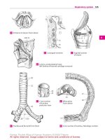
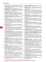
Bạn đang xem bản rút gọn của tài liệu. Xem và tải ngay bản đầy đủ của tài liệu tại đây (1.53 MB, 51 trang )
247
1
2
3
4
5
6
7
8
9
10
11
12
13
14
15
16
17
18
19
20
21
22
23
24
25
a
a
a
Veins
2222
29
32
28
25
28
21
33; 34
23
30; 31
35
28a
26
27
17
14
12
10
15
18
19
16
2
8
6
4
7
1
14
8
5
3
9
12
11
23
10
21
10
13
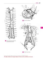
Veins of posterior thoracic
and abdominal walls
A
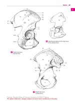
Veins of vertebrae
B
Inferior vena cava
C
Feneis, Pocket Atlas of Human Anatomy © 2000 Thieme
All rights reserved. Usage subject to terms and conditions of license.
248
1
2
3
4
5
6
7
8
9
10
11
12
13
14
15
16
17
18
19
20
21
22
23
24
25
1 PORTAL VEIN OF LIVER. Vena portae hepatis. It
brings blood from the intestinal tract to the
liver. Forms important anastomoses to
esophageal veins, rectal venous plexus and the
superficial veins of the abdominal skin. A
2 Right branch. Ramus dexter. Strong shorter
right branch of portal vein. It forms bifurcations
in the right lobe of the liver that extend as far as
the interlobular veins. A
3
Anterior branch. Ramus anterior. It supplies
the anterior part of the right lobe. A
4
Posterior branch. Ramus posterior. It supplies
the posterior part of the right lobe. A
5 Lef t branch. Ramus sinister. Longer and some-
what more slender branch supplying the left
lobe as well as the caudate and quadrate lob es.
A
6
Transverse par t. Pars transversa. Initial seg-
ment of left branch coursing transversely into
liver hilum. A
7
Caudate branches. Rami caudati. Twigs to cau-
date lobe. A
8
Umbilical par t. Pars umbilicus. Sagittal con-
tinuation of the left branch into the left lobe. A
9
[Ductus venosus]. Embryonic vein uniting
umbilical vein and inferior vena cava. Bypasses
the liver. B
10
Ligamentum venosum. Lig. venosum. Connec-
tive tissue vestige of the ductus venosus in the
groove for the ligamentum venosum. B
11
Lateral branches. Rami laterales. Branches to the
quadrate lobe and part of the caudate lobe.
12
Left umbilical vein. V. umbilicalis sinistra.
Embryonic vein joining the portal vein in the
liver. It carries cord blood to the right atrium
partly via the ductus venosus and inferior vena
cava. B
13
Round ligament of liver. Lig. teres hepatis. Con-
nective tissue remains of left umbilical vein. A
14
Medial branches. Rr. mediales. Branches of the
umbilical part extending to anterior portion of
left lobe of liver. A
15
Cystic vein. V. cystica. Vein from gallbladder
emptying into right branch of portal vein. A
16
Paraumbilical veins. Vv. paraumbilicales.
Small veins around the round ligament. They
form anastomoses between the left branch of
the portal vein and subcutaneous abdominal
veins. A
17
Left gastric vein. V. gastrica sinistra. Compan-
ion vein of left gastric artery. A
18
Right gastric vein. V. gastrica dextra. Compan-
ion vein of right gastric artery. A
19
Prepyloric vein. V. praepylorica. Branch from
the anterior part of pylorus to the right gastric
vein or portal vein. A
20 Superior mesenteric vein. V. mesenterica su-
perior. Its drainage area extends from the distal
half of the duodenum to the left colic flexure. It
joins the splenic vein to form the portal vein. A
21 Jejunal veins. Vv. jejunales. Branches from the
jejunum and ileum. A
21 a Ileal branches. Vv. ileales. A
22 Right gastro-omental (gastro-epiploic) vein.
V. gastro-omentalis (epiploica) dextra. Com-
panion vein of right gastro-omental artery. A
23 Pancreatic veins. Vv. pancreaticae. Direct
branches from the pancreas. A
24 Pancreaticoduodenal veins. Vv. pancreati-
coduodenales. Companion veins of pancreati-
coduodenal arteries. A
25 Ileocolic vein. V. ileocolica. Branch from the
ileocecal region. A
26
Appendicular vein. V. appendicularis. Vein
from the vermiform appendix. A
27 Right colic vein. V. colica dextra. Vein from the
ascending colon. A
28 Middle colic vein. V. colica media (intermedia).
Vein of transverse colon. It can also drain into
the superior and inferior mesenteric veins. A
29 Splenic vein. V. splenica. It is found in the
phrenicolic ligament and behind the pancreas.
It joins the superior mesenteric vein to form
the portal vein. A
30 Pancreatic veins. Vv. pancreaticae. They open
directly into the splenic vein. A
31 Short gastric veins. Vv. gastricae breves. They
course in the gastrosplenic ligament. A
32 Left gastro-omental (gastro-epiploic) vein. V.
gastro-omentalis (epiploica) sinistra. Compan-
ion vein of left gastro-omental vein. A
33 Inferior mesenteric vein. V. mesenterica infe-
rior. Extends from the left third of the colon to
the upper rectum and opens into the splenic
vein. A
34
Left colic vein. V. colica sinistra. Arises from
the descending colon. A
35
Sigmoid veins. Vv. sigmoideae. They drain the
sigmoid colon. A
36
Superior rectal vein. V. rectalis superior.
Branch from the upper rectum. A
37 COMMON ILIAC VEIN. V. iliaca communis.
Venous trunk reaching from L4 to the sacroiliac
joint. It unites with the contralateral vein to
form the inferior vena cava. A
38 Median sacral vein. V. sacralis mediana. Un-
paired branch that joins the left common iliac
vein. A
39 Iliolumbar vein. V. iliolumbalis. Accompanying
vein of the iliolumbar artery. It opens into the
internal or common iliac vein. A
Veins
Feneis, Pocket Atlas of Human Anatomy © 2000 Thieme
All rights reserved. Usage subject to terms and conditions of license.
249
1
2
3
4
5
6
7
8
9
10
11
12
13
14
15
16
17
18
19
20
21
22
23
24
25
a
a
a
Veins
2
3
4
5
6
7
8
14
17
18
19
13
16
15
24
20
23
28
22
34
33
30
32
31
29
27
21
21
34
35
37
36
38
39
26
21a
25
9(10)
12
1
Portal veinA
Veins of fetal liver
from below and behind
B
Feneis, Pocket Atlas of Human Anatomy © 2000 Thieme
All rights reserved. Usage subject to terms and conditions of license.
250
1
2
3
4
5
6
7
8
9
10
11
12
13
14
15
16
17
18
19
20
21
22
23
24
25
1 INTERNAL ILIAC [[HYPOGASTRIC]] VEIN. V. iliaca
interna [[v. hypogastrica]]. Short trunk receiving
veins from the pelvic viscera and perineum. A C
2
Superior gluteal veins. Vv. glutaeales
5
super-
iores. Companion veins of superior gluteal
artery passing through the upper division of
the greater sciatic foramen [[suprapiriform
foramen]] to the pelvis. They converge to form a
trunk which opens into the internal iliac vein. A
3
Inferior gluteal veins. Vv. glutaeales
5
inferi-
ores. Companion veins of inferior gluteal artery
passing through the lower division of the
greater sciatic foramen [[infrapiriform fora-
men]] into the pelvis. They unite to form a trunk
and open into the internal iliac vein. A C
4
Obturator veins. Vv. obturatoriae. They enter
the pelvis via the obturator foramen and usu-
ally open into both the internal iliac and com-
mon iliac veins. A
5
Lateral sacral veins. Vv. sacrales laterales.
Lateral branches from the sacral venous plexus.
A
6
Sacral venous plexus. Plexus venosus sacralis.
Venous network lying in front of the sacrum. A
7
Rectal venous (hemorrhoidal) plexus. Plexus
venosus rectalis [[plexus haemorrhoidalis]].
Plexus surrounding the rectum. A
8
Vesical veins. Vv. vesicales. Veins from the
vesical venous plexus. A
9
Vesical venous plexus. Plexus venosus vesi-
calis. Extends from the base of the bladder to
communicate with the prostatic or vaginal
venous plexus. A C
10
Prostatic venous plexus. Plexus venosus pros-
taticus. It surrounds the prostate and unites
with the neighboring vesical venous plexus. C
11
Deep dorsal vein of penis. V. dorsalis pro-
funda penis. Subfascial vein of the dorsum of
the penis that passes below the symphysis be-
tween the arcuate ligament of the pubis and
the transverse perineal ligament to enter the
prostatic venous plexus. It lies between the
deep fascia of the penis and the tunica al-
buginea and is usually not paired. C. See also
p. 165 B
12
Deep dorsal vein of clitoris. V. dorsalis pro-
funda clitoridis. Subfascial vein of dursum of
clitoris opening into vesical venous plexus. B
13
Uterine veins. Vv. uterinae. Connecting veins
that join the uterine venous plexus and internal
iliac vein. A
14 Uterine venous plexus. Plexus venosus uter-
inus. Venous network primarily at the root of
the broad ligament. It communicates with the
vaginal venous plexus. A
15 Vaginal venous plexus. Plexus venosus vagi-
nalis. Venous network around the vagina with
numerous connections to the surrounding
venous plexus. A
16 Internal pudendal vein. V. pudenda interna. It
runs in the lateral wall of the ischioanal fossa
and enters the pelvis via the lower division of
the greater sciatic foramen [[infrapiriform fora-
men]]. A B C
17
Deep veins of penis. Vv. profundae penis. They
arise from the roots of the corpus cavernosum
and corpus spongiosum and drain into the
prostatic venous plexus via the deep dorsal
vein of the penis. C
18
Deep veins of clitoris. Vv. profundae clitoridis.
Equivalent to the deep veins of the penis. B
19
Middle rectal veins. Vv. rectales mediae.
Branches from the rectal venous plexus located
in the lesser pelvis. They anastomose with the
superior rectal vein and the inferior rectal
veins. A C
20
Inferior rectal veins. Vv. rectales inferiores.
Arise from the anal region, join the internal pu-
dendal vein and anastomose with the middle
rectal veins and the superior rectal vein. B C
21 Posterior scrotal/labial veins. Vv. scrotales/
labiales posteriores. Arise from the scrotum or
labia and join the internal pudendal vein. B C
22 Vein of bulb of penis/vestibule. V. bulbi penis/
vestibuli. Arise from the bulb of the corpus
spongiosum and convey blood either to the
deep dorsal vein of the penis (clitoris) or into
the internal pudendal vein. B C
23 External iliac vein. V. iliaca externa. Arises from
the upper end of the femoral vein below the in-
guinal ligament and ends where it joins the in-
ternal iliac vein to form the common iliac vein.
A
24 Inferior epigastric vein. V. epigastrica inferior.
Arises from the posterior side of the anterior
abdominal wall and extends as a companion
vein of the inferior epigastric artery. A
24 a
Pubic branch (accessory obturator vein). R.
pubicus (v. obturatoria accessoria). It anasto-
moses with the branch of the obturator vein at
the inner surface of the pubis. A
25 Deep circumflex iliac vein. V. circumflexa iliaca
profunda. Companion vein of the deep circum-
flex iliac artery. A
Veins
Feneis, Pocket Atlas of Human Anatomy © 2000 Thieme
All rights reserved. Usage subject to terms and conditions of license.
251
1
2
3
4
5
6
7
8
9
10
11
12
13
14
15
16
17
18
19
20
21
22
23
24
25
a
a
a
Veins
20
16
22
21
17
10
9
19
3
1
11
21
12
18
22
16
20
20
1
2
23
5
6
3
4
13
19
16
9
8
14
15
7
25
24
24a
Pelvic veins medial viewA
Veins of female perineum
B Veins of male urogenital organsC
Feneis, Pocket Atlas of Human Anatomy © 2000 Thieme
All rights reserved. Usage subject to terms and conditions of license.
252
1
2
3
4
5
6
7
8
9
10
11
12
13
14
15
16
17
18
19
20
21
22
23
24
25
VEINS OF LOWER LIMBS. Venae membri inferi-
oris.
0a Venae superficiales membri inferioris. Super-
ficial veins of lower limbs.
0b Venae profundae membri inferioris. Deep
veins of lower limbs.
1 Femoral vein. V. femoralis. A companion vein
of the femoral artery that extends from the hia-
tus tendineus of the adductor canal to the in-
guinal ligament. A
2
External pudendal veins. Vv. pudendae ex-
ternae. Individual branches from the external
genitalia. A
3
Superf icial circumflex iliac vein. V. cir-
cumflexa iliaca superficialis. Subcutaneous
companion vein of the superficial circumflex
iliac artery. A
4
Superf icial epigastric vein. V. epigastrica su-
perficialis. Subcutaneous companion vein of
the superficial epigastric artery. A
5
Superf icial dorsal veins of penis/clitoris. Vv.
dorsales superficiales penis/clitoridis. Paired
epifascial veins of the penis (clitoris) that drain
into the femoral vein or external pudendal
veins. They run between the superf icial and
deep fasciae of the penis. A. See also p. 165 B
6
Anterior scrotal/labial veins. Vv. scrotales/
labiales anteriores. Arise from the scrotum or
labia majora and open into the femoral vein or
the external pudendal veins. A
7 Greater saphenous vein. V. saphena magna.
Arises from the medial side of the foot and as-
cends medially. This vein is provided with
valves and receives most of the medial superfi-
cial cutaneous veins. It drains into the femoral
vein via the saphenous opening. A B C D
8
Accessory saphenous vein. V. saphena acces-
soria. Connecting branch that occasionally joins
the small saphenous vein to the great
saphenous vein. It may receive blood from the
thigh except for the deep and lateral regions. It
sometimes runs parallel to the great saphenous
vein before entering the latter. A
9 Accompanying vein of the profunda femoris
artery. V. profunda femoris. A
10
Medial circumflex femoral veins. Vv. cir-
cumflexae mediales femorales. Companion
veins of the corresponding artery. A
11
Lateral circumflex femoral veins. Vv. cir-
cumflexae laterales femorales. Companion
veins of the corresponding artery. A
12
Perforating veins. Vv. perforantes. Arise from
the ischiocrural musculature, penetrate the ad-
ductors and open into the profunda femoris
vein. A
13 Popliteal vein. V. poplitea. From its origin be-
tween the popliteal artery and tibial nerve, it
extends from the union of the anterior and
posterior tibial veins to the hiatus tendineus of
the adductor canal. C
13 a Sural veins. Venae surales. Companion veins of
the corresponding arteries.
14 Genicular veins. Vv. geniculares. Usually five
veins arising from the knee. A
15 Small saphenous vein. V. saphena parva. It
arises from the lateral margin of the foot,
passes along the posterior side of the lower leg
and drains into the popliteal vein. A B C D
16 Anterior tibial veins. Vv. tibiales anteriores.
Companion veins of the anterior tibial artery. A
BC
17
Dorsal venous network of foot. Rete veno-
sum dorsale pedis. Network of veins on the
dorsum of the foot that drain into the great and
small saphenous veins and anterior tibial veins.
B
18
Dorsal venous arch of foot. Arcus venosus
dorsalis pedis. Venous arch on the dorsum of
the foot receiving the dorsal metatarsal veins of
the foot. It also serves as the main outlet for
blood from the sole of the foot. B C D
19
Dorsal digital veins of foot. Vv. digitales dor-
sales pedis. Veins on the dorsum of the toes. B
20
Dorsal metatarsal veins. Vv. metatarsales
dorsales. Companion veins of corresponding
arteries. They arise from the dorsal digital veins
of the foot. B D
21 Posterior tibial veins. Vv. tibiales posteriores.
Veins accompanying the posterior tibial artery.
C
22
Peroneal (fibular) veins. Vv. fibulares. Com-
panion veins of the fibular artery found partly
beneath the flexor hallucis longus. C
23
Plantar venous network. Rete venosum plan-
tare. Dense subcutaneous network of veins on
the sole of the foot. C
24
Plantar venous arch. Arcus venosus plantaris.
Venous arch accompanying the arterial plantar
arch. C
25
Plantar metatarsal veins. Vv. metatarsales
plantares. Veins accompanying the correspond-
ing arteries. C
26
Plantar digital veins. Vv. digitales plantares.
Veins on the flexor side of the toes. C
26 a
Intercapitular veins. Vv. intercapitulares.
Veins that connect the plantar and dorsal
venous arches. D
26 b Lateral marginal vein. V. marginalis lateralis.
Anastomotic vein as in 26 a. It drains into the
small saphenous vein. D
26 c Medial marginal vein. V. marginalis medialis.
Anastomotic vein as in 26 a. It drains into the
great saphenous vein. D
27
Perforating veins. Vv. perforantes. Veins that
connect the cutaneous and subfascial veins
especially on the lower leg. Their valves pre-
vent the flow of blood from the deep veins to
the epifascial veins.
Veins
Feneis, Pocket Atlas of Human Anatomy © 2000 Thieme
All rights reserved. Usage subject to terms and conditions of license.
253
1
2
3
4
5
6
7
8
9
10
11
12
13
14
15
16
17
18
19
20
21
22
23
24
25
a
a
a
Veins
7
26c
20
26b
26a
26a
15
18
13
7
21
23
24
25
26
15
16
22
18
16
15
17
18
20
19
7
3
1
4
2
5
6
10
7
9
12
12
8
7
14
1
11
11
14
16
15
Veins of lower limb,
anterior view
A
Veins on dorsum of foot
B
Veins of leg and sole of foot
C
Veins on dorsum of foot
with venous arch
D
Feneis, Pocket Atlas of Human Anatomy © 2000 Thieme
All rights reserved. Usage subject to terms and conditions of license.
254
1
2
3
4
5
6
7
8
9
10
11
12
13
14
15
16
17
18
19
20
21
22
23
24
25
1 LYMPHATIC SYSTEM. Systema lymphaticum.
2 Lymphatic vessels. Vasa lymphatica.
3 Lymphatic capillary. Vas lymphocapillare. Any
of the vessels of the lymphatic system that
form closed networks and have permeable
walls. C
4 Lymphatic capillary network. Rete lympho-
capillare. Network of lymphocapillary veins. C
5 Lymphatic vessel. Vas lymphaticum. Any of the
valvular lymphatic vessels that communicate
with the lymphocapillary vessels. Their thin
walls are sparsely lined with smooth muscles. C
6
Lymphatic plexus. Plexus lymphaticus. Net-
work of lymphatic vessels lying deeper than
the lymphocapillary vessels. In the outer layers
of the skin, it lies within and directly below the
corium. C
7 Superficial lymphatic vessel. Vas lymphaticum
superficiale. It is situated superficially on the
fascia of the limbs.
8 Deep lymphatic vessel. Vas lymphaticum pro-
fundum. It lies beneath the fascia of the limbs
and often, but not always, accompanies blood
vessels.
9 Lymphatic trunks. Trunci lymphatici. Five
main lymphatic branches of the lymph-vascu-
lar system.
10 Right/left lumbar trunk. Truncus lumbaris
dexter/sinister. Main branch which brings
lymph to the cisterna chyli from the legs, pelvic
viscera, urogenital system and parts of the
abdominal wall and the abdominal viscera. B
11 Intestinal trunks. Trunci intestinales. Main
conduits which transport lymph to the cisterna
chyli from the supply region of the superior and
inferior mesenteric arteries. B
12 Right/left bronchomediastinal trunk. Truncus
bronchomediastinalis dexter/sinistra. It col-
lects lymph from the heart, lungs and medi-
astinum. On the left side it opens into the
thoracic duct, on the right side, the right lym-
phatic duct. Often, however, both may open
directly into the subclavian veins. B
13 Right/left subclavian trunk. Truncus sub-
clavius dexter/sinister. Arises from the arm, ac-
companies the subclavian vein and usually
opens on the right side into the right lymphatic
duct and on the left side into the angle between
the left subclavian vein and internal jugular
vein. B
14 Right/left jugular trunk. Truncus jugularis
dexter/sinister. Accompanies the internal jugu-
lar vein and passes to the angle between the in-
ternal jugular and subclavian veins (venous
angle). B
15 Lymphatic ducts. Ductus lymphatici. The main
drainage ducts of the lymphatic system.
16 Right lymphatic duct (right thoracic duct).
Ductus lymphaticus dexter (ductus thoracicus
dexter). It is formed by the union of the right
jugular, subclavian and bronchomediastinal
trunks. It may be absent. B
17 Thoracic duct. Ductus thoracicus. Arises from
the cisterna chyli a short distance below the di-
aphragm, courses upward behind the aorta and
opens into the venous angle, i. e., the angle be-
tween the left internal jugular and subclavian
veins. B
18
Arch of thoracic duct. Arcus ductus thoracici.
Arch formed by the thoracic duct before enter-
ing the venous angle. B
19 Cer vical part. Pars cervicalis. Short cervical
segment in front of C7. B
20
Thoracic part. Pars thoracica. It begins at the
aortic hiatus and ends at the upper margin of
T1. B
21 Abdominal par t. Pars abdominalis. Very short
segment in front of L1. B
22 Cisterna chyli. Variable dilatation at the origin
of the thoracic duct. It receives the lumbar and
intestinal trunks. B
23 Lymph node. Nodus lymphaticus (Lym-
phonodus). Lymphoreticular filtering organ, 1−
25 mm in diameter, within the lymphatic ves-
sels. Since lymph must usually traverse two
lymph nodes before arriving in the blood
stream at the venous angle, there is double pro-
tection against the invasion of pathogens or
tumor cells into the blood stream. A
24 Afferent lymphatic vessels. Vas lymphaticum
afferens. Any of the vessels that carry lymph to
a lymph node; located on the convex surface of
the node. A
25 Efferent lym phatic vessel. Vas lymphaticum
efferens. Any of the vessels that carry lymph
away from a lymph node; located on the hilum
of the node. A
26 Cortex. Part of the lymphoreticular tissue pro-
ximal to the capsule. A
27 Medulla. Lymphoreticular tissue between cor-
tex and hilum. A
28 Hilum. Somewhat retracted area where blood
vessels enter and where blood and lymphatic
vessels exit. A
29
Lymphatic nodule. Nodulus lymphaticus
(lymphonodulus). Spherical condensation of
lymphoreticular tissue predominantly occupy-
ing the cortex. It exhibits a lighter central area
(“reaction center”). A
Lymphatic system
Feneis, Pocket Atlas of Human Anatomy © 2000 Thieme
All rights reserved. Usage subject to terms and conditions of license.
255
1
2
3
4
5
6
7
8
9
10
11
12
13
14
15
16
17
18
19
20
21
22
23
24
25
a
a
a
28
27
27
24
26
29
25
14
13
1212
14
13
1819
16
20
21
22
11
11
10
3
4
6
6
4
5
5
Section of lymph
node
A
Lymphatic vessels
of the trunk
B
Lymphatic vessels
of small intestine
C
Lymphatic system
Feneis, Pocket Atlas of Human Anatomy © 2000 Thieme
All rights reserved. Usage subject to terms and conditions of license.
256
1
2
3
4
5
6
7
8
9
10
11
12
13
14
15
16
17
18
19
20
21
22
23
24
25
1 REGIONAL LYMPH NODES. Nodi lymphatici re-
gionales.
2 Head and neck. Caput et collum.
3 Occipital lymph nodes. Nodi lymphatici occipi-
tales. One to three lymph nodes lying close to
the margin of the trapezius. Afferents: scalp,
deep cervical muscles. Efferents: deep cervical
lymph nodes. A
4 Mastoid [retroauricular] lymph nodes. Nodi
lymphatici mastoidei [[retroauriculares]]. Usu-
ally two nodes on the mastoid process. Affer-
ents: posterior surface of pinna, posterior wall
of external acoustic meatus and corresponding
parts of scalp. Efferents: deep cervical lymph
nodes. A
5 Superficial partodi lymph nodes. Nodi lym-
phatici parotidei superf iciales. They lie on the
parotid fascia in front of the tragus. Afferents:
junction of temporal region and anterior sur-
face of pinna. Efferents: deep cervical lymph
nodes. A
6 Deep parotid lymph nodes. Nodi lymphatici
parotidei profundi. Group beneath the parotid
fascia. Afferents: tympanic cavity, external
acoustic meatus, frontotemporal region, eye-
lids, root of nose, and sometimes the posterior
floor of the nose and nasopharyngeal cavity.
Efferents: deep cervical lymph nodes. A
7
Preauricular lymph nodes. Nodi lymphatici
prae-auriculares. Group located in front of the
pinna. A
8
Infra-auricular lymph nodes. Nodi lymphat-
ici infra-auriculares. Group beneath the pinna.
A
9
Intraglandular lymph nodes. Nodi lymphatici
intraglandulares. Group situated directly
within the parotid. A
10 Facial lymph nodes. Nodi lymphatici faciales.
Variable lymph nodes that receive lymph from
the eyelids, nose and the rest of the face and
buccal mucosa. Efferents: submandibular
lymph nodes. The vessels accompany the facial
artery.
11
Buccinator node. [Nodus buccinatorius].
Lymph node situated deep within the buccina-
tor muscle. A
12
Nasolabial node. [Nodus nasolabialis]. Lymph
node located below the nasolabial fold. A
13
Malar node. [Nodus malaris]. Superficial
lymph node of the cheek.
14
Mandibular node. [Nodus mandibularis].
Lymph node located on the mandible. A
14 a Lingual lymph nodes. Nodi lymphatici lingu-
ales. Nodes located on the hyoglossus muscle.
They drain lymph from the lower surface and
lateral margin to tongue as well as the medial
anterior two-thirds of its dorsal surface.
15 Submental lymph nodes. Nodi lymphatici sub-
mentales. Nodes between the anterior bellies
of the digastric muscles. Afferents: middle of
lower lip, floor of mouth and tip of tongue.
Efferents: deep cervical and submental lymph
nodes. B
16 Submandibular lymph nodes. Nodi lymphatici
submandibulares. Nodes between the
mandible and submandibular gland that serve
as first and second filter stations. Direct affer-
ent area: inner canthus of eye, cheek, side of
nose, upper lip, lateral lower lip, gingiva and
anterior lateral margin of tongue. Indirect affer-
ents: facial and submental lymph nodes. Effer-
ents: deep cervical lymph nodes. B
17 Anterior cervical lymph nodes. Nodi lymphat-
ici cervicales anteriores.
18
Superf icial (anterior jugular) lymph nodes.
Nodi lymphatici superficiales (jugulares anteri-
ores). Nodes on the internal jugular vein. Affer-
ent region: skin of anterior side of neck. Effer-
ents: bilateral deep cervical lymph nodes. A
19
Deep lymph nodes. Nodi lymphatici profundi.
Anterior group.
19 a
Infrahyoid lymph nodes. Nodi lymphatici in-
frahyoidei. They lie in the midline below the
body of the hyoid bone. Afferent areas: larny-
geal vestibule, piriform recess and adjacent hy-
popharynx. Efferents: deep cervical lymph
nodes. B
20
Prelaryngeal lymph nodes. Nodi lymphatici
praelaryngeales
3
. Nodes on the cricothyroid
ligament. Afferent area: lower half of larynx.
Efferents: deep cervical lymph nodes. B
21
Thyroid lymph nodes. Nodi lymphatici thyroidei.
Nodes on the thyroid gland. Efferents: as in 20.
B
22
Pretracheal lymph nodes. Nodi lymphatici pre-
tracheales. Nodes in front of the trachea. Affer-
ent regions: trachea and larynx. Efferents: deep
cervical lymph nodes. B
23
Paratracheal lymph nodes. Nodi lymphatici par-
atracheales. Nodes beside the trachea. Actions
similar to those of 22. B
23 a
Retropharyngeal lymph nodes. Nodi lymphatici
retropharyngeales. Deep cervical lymph nodes
in front of the arch of the atlas. See p. 258.13
Lymphatic system
Feneis, Pocket Atlas of Human Anatomy © 2000 Thieme
All rights reserved. Usage subject to terms and conditions of license.
257
1
2
3
4
5
6
7
8
9
10
11
12
13
14
15
16
17
18
19
20
21
22
23
24
25
a
a
a
19a
3
8
4
5
7
6
9
11
12
14
18
258.2
16
15
20
21
22
23
Lymph nodes at surface of
neck and head
A
Deep lymph nodes of neck
B
Lymphatic system
Feneis, Pocket Atlas of Human Anatomy © 2000 Thieme
All rights reserved. Usage subject to terms and conditions of license.
258
1
2
3
4
5
6
7
8
9
10
11
12
13
14
15
16
17
18
19
20
21
22
23
24
25
1 Lateral cervical lymph nodes. Nodi lymphatici
cervicales laterales. Groups of lymph nodes
lateral to the neck which are classified as fol-
lows.
2
Superf icial lymph nodes. Nodi lymphatici su-
perficiales. Nodes on the external jugular vein.
Afferent regions: lower pinna and area below
the parotid. Efferents: deep cervical lymph
nodes. See p. 257 A
3
Superior deep lymph nodes. Nodi lymphatici
profundi superiores. Nodes that form the sec-
ond filter station for almost all of the head
lymph nodes but also receive direct peripheral
tributaries from their surroundings. Efferents:
jugular trunk. A
4
Lateral lymph nodes. Nodi lymphatici laterales.
They lie lateral to the internal jugular vein. A
5
Anterior lymph nodes. Nodi lymphatici anteri-
ores. Group of lymph nodes in front of the in-
ternal jugular vein. A
6
Jugulodigastric node. Nodus jugulodigastricus. It
is the most cranial of the deep cervical nodes
and is palpable when the tonsil, tongue or
pharynx is inflamed. A
7
Inferior deep cervical lymph nodes. Nodi
lymphatici profundi inferiores. They form the
second f ilter station for the lymph nodes of the
cervical viscera and the last filter station for the
lymph nodes of the head. They also receive
direct tributaries. Efferents: jugular trunk.
8
Jugulo-omohyoid node. Nodus juguloomohy-
oideus. Node between the omohyoid muscle
and internal jugular vein. Afferent area: tongue.
A
9 Lateral lymphatic nodes. Nodi lymphatici later-
ales. They lie lateral to the internal jugular vein.
A
10
Anterior lymph nodes. Nodi lymphatici anteri-
ores. Group in front of the internal jugular vein.
A
11
Supraclavicular lymph nodes. Nodi lymphatici
supraclaviculares. Nodes of the same group lo-
cated above the clavicle. A
12
Accessory nodes. Nodi accessorii.
13
Retropharyngeal lymph nodes. Nodi lym-
phatici retropharyngeales. Deep cervical lymph
nodes at the level of the lateral mass of the atlas
and at the lateral margin of the longus capitis
muscle. A B
14 Upper limb. Membrum superius.
15 Axillary lymphatic plexus. Plexus lymphaticus
axillaris. Netlike connections of 20−30 axillary
lymph nodes via their lymphatic vessels. C
16 Axillary lymph nodes. Nodi lymphatici axil-
lares. Nodes in the axilla. C
17
Apical lymph nodes. Nodi lymphatici apicales.
Nodes medial to the axillary vein that extend
from the upper margin of the prectoralis minor
to the apex of the axilla. Afferent areas: upper
lateral part of the breast and all remaining axil-
lary lymph nodes. Efferents: (left) subclavian
trunk to the thoracic duct or subclavian vein;
(right) into the vein directly or after joining the
jugular trunk. C
18
Brachial lymph nodes. Nodi lymphatici
brachiales. Nodes along the axillary artery that
drain lymph from the arm. C
19
Subscapular lymph nodes. Nodi lymphatici
subscapulares. Nodes alongside the subscapu-
lar artery that drain lymph from the posterior
thorax and shoulder as well as from the lower
nuchal region. C
20
Pectoral lymph nodes. Nodi lymphatici pec-
torales. Nodes along the lateral margin of the
pectoralis minor that drain lymph from the
anterior and lateral wall of the trunk as far as
the navel, as well as the central and lateral part
of the breast. C
21
Central lymph nodes. Nodi lymphatici cen-
trales. Nodes in the fat of the axillar that filter
lymph from the brachial, subscapular and pec-
toral lymph nodes. C
22
Interpectoral lymph nodes. Nodi lymphatici
interpectorales. Small group of nodes situated
between the pectoralis major and minor. Affer-
ent area: mammary gland. Efferents: apical
lymph nodes. C
23
Deltopectoral (infraclavicular) lymph
nodes.
Nodi lymphatici deltopectorales (infra-
claviculares). Nodes on the cephalic vein in the
deltopectoral groove that receive lymph from
the arm. C
24 Brachial lymph nodes. Nodi lymphatici
brachiales. Single lymph nodes along the
brachial vessels.
25 Cubital lymph nodes. Nodi lymphatici cubi-
tales. One or two lymph nodes on the brachial
artery in the cubital fossa. C
26 Supratrochlear lymph nodes. Nodi lymphatici
supratrochlearis. Nodes that lie medial to the
basilic vein and above the elbow joint. C
27 Superficial lymph nodes. Nodi lymphatici su-
perficiales.
28 Deep lymph nodes. Nodi lymphatici profundi.
Individual lymph nodes that follow the course
of the deep lymphatic vessels.
Lymphatic system
Feneis, Pocket Atlas of Human Anatomy © 2000 Thieme
All rights reserved. Usage subject to terms and conditions of license.
259
1
2
3
4
5
6
7
8
9
10
11
12
13
14
15
16
17
18
19
20
21
22
23
24
25
a
a
a
Deep lymph nodes of neckA
Neck, anterior view
B
Lymph nodes of arm, axilla and chest
C
6
13
4
9
5
8
13
25
26
23
2115
22
17
19
18
20
10
11
Lymphatic system
Feneis, Pocket Atlas of Human Anatomy © 2000 Thieme
All rights reserved. Usage subject to terms and conditions of license.
260
1
2
3
4
5
6
7
8
9
10
11
12
13
14
15
16
17
18
19
20
21
22
23
24
25
1 Thorax.
2 PARAMAMMARY LYMPH NODES. Nodi lym-
phatici paramammarii. Lymph nodes on the
lateral margin of the mammary gland. A
3 Parasternal lymph nodes. Nodi lymphatici
parasternales. Nodes located along the course
of the internal thoracic vessels. Afferent re-
gions: mammary gland, intercostal spaces, part
of the liver and diaphragm. Efferents: either
directly into the respective subclavian vein or
internal jugular vein or into the thoracic duct or
subclavian trunk. A
4 Intercostal lymph nodes. Nodi lymphatici in-
tercostales. Nodes located in the paravertebral
portion of the intercostal spaces. Afferent area:
pleura and intercostal spaces. D
5 Paravertebral lymph nodes. Nodi lymphatici
praevertebrales.
3
Nodes located between the
esophagus and vertebral column. Afferent re-
gions: surroundings, if not drained by other
vessels. C D
6 Superior phrenic lymph nodes. Nodi lymphat-
ici phrenici superiores. Nodes situated behind
the cartilage-bone boundary of the 7
th
rib at the
site where the aorta opens into the diaphragm
and along the inferior vena cava. Afferent areas:
liver and diaphragm. D
7 Prepericardial lymph nodes. Nodi lymphatici
prepericardiales. Nodes located between the
sternum and pericardium. Afferent regions:
sternum and anterior pericardium. Efferents:
parasternal lymph nodes. B
8 Lateral pericardial lymph nodes. Nodi lym-
phatici pericardiales laterales. Nodes found be-
tween the pericardium and mediastinal pleura.
B
9 Anterior mediastinal lymph nodes. Nodi lym-
phatici mediastinales anteriores. Nodes along
the brachiocephalic veins, in front of the arch of
the aorta and its branches. Afferent regions:
thymus, pericardium and parasternal lymph
nodes. Efferents: bronchomediastinal trunk. B
10 Ligamentum arteriosum node. [Nodus liga-
menti arteriosi]. Node occasionally found along
the lig. arteriosum. B
11 Posterior mediastinal lymph nodes. Nodi lym-
phatici mediastinales posteriores. Nodes sit-
uated in the superior and posterior medi-
astinum. They receive lymph from the follow-
ing organs: lungs, bronchi, trachea, esophagus,
pericardium, diaphragm and diaphragmatic
surface of the liver. They pass efferent vessels to
the thoracic and bronchomediastinal ducts. The
posterior mediastinal lymph nodes are sub-
divided into the following groups.
12
Pulmonary juxtaesophageal lymph nodes.
Nodi lymphatici juxtaoesophageales pul-
monales. Group situated beside the esophagus;
they serve the lungs. C
13
Tracheobronchial lymph nodes. Nodi lym-
phatici tracheobronchiales. Group located
along the bronchi at the site where they enter
into the lungs. C
14
Superior tracheobronchial lymph nodes. Nodi
lymphatici tracheobronchiales superiores.
Group situated cranially on the stem bronchi
and the trachea. C
15
Inferior tracheobronchial lymph nodes. Nodi lym-
phatici tracheobronchiales inferiores. Group lo-
cated caudal to the tracheal bifurcation. C
16
Paratracheal lymph nodes. Nodi lymphatici
paratracheales. Group situated along the tra-
chea. C
17
Node of arch of azygos vein. [Nodus arcus
venae azygos]. Lymph node occasionally pre-
sent at the arch which the azygos vein forms
around the hilum of the right lung before join-
ing the superior vena cava. B
Lymphatic system
Feneis, Pocket Atlas of Human Anatomy © 2000 Thieme
All rights reserved. Usage subject to terms and conditions of license.
261
1
2
3
4
5
6
7
8
9
10
11
12
13
14
15
16
17
18
19
20
21
22
23
24
25
a
a
a
3
2
9
9
17
10
78
14
15
13
16
12
5
5
4
6
Lymph nodes of arm, axilla and thoraxA
Lymph nodes in thorax
B
Lymph nodes in thorax
C
Lymph nodes in thoraxD
Lymphatic system
Feneis, Pocket Atlas of Human Anatomy © 2000 Thieme
All rights reserved. Usage subject to terms and conditions of license.
262
1
2
3
4
5
6
7
8
9
10
11
12
13
14
15
16
17
18
19
20
21
22
23
24
25
1 Abdomen − parietal lymph nodes. Nodi lym-
phatici parietales. Lymph nodes in the abdom-
inal wall.
2 Lef t lumbar lymph nodes. Nodi lymphatici
lumbales (lumbares) sinistri. Nodes situated
along the abdominal aorta. They mainly serve
as secondar y filter stations for lymph nodes lo-
cated further below, but also as primary filter
stations for the suprarenal gland, kidney, ure-
ter, testis, ovary, uterine tubes, fundus of uterus
and abdominal wall. Efferents: primarily into
the lumbar trunk. They can be divided into the
following three groups.
3
Lateral aortic lymph nodes. Nodi lymphatici
aortici laterales. Group on the left side of the
aorta. A
4
Pre-aortic lymph nodes. Nodi lymphatici
preaortici. Group situated in front of the aorta. A
5
Postaortic lymph nodes. Nodi lymphatici
postaortici. This group lies between the aorta
and vertebral column. A
6 Intermediate lumbar lymph nodes. Nodi lym-
phatici lumbales (lumbares) intermedii. Group
situated between the aorta and inferior vena
cava. They function as described in 2−A
7 Right lumbar lymph nodes. Nodi lymphatici
lumbales (lumbares) dextri. Group situated
along the inferior vena cava. Theyfunctionas de-
scribed in (2) andare subdivided into the follow-
ing three groups.
8
Lateral caval lymph nodes. Nodi lymphatici
cavales laterales. Nodes on the right side of the
inferior vena cava. A
9
Precaval lymph nodes. Nodi lymphatici pre-
cavales.Nodessituatedinfrontofthevenacava.A
10
Postcaval lymph nodes. Nodi lymphatici
postcavales. Nodes located behind the vena
cava. A
11 Inferior phrenic lymph nodes. Nodi lymphatici
phrenici inferiores. Nodes situated along the
inferior surface of the diaphragm near the aor-
tic opening. A C
12 Inferior epigastric lymph nodes. Nodi lym-
phatici epigastrici inferiores. Three or four
nodes along the inferior epigastric artery that
receive lymph from its supply area. B
13 Visceral nodes. Abdomen − nodi lymphatici
viscerales. Lymph nodes of the abdominal
viscera.
14 Celiac lymph nodes. Nodi lymphatici coeliaci.
Nodes situated the celiac trunk. They form the
secondary filter station for the stomach,
duodenum, liver, gallbladder, pancreas and
spleen.Efferents:someform theintestinaltrunk,
some pass directly into the cisterna chyli. A C
15 Gastric lymph nodes [right and left].Nodilym-
phatici gastrici [dextri/sinistri]. From the lesser
curvature of the stomach, they follow the course
of the right and left gastric arteries. Afferent re-
gion: stomach. Efferents: celiac lymph nodes. C
16 [Anulus lymphaticus cardiae]. Lymphatic ring
occasionally present around the cardia of the
stomach. C
17 Gastro-omental lymph nodes [right and left].
Nodi lymphatici gastro-omentales [dextri/sin-
istri]. Nodes located along the course of the
right and left gastro-omental arteries at the
greater curvature of the stomach. Their affer-
ents receive lymph from the stomach and the
greater omentum and their efferents convey
lymph along the right side to the lymph nodes
of the liver and along the left side to the lymph
nodes of the spleen and pancreas. C
18 Pyloric lymph nodes. Nodi lymphatici pylorici.
Situated around the pylorus. Their efferents
drain into the hepatic or celiac lymph nodes.
19
Suprapyloric node. [Nodus suprapyloricus].
Node situated above the pylorus. C
20
Subpyloric nodes. [Nodi subpylorici]. Nodes
situated caudal to the pylorus. C
21
Retropyloric nodes. [Nodi retropylorici].
Group of nodes located dorsal to the pylorus. C
22 Pancreatic lymph nodes. Nodi lymphatici pan-
creatici. Situated along the upper and lower
margins of the pancreas. Their efferent vessels
convey lymph to the splenic lymph nodes,
mesenteric lymph nodes and pancreati-
coduodenal lymph nodes.
23
Superior pancreatic lymph nodes. Nodi lym-
phatici pancreatici superiores. Group of nodes
located along the upper margin of the pancreas.
AC
24
Inferior pancreatic lymph nodes. Nodi lym-
phatici pancreatici inferiores. Group of nodeslo-
catedalongthelowermarginofthe pancreas. A C
25 Splenic (lienal) lymph nodes. Nodi lymphatici
splenici (lienales). Nodes near the hilum of the
spleen that convey their lymph to the celiac
lymph nodes. A C
26 Pancreaticoduodenal lymph nodes. Nodi lym-
phatici pancreaticoduodenales. Small nodes be-
tween the pancreas and duodenum. Afferent re-
gions: duodenum and pancreas.
27
Superior pancreaticoduodenal lymph
nodes.
Nodi lymphatici pancreaticoduodenales
superiores. Cranially situated group of nodes.
Efferents: hepatic nodes. C
28
Inferior pancreaticoduodenal lymph nodes.
Nodi lymphatici pancreaticoduodenales inferi-
ores. Caudal group of lymph nodes. Efferents:
mesenteric lymph nodes. C
29 Hepatic lymph nodes. Nodi lymphatici hepat-
ici. Nodes situatednear the hilum of the liverand
in the hepatoduodenal ligament. Their lymph is
taken partly from the liver, partly from adjacent
lymph nodes and transported to the celiac
lymph nodes.
30 Cystic node. Nodus cysticus. Larger lymph node
near the neck of the gallbladder. C
31 Foraminal node. Nodus foraminalis. Larger
lymph node near the epiploic foramen. C
Lymphatic system
Feneis, Pocket Atlas of Human Anatomy © 2000 Thieme
All rights reserved. Usage subject to terms and conditions of license.
263
1
2
3
4
5
6
7
8
9
10
11
12
13
14
15
16
17
18
19
20
21
22
23
24
25
a
a
a
11
25
9
6
4
9
24
23
3
5
8
10
14
12
16
11
15
14
25
23
30
31
19
21
20
27
28
24
17
Deep lymph nodes at abdominal cavityA
Anterior abdominal wall from behindB
Lymph nodes in
upper abdomen
C
Lymphatic system
Feneis, Pocket Atlas of Human Anatomy © 2000 Thieme
All rights reserved. Usage subject to terms and conditions of license.
264
1
2
3
4
5
6
7
8
9
10
11
12
13
14
15
16
17
18
19
20
21
22
23
24
25
1 Mesenteric lymph nodes. Nodi lymphatici
mesenterici. Numerous (100−150) lymph nodes
important for the prevention of lipidemia. Their
efferent vesselsdrainlymph via the celiac lymph
nodes.
2
Juxtaintestinal lymph nodes. Nodi lymphatici
juxtaintestinales. Subgroup of mesenteric
lymph nodes located close to the smallintestine.
3
Superior [central] lymph nodes. Nodi lym-
phatici superiores [centrales]. Subgroup of
mesenteric lymph nodes located near the stem
of the superior mesenteric artery. A
4 Ileocolic lymph nodes. Nodi lymphatici ileo-
colici.Groupofnodes situated alongtheileocolic
artery. Efferents: celiac lymph nodes. A
5 Prececal lymph nodes. Nodi lymphatici precae-
cales. Nodes situated along the anterior cecal
artery. A
6 Retrocecal lymph nodes. Nodi lymphatici ret-
rocaecales. They situated along the posterior
cecal artery. A
7 Appendicular lymph nodes. Nodi lymphatici
appendiculares. Nodes situated along the ap-
pendicular artery. They are absent in 33−50% of
cases. A
8 Mesocolic lymph nodes. Nodi lymphatici me-
socolici. Nodes for the greater part of the colon.
Predominantly located in the mesocolon. Their
efferents convey lymph to the celiac lymph
nodes.
9
Paracolic lymph nodes. Nodi lymphatici para-
colici. Subgroup of mesocolic lymph nodes lo-
cated along the colon. A
10
Right/middle/left colic lymph nodes. Nodi
lymphatici colici [dextri/medii/sinistri]. Sub-
group of mesocolic lymph nodes situated along
the stems of the right, middle and left colic ar-
teries. A
11 Inferior mesenteric lymph nodes. Nodi lym-
phatici mesenterici inferiores. Nodes located
along the inferior mesenteric artery. Afferent
areas: part of the descending colon, sigmoid
and part of the rectum. Efferents: pre-aortic
lymph nodes at the level of the inferior mesen-
teric artery. A
12
Sigmoid lymph nodes. Nodi lymphatici sig-
moidei. Nodes located along the sigmoid
artery. They drain the sigmoid and the adjoin-
ing segment of colon. A
13
Superior rectal lymph nodes. Nodi lymphat-
ici rectales superiores. Nodes located along
the superior rectal artery for drainage of the
rectum. A
14 Parietal nodes of pelvis. Pelvis − nodi lym-
phatici parietales. Lymph nodes along the
wall of pelvis.
15 Common iliac lymph nodes. Nodi lymphatici
iliaci communes. Group of nodes situated
along the internal iliac vein. They serve as the
second station for the lymph nodes of the
pelvic organs, (= pelvic), interior pelvic wall,
abdominal wall up to the navel, hip muscles,
and gluteal muscles. Efferents: lumbar lymph
nodes and lumbar trunk. Individually, the fol-
lowing subgroups are distinguished.
16
Medial common iliac lymph nodes. Nodi
lymphatici iliaci communes mediales. Sub-
group situated medial to the vascular cord. B
17
Intermediate common iliac lymph nodes.
Nodi lymphatici iliaci communes intermedii.
Subgroup situated between the medial and
lateral groups behind the vascular cord. B
18
Lateral common iliac lymph nodes. Nodi
lymphatici iliaci communes laterales. Sub-
group located lateral to the vascular cord. B
19
Subaortic common iliac lymph nodes. Nodi
lymphatici iliaci communes subaortici. Sub-
group located caudal to the aortic bifurcation
in front of L4. B
20
Promontory common iliac lymph nodes.
Nodi lymphatici iliaci communes promontorii.
Subgroup situated in front of the promontory. B
21 External iliac lymph nodes. Nodi lymphatici il-
iaci externi. Group of nodes located along the
external iliac vessels. They are the first lymph
station for a part of the urinary bladder and
vagina, and second lymph station for the ingui-
nal lymph nodes. Efferents: common iliac
lymph nodes. They are divided into the follow-
ing groups.
22
Medial external iliac lymph nodes. Nodi lym-
phatici iliaci externi mediales. Group located
medial to the vascular cord. B
23
Intermediate external iliac lymph nodes.
Nodi lymphatici iliaci externi intermedii. Group
located between the lateral and medial groups
and behind the artery. B
24
Lateral external iliac lymph nodes. Nodi
lymphatici iliaci externi laterales. Group lo-
cated lateral to the vascular bundle. B
25
Medial lacunar node. [Nodus lacunaris medi-
alis]. Node situated in the vascular lacuna me-
dial to the vascular cord. B
26
Intermediate lacunar node. [Nodus lacunaris
intermedius]. When present, it lies in the
middle of the vascular lacuna. B
27
Lateral lacunar node. [Nodus lacunaris later-
alis]. Node situated laterally in the vascular
lacuna. B
28
Interiliac external iliac lymph nodes. Nodi
lymphatici iliaci externi interiliaci. Lymph
nodes in the bifurcation between the internal
and external iliac arteries. B
29
Obturator external iliac lymph nodes. Nodi
lymphatici iliaci externi obturatorii. Group lo-
cated at the obturator artery. B
Lymphatic system
Feneis, Pocket Atlas of Human Anatomy © 2000 Thieme
All rights reserved. Usage subject to terms and conditions of license.
265
1
2
3
4
5
6
7
8
9
10
11
12
13
14
15
16
17
18
19
20
21
22
23
24
25
a
a
a
3
10
10
11
12
20
13
7
6
5
4
9
9
9
19
2016
19
16
28
29
22
25
27
26
18
17
24
23
Lymph nodes
of abdominal cavity
A
Lymph nodes
along pelvic vessels
B
Lymphatic system
Feneis, Pocket Atlas of Human Anatomy © 2000 Thieme
All rights reserved. Usage subject to terms and conditions of license.
266
1
2
3
4
5
6
7
8
9
10
11
12
13
14
15
16
17
18
19
20
21
22
23
24
25
1 Internal iliac lymph nodes. Nodi lymphatici ili-
aci interni. Located along the internal iliac
artery; they drain the pelvic organs, the deep
perineal region and both the external and inter-
nal walls of the pelvis. Their efferents com-
municate with the common iliac lymph nodes.
2
Superior gluteal lymph nodes. Nodi lymphat-
ici gluteales superiores. Nodes for the pelvic
wall located at the superior gluteal artery. A
3
Inferior gluteal lymph nodes. Nodi lymphat-
ici gluteales inferiores. Nodes for the prostate
and proximal urethra are situated along the in-
ferior gluteal artery. A
4
Sacral lymph nodes. Nodi lymphatici sacrales.
Nodes for the prostate and cervix found along
the sacrum. A
5 Visceral nodes of pelvis. Pelvis − nodi lympha-
tici viscerales.
6 Perivesicular lymph nodes. Nodi lymphatici
paravesiculares. Nodes for the urinary bladder
and partly also for the prostate located along
the bladder. A
7
Prevesicular lymph nodes. Nodi lymphatici
prevesiculares. Subgroup located between the
urinary bladder and symphysis. A
8
Postvesicular lymph nodes. Nodi lymphatici
postvesiculares. Subgroup behind the urinary
bladder. A
9
Lateral vesicular lymph nodes. Nodi lym-
phatici vesiculares laterales. Nodes situated at
the lower end of the medial − formerly lateral −
umbilical ligament. A
10 Parauterine lymph nodes. Nodi lymphatici
parauterini. Nodes for the cervix uteri situated
beside the uterus. A
11 Paravaginal lymph nodes. Nodi lymphatici
paravaginales. Located beside the vagina; they
drain lymph from part of this organ. A
12 Pararectal (anorectal) lymph nodes. Nodi
lymphatici pararectales (anorectales). Located
lateral to the musculature of the rectum; they
drain this organ and a part of the vagina. A
13 Lower limb. Membrum inferius.
14 Superficial inguinal lymph nodes. Nodi lym-
phatici inguinales superficiales. Group of nodes
located in the subcutaneous adipose tissue,
thus on the fascia lata. They drain the anus, per-
ineum, external genitalia, abdominal wall and
surface of the leg. Efferents: external iliac
lymph node.
15
Superomedial superf icial inguinal lymph
nodes.
Nodi lymphatici superomediales. Me-
dial portion of the group of nodes located along
the inguinal ligament. B
16
Superolateral superf icial inguinal lymph
nodes.
Nodi lymphatici superolaterales. Lateral
portion of group located below the inguinal
ligament. B
Lymphatic system
17 Inferior lymph nodes. Nodi lymphatici inferi-
ores. Group arranged along a vertical line at the
proximal end of the great saphenous vein. They
drain the superficial lymphatic vessels of the
leg. B
18 Deep inguinal lymph nodes. Nodi lymphatici
inguinales profundi. Positioned below the fas-
cia lata at the level of the saphenous hiatus. The
uppermost node can be especially large and
may occupy the femoral canal (Rosenmüller’s
node). Afferent region: deep lymphatic vessels
of the leg. Efferents: external iliac lymph nodes.
B
19 Superficial popliteal lymph nodes. Nodi lym-
phatici popliteales superficiales. They lie at the
proximal end of the small saphenous vein and
receive lymph from the lateral margin of the
foot and calf. Their efferents pass anteriorly
through the hiatus tendineus into the deep in-
guinal lymph nodes. C
20 Deep popliteal lymph nodes. Nodi lymphatici
popliteales profundi. Situated between the
knee joint capsule and the popliteal artery.
They receive lymph from the posterior side of
the lower leg and their efferents convey it ante-
riorly through the hiatus tendineus to the deep
inguinal lymph nodes. C
21 Anterior tibial node. [Nodus tibialis anterior].
Lymph node occasionally present along the
anterior tibial artery.
22 Posterior tibial node. [Nodus tibialis poste-
rior]. Lymph node occasionally present along
the posterior tibial artery.
23 Fibular node. [Nodus fibularis]. Lymph node
occasionally present along the peroneal (fibu-
lar) artery.
Feneis, Pocket Atlas of Human Anatomy © 2000 Thieme
All rights reserved. Usage subject to terms and conditions of license.
267
1
2
3
4
5
6
7
8
9
10
11
12
13
14
15
16
17
18
19
20
21
22
23
24
25
a
a
a
4
2
10
7
3
9
8
12
11
9
18
17
16 15
20
19
Lymph nodes in the female pelvisA
Lymph nodes in the inguinal region
B Lymph nodes in the popliteal fossaC
Lymphatic system
Feneis, Pocket Atlas of Human Anatomy © 2000 Thieme
All rights reserved. Usage subject to terms and conditions of license.
268
1
2
3
4
5
6
7
8
9
10
11
12
13
14
15
16
17
18
19
20
21
22
23
24
25
1 SPLEEN. Splen (lien). Lymphoreticular organ
within the circulatory system. Actions:
phagocytosis and destruction of red blood cells,
lymphopoiesis, blood filtration and synthesis of
antibodies. A B
2 Accessory spleen. [Splen accessorius]. Small is-
lands of splenic tissue mostly in the greater
omentum or gastrosplenic ligament.
3 Diaphragmatic surface. Facies diaphragmatica.
Convex surface facing the diaphragm.
4 Visceral surface. Facies visceralis. Concave sur-
face facing the viscera. A
5
Renal surface. Facies renalis. Lower surface in
contact with the kidney. A
6
Gastric sur face. Facies gastrica. Upper surface
in contact with the stomach. A
7
Colic sur face. Facies colica. Surface in contact
with the colon. A
8 Posterior end. Extremitas posterior. A
9 Anterior end. Extremitas anterior. A
10 Upper margin. Margo superior. Border be-
tween gastric and diaphragmatic surfaces. A
11 Lower margin. Margo inferior. Border between
diaphragmatic and renal surfaces. A
12 Hilum of spleen. Hilum spenicum. Entry and
exit site of vessels between the gastric and
renal surfaces. A
13 Peritoneal covering. Tunica serosa. B
14 Connective tissue capsule of spleen. Tunica fi-
brosa. B
15 Trabeculae of spleen. Trabeculae splenicae
[lienales]. Connective tissue partitions pene-
trating into the spleen from the hilum and cap-
sule and containing blood vessels. B
16 Splenic pulp. Pulpa splenica [lienalis]. It com-
prises white pulp (lymphoreticular tissue in
form of arterial sheaths) and red pulp (venous
sinuses with erythrocytes, reticular tissue). B
17 Splenic sinus. Sinus splenica [lienalis]. Thin-
walled venous spaces with multiple anasto-
moses situated in the red pulp. B
18 Splenic [lienal] branches. Rami splenica [lien-
ales]. Branches formed by the splenic artery
before entering the spleen.
19 Penicilli. Brush-like arterial branches between
the nodular arteries (in white pulp) and capil-
laries (or ”sheathed capillaries”). B
20 Lymphatic nodules (follicles) of spleen.
Foliculi lymphatici splenici [lienales] (lym-
phonoduli splenici). Spherical or cylindrical ag-
gregations of lymphoreticular tissue around an
artery (Malpighian corpuscle). They are visible
to the naked eye. B
21
NERVOUS SYSTEM. Systema nervosum.
22 MENINGES. Connective tissue sheaths sur-
rounding the central nervous system: dura
mater, arachnoid and pia mater.
23 Cranial dura mater [[Pachymeninx]]. Dura
mater cranialis (encephali). Tough f ibrous sheet
forming a supporting capsule for the brain and
at the same time forming the periosteum for
the inner aspect of the skull. E
24 Falx cerebri. Sickle-shaped part of the dura
projecting downward into the longitudinal
cerebral fissure. C
25 Tentorium cerebelli. Dural sheet spreading out
between the ridge of the petrous part of the
temporal bone and the transverse sinus. It sup-
ports the occipital lobes. C
26
Tentorial notch. Incisura tentorii. Opening in
the tentorium cerebelli for passage of the
brainstem. C
27 Falx cerebelli. Small, sickle-shaped dural sheet
between the right and left cerebellar hemi-
spheres. C
28 Diaphragma sellae. Small horizontal sheet of
dura spreading out between the clinoid
processes above the hypophysis. C
29 Cavitas trigeminalis (Cavum trigeminale).
Outpocketing of the dura enclosing the trigemi-
nal ganglion. C
30 Subdural space. Spatium subdurale. Capillary
space between the dura and arachnoid. It may
be distended, for example, due to hemorrhage.
E
31 Spinal dura mater. Dura mater spinalis. It is
separated from the wall of the vertebral canal
by an epidural space. D
32 External filum terminale. Filum terminale ex-
ternum (durale). Filamentous end of the dura
mater fused with the filum terminale. It ex-
tends from S2−3 to Co 2.
33 Epidural space. Spatium epidurale (peridu-
rale). Space between the spinal dura mater and
the wall of the vertebral canal. It is filled with
fat and venous plexuses. D
Spleen, meninges
Feneis, Pocket Atlas of Human Anatomy © 2000 Thieme
All rights reserved. Usage subject to terms and conditions of license.
269
1
2
3
4
5
6
7
8
9
10
11
12
13
14
15
16
17
18
19
20
21
22
23
24
25
a
a
a
28 29
27
25
24
26
25
4
6
8
10
5
7
9
11
12
17 13
14
19
20
16
33 31
23
30
30
SpleenA
Spleen, histologic section, schematic
B
Falx of cerebrum and tentorium of cerebellumC
Spinal meninges
D
Cranial meninges
E
Spleen, meninges
Feneis, Pocket Atlas of Human Anatomy © 2000 Thieme
All rights reserved. Usage subject to terms and conditions of license.
270
1
2
3
4
5
6
7
8
9
10
11
12
13
14
15
16
17
18
19
20
21
22
23
24
25
1 Cranial arachnoid. Arachnoidea mater cranialis
(encephali). Thin, avascular membrane attach-
ing to the cranial dura only by surface adhesion
and communicating with the pia mater by con-
nective tissue fibers. D
2 Subarachnoid space. Spatium sub-
arachnoideum. Space between flat portion of
arachnoid and pia mater. It is filled with
arachnoidal connective tissue fibers and cere-
brospinal fluid. D
3
Cerebrospinal fluid. Liquor cerebrospinalis.
Protein-poor fluid secreted by the choroid
plexus with a cell content of 2−6 per mm. It
flows into the subarachnoid space through
openings in the fourth ventricle.
4 Subarachnoid cisterns. Cisternae sub-
arachnoideae. Expansions of the subarachnoid
space containing cerebrospinal fluid.
5 Cerebellomedullary cistern (cisterna magna).
Cisterna cerebellomedullaris (magna). Space
between the cerebellum and medulla oblon-
gata filled with cerebrospinal fluid. It com-
municates with the fourth ventricle by a me-
dian aperture. It is accessible through the fora-
men magnum. B
6 Cisterna of lateral fossa of cerebrum. Cisterna
fossae lateralis cerebri. Space between the in-
sula, temporal, frontal and parietal lobes. It is
filled with cerebrospinal fluid and is accessible
through the lateral sulcus. It contains branches
of the middle cerebral and insular arteries. C
7 Chiasmatic cistern. Cisterna chiasmatica. En-
larged space around the optic chiasma filled
with cerebrospinal fluid. B
8 Interpeduncular cistern. Cisterna inter-
peduncularis. Space situated behind the chias-
matic cistern and bordered laterally by the
temporal lobe and the cerebral crura. It is filled
with cerebrospinal fluid and contains the
oculomotor nerve, branches of the basilar
artery, the origin of the superior cerebellar
artery and the posterior cerebral artery. B
9 Ambient cistern. Cisterna ambiens. Enlarged
cerebrospinal fluid-filled space lateral to the
cerebral crus. It contains the posterior cerebral
artery, superior cerebellar artery, basal vein
(Rosenthal’s) and the trochlear nerve. F
10 Cisterna pericallosa. Space filled with cere-
brospinal fluid along the corpus callosum. F
11 Pontocerebellar cistern. Cisterna pontocere-
bellaris. Expanded space in the cerebellopon-
tine angle filled with cerebrospinal fluid. It
communicates with the 4
th
ventricle by a
lateral aperture. E
12 Arachnoid granulations. Granulationes
arachnoideae. Avascular, villous-like outpock-
etings of the subarachnoid space into the sagit-
tal sinus and diploic veins. They are more pro-
nounced after the tenth year of life and are con-
cerned in the excretion of cerebrospinal fluid. D
13 Spinal arachnoid. Arachnoidea mater spinalis.
Thin avascular membrane attached to the dura
mater by surface adhesion and to the pia mater
by its connective tissue fibers. A
14 Subarachnoid space. Spatium sub-
arachnoideum. Space between the flat part of
the arachnoid and the pia mater. It is filled with
arachnoidal connective tissue fibers and cere-
brospinal fluid. A
15
Cerebrospinal fluid. Liquor cerebrospinalis.
Fluid secreted predominantly by the choroid
plexus. It is protein-poor and has a cell content
of 2−6 per mm.
Meninges
Feneis, Pocket Atlas of Human Anatomy © 2000 Thieme
All rights reserved. Usage subject to terms and conditions of license.
271
1
2
3
4
5
6
7
8
9
10
11
12
13
14
15
16
17
18
19
20
21
22
23
24
25
a
a
a
10
9
11
12
12
2
1
6
8
7
5
13
14
Spinal meningesA
Sagittal section with choroid plexus
B
Cerebrum, lateral view
C
Meninges
D
Pontocerebellar cistern
E System of cisterns in sagittal planeF
Meninges
Feneis, Pocket Atlas of Human Anatomy © 2000 Thieme
All rights reserved. Usage subject to terms and conditions of license.