Báo cáo y học: " Antiviral properties of two trimeric recombinant gp41 proteins" pot
Bạn đang xem bản rút gọn của tài liệu. Xem và tải ngay bản đầy đủ của tài liệu tại đây (452.18 KB, 12 trang )
BioMed Central
Page 1 of 12
(page number not for citation purposes)
Retrovirology
Open Access
Research
Antiviral properties of two trimeric recombinant gp41 proteins
Delphine Delcroix-Genête
†1
, Phenix-Lan Quan
†1,2
, Marie-Gaëlle Roger
3
,
Uriel Hazan*
1,4
, Sébastien Nisole
1,4
and Cécile Rousseau
1
Address:
1
Institut Cochin, Department of Infectious Diseases, 22 rue Méchain, 75014 Paris, France, INSERM, U 567, CNRS, UMR 8104, Faculté de
Médecine René Descartes, UMR-S 8104, 75014 Paris, France,
2
Mymetics Corporation, 14, rue de la Colombière, 1260 Nyon, Switzerland,
3
Protein'eXpert SA, 15, rue des Martyrs, 38027 Grenoble, France and
4
Université Paris 7-Denis Diderot, UFR de Biochimie, 2 Place Jussieu, 75251
Paris, France
Email: Delphine Delcroix-Genête - ; Phenix-Lan Quan - ; Marie-
Gaëlle Roger - ; Uriel Hazan* - ;
Sébastien Nisole - ; Cécile Rousseau -
* Corresponding author †Equal contributors
Abstract
Background: As it is the very first step of the HIV replication cycle, HIV entry represents an
attractive target for the development of new antiviral drugs. In this context, fusion inhibitors are
the third class of anti-HIV drugs to be used for treatment, in combination with nucleoside analogues
and antiproteases. But the precise mechanism of HIV fusion mechanism is still unclear. Gp41
ectodomain-derived synthetic peptides represent ideal tools for clarifying this mechanism, in order
to design more potent anti-HIV drugs.
Results: Two soluble trimeric recombinant gp41 proteins, termed Rgp41B and Rgp41A were
designed. Both comprise the N- and C-terminal heptad repeat regions of the ectodomain of HIV-
1 gp41, connected by a 7-residue hydrophilic linker, in order to mimic the trimeric fusogenic state
of the transmembrane glycoprotein. Both recombinant proteins were found to inhibit HIV-1 entry
into target cells in a dose-dependent manner. Rgp41A, the most potent inhibitor, was able to inhibit
both X4 and R5 isolates into HeLa cells and primary T lymphocytes. X4 viruses were found to be
more susceptible than R5 isolates to inhibition by Rgp41A. In order to elucidate how the trimeric
recombinant gp41 protein can interfere with HIV-1 entry into target cells, we further investigated
its mode of action. Rgp41A was able to bind gp120 but did not induce gp120-gp41 dissociation.
Furthermore, this inhibitor could also interfere with a late step of the fusion process, following the
mixing of lipids.
Conclusion: Taken together, our results suggest that Rgp41A can bind to gp120 and also interfere
with a late event of the fusion process. Interestingly, Rgp41A can block membrane fusion without
preventing lipid mixing. Although further work will be required to fully understand its mode of
action, our results already suggest that Rgp41A can interfere with multiple steps of the HIV entry
process.
Published: 03 March 2006
Retrovirology2006, 3:16 doi:10.1186/1742-4690-3-16
Received: 31 January 2006
Accepted: 03 March 2006
This article is available from: />© 2006Delcroix-Genête et al; licensee BioMed Central Ltd.
This is an Open Access article distributed under the terms of the Creative Commons Attribution License ( />),
which permits unrestricted use, distribution, and reproduction in any medium, provided the original work is properly cited.
Retrovirology 2006, 3:16 />Page 2 of 12
(page number not for citation purposes)
Background
The discovery of powerful antiviral compounds in the 90's
raised hopes for the eradication of human immunodefi-
ciency virus (HIV). However, AIDS still remains a major
health problem throughout the world and despite the
considerable success of highly active antiretroviral therapy
(HAART), the identification of novel targets for therapy is
sorely needed [1,2]. Indeed, although current drugs suc-
ceed in decreasing and controlling viral replication, com-
plete eradication of the virus is still out of reach [3,4]. The
persistence of virus even after long periods of treatment
mainly results from the presence of cellular reservoirs that
contain transcriptionally competent latent viruses capable
of producing new infectious particles after cellular activa-
tion [4-6]. These latently infected cells are a permanent
source of virus that lead to a rebound of the viral load after
interruption of HAART [3,7]. Furthermore, patients often
stop treatment due to the onset of side effects and viral
resistance often develops, making one or more of the
drugs ineffective. It is now clear that an effective treatment
against HIV will require the use of multiple drugs target-
ing different stages of the replicative HIV-1 cycle. In this
context, HIV entry represents an attractive target, as it is
the earliest event of the infection cycle [1,8].
HIV entry is a multistep process involving complex inter-
actions between the viral envelope glycoproteins and
receptor molecules expressed at the surface of target cells
[9-11]. Envelope glycoproteins consist of trimers of two
noncovalently associated subunits, gp120 and gp41, gen-
erated by the proteolytic cleavage of a precursor protein,
gp160. Whereas the surface subunit, gp120, is responsible
for the binding to cell surface receptors, CD4 and a chem-
okine receptor, the transmembrane glycoprotein, gp41,
promotes the direct fusion of viral and cellular mem-
branes, allowing the viral core to enter the cytoplasm of
the target cell [9,11].
The ectodomain of gp41 contains a hydrophobic N-termi-
nus, referred to as the fusion peptide [12], and two heptad
repeat regions, N-HR and C-HR (also designated N36 and
C34) located at the N- and C-terminal of the gp41 ectodo-
main, respectively [13,14]. The sequential binding of
gp120 to the cellular receptors triggers conformational
changes in gp41, which adopts a conformation known as
the pre-hairpin intermediate state, leading to the insertion
of the hydrophobic N-terminal fusion peptide into the
membrane of the target cell. Subsequently, the N- and C-
terminal heptad repeat segments fold in an antiparallel
manner to create a six-helix bundle (6HB) composed of
an internal trimeric coiled-coil of N-terminal helices sur-
rounded by three C-terminal HR helices that pack into the
grooves of the coiled coil [15-18]. This transition from the
prehairpin intermediate state to the stable 6HB structure
brings the viral and cellular membranes into close prox-
imity and allows membrane fusion [19,20].
Synthetic peptides corresponding to the N-HR and C-HR
of gp41 block fusion and viral infection by binding to the
transiently exposed HRs of gp41 during conformational
changes, thus preventing 6HB formation [21]. C-peptides
are based on the gp41 C-HR sequence and target the N-HR
[22,23], whereas N-HR derived peptides are believed to
bind the C-HR [24,25]. Both N- and C-HR derived pep-
tides are able to block gp41-induced fusion, but C-pep-
tides are more potent inhibitors. T-20 (also known as DP-
178, Fuzeon
®
or Enfuvirtide) is a synthetic peptide corre-
sponding to 36 conserved residues within C-HR. This pep-
tide potently inhibits viral entry and membrane fusion of
both laboratory-adapted strains and primary isolates of
HIV-1 [26,27] and was the first HIV fusion inhibitor to be
approved for treatment of HIV-1 infection (for a review,
see [28]). This inhibitor is believed to prevent 6HB forma-
tion by binding to the N-HR of gp41 [21,29]. However, as
for other anti-HIV agents, resistant strains emerge [26,30],
underlining the need for additional HIV fusion inhibitors.
Such inhibitors would represent ideal tools to further
investigate the mechanisms involved in gp41-mediated
fusion and may open new avenues for the development of
anti-HIV drugs.
In this study, we present the design of two soluble gp41-
derived trimeric recombinant proteins produced in E. coli,
which were termed Rgp41A and Rgp41B. Each of these
two proteins are constituted of an N-domain spanning the
N-HR (N36) and a C-domain spanning the C-HR (C34),
associated via a 7-residue linker. Both recombinant pro-
teins fold spontaneously into trimers and inhibit HIV-1
entry into target cells in a dose-dependent manner.
Rgp41A, the most potent inhibitor, was able to inhibit
viral entry of both X4 and R5 isolates into HeLa-CD4 or
HeLa-CD4-CCR5 cells or into primary T lymphocytes.
However, as previously described for T-20 [31-33],
Rgp41A is a more potent inhibitor against X4 viruses. In
order to elucidate the mechanism by which Rgp41A inter-
feres with HIV-1 entry into target cells, we further investi-
gated its mode of action. We show that Rgp41A is able to
bind gp120 but this binding did not seem to induce
gp120-gp41 dissociation. Furthermore, we show that this
inhibitor interferes with a late step of the fusion process,
following the mixing of lipids. Together, our observations
suggest that Rgp41A inhibits HIV-1 entry by acting at dif-
ferent stages of the entry process.
Results
Characterization of recombinant proteins
Two recombinant proteins derived from the HIV-1 gp41
ectodomain were designed in order to mimic the trimeric
fusogenic state of HIV-1 gp41 ectodomain and were
Retrovirology 2006, 3:16 />Page 3 of 12
(page number not for citation purposes)
referred to as Rgp41A and Rgp41B. Rgp41A comprises an
N-domain of 59 residues, spanning the N-HR (or N36
peptide) and a C-domain of 54 residues, including the C-
HR (or C34 peptide), whereas the N and C-domains of
Rgp41B are 53 and 47 amino-acid long, respectively. Fur-
thermore, thirteen (Rgp41A) or twenty five (Rgp41B)
amino acids have been deleted between the N and C-
domains, including the disulfide bridge. This gap was
then replaced by an hydrophilic linker
(
NH2
SGGRGGS
COOH
) for maintaining the N and C-
domains connected. A 6xHIS tag was added at the C-ter-
minal end of both constructs (LEHHHHHH) in order to
allow their purification by immobilized metal ion affinity
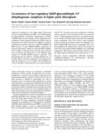
chromatography (IMAC). Figure 1A shows a schematic
representation of both constructs. SDS-PAGE analysis of
purified Rgp41A and Rgp41B revealed apparent molecu-
lar weights of 15 and 14 kD, respectively (Figure 1B). Both
recombinant proteins were analyzed by gel filtration in
order to determine their oligomeric state. As shown by
their elution profiles on a Superdex 75 column, that cor-
responding to an apparent molecular weight of 50 kDa,
both proteins appear to fold spontaneously into trimers.
As expected, circular dichroism spectra of both trimeric
recombinant proteins confirmed the presence of a high
proportion of α-helix (not shown)[17,18].
Recombinant gp41 proteins inhibit HIV-1 entry into CD4+
HeLa cells
Recombinant gp41 proteins were first tested for their abil-
ity to inhibit the infection of HeLa P4.2 cells by HIV-1 iso-
lates. In the first set of experiments, we assayed the
concentration-dependent inhibitory effect of Rgp41A and
Rgp41B on HIV-1 particles pseudotyped with the enve-
lope glycoproteins from the X4 isolate HIV-1 LAI or the R5
Gp41-derived recombinant proteinsFigure 1
Gp41-derived recombinant proteins. A. Schematic representation of Rgp41A (N59(L7)C54) and Rgp41B (N54(L7)C47)
synthetic trimeric peptides, derived from the HIV-1 gp41 ectodomain. Grey boxes represent the position of the N36 and C34
peptides. For comparison, the sequence of T20 is also indicated. B. Characterization of recombinant proteins. SDS-PAGE anal-
ysis and gel filtration on a Superdex 75 column. The elution profiles of Rgp41 proteins were compared to a calibration curve
realized with standard globular proteins. An elution volume of 10.0 ml corresponds to an apparent molecular weight of 50 kDa.
0.5
0.4
0.3
0.2
0.1
0
Rgp41B
97
66
45
30
20,1
14,4
Trimeric form
00 5.0 10.0 15.0 20.0
200
220
240
260
280
180
160
Elution volume
mS/cmA280nm
0.5
0.4
0.3
0.2
0.1
0
Rgp41A
97
66
45
30
20,1
14,4
Trimeric form
00 5.0 10.0 15.0 20.0
246
248
250
252
254
256
258
Elution volume
mS/cmA280nm
540 QARQLLSGIVQQQNNLLRAIEAQQHLLQLTVWGIKQLQARILAVERYLKDQQL 593
618 SLEQIWNHTTWMEWDREINNYTSLIHSLIEESQNQQEKNEQELLELD 664
N36 C34
Fusion
peptide
TM
546 581 628 661 684 705
NH2 COOH
540 593
618 664
512 527
LGIDGS SGGRGGS NASWSNK LEHHHHHHM
Rgp41A
LEHHHHHHM
Rgp41B
SGGRGGS
T20
638 YTSLIHSLIEESQNQQEKNEQELLELDKWASLWNWF 675
Retrovirology 2006, 3:16 />Page 4 of 12
(page number not for citation purposes)
strain HIV-1 ADA. For this purpose, viruses were mixed
with increasing doses of recombinant proteins prior to
infection. Cells were incubated with the mixes for 4 h and
rinsed several times to remove free viruses and recom-
binant proteins. After an incubation of 48 h at 37°C, virus
replication was estimated by measuring luciferase activity
in cell extracts. Since the buffers used for the solubiliza-
tion of recombinant gp41 proteins showed some cyto-
pathic effect, resulting in an artefactual decrease of the
luciferase signal, the inhibitory effect of recombinant pro-
teins was systematically compared to the same volume of
buffer. Figure 2 shows the results of a typical experiment.
Both constructs significantly inhibited entry of the X4
pseudotyped virus into host cells, whereas only Rgp41A
has the capacity to also inhibit the entry of particles pseu-
dotyped with R5 ADA Env. As expected, gp41-derived
trimeric proteins had no effect on Vesicular Stomatitis
Virus (VSV) envelope-pseudotyped viruses (not shown).
IC50 values were calculated from these curves and
reported in Table 1. Rgp41 showed IC50 values of 56 and
156 nM for HIV-1 LAI and ADA-pseudotyped viruses,
respectively. Rgp41B has an IC50 value of 429 nM for LAI-
pseudotyped virus. Similar experiments were performed
on X4 laboratory-adapted viruses HIV-1 LAI and NDK or
R5 HIV-1 YU2 and ADA. As for pseudotyped viruses,
Rgp41A showed a better inhibitory effect than Rgp41B.
The results are summarized in Table 1. Surprisingly,
whereas Rgp41A showed an IC50 value of 156 nM on
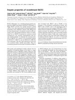
Inhibition of HIV-1 entry into CD4+ HeLa cells by gp41-derived recombinant proteinsFigure 2
Inhibition of HIV-1 entry into CD4+ HeLa cells by gp41-derived recombinant proteins. HIV-1 particles pseudo-
typed with envelope glycoproteins from either the X4 isolate HIV-1 LAI or the R5 isolate HIV-1 ADA were pre-incubated with
increasing concentrations of Rgp41A or Rgp41B before being added to HeLa-CD4-LTR-LacZ cells. For infection with HIV-1
ADA pseudotyped virus, cells were transfected with pCMV-CCR5 48 h before infection. Fourty-eight hours post-infection,
viral entry and replication was monitored by measuring luciferase activity in cell extracts. For each experiment, the inhibitory
effect of recombinant proteins was compared to the effect of the same volume of solubilization buffer. NLI: Normalized Luci-
ferase Index. The average ± SD of triplicate samples is shown. Results represent the average ± SD of a typical experiment per-
formed in duplicate, representative of at least 3 independent experiments.
0.0E+00
2.0E+05
4.0E+05
6.0E+05
8.0E+05
1.0E+06
1.2E+06
1.4E+06
1.6E+06
0 5 10 15 20 25 30 35 40 45 50
0.0E+00
2.0E+05
4.0E+05
6.0E+05
8.0E+05
1.0E+06
1.2E+06
0.0E+00
2.0E+06
4.0E+06
6.0E+06
8.0E+06
1.0E+07
1.2E+07
1.4E+07
1.6E+07
0.0E+00
2.0E+06
4.0E+06
6.0E+06
8.0E+06
1.0E+07
1.2E+07
1.4E+07
1.6E+07
Buffer
Rgp41A
Buffer
Rgp41A
Rgp41A (Pg/ml)
Rgp41A (Pg/ml)
Rgp41B (Pg/ml)
Rgp41B (Pg/ml)
Env X4 HIV-1 LAI
EnvR5HIV-1 ADA
EnvX4HIV-1 LAI
Env R5 HIV-1 ADA
NLI
NLI
NLI
NLI
Buffer
Rgp41B
0 5 10 15 20 25 30 35 40 45 50
0 5 10 15 20 25 30 35 40 45
0 5 10 15 20 25 30 35 40 45
Buffer
Rgp41B
Retrovirology 2006, 3:16 />Page 5 of 12
(page number not for citation purposes)
ADA pseudotyped HIV-1 particles, it displayed no inhibi-
tory effect on the corresponding wild-type virus HIV-1
ADA. In contrast, Rgp41A inhibitory effects on the X4
strain HIV-1 NDK and the R5 strain HIV-1 YU2 were weak
but significant, with IC50 values of 844 and 489 nM,
respectively. Also shown for comparison in Table 1 are the
IC50 values for T-20 on each virus. This inhibitor is
approximately 25-fold more effective than Rgp41A to
block HIV-1 LAI entry into HeLa-CD4 cells. Consistent
with previous data, T-20 is not as effective on R5 isolates,
such as YU2 and ADA, as on X4 viruses, such as HIV-1 LAI
and NDK [31].
Rgp41A also blocks entry of X4 viruses into PBL
Antiviral properties of the Rgp41A were tested on the
infection of PBLs by HIV-1 laboratory-adapted strains.
Similar experiments were performed in parallel with T-20
(Table 2). In this model, the Rgp41A significantly blocked
the entry of X4 viruses into host cells but had no effect on
the R5 virus tested, suggesting that antiviral properties of
the Rgp41A not only depend on the virus strain but also
on the cell type. Calculation of IC50 values (Table 2)
revealed that HIV-1 NDK was approximately 4 times more
susceptible to inhibition by T-20 than by Rgp41A. IC50
values of Rgp41A on HIV-1 LAI and NDK are 356 and 322
nM, respectively.
Rgp41A can interact with soluble monomeric gp120
In order to determine the mechanism of action of the
most potent trimeric recombinant protein, Rgp41A, we
first tested its ability to bind HIV-1 gp120. Some HIV-1
entry inhibitors act by binding to the envelope glycopro-
teins in order to interfere with their interaction with cellu-
lar receptors. This is the case for sCD4 and also for T-20,
which was recently shown to interact with gp120 of X4
viruses, and to a lesser extent with gp120 of R5 viruses
[33]. This interaction probably contributes to the mecha-
nism by which T-20 blocks entry of X4 viruses into host
cells [33]. Since both Rgp41 constructs contain the C34
sequence of T-20, we tested their ability to interact with a
soluble monomeric recombinant gp120 from the X4 virus
HIV-1
IIIB
. For this purpose, 96-well plates were coated
with various doses of the Rgp41 proteins and then incu-
bated with the monomeric gp120. The amount of gp120
bound to Rgp41 proteins was determined using anti-
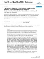
gp120 antibodies. As shown on Figure 3, the monomeric
gp120 bound to both Rgp41 proteins in a dose dependent
manner. Interestingly, Rgp41B retained significantly less
Table 1: IC50 values of Rgp41A and Rgp41B on the entry of pseudotyped or laboratory-adapted HIV-1 isolates into HeLa-CD4-LTR-
LacZ cells.
Rgp41A Rgp41B T-20
µg/ml nM µg/ml nM µg/ml nM
Pseudotyped LAI 2.5
a
56 18 429 / /
ADA 7 156 NE NE / /
Laboratory-
adapted
LAI 13 289 42* 1000 0.05 11
NDK 38* 844 70* 1667 0.04 9
YU2 22 489 NE NE 2 444
ADA NE
b
NE NE NE 1.75 389
a
These values correspond to the amount of recombinant proteins resulting in a 50% decrease in reporter activity compared to the value in the
absence of inhibitor. Luciferase and β-Galactosidase were used as reporters of infection in the case of pseudotyped viruses and laboratory-adapted
strains, respectively. This table shows the averages of a typical experiment performed in duplicate. *: Estimated from exponential regression
analysis.
b
NE:No effect.
Table 2: IC50 values of Rgp41A and T-20 on the entry of adapted HIV-1 isolates into PBL.
Rgp41A T-20
µg/ml nM µg/ml nM
LAI 16
a
356 / /
NDK 14.5 322 0.38 84
YU2 NE
b
NE 1.05 233
a
These values represent the amount of recombinant proteins giving a 50% reduction of the amount of p24 in cell extracts, compared to the value in
the absence of inhibitor and correspond to the mean value of a typical experiment. IC50 in nM correspond to the trimeric form of Rgp41A.
b
NE:
No effect.
Retrovirology 2006, 3:16 />Page 6 of 12
(page number not for citation purposes)
gp120 than Rgp41A, suggesting a higher affinity of gp120
for Rgp41A than for Rgp41B.
Rgp41A does not induce the release of gp120 from HeLa
cells expressing HIV envelope
Since recombinant gp41 appeared to be able to interact
with a soluble monomeric gp120, we investigated
whether this interaction could lead to gp120 release from
the surface of the virus. Such a phenomenon has been
reported for sCD4 and proposed to explain at least part of
its antiviral properties [37-39]. To test gp120 shedding
induced by recombinant gp41, HeLa cells expressing the
env gene from the LAI virus (HeLa-LAI) were incubated
with Rgp41A or sCD4. After incubation, the amount of
gp120 present in the supernatant was measured by ELISA.
As shown in Figure 4, Rgp41A did not induce the release
of gp120 from the HeLa-LAI cells, in comparison to the
control, whereas sCD4 induced the release of a significant
amount of gp120. This result suggests that Rgp41A inhib-
its HIV entry into host cells by a mechanism that does not
involve gp120 shedding.
Rgp41A inhibits the fusion between cells expressing the
env gene and target cells expressing HIV receptors
As cell-to-cell fusion experiments could be convenient
models to analyze the mechanism by which Rgp41 pro-
teins inhibit virus entry into host cells, we tested the effi-
cacy of Rgp41A to inhibit the fusion between HeLa cells
expressing the env gene of various HIV strains (HeLa-Env
cells) and HeLa P4.2 cells expressing HIV receptors (target
cells). For this purpose, HeLa-Env cells were incubated
with Rgp41A prior to incubation with target cells, at a con-
centration that inhibits 90% of HIV-1 LAI infection
(IC90). Cell fusion was monitored by measuring the β-
galactosidase activity. As shown in Figure 5, the Rgp41A
buffer appears to partially inhibit cell-to-cell fusion, prob-
ably reflecting its cytotoxicity. Indeed, we observed 45 to
55% reduction of β-galactosidase activity with Rgp41A
solubilization buffer. In comparison with the buffer
alone, Rgp41A inhibited nearly 4 times the fusion
between HeLa-Env cells expressing the X4 HIV envelopes
(LAI and NDK) and target cells, but had no significant
effect on cells expressing a R5 envelope (ADA). Thus, in
this model, Rgp41A activity seems to be restricted to the
X4 HIV envelopes tested. For comparative purposes, we
also included T20 in this experiment. At a concentration
that inhibits 90% of HIV-1 LAI infection (0.2 µg/ml), T20
inhibits only around 50% of syncytia formation (Figure
5).
Cell to cell fusion is inhibited by Rgp41A at a late stage
during the fusion process
T-20 was recently shown to inhibit the membrane fusion
process at a late stage, after the exchange of lipids between
env expressing cells and target cells [35]. We investigated
at which step of the fusion process Rgp41A acts. For this
purpose, HeLa cells expressing the X4 LAI envelope and
HeLa P4.2 target cells were labelled with two different
hydrophobic fluorescent probes, DiO and DiI, respec-
tively. Labelled HeLa-LAI cells were pre-incubated with
Rgp41A, T-20, PBS or Rgp41A buffer, and then incubated
with labelled target cells. After 6 h at 37°C, the amount of
double fluorescent cells was measured by flow cytometry
analysis. Double fluorescent cells result from an exchange
of membrane lipids during the fusion process between
HeLa-LAI and target cells. In parallel, the fusion efficiency
was evaluated by measuring syncytia formation using an
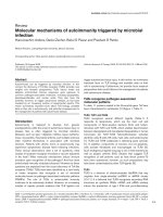
X-Gal assay. As shown in figure 6, the percentage of dou-
ble fluorescent cells was about 12% when the cells were
incubated with PBS, and the X-Gal assay showed the for-
mation of many large syncytia, as expected. No significant
difference was observed when the buffer of Rgp41A was
used. At low dose (10 nM), T-20 had a limited effect on
lipid exchange since about 7% of double fluorescent cells
were observed, which corresponds to a reduction of about
37% of membrane exchange. However, it blocked syncy-
tia formation, as shown by the small number and size of
syncytia on the plate. In contrast, at higher dose (400
nM), T-20 has completely abolished syncytia formation
and more than 95% of lipid exchange. Rgp41A signifi-
cantly inhibited syncytia formation but did not inhibit the
exchange of lipids, since the treatment of cells with
Rgp41A did not significantly modify the amount of dou-
In vitro interaction between Rgp41 proteins and soluble mon-omeric gp120Figure 3
In vitro interaction between Rgp41 proteins and solu-
ble monomeric gp120. Ninety-six well plates were coated
with increasing doses of recombinant gp41 proteins (0, 50,
100 or 200 ng) and then incubated with 2 ng of soluble
gp120. Bound gp120 was revealed using specific anti-gp120
antibodies and HRP-conjugated secondary antibodies. Black
bars: wells coated with Rgp41A, gray bars: wells coated with
Rgp41B. Results represent the average of two independent
experiments. Standard deviations are indicated by error bars.
0
0.2
0.4
0.6
0.8
1
1.2
200 100 50 0
Bound gp120, absorbance at 405nm
Rgp41A
Rgp41B
Amount of Rgp41 (ng)
Retrovirology 2006, 3:16 />Page 7 of 12
(page number not for citation purposes)
ble fluorescent cells. Thus, these results suggest that
Rgp41A, as for T-20 at low dose, inhibits the fusion proc-
ess at a late stage after the mixing of lipids, since it
appeared to block the formation of syncytia without pre-
venting the exchange of lipids between HeLa-LAI cells and
CD4-expressing cells.
Discussion
In this study, two soluble trimeric HIV-1 gp41 recom-
binant proteins were shown to inhibit the HIV-1 fusion
process. Both constructs comprise N and C domains con-
nected by a 7-residue hydrophilic linker and were shown
to fold spontaneously into a trimer, confirming that these
proteins may mimic the six-helix bundle of the gp41 ecto-
domain in its fusogenic state [15-18,40]. Lu et al. previ-
ously described a gp41-derived construct named
N34(L6)C28, formed by the gp41-derived N34 and C28
peptides associated by a 6 residue linker. This protein,
which has the same overall structure as Rgp41B
(N53(L7)C47) and Rgp41A (N59(L7)C54), was found to
form highly thermostable α-helical trimers [41].
Infection experiments with pseudotyped or wild-type
HIV-1 viruses on HeLa-CD4 or HeLa-CD4-CCR5 cells
revealed that both Rgp41A and Rgp41B have the capacity
to interfere with HIV-1 entry into target cells. It should be
noted that both Rgp41 trimeric proteins have the pro-
pency to aggregate in solution, especially Rgp41A, pre-
sumably because of the presence of the N-HR [16]. The
high degree of insolubility of Rgp41A lead us to use a sol-
ubilization buffer that was found to be toxic for the cells.
Therefore, Rgp41A apparent effect on HIV-1 entry was sys-
tematically compared to the effect of its solubilization
buffer alone.
The most potent inhibitor is Rgp41A, which inhibits the
infection of HeLa-CD4 cells by HIV-1 LAI and ADA Env
pseudotyped viruses with IC50 values of 56 and 156 nM,
respectively. Its efficacy on wild-type viruses appeared
weaker, with IC50 values from 289 nM for HIV-LAI to 844
nM for HIV-1 NDK. For comparison, N34(L6)C28 IC50
value on HIV-1 IIIB infectivity is 1.5 µM [42], indicating
that the Rgp41A is about 5 times more efficient than
N34(L6)C28 to inhibit X4 viruses infection.
In contrast, Rgp41A displayed no effect on the R5 strain
HIV-1 ADA. Interestingly, HIV-1 ADA has been previously
reported to be particularly resistant to different entry
inhibitors [43,44]. This HIV-1 isolate appears therefore to
behave differently compared to other known HIV-1 iso-
lates for a reason that remains to be investigated. More
generally, R5 isolates appeared less susceptible to Rgp41A
inhibition than X4 strains. This higher sensitivity of X4
viruses has been previously observed with the C-HR
derived fusion inhibitor, T-20 [31,32]. The second con-
struct, Rgp41B, which contains shorter N and C domains,
inhibits poorly X4 HIV-1 isolates LAI and NDK, at micro-
molar concentrations, and is inactive against R5 strains.
We next examined the mechanism by which Rgp41A
interferes with HIV-1 entry into target cells. We demon-
strate that the trimeric recombinant protein was able to
interact with monomeric gp120 derived from an X4 HIV-
1 isolate. However, this binding does not seem to result in
the release of gp120, a mechanism partly responsible for
inhibition of infection by sCD4 [37-39]. It may thus be
possible that Rgp41A can bind preferentially to gp120 of
X4 viruses, preventing its interaction with CD4 and/or
CXCR4. This hypothesis would explain its better efficacy
against X4 viruses entry, although this has yet to be fully
investigated. Interestingly, the fusion inhibitor T-20 was
also shown to bind gp120 of X4 HIV-1 strains in a CD4-
induced, V3 loop dependent manner [33]. This binding
was shown to prevent the interaction of gp120-CD4 com-
plexes with the CXCR4 coreceptor.
In order to determine whether Rgp41A can affect different
steps of the entry process in a similar way as T-20 [33,45],
we tested the ability of Rgp41A to block the fusion proc-
ess. As expected, the trimeric molecule revealed its capac-
ity to potently inhibit the fusion between X4 Env-
expressing cells and target cells expressing CD4 and
CXCR4. This inhibition occurs at a late stage of this proc-
ess, as revealed by the incapacity of Rgp41A to prevent
membrane exchanges, even at high concentrations that
efficiently block the formation of syncytia. Once again,
this observation is reminiscent of previous results
Gp120 sheddingFigure 4
Gp120 shedding. HeLa-Env or HeLa∆20 were incubated
for 6 h at 37°C with Rgp41A (0.03 µg/µl), Rgp41A buffer
(same volume), sCD4 or PBS. Release of gp120 from the cell
surface was quantified by ELISA. Values represent averages of
duplicate samples from a typical experiment.
0
100
200
300
400
500
600
Rgp41A Buffer PBS sCD4 Rgp41A Buffer PBS sCD4
HeLa LAI
HeLa 'Env
gp120 concentration (ng/ml)
Retrovirology 2006, 3:16 />Page 8 of 12
(page number not for citation purposes)
described for T-20 at low concentrations [35,46,47]. T-20
is believed to act by binding to the transiently exposed tri-
ple-stranded coiled-coil of NH2-terminal helices, thus
preventing the 6-HB formation. This mechanism of action
is in agreement with the finding that the fusion pore
forms before the folding of the 6HB has been completed
[48]. Unlike T-20 and other previously described fusion
inhibitors, which are small gp41-derived peptides capable
of binding to the transiently exposed HRs of gp41,
Rgp41A is a rather large molecule (approximately 50 kD).
The expected conformation for Rgp41A is a trimer of hair-
pins, mimicking the fusogenic conformation of HIV-1
gp41. Whether its large size allow the trimeric protein to
gain access to gp41 during its conformational changes
remains to be elucidated. However, the fact Rgp41A
inhibits fusion without any effect on the lipid-mixing sug-
gest it might also interfere with the 6HB formation.
N34(L6)C28 has also been found to inhibit HIV-1 Env-
mediated membrane fusion, in agreement with our results
[41]. Interestingly, the potency of these trimeric HR1-HR2
proteins to inhibit HIV-1 entry appears proportional to
the the length of the N- and C-terminal domains, the less
and most potent inhibitor being N34(L6)C28 and
Rgp41A, respectively [41]. Synthetic peptides correspond-
ing to the N-HR and C-HR of gp41 block fusion by bind-
ing to the transiently exposed HRs of gp41 during
conformational changes, thus preventing 6HB formation
[21]. C peptides are potent inhibitors of HIV-1 infectivity
with activity at nanomolar concentrations, whereas N-
peptides are relatively poor inhibitors, presumably due to
their tendency to aggregate in solution [16]. Many groups
have tried to design more potent inhibitors by combining
multiple HR1 and HR2, such as N(CCG)-gp41 (HR1-
HR1-HR2)[49], 5-Helix (HR1-HR2-HR1-HR2-HR1) [25],
HR121 (HR1-HR2-HR1)[41], HR212 (HR2-HR1-
HR2)[41] or other N-peptides-derived inhibitors such as
IQN17 or IQN23 [24]. Although some of these constructs
have a strong inhibitory effect, their precise mode of
action is still unclear.
In the case of Rgp41A and B, the only difference between
the two trimeric molecules is the lenght of the N- and C-
terminal gp41-derived domains, which differ by only 6
and 7 residues, respectively. In consequence, it would be
interesting to explain the reason why the inhibitory effect
of Rgp41A on HIV entry is systematically much higher
than its B counterpart, despite the fact they both have the
same overall structure. In this context, the synthetis of
intermediate constructs containing N- and C-terminal
domains of increasing lenghts would be particularly
informative in order to identify the determinants of this
difference of antiviral activity.
Conclusion
Both Rgp41 proteins were found to inhibit HIV-1 entry
into target cells in a dose-dependent manner. Rgp41A, the
most potent inhibitor, was found to inhibit both X4 and
R5 isolates into HeLa cells and primary T lymphocytes.
Rgp41A was able to bind gp120 but did not induce
gp120-gp41 dissociation. Furthermore, this inhibitor
interferes with a late step of the fusion process, following
the mixing of lipids.
Considering our results, it is also possible that Rgp41A,
like T-20, may act at different stages of the entry process.
Although the precise mechanism of action of these HIV
entry inhibitors will be difficult to unravel, it will
undoubtedly help to elucidate the complex mechanisms
involved during HIV entry process.
Materials and methods
Cell lines and plasmids
HeLa-CD4-LTR-LacZ (also referred as HeLa P4.2) cells sta-
bly express the human CD4 molecule and contain the β-
galactosidase encoding gene (lacZ) under the transcrip-
tional control of the HIV-1 long terminal repeat (LTR).
They were kindly provided by Dr M. Alizon (Institut
Cochin, Paris, France). HeLa-Env/ADA (or HeLa-ADA)
cells stably express the envelope of the R5 tropic HIV
strain ADA. HeLa-Env/LAI (or HeLa-LAI) and HeLa-Env/
NDK (or HeLa-NDK) cells stably express LAI and NDK env
Cell-to-cell fusion inhibition by the Rgp41AFigure 5
Cell-to-cell fusion inhibition by the Rgp41A. HeLa-Env/
LAI, HeLa-Env/NDK, and HeLa-Env/ADA were incubated
with target cells expressing the HIV receptors in the pres-
ence of PBS, Rgp41A (0.03 µg/µl), Rgp41A solubilization
buffer or T-20 (0.2 µg/ml). Syncytia formation was evaluated
by measuring the β-galactosidase activity after lysis of the
cells. The results are expressed as percentage of the β-galac-
tosidase activity observed in the control with PBS. Results
represent the average of three independent experiments
performed in duplicate. Standard deviations are indicated by
error bars.
% of fusion
0
20
40
60
80
100
120
Buffer Rgp41APBS
HeLa LAI
HeLa NDK
HeLa ADA
T20
Retrovirology 2006, 3:16 />Page 9 of 12
(page number not for citation purposes)
genes from X4 viruses LAI and NDK, respectively.
HeLa∆20 cells are derived from HeLa-Env/LAI cells and
contain a deletion in the env gene. Both cell lines were
kindly provided by Dr M. Alizon (Institut Cochin, Paris,
France). HeLa-Env and HeLa∆20 cells also stably express
the Tat HIV protein. All adherent cell lines were grown in
Dulbecco's modified Eagle's medium (DMEM, Invitro-
gen) supplemented with 5% fetal calf serum (Invitrogen),
50 U/ml penicillin, 50 µg/ml streptomycin (Invitrogen)
and 2 mM glutamine (Invitrogen).
The pCMV-CCR5 plasmid (kindly given by Dr. T. Dragic,
New York, USA) contains the CCR5 gene under the con-
trol of the CMV promoter. The proviral plasmid pNL4.3-
∆env-Luc contains the NL4.3 env-deleted provirus includ-
ing the luciferase reporter gene inserted in the nef ORF
[34]. The LAI and ADA8 expression plasmids harbor the
LAI and ADA8 env genes, respectively, under the control of
the HIV-1 LTR. The pEnv-VSV-G plasmid encoding VSV-G
envelope was a gift from Dr. P. Sonigo (Institut Cochin,
Paris, France). The pADA, pJRCSF and pYU2 proviral plas-
mids encode proviral genomes of R5 tropic viruses,
whereas the pNL4.3 and pNDK proviral plasmids encode
proviral genomes of X4 tropic viruses.
Antibodies and chemical reagents
The sheep anti-gp120 monoclonal antibody D7324
(Aalto) was raised against a C-terminal peptide of the
gp120. Sera from HIV+ patients were a gift from Professor
J.C. Nicolas (Tenon hospital, Paris, France). HRP-coupled
anti-human and anti-sheep antibodies were purchased
from Caltag and DAKO respectively. The HRP substrate
ABTS from Roche was used at a concentration of 1 mg/ml.
The fluorescent hydrophobic probes DiO and DiI were
purchased from Sigma Aldrich. The fusion inhibitor T-20
and the soluble CD4 (sCD4) were obtained through the
NIH AIDS Research and Reference Reagent Program. The
recombinant protein gp120 HIV-1
IIIB
was purchased from
Advanced Biotechnologies Incorporated.
Production and purification of soluble trimeric
recombinant gp41 proteins
The trimeric recombinant protein Rgp41A and Rgp41B
were provided by Protein'eXpert (Grenoble, France) and
produced as follow. Briefly, HIV-1 gp41 sequences corre-
sponding to Rgp41A and Rgp41B were cloned between
the NdeI and the XhoI sites of the pET21b and pET20b
expression vectors (Novagen), respectively, allowing the
production of recombinant protein harboring a 6xHIS tag
at their C-terminus. Competent Escherichia coli
BL21(DE3) were transformed with each vector and grown
in LB medium at 37°C until an absorbance of 0.6 at 600
nm was reached. The production of recombinant protein
was then induced by adding 1mM IPTG. Two hours after
induction, bacteria were harvested and lysed in protein
buffer (50 mM Tris-HCl, 300 mM NaCl, pH 8) by sonica-
tion. The suspension was then centrifuged at 40 000 g for
30 min at 4°C to separate the soluble proteins (superna-
tant) from the insoluble proteins and cell debris (pellet).
Rgp41 proteins were purified from supernatant by affinity
chromatography using Chelating Sepharose™ Fast Flow
(Amersham Biosciences) and eluted using elution buffer
(50 mM Tris-HCl, 300 mM NaCl, imidazole 500 mM, pH
8). Rgp41A-containing fractions were pooled and dia-
lyzed against 50 mM Tris-HCl, 200 mM NaCl, 200 mM
imidazole, pH 8. Rgp41B-containing fractions were
pooled and dialyzed against 50 mM Tris-HCl, 200 mM
NaCl, pH 8. Purity of the recombinant proteins was
checked by 12% SDS-PAGE. The oligomeric status of the
recombinant protein was determined by gel filtration
chromatography using Superdex 75 HR 10/30 (Amer-
sham Biosciences). Columns were equilibrated and eluted
with 50 mM Tris-HCl, 200 mM NaCl; 200 mM imidazole;
5% glycerol; pH 8 in the case of Rgp41A and with 50 mM
Tris-HCl, 200 mM NaCl, 5% glycerol, pH 8 in the case of
Rgp41B. The calibration curve was obtained with standard
globular proteins.
Rgp41A and Rgp41B were patented by Mymetics Corpora-
tion (ref PCT/IB2004/002433).
lipid exchange and syncytia formation during cell-to-cell fusion experimentsFigure 6
lipid exchange and syncytia formation during cell-to-
cell fusion experiments. HeLa-Env/LAI and target cells
were labeled with DiO and DiI, respectively. Co-cultures of
labeled cells were performed in the presence of PBS (con-
trol), Rgp41A buffer, Rgp41A or T-20. After 6 h, lipid
exchange between both types of cells was evaluated by flow
cytometry analysis. In parallel, an X-Gal assay was performed
to estimate the fusion efficiency between Env- and CD4-
expressing cells in the presence or absence of inhibitors.
Indicated percentages correspond to the proportion of dou-
ble-positive gated cells. -: absence of syncytia, +/-: presence
of small syncytia, +: presence of many large syncytia.
PBS T-20 10 nM
T-20 400 nM
Buffer Rgp41A
Rgp41A 1.1 PM
12.5%
12.2%
7.63%
12.1%
0.56%
+
+
+/-
-
+/-
Retrovirology 2006, 3:16 />Page 10 of 12
(page number not for citation purposes)
Production of HIV-1 pseudotyped and laboratory-adapted
strains
Stocks of pseudotyped viruses were generated by co-trans-
fecting HEK293 cells with the proviral plasmid pNL4.3-
∆env-Luc and one of the env encoding plasmids. Stocks of
adapted laboratory viruses were obtained by transfecting
HEK 293 cells with the proviral plasmids pADA, pYU2,
pNL4.3 or pNDK. Forty eight hours after transfection, the
supernatant containing viruses was filtered and virus
stocks were titrated by p24 ELISA (Coulter).
Production and activation of PBL
Peripheral blood mononuclear cells were isolated from
human blood on Ficoll (Ficoll-Paque PLUS, Amersham
Biosciences), washed several times in PBS, EDTA 0.3 mM
and stored frozen in fetal calf serum supplemented with
10% DMSO. For peripheral blood lymphocytes (PBL)
production, stocks were quickly thawed and washed in
RPMI, 10% fetal calf serum. Cells were cultured in 6-well
plates in RPMI, 10% fetal calf serum. PBL were activated
with 5 µg/ml PHA (DIFCO) and, three days later, with 40
U/ml IL-2 (Proleukin, Chiron). After two weeks of IL-2
induced proliferation, cells were used for infection exper-
iments.
Cell infections
HeLa P4.2 transfected with pCMV-CCR5 or PBL were cul-
tured in 48-well plates (about 5 × 10
4
cells per well). Prior
to infection, fixed concentrations of pseudotyped or labo-
ratory-adapted viruses were incubated for 15 min with a
range of Rgp41 concentrations (from 0.0025 to 0.03 µg/
µl) or with the same volume of Rgp41 specific solubiliza-
tion buffer. After incubation, mixes were added to the
cells. The amount of viruses added per well was equivalent
to 10 ng of p24. Four hours post-infection, cells were
washed several times to remove free viruses and recom-
binant proteins and cultured for 48 h. Viral infectivity was
monitored by measuring the luciferase activity in cell
lysates in the case of HeLa cells infections by pseudotyped
viruses. For infections with laboratory adapted HIV-1
strains, infection was monitored by measuring the β-
galactosidase activity in cell lysates. Finally, PBL infections
were followed by measuring the amount of p24 in the
supernatant.
Cell-to-cell fusion assay
HeLa-Env cells were seeded in 48-well plates (10
5
cells per
well) with either Rgp41A (0.03 µg/µl), Rgp41A buffer or
PBS. Fifteen minutes later, target cells (HeLa P4.2 or HeLa
P4.2 transfected with pCMV-CCR5) were added to the
wells (10
5
per well) and co-cultures were incubated for 6
h at 37°C.
Beta-galactosidase assay
Cells grown in 48-well plates were lysed in 200 µl of lysis
buffer (60 mM Na
2
HPO
4
, 40 mM NaH
2
PO
4
, 10 mM KCl,
10 mM MgSO
4
, 2.5 mM EDTA, 1.25‰ NP40, 50 mM β-
mercaptoethanol) for 10 min. An equivalent volume of
reaction buffer (61.9 mM Na
2
HPO
4
, 18.1 mM NaH
2
PO
4
,
10 mM MgCl
2
, 10 mM β-mercaptoethanol, 6 mM chlo-
rophenol-β-D-galactose) was then added to the lysate.
Kinetics were performed by measuring the absorbance at
575 nm for 30 min. The β-galactosidase activity corre-
sponds to the slope of the curve.
Luciferase assay
Cells grown in 48-well plates were lysed in 200 µl of lysis
buffer (25 mM Tris pH 7.8, 8 mM MgCl
2
, 2 mM DTT, 1%
Triton X-100, 15% glycerol) before adding 100 µl of lysis
buffer containing 0.25 mM luciferin and 1 mM ATP. Luci-
ferase activity was measured on a Berthold Luminometer
(Lumat LB9507).
p24 titration
The p24 protein was titrated using the HIV-1 p24 Antigen
Assay Kit (Coulter), according to the supplier's instruc-
tions. Briefly, infected cells or viral stocks were lysed in
Triton X-100 and the lysates introduced into wells pre-
coated with mouse anti-p24 monoclonal antibodies.
Bound p24 was revealed using biotin-coupled human
anti-HIV IgG followed by HRP-coupled streptavidin. HRP
reaction was initiated by adding the HRP substrate into
the wells and stopped 30 min later with the stopping
buffer. The absorbance at 450 nm was determined. Puri-
fied p24 was used to generate standard curves.
Interaction of Rgp41 proteins with monomeric gp120
Protein Immobilizer plates (EXIQON) were coated with
Rgp41 proteins (200, 100 or 50 ng in 15 mM Na
2
CO
3
, 35
mM NaHCO
3
per well). After overnight coating at 4°C,
wells were washed several times with PBS-Tween (PBS 1×,
0.05% Tween 20), saturated with PBS containing 10%
fetal calf serum for 2 h and washed again with PBS-Tween.
Recombinant monomeric gp120 derived from HIV-1
IIIB
diluted in PBS-Tween was then added to the well (2 ng per
well). Plates were incubated for 2 h at room temperature
and washed several times with PBS-Tween. gp120 bound
to Rgp41 was labeled with anti-gp120 antibodies D7324
diluted in PBS-Tween for 2 h at room temperature fol-
lowed by HRP-conjugated secondary antibodies diluted
in PBS-Tween for an additional hour. Plates were washed
extensively with PBS-Tween. The HRP substrate was then
added to the wells and the absorbance at 405 nm was
measured 10 min later.
gp120 release from HeLa-Env cells
HeLa-Env and HeLa∆20 cells (4 × 10
6
cells per tube in 200
µl DMEM, 10% fetal calf serum) were incubated with
Retrovirology 2006, 3:16 />Page 11 of 12
(page number not for citation purposes)
Rgp41A (0.03 µg/µL), Rgp41A buffer, sCD4 (50 µg/ml) or
PBS at 37°C. Six hours later, supernatants were harvested
to quantify the amount of gp120 released. For this pur-
pose, 96-well Protein Immobilizer plates (EXIQON) were
coated overnight at 4°C with anti-gp120 antibodies
D7324 diluted in 15 mM Na
2
CO
3
, 35 mM NaHCO
3
, pH
9.6. Wells were rinsed several times with PBS-Tween.
Supernatants were then deposited into the wells and incu-
bated for 2 h at room temperature. After several washes, a
human anti-HIV serum was added into the wells and incu-
bated for 2 h at room temperature. Wells were washed and
HRP-coupled secondary antibodies diluted in PBS-Tween
were added into the wells for an additional hour at room
temperature. Wells were washed extensively before addi-
tion of the HRP substrate. The absorbance at 405 nm was
measured 1 h after initiation of the reaction. Standard
curves were obtained with purified HIV-1
IIIB
gp120.
Lipid mixing analysis
HeLa-Env cells and HeLa P4.2 target cells were labeled
with 2 µM DiO and DiI, respectively, as previously
described [35]. After cell labeling, a cell-to-cell fusion
assay was performed as described above, in the presence
of Rgp41A (50 µg/ml), Rgp41A buffer, T-20 (10 nM or
400 nM) or PBS. Co-cultures were incubated for 6 h at
37°C. Cells were then detached from wells with PBS, 15
mM citrate, pH 7 and fixed with PBS 2% formaldehyde.
Double fluorescent cells, containing both DiO and DiI,
were detected by two color XL
2
Beckman Coulter cytome-
ter using the System II™ software. At least 10
4
cells were
counted for each sample. In parallel, some cells were sub-
jected to an X-Gal assay in order to estimate cell to cell
fusion efficiency, as described previously [36].
Competing interests
The author(s) declare that they have no competing inter-
ests.
Authors' contributions
DDG carried out HIV infections, cell to cell fusion experi-
ments, ELISA and lipid mixing assays. PLQ participated in
HIV inhibition experiments and also in the experimental
design and data analysis. MGR produced and performed
the structural analysis of Rgp41 proteins. UH conceived of
the study and participated in its design and coordination,
as well as in the writing of the manuscript. SN participated
in the data analysis and drafted the manuscript. CR partic-
ipated in the experimental design and data analysis and
also performed HIV infection and cell to cell fusion exper-
iments. All authors have read and approved the final man-
uscript.
Acknowledgements
We thank Laura Burleigh and Ara Hovanessian for helpful discussion and
critical reading of the manuscript. We thank the NIH AIDS Research and
Reference Reagent Program for the kind gift of reagents. P-L.Q. is sup-
ported by a grant from Mymetics Corporation. Mymetics Corporation sup-
ported this work.
References
1. Moore JP, Stevenson M: New targets for inhibitors of HIV-1 rep-
lication. Nat Rev Mol Cell Biol 2000, 1(1):40-49.
2. Fauci AS: HIV and AIDS: 20 years of science. Nat Med 2003,
9(7):839-843.
3. Chun TW, Davey RT, Engel D, Lane HC, Fauci AS: Re-emergence
of HIV after stopping therapy. Nature 1999,
401(6756):874-875.
4. Finzi D, Blankson J, Siliciano JD, Margolick JB, Chadwick K, Pierson T,
Smith K, Lisziewicz J, Lori F, Flexner C, Quinn TC, Chaisson RE,
Rosenberg E, Walker B, Gange S, Gallant J, Siliciano RF: Latent
infection of CD4(+) T cells provides a mechanism for lifelong
persistence of HIV-1, even in patients on effective combina-
tion therapy. Nature Medicine 1999, 5(5):512-517.
5. Pomerantz RJ: Eliminating HIV-1 reservoirs. Curr Opin Investig
Drugs 2002, 3(8):1133-1137.
6. Hamer DH: Can HIV be cured? Mechanisms of HIV persist-
ence and strategies to combat it. Curr HIV Res 2004,
2(2):99-111.
7. Chun TW, Fauci AS: Latent reservoirs of HIV: obstacles to the
eradication of virus. Proc Natl Acad Sci U S A 1999,
96(20):10958-10961.
8. Pierson TC, Doms RW: HIV-1 entry and its inhibition. Curr Top
Microbiol Immunol 2003, 281:1-27.
9. Wyatt R, Sodroski J: The HIV-1 envelope glycoproteins:
fusogens, antigens, and immunogens. Science 1998,
280(5371):1884-1888.
10. Altmeyer R: Virus attachment and entry offer numerous tar-
gets for antiviral therapy. Curr Pharm Des 2004,
10(30):3701-3712.
11. Clapham PR, McKnight A: Cell surface receptors, virus entry
and tropism of primate lentiviruses. J Gen Virol 2002, 83(Pt
8):1809-1829.
12. Gallaher WR: Detection of a fusion peptide sequence in the
transmembrane protein of human immunodeficiency virus.
Cell 1987, 50(3):327-328.
13. Delwart EL, Mosialos G, Gilmore T: Retroviral envelope glyco-
proteins contain a 'leucine zipper'-like repeat. AIDS Res Hum
Retroviruses 1990, 6(6):703-706.
14. Chambers P, Pringle CR, Easton AJ: Heptad repeat sequences are
located adjacent to hydrophobic regions in several types of
virus fusion glycoproteins. J Gen Virol 1990, 71 ( Pt
12):3075-3080.
15. Chan DC, Fass D, Berger JM, Kim PS: Core structure of gp41
from the HIV envelope glycoprotein. Cell 1997, 89(2):263-273.
16. Lu M, Blacklow SC, Kim PS: A trimeric structural domain of the
HIV-1 transmembrane glycoprotein. Nat Struct Biol 1995,
2(12):1075-1082.
17. Tan K, Liu J, Wang J, Shen S, Lu M: Atomic structure of a ther-
mostable subdomain of HIV-1 gp41. Proc Natl Acad Sci U S A
1997, 94(23):12303-12308.
18. Weissenhorn W, Dessen A, Harrison SC, Skehel JJ, Wiley DC:
Atomic structure of the ectodomain from HIV-1 gp41.
Nature 1997, 387(6631):426-430.
19. Eckert DM, Kim PS: Mechanisms of viral membrane fusion and
its inhibition. Annu Rev Biochem 2001, 70:777-810.
20. Gallo SA, Finnegan CM, Viard M, Raviv Y, Dimitrov A, Rawat SS, Puri
A, Durell S, Blumenthal R: The HIV Env-mediated fusion reac-
tion. Biochim Biophys Acta 2003, 1614(1):36-50.
21. Chan DC, Kim PS: HIV entry and its inhibition. Cell 1998,
93(5):681-684.
22. He Y, Vassell R, Zaitseva M, Nguyen N, Yang Z, Weng Y, Weiss CD:
Peptides trap the human immunodeficiency virus type 1
envelope glycoprotein fusion intermediate at two sites. J Virol
2003, 77(3):1666-1671.
23. Rimsky LT, Shugars DC, Matthews TJ: Determinants of human
immunodeficiency virus type 1 resistance to gp41-derived
inhibitory peptides. J Virol 1998, 72(2):986-993.
24. Eckert DM, Kim PS: Design of potent inhibitors of HIV-1 entry
from the gp41 N-peptide region. Proc Natl Acad Sci U S A 2001,
98(20):11187-11192.
Publish with BioMed Central and every
scientist can read your work free of charge
"BioMed Central will be the most significant development for
disseminating the results of biomedical research in our lifetime."
Sir Paul Nurse, Cancer Research UK
Your research papers will be:
available free of charge to the entire biomedical community
peer reviewed and published immediately upon acceptance
cited in PubMed and archived on PubMed Central
yours — you keep the copyright
Submit your manuscript here:
/>BioMedcentral
Retrovirology 2006, 3:16 />Page 12 of 12
(page number not for citation purposes)
25. Root MJ, Kay MS, Kim PS: Protein design of an HIV-1 entry
inhibitor. Science 2001, 291(5505):884-888.
26. Kilby JM, Hopkins S, Venetta TM, DiMassimo B, Cloud GA, Lee JY,
Alldredge L, Hunter E, Lambert D, Bolognesi D, Matthews T, Johnson
MR, Nowak MA, Shaw GM, Saag MS: Potent suppression of HIV-
1 replication in humans by T-20, a peptide inhibitor of gp41-
mediated virus entry. Nat Med 1998, 4(11):1302-1307.
27. Wild CT, Shugars DC, Greenwell TK, McDanal CB, Matthews TJ:
Peptides corresponding to a predictive alpha-helical domain
of human immunodeficiency virus type 1 gp41 are potent
inhibitors of virus infection. Proc Natl Acad Sci U S A 1994,
91(21):9770-9774.
28. Matthews T, Salgo M, Greenberg M, Chung J, DeMasi R, Bolognesi D:
Enfuvirtide: the first therapy to inhibit the entry of HIV-1
into host CD4 lymphocytes. Nat Rev Drug Discov 2004,
3(3):215-225.
29. Kliger Y, Shai Y: Inhibition of HIV-1 entry before gp41 folds into
its fusion-active conformation. J Mol Biol 2000, 295(2):163-168.
30. Wei X, Decker JM, Liu H, Zhang Z, Arani RB, Kilby JM, Saag MS, Wu
X, Shaw GM, Kappes JC: Emergence of resistant human immu-
nodeficiency virus type 1 in patients receiving fusion inhibi-
tor (T-20) monotherapy. Antimicrob Agents Chemother 2002,
46(6):1896-1905.
31. Derdeyn CA, Decker JM, Sfakianos JN, Wu X, O'Brien WA, Ratner
L, Kappes JC, Shaw GM, Hunter E: Sensitivity of human immun-
odeficiency virus type 1 to the fusion inhibitor T-20 is modu-
lated by coreceptor specificity defined by the V3 loop of
gp120. J Virol 2000, 74(18):8358-8367.
32. Derdeyn CA, Decker JM, Sfakianos JN, Zhang Z, O'Brien WA, Ratner
L, Shaw GM, Hunter E: Sensitivity of human immunodeficiency
virus type 1 to fusion inhibitors targeted to the gp41 first
heptad repeat involves distinct regions of gp41 and is consist-
ently modulated by gp120 interactions with the coreceptor.
J Virol 2001, 75(18):8605-8614.
33. Yuan W, Craig S, Si Z, Farzan M, Sodroski J: CD4-induced T-20
binding to human immunodeficiency virus type 1 gp120
blocks interaction with the CXCR4 coreceptor. J Virol 2004,
78(10):5448-5457.
34. Moore JP, McKeating JA, Weiss RA, Sattentau QJ: Dissociation of
gp120 from HIV-1 virions induced by soluble CD4. Science
1990, 250(4984):1139-1142.
35. Hart TK, Kirsh R, Ellens H, Sweet RW, Lambert DM, Petteway SRJ,
Leary J, Bugelski PJ: Binding of soluble CD4 proteins to human
immunodeficiency virus type 1 and infected cells induces
release of envelope glycoprotein gp120. Proc Natl Acad Sci U S
A 1991, 88(6):2189-2193.
36. Sattentau QJ, Moore JP: Conformational changes induced in the
human immunodeficiency virus envelope glycoprotein by
soluble CD4 binding. J Exp Med 1991, 174(2):407-415.
37. Bar S, Alizon M: Role of the ectodomain of the gp41 trans-
membrane envelope protein of human immunodeficiency
virus type 1 in late steps of the membrane fusion process. J
Virol 2004, 78(2):811-820.
38. Furuta RA, Wild CT, Weng Y, Weiss CD: Capture of an early
fusion-active conformation of HIV-1 gp41. Nat Struct Biol 1998,
5(4):276-279.
39. Lu M, Ji H, Shen S: Subdomain folding and biological activity of
the core structure from human immunodeficiency virus type
1 gp41: implications for viral membrane fusion. J Virol 1999,
73(5):4433-4438.
40. Ji H, Shu W, Burling FT, Jiang S, Lu M: Inhibition of human immu-
nodeficiency virus type 1 infectivity by the gp41 core: role of
a conserved hydrophobic cavity in membrane fusion. J Virol
1999, 73(10):8578-8586.
41. Ghorpade A, Xia MQ, Hyman BT, Persidsky Y, Nukuna A, Bock P,
Che M, Limoges J, Gendelman HE, Mackay CR: Role of the beta-
chemokine receptors CCR3 and CCR5 in human immunode-
ficiency virus type 1 infection of monocytes and microglia. J
Virol 1998, 72(4):3351-3361.
42. Nisole S, Krust B, Callebaut C, Guichard G, Muller S, Briand JP, Hov-
anessian AG: The anti-HIV pseudopeptide HB-19 forms a
complex with the cell-surface-expressed nucleolin independ-
ent of heparan sulfate proteoglycans. J Biol Chem 1999,
274(39):27875-27884.
43. Liu S, Lu H, Niu J, Xu Y, Wu S, Jiang S: Different from the HIV
fusion inhibitor C34, the anti-HIV drug Fuzeon (T-20) inhib-
its HIV-1 entry by targeting multiple sites in gp41 and gp120.
J Biol Chem 2005, 280(12):11259-11273.
44. Kliger Y, Gallo SA, Peisajovich SG, Munoz-Barroso I, Avkin S, Blumen-
thal R, Shai Y: Mode of action of an antiviral peptide from HIV-
1. Inhibition at a post-lipid mixing stage. J Biol Chem 2001,
276(2):1391-1397.
45. Munoz-Barroso I, Durell S, Sakaguchi K, Appella E, Blumenthal R:
Dilation of the human immunodeficiency virus-1 envelope
glycoprotein fusion pore revealed by the inhibitory action of
a synthetic peptide from gp41. J Cell Biol 1998, 140(2):315-323.
46. Markosyan RM, Cohen FS, Melikyan GB: HIV-1 envelope proteins
complete their folding into six-helix bundles immediately
after fusion pore formation. Mol Biol Cell 2003, 14(3):926-938.
47. Louis JM, Bewley CA, Clore GM: Design and properties of
N(CCG)-gp41, a chimeric gp41 molecule with nanomolar
HIV fusion inhibitory activity. J Biol Chem 2001,
276(31):29485-29489.
48. Connor RI, Sheridan KE, Ceradini D, Choe S, Landau NR: Change in
coreceptor use correlates with disease progression in HIV-1-
infected individuals. In J Exp Med Volume 185. Issue 4 1114 First
Ave, 4TH Fl, New York, NY 10021 , Rockefeller Univ Press;
1997:621-628.
49. Dumonceaux J, Nisole S, Chanel C, Quivet L, Amara A, Baleux F, Bri-
and P, Hazan U: Spontaneous mutations in the env gene of the
human immunodeficiency virus type 1 NDK isolate are asso-
ciated with a CD4-independent entry phenotype. J Virol 1998,
72(1):512-519.