Báo cáo khoa học: "opeptin, a novel prognostic biomarker in ventilator-associated pneumonia" ppsx
Bạn đang xem bản rút gọn của tài liệu. Xem và tải ngay bản đầy đủ của tài liệu tại đây (329.07 KB, 9 trang )
Open Access
Available online />Page 1 of 9
(page number not for citation purposes)
Vol 12 No 1
Research
Copeptin, a novel prognostic biomarker in ventilator-associated
pneumonia
Renato Seligman
1,2
, Jana Papassotiriou
3
, Nils G Morgenthaler
3
, Michael Meisner
4
and
Paulo JZ Teixeira
2
1
Hospital de Clínicas de Porto Alegre, Rua Ramiro Barcelos 2350, 90035-003 Porto Alegre, Brazil
2
Universidade Federal do Rio Grande do Sul, Rua Ramiro Barcelos 2400 – 4o Andar, 90035-003 Porto Alegre, Brazil
3
Research Department, BRAHMS AG, Neuendorfstrasse 25, D-16761 Hennigsdorf bei Berlin, Germany
4
Hospital of Dresden–Neustadt, Industriestrasse 40, D-01129 Dresden, Germany
Corresponding author: Renato Seligman,
Received: 15 Oct 2007 Revisions requested: 13 Nov 2007 Revisions received: 16 Jan 2008 Accepted: 5 Feb 2008 Published: 5 Feb 2008
Critical Care 2008, 12:R11 (doi:10.1186/cc6780)
This article is online at: />© 2008 Seligman et al.; licensee BioMed Central Ltd.
This is an open access article distributed under the terms of the Creative Commons Attribution License ( />),
which permits unrestricted use, distribution, and reproduction in any medium, provided the original work is properly cited.
Abstract
Background The present study sought to investigate the
correlation of copeptin with the severity of septic status in
patients with ventilator-associated pneumonia (VAP), and to
analyze the usefulness of copeptin as a predictor of mortality in
VAP.
Methods The prospective observational cohort study was
conducted in a teaching hospital. The subjects were 71 patients
consecutively admitted to the intensive care unit from October
2003 to August 2005 who developed VAP. Copeptin levels
were determined on day 0 and day 4 of VAP. Patients were
followed for 28 days after the diagnosis, when they were
considered survivors. Patients who died before day 28 were
classified as nonsurvivors. There were no interventions.
Results Copeptin levels increased from sepsis to severe sepsis
and septic shock both on day 0 and day 4 (P = 0.001 and P =
0.009, respectively). Variables included in the univariable
logistic regression analysis for mortality were age, gender, Acute
Physiology and Chronic Health Evaluation II score and ln
copeptin on day 0 and day 4. Mortality was directly related to ln
copeptin levels on day 0 and day 4, with odds ratios of 2.32
(95% confidence interval, 1.25 to 4.29) and 2.31 (95%
confidence interval, 1.25 to 4.25), respectively. In a multivariable
logistic regression model for mortality, only ln copeptin on day 0
with odds ratio 1.97 (95% confidence interval, 1.06 to 3.69)
and ln copeptin on day 4 with odds ratio 2.39 (95% confidence
interval, 1.24 to 4.62) remained significant.
Conclusion Our data demonstrate that copeptin levels increase
progressively with the severity of sepsis and are independent
predictors of mortality in VAP.
Introduction
Arginine vasopressin (AVP), produced by hypothalamic neu-
rons, is stored and released from the posterior pituitary gland
following different stimuli such as hypotension, hypoxia, hyper-
osmolarity, acidosis and infections [1]. AVP has vasoconstric-
tor and antidiuretic properties and has potency to restore
vascular tone in vasodilatory hypotension [2]. AVP is derived
from a larger precursor (preproAVP) along with two other pep-
tides of unknown function, neurophysin II and copeptin, the
carboxy-terminal part of the precursor [3]. Measurement of
AVP levels has limitations due to its short half-life and instabil-
ity. Copeptin is a more stable peptide. Copeptin concentra-
tions mirror that of AVP and are also elevated in sepsis and
septic shock [4]. In critically ill patients, copeptin values
increased significantly with the severity of the disease [4-6].
The role of copeptin is as yet unclear. Copeptin was recently
suggested to play an important role in the correct structural
formation of the AVP precursor, as a prerequisite for its effi-
cient proteolytic maturation [7].
APACHE = Acute Physiology and Chronic Health Evaluation; AVP = arginine vasopressin; CPIS = Clinical Pulmonary Infection Score; ICU = intensive
care unit; QEA = quantitative endotracheal aspirate; VAP = ventilator-associated pneumonia.
Critical Care Vol 12 No 1 Seligman et al.
Page 2 of 9
(page number not for citation purposes)
In septic patients, copeptin was higher on admission in non-
survivors as compared with survivors, suggesting copeptin
may be a prognostic marker in sepsis [5].
Stolz and collaborators assessed the prognostic value of
copeptin in acute exacerbation of chronic obstructive pulmo-
nary disease [8]. Copeptin was predictive for long-term clinical
failure independent of age, comorbidity, hypoxemia, and lung
functional impairment. In that study copeptin was a prognostic
marker for short-term and long-term prognosis in patients with
acute exacerbation of chronic obstructive pulmonary disease
requiring hospitalization [8].
Muller and collaborators studied copeptin in community-
acquired pneumonia patients. Copeptin levels increased with
increasing severity of community-acquired pneumonia. In
patients who died, the copeptin levels on admission were sig-
nificantly higher compared with levels in survivors [6].
No published information exists to date about the behavior of
copeptin in patients with ventilator-associated pneumonia
(VAP). The present study aimed to investigate the correlation
of copeptin with the severity of septic status in patients with
VAP, and to analyze the usefulness of copeptin as a predictor
of mortality in VAP.
Materials and methods
The study was conducted in the clinical/surgical 26-bed inten-
sive care unit (ICU) of the Hospital de Clínicas de Porto Ale-
gre, a tertiary-care–teaching institution with 744 hospital
beds.
All patients consecutively admitted to the ICU suspected of
VAP were eligible for this prospective observational cohort
study. Patients at least 18 years old were recruited. The exclu-
sion criteria were a previous diagnosis of AIDS or neutropenia
<500 cells/ml. Pneumonia was considered ventilator-associ-
ated when it occurred after 48 hours of mechanical ventilation
and was judged to not have been incubating before starting
mechanical ventilation. VAP was considered early-onset when
it occurred during the first 4 days of mechanical ventilation and
was considered late-onset when it developed 5 days or more
after the initiation of mechanical ventilation [9]. The Acute
Physiology and Chronic Health Evaluation (APACHE) II score
was calculated during the first 24 hours of admission to the
ICU [10]. Patients were considered immunosuppressed when
they had received chemotherapy within the preceding 45
days, or had neutropenia less than 1,000/mm
3
.
Diagnosis of pneumonia was suspected when a patient devel-
oped a new and persistent radiographic infiltrate plus two of
the following signs/symptoms: body temperature >38°C or
<36°C; white blood cells >11,000 or <4,000/mm
3
; and mac-
roscopically purulent tracheal aspirate [11]. Purulent endotra-
cheal aspirate was defined on inspection by the assistant
team. The axillary temperature used was the highest in the pre-
vious 24 hours before inclusion into the study.
A chest X-ray scan, arterial blood gases, complete blood
count, creatinine, total bilirubin, and albumin were obtained by
the time VAP was suspected (D0) and were repeated on the
fourth day of treatment (D4). Quantitative endotracheal aspi-
rate (QEA) was obtained on D0, repeated on the third day
after the diagnosis (D3) and then obtained weekly. Sterile
endotracheal aspirates were obtained with a suction catheter
adapted to a mucus collector without saline instillation, and
two samples of hemocultures were collected from different
veins with a 15-minute interval before starting antimicrobial
treatment.
The Clinical Pulmonary Infection Score (CPIS) [12], modified
as described by Singh and colleagues [13], was calculated on
the basis of data on D0 and D3. Patients were assumed to
have VAP when the CPIS was 7 points or more. The CPIS was
calculated with data from D0, adding points for microbiologi-
cal results and progression of pulmonary infiltrate on a new
chest X-ray scan on D3. To calculate the CPIS on D3, data
from D3 were used.
For a diagnosis of VAP there should be no evidence of another
medical condition to which the presenting symptoms, signs or
radiological findings could be attributed. A Sequential Organ
Failure Assessment score was calculated on D0 and D4. QEA
was considered positive when values were at least 10
5
colony-
forming units/ml.
All patients with a clinical suspicion of VAP, later confirmed by
a CPIS of at least 7 points and fulfilling inclusion criteria, were
included and received empirical antimicrobial therapy on D0.
The choice of antibiotics and changes rested solely with the
critical care team or primary service caring for the patient.
Modifications to empirical therapy were based on the results
of QEA and hemocultures. Mechanical ventilation, physiother-
apy and airway management were performed in accordance
with a standard protocol in all patients.
Patients were classified at the time of VAP diagnosis into
those with sepsis, those with severe sepsis and those with
septic shock, which were defined according to international
criteria [14,15].
Patients' progress was followed until the 28th day (D28) after
the diagnosis of VAP. Patients who survived until follow up
were counted as survivors. Assuming crude mortality, patients
who died before D28 were nonsurvivors. Patients discharged
from the ICU before D28 were also considered survivors. All
patients with VAP were reviewed by one of the investigators to
confirm the diagnosis on the basis of predetermined criteria.
Available online />Page 3 of 9
(page number not for citation purposes)
Seventy-one patients enrolled from October 2003 to August
2005 constituted the study population. The research protocol
was reviewed and approved by the Human Research Commit-
tee from the Hospital de Clínicas de Porto Alegre, and
informed written consent was obtained from patients' repre-
sentatives before enrollment. The study protocol conforms to
the ethical guidelines of the Declaration of Helsinki.
Trained investigators collected data on D0, on D3, on D4, and
weekly until D28. The recorded data included age, sex, cause
of ICU admission, arterial partial pressure of oxygen/fraction of
inspired oxygen, APACHE II score, Sequential Organ Failure
Assessment score, CPIS, comorbidities including chronic
obstructive pulmonary disease, whether an active smoker, his-
tory of congestive heart failure, history of malignancy, immuno-
suppression, albumin, use of histamine type-2 receptor
antagonist, use of proton pump inhibitor, use of corticoster-
oids, dialysis, central vein catheterization, urinary tract cathe-
terization, duration of mechanical ventilation, duration of stay in
ICU before VAP, cardiopulmonary resuscitation, intubation
(orotracheal versus nasotracheal), and tracheotomy.
Adequacy of the empirical antimicrobial treatment was
recorded on the basis of microbiological results. Adequate
antibiotic therapy was defined as coverage of all the patho-
gens isolated (from the QEA culture or from blood), by at least
one antimicrobial administered by the onset of VAP, deter-
mined by the sensitivity pattern in the antibiogram [16]. Treat-
ment was considered adequate when cultures were negative.
Blood was drawn when a diagnosis of VAP was clinically sus-
pected, before empirical antibiotic treatment was started.
Samples of serum were prepared and frozen immediately after
blood was drawn, and then stored at -80°C in the Hospital de
Clínicas de Porto Alegre research laboratory. Assays were
performed in batches at the end of the study period.
Copeptin measurements were performed in D0 and D4 sam-
ples using a new sandwich immunoluminometric assay, as
described recently [17]. Briefly, two polyclonal antibodies to
the C-terminal region (covering amino acids 132 to 164 of pre-
proAVP) were used. One antibody is bound to polystyrene
tubes, and the other is labeled with acridinium ester for chemi-
luminescence detection. The assay requires 50 μl serum or
plasma and yields results within 3 hours. In contrast to meas-
urements of mature AVP, no extraction step prior to measure-
ment is needed and the analyte shows ex vivo stability for at
least 7 days at room temperature and for 14 days at 4°C. The
assay has a functional assay sensitivity (defined as the lowest
value with an interassay coefficient of variation <20%) of 2.25
pmol/l. The median copeptin level in 359 healthy individuals in
previous investigations was 4.2 pmol/l [17].
Copeptin measurements were performed in the Research
Department of BRAHMS AG (Biotechnology Centre, Hen-
nigsdorf/Berlin, Germany). Laboratory measurements were
performed in a blinded fashion without knowledge of the clini-
cal status of the patient.
Statistical analysis
Continuous baseline data are expressed as the means ±
standard deviation. Categorical variables were compared with
the chi-squared test. Comparison of the copeptin levels
between survivors and nonsurvivors was analyzed by the
Mann–Whitney test. Comparison of the copeptin levels in dif-
ferent septic status patients was analyzed by the Kruskal–Wal-
lis test. For these analyses, two-tailed tests and P ≤ 0.05 were
considered statistically significant.
Logistic regression analysis was used to determine the rela-
tion of risk factors to clinical outcome. We performed logarith-
mic transformation of copeptin values in the regression
models, since they have a nonparametric distribution. In a mul-
tivariable model we considered significant variables with bio-
logical importance. Variables with P < 0.20 in univariable
logistic regression were entered into the multivariable model.
In the multivariable model we considered as significant those
variables with P < 0.05.
SPSS 11.0 for Windows (SPSS Inc., Chicago, IL, USA) was
used for statistical analysis.
Results
Seventy-one patients were included in the study. Forty-five
patients were survivors and 26 were nonsurvivors. Detailed
baseline characteristics of the study population, stratified as
survivors or nonsurvivors, are presented in Table 1. Microbio-
logical identification of VAP is presented in Table 2.
Eight patients were not included in the D4 analysis because
six patients died before D4, one patient left the ICU before D4
and the copeptin measurement for one patient was not per-
formed because a serum sample was not available.
Accuracy of copeptin to predict mortality in VAP patients on
D0 and D4 was assessed by receiver operating characteristic
curve analysis, as shown in Figure 1. The data are presented
in Table 3. Copeptin had the slightly higher accuracy on D4
compared with D0. The area under the curve for Copeptin on
D0 was 0.70 (standard deviation, 0.06; P = 0.006). For a
threshold of 64.8 pmol/l (minimal false negative and false pos-
itive results), the sensitivity was 0.69 and the specificity was
0.69. The area under the curve for copeptin on D4 was 0.72
(standard deviation, 0.07; P = 0.006). Using a cutoff level of
43.0 pmol/l, the sensitivity was 0.80 and the specificity was
0.60.
Copeptin levels were lower in survivors compared with non-
survivors on D0 (44.7 pmol/l and 74.2 pmol/l, respectively; P
= 0.006). Similar results were found on D4 (34.5 pmol/l and
Critical Care Vol 12 No 1 Seligman et al.
Page 4 of 9
(page number not for citation purposes)
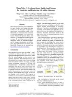
72.3 pmol/l, respectively; P = 0.006), as shown in Figure 2
and Table 4.
The influence of septic status on copeptin levels is shown in
Table 5 and Figure 3. Values were higher in the septic shock
group both for D0 and D4. Copeptin levels increased from
sepsis to severe sepsis to septic shock both on D0 (41.2
pmol/l, 64.8 pmol/l, 84.2 pmol/l, respectively; P = 0.001) and
on D4 (25.3 pmol/l, 68.7 pmol/l, 91.8 pmol/l, respectively; P
= 0.009).
Logistic regression analysis was used to determine the rela-
tion of risk factors to mortality. The variables included in the
univariable logistic regression analysis for mortality were age,
gender, APACHE II score, ln copeptin on D0 and ln copeptin
on D4. In univariable analysis, ln copeptin on D0 (odds ratio,
2.32) and ln copeptin on D4 (odds ratio, 2.31) were predictors
of mortality. There was a trend to significance for age, gender
and APACHE II score.
The multivariable logistic regression model for mortality
included the variables from the univariable analysis. The only
variables that remained as independent predictors of death
were ln copeptin D0 with an odds ratio of 1.97 (95% confi-
dence interval, 1.06 to 3.69; P = 0.03), and ln copeptin D4
with an odds ratio of 2.39 (95% confidence interval, 1.24 to
4.62; P = 0.01) (Tables 6 and 7).
Discussion
The current study demonstrates that copeptin levels are signif-
icantly higher in nonsurviving VAP patients compared with sur-
vivors. In multivariate logistic regression models of predictors
of death, including age, sex, APACHE II score and copeptin
level on the day of diagnosis of VAP (D0) and on day 4 (D4),
Table 1
Baseline characteristics of 71 patients who developed ventilator-associated pneumonia
Parameter Survivors (n = 45) Nonsurvivors (n = 26) Total (n = 71) P value
Age (years) 58 ± 14 64 ± 16 60 ± 15 0.12
Acute Physiology and Chronic Health Evaluation II score 18 ± 6 22 ± 9 19 ± 7 0.06
Albumin level (mg/dl) 2.8 ± 0.6 2.4 ± 0.5 2.7 ± 0.6 0.01
Gender (%) 0.09
Male 66.7 46.2 59.2
Female 33.3 53.8 40.8
Origin (%) 0.25
Medical 51.1 65.4 56.3
Surgical 48.9 34.6 43.7
Onset (%)
a
0.93
Early onset 22.2 23.1 22.5
Late onset 77.8 76.9 77.5
Chronic obstructive pulmonary disease (%) 17.7 26.9 19.7 0.59
Congestive heart failure (%) 17.8 26.9 21.1 0.37
Malignancy (%) 13.3 15.4 14.1 0.81
Histamine type-2 receptor antagonist (%) 66.7 57.7 63.4 0.45
Proton pump inhibitor (%) 22.2 34.6 26.8 0.26
Corticosteroids (%) 13.3 19.2 15.5 0.51
Dialysis (%) 11.1 19.2 14.1 0.35
Smoker (%) 37.8 38.5 38.0 0.95
Septic status (%) 0.01
Sepsis 66.7 15.4 47.9
Severe sepsis 28.9 30.8 29.6
Septic shock 4.4 53,8 22.5
Data presented as the mean ± standard deviation or the percentage.
a
Early onset is defined as occurring during the first 4 days of mechanical
ventilation, and late onset as occurring 5 days or more after mechanical ventilation.
Available online />Page 5 of 9
(page number not for citation purposes)
Table 2
Microbiological identification in 71 ventilator-associated pneumonia patients and mortality
a
Microorganism Survivors (n = 56
b
) Nonsurvivors (n = 31
b
) Total (n = 87
b
)
Pseudomonas aeruginosa 9 (16.1) 6 (19.4) 15 (17.2)
Staphylococcus aureus oxacillin resistant 8 (14.3) 5 (16.1) 13 (14.9)
Staphylococcus aureus oxacillin sensitive 7 (12.5) 1 (3.2) 8 (9.2)
Stenotrophomonas maltophilia 3 (5.4) 3 (9.7) 6 (6.9)
Acinetobacter sp 4 (7.1) 1 (3.2) 5 (5.7)
Klebsiella pneumoniae 2 (3.6) 3 (9.7) 5 (5.7)
Enterobacter sp 4 (7.1) 0 4 (4.6)
Haemophilus sp 4 (7.1) 0 4 (4.6)
Escherichia coli 0 2 (6.5) 2 (2.3)
Citrobacter koseri 2 (3.6) 0 2 (2.3)
Proteus mirabilis 2 (3.6) 0 2 (2.3)
Other 5 (8.9) 1 (3.2) 6 (6.9)
Nonidentified 6 (10.7) 9 (29.0) 15 (17.2)
Data presented as the frequency (%). Not all percentages add up to 100 because of rounding.
a
Positive quantitative endotracheal aspirate when
≥10
5
colony-forming units/ml.
b
We identified more than one microorganism in 11 patients who survived and in five patients who died. In total, 16
patients had more than one microorganism identified.
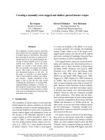
Figure 1
Receiver operating characteristic analysis of copeptin with respect to mortality prediction in ventilator-associated pneumonia patientsReceiver operating characteristic analysis of copeptin with respect to mortality prediction in ventilator-associated pneumonia patients. Data on the
day of diagnosis of ventilator-associated pneumonia (D0) and on day 4 (D4) are shown.
Critical Care Vol 12 No 1 Seligman et al.
Page 6 of 9
(page number not for citation purposes)
Table 3
Prediction of mortality in patients with ventilator-associated pneumonia (n = 71): area under the curve of receiver operating curve
characteristic plot analysis
Variable Threshold (pmol/l)
a
Sensitivity Specificity Area under the curve Standard error Asymptotic significance
Copeptin on day 0 64.8 0.69 0.69 0.70 0.06 0.006
Copeptin on day 4 43.0 0.80 0.60 0.72 0.07 0.006
a
Optimal cutoff value (minimal false negative and false positive results).
Figure 2
Copeptin levels in survivors and nonsurvivorsCopeptin levels in survivors and nonsurvivors. Box plots showing copeptin levels in survivors and nonsurvivors on day 0 (D0) and on day 4 (D4).
Boxes represent the 25th to 75th percentiles. Circles and asterisks represent outliers.
Table 4
Comparison of copeptin levels between survivors and nonsurvivors (Mann–Whitney test)
Variable Median (pmol/l) Interquartile range P value
Copeptin on day 0 Survivor 44.7 7.8 to 81.6 0.006
Nonsurvivor 74.2 12.3 to 136.1
Copeptin on day 4 Survivor 34.5 2.6 to 66.4 0.006
Nonsurvivor 72.3 38.6 to 106.0
Available online />Page 7 of 9
(page number not for citation purposes)
copeptin was the only parameter that remained an independ-
ent predictor.
The role of neuroendocrine regulation in sepsis is under inves-
tigation. The hypothalamus is the integrating center for stress
responses, and corticotropin-releasing hormone and vaso-
pressin neurones convert stress signals to hormonal outputs
[1]. Vasopressin is released into the portal circulation with cor-
ticotropin-releasing hormone in response to stress, and poten-
tiates corticotropin-releasing hormone-induced ACTH
secretion; thus, vasopressin and corticotropin-releasing
hormone are both endogenous releasing peptides for ACTH
[18]. Why we need both peptides for the regulation of gluco-
corticoid secretion, however, is not clear.
VAP and sepsis impose stress on patients, potentially promot-
ing cardiovascular instability and an elevated demand for vaso-
pressin and glucocorticoid secretion. Our data showed that
copeptin levels increased progressively with the severity of
sepsis. The levels appear to be consistent with findings
reported elsewhere about copeptin levels in septic patients [4-
6,19,20]. Septic shock VAP patients presented the highest
values of copeptin and highest mortality in our sample. Septic
shock and copeptin were colinear variables, and competed in
the multivariate analysis, since they express the same phenom-
enon, as clinical and laboratorial variables, respectively. Since
only 22.5% of our patients presented septic shock on D0, ele-
vated copeptin levels at the diagnosis of VAP in a patient with-
out septic shock could be helpful in prognostic assessment.
Copeptin might be used as a surrogate marker of the stimu-
lated neuroendocrine regulation in septic patients.
Relatively low vasopressin plasma concentrations are sus-
pected to contribute to cardiovascular failure in vasodilator
shock [21-23], and copeptin levels are elevated in this condi-
tion. It was shown in a previous study that the AVP plasma
concentration is lower in sepsis than the corresponding
copeptin values [19]. As both peptides are initially secreted in
an equimolar ratio, this could be an indication that AVP is rap-
idly consumed in extreme physiological conditions, thus result-
ing in a relative AVP insufficiency. It is possible that AVP
therapy could be useful particularly in those patients who have
this discrepant copeptin/AVP ratio. High copeptin in the
presence of vasodilatory shock may indicate insufficient
endogenous AVP production and may warrant exogenous
substitution. This hypothesis needs to be addressed in a future
prospective study, however, as we did not measure AVP in the
present study.
Further studies must evaluate whether implementation of
exogenous vasopressin therapy in patients with vasodilatory
shock should also be guided by endocrinologic investigation
or exclusively by cardiovascular investigations.
Figure 3
Copeptin levels in septic patients, severe sepsis patients, and septic shock patientsCopeptin levels in septic patients, severe sepsis patients, and septic
shock patients. Box plots showing copeptin levels in septic patients,
patients with severe sepsis and patients with septic shock on day 0
(D0) and on day 4 (D4). Boxes represent the 25th to 75th percentiles.
Circles and asterisks represent outliers.
Table 5
Comparison of copeptin levels between different septic statuses (Kruskal–Wallis test)
Variable Median (pmol/l) Interquartile range P value
Copeptin on day 0 Sepsis 41.2 17.2 to 65.9 0.001
Severe sepsis 64.8 23.7 to 105.9
Septic shock 84.2 15.6 to 152.8
Copeptin on day 4 Sepsis 25.3 2.1 to 48.5 0.009
Severe sepsis 68.7 39.3 to 98.1
Septic shock 91.8 35.9 to 144.7
Critical Care Vol 12 No 1 Seligman et al.
Page 8 of 9
(page number not for citation purposes)
In the present study we demonstrated copeptin is an inde-
pendent predictor of mortality in VAP. In such a condition it is
essential to assess the disease severity to optimize clinical
decision-making and therapy.
Our data should be interpreted in light of certain limitations.
First, copeptin comes from the same precursor as mature
AVP, which is already well associated with hemodynamic
changes and patient outcome. The measurement of mature
AVP, however, is subject to considerable challenges, and has
therefore not reached clinical routine in the context of rapid
measurements in the ICU setting. Here the stability and longer
ex vivo half-life of copeptin is a practical advantage, which
makes it easier to determine in the clinical laboratory. Second,
our sample size is not large enough for a stronger analysis, and
adrenal failure was therefore not assessed. Third, our study
was not designed to include a control group, which limits pos-
itive and negative predictive value assessment. Finally, the use
of crude mortality instead of attributable mortality is another
limitation, but it also avoids variability and confounding
interpretation.
Prior to the present study no published information existed
about the behavior of copeptin in patients with VAP. Copeptin
may represent a novel tool to assess prognosis in VAP. Addi-
tional studies are warranted to investigate these findings and
to further define the potential impact of strategies based on
biomarkers in improving VAP outcomes.
Conclusion
Copeptin levels increase progressively with the severity of
sepsis in VAP patients and are independent predictors of mor-
tality in this condition.
Competing interests
NGM and JP are employees of BRAHMS AG (Hennigsdorf/
Berlin, Germany), the manufacturer of the copeptin assay. MM
has received remuneration for holding lectures on the topic of
inflammation markers by BRAHMS AG, Germany. The authors
declare that there are no further competing interests.
Authors' contributions
RS developed the study design, coordinated its implementa-
tion and was responsible for patient recruitment as well as
Table 6
Odds ratios for mortality in 71 ventilator-associated pneumonia patients on day 0: univariable and multivariable logistic regression
analysis
Parameter Univariable analysis Multivariable analysis
Odds ratio (95%
confidence interval)
P value Odds ratio (95%
confidence interval)
P value
Age 1.03 (0.99 to 1.06) 0.12 1.01 (0.98 to 1.05) 0.55
Gender, female 2.33 (0.87 to 6.28) 0.09 1.80 (0.62 to 5.23) 0.28
APACHE II score 1.07 (1.00 to 1.15) 0.06 1.05 (0.97 to 1.13) 0.23
ln copeptin on day 0 2.32 (1.25 to 4.29) 0.008 1.97 (1.06 to 3.69) 0.03
Key messages
• Copeptin levels increase progressively with the severity
of sepsis and are independent predictors of mortality in
VAP.
Table 7
Odds ratios for mortality in 71 ventilator-associated pneumonia patients on day 4: univariable and multivariable logistic regression
analysis
Parameter Univariable analysis Multivariable analysis
Odds ratio (95%
confidence interval)
P value Odds ratio (95%
confidence interval)
P value
Age 1.03 (0.99 to 1.06) 0.12 1.00 (0.97 to 1.05) 0.84
Gender, female 2.33 (0.87 to 6.28) 0.09 2.52 (0.75 to 8.45) 0.13
APACHE II score 1.07 (1.00 to 1.15) 0.06 1.07 (0.98 to 1.18) 0.14
ln copeptin on day 4 2.31 (1.25 to 4.25) 0.007 2,39 (1.24 to 4.62) 0.01
Available online />Page 9 of 9
(page number not for citation purposes)
data collection. NGM and JP carried out laboratory tests. RS
and PJZT carried out the statistical analysis. RS, NGM, JP, MM
and PJZT participated in interpretation/discussion of results
and drafted and revised the manuscript. All authors read and
approved the final manuscript.
Acknowledgements
The present study was supported by grants from Fundo de Incentivo a
Pesquisa – FIPEHCPA, Porto Alegre, Brazil, and was performed in the
Hospital de Clínicas de Porto Alegre.
References
1. Itoi K, Jiang YQ, Iwasaki Y, Watson SJ: Regulatory mechanisms
of corticotropin-releasing hormone and vasopressin gene
expression in the hypothalamus. J Neuroendocrinol 2004,
16:348-355.
2. Asfar P, Hauser B, Radermacher P, Matejovic M: Catecholamines
and vasopressin during critical illness. Crit Care Clin 2006,
22:131-149.
3. De Bree FM, Burbach JP: Structure–function relationships of
the vasopressin prohormone domains. Cell Mol Neurobiol
1998, 18:173-191.
4. Struck J, Morgenthaler NG, Bergmann A: Copeptin, a stable pep-
tide derived from the vasopressin precursor, is elevated in
serum of sepsis patients. Peptides 2005, 26:2500-2504.
5. Morgenthaler NG, Muller B, Struck J, Bergmann A, Redl H, Christ-
Crain M: Copeptin, a stable peptide of the arginine vasopressin
precursor, is elevated in hemorrhagic and septic shock. Shock
2007, 28:219-226.
6. Muller B, Morgenthaler N, Stolz D, Schuetz P, Muller C, Bingisser
R, Bergmann A, Tamm M, Christ-Crain M: Circulating levels of
copeptin, a novel biomarker, in lower respiratory tract
infections. Eur J Clin Invest 2007, 37:145-152.
7. Barat C, Simpson L, Breslow E: Properties of human vaso-
pressin precursor constructs: inefficient monomer folding in
the absence of copeptin as a potential contributor to diabetes
insipidus. Biochemistry 2004, 43:8191-8203.
8. Stolz D, Christ-Crain M, Morgenthaler NG, Leuppi J, Miedinger D,
Bingisser R, Müller C, Struck J, Müller B, Tamm M: Copeptin, C-
reactive protein, and procalcitonin as prognostic biomarkers in
acute exacerbation of COPD. Chest 2007, 131:1058-1067.
9. Langer M, Cigada M, Mandelli M, Mosconi P, Tognoni G: Early
onset pneumonia: a multicenter study in intensive care units.
Intensive Care Med 1987, 13:342-346.
10. Knaus WA, Draper EA, Wagner DP, Zimmerman JE: APACHE II: a
severity of disease classification system. Crit Care Med 1985,
13:818-829.
11. Fabregas N, Ewig S, Torres A, El Ebiary M, Ramirez J, de la Bel-
lacasa JP, Bauer T, Cabello H: Clinical diagnosis of ventilator
associated pneumonia revisited: comparative validation using
immediate post-mortem lung biopsies. Thorax
1999,
54:867-873.
12. Pugin J, Auckenthaler R, Mili N, Janssens JP, Lew PD, Suter PM:
Diagnosis of ventilator-associated pneumonia by bacterio-
logic analysis of bronchoscopic and nonbronchoscopic 'blind'
bronchoalveolar lavage fluid. Am Rev Respir Dis 1991,
143:1121-1129.
13. Singh N, Rogers P, Atwood CW, Wagener MM, Yu VL: Short-
course empiric antibiotic therapy for patients with pulmonary
infiltrates in the intensive care unit. A proposed solution for
indiscriminate antibiotic prescription. Am J Respir Crit Care
Med 2000, 162:505-511.
14. American College of Chest Physicians/Society of Critical Care
Medicine Consensus Conference: Definitions for sepsis and
organ failure and guidelines for the use of innovative thera-
pies in sepsis. Crit Care Med 1992, 20:864-874.
15. Bone RC, Balk RA, Cerra FB, Dellinger RP, Fein AM, Knaus WA,
Schein RM, Sibbald WJ: Definitions for sepsis and organ failure
and guidelines for the use of innovative therapies in sepsis.
The ACCP/SCCM Consensus Conference Committee. Ameri-
can College of Chest Physicians/Society of Critical Care
Medicine. Chest 1992, 101:1644-1655.
16. Luna CM, Blanzaco D, Niederman MS, Matarucco W, Baredes
NC, Desmery P, Palizas F, Menga G, Rios F, Apezteguia C: Reso-
lution of ventilator-associated pneumonia: prospective evalu-
ation of the clinical pulmonary infection score as an early
clinical predictor of outcome. Crit Care Med 2003, 31:676-682.
17. Morgenthaler NG, Struck J, Alonso C, Bergmann A: Assay for the
measurement of copeptin, a stable peptide derived from the
precursor of vasopressin. Clin Chem 2006, 52:112-119.
18. Itoi K, Seasholtz AF, Watson SJ: Cellular and extracellular regu-
latory mechanisms of hypothalamic corticotropin-releasing
hormone neurons. Endocr J 1998, 45:13-33.
19. Jochberger S, Morgenthaler NG, Mayr VD, Luckner G, Wenzel V,
Ulmer H, Schwarz S, Hasibeder WR, Friesenecker BE, Dünser
MW: Copeptin and arginine vasopressin concentrations in crit-
ically ill patients. J Clin Endocrinol Metab 2006, 91:4381-4386.
20. Jochberger S, Luckner G, Mayr VD, Wenzel V, Morgenthaler NG,
Friesenecker BE, Hasibeder WR, Dünser MW: Course of vaso-
pressin and copeptin plasma concentrations in a patient with
severe septic shock.
Anaesth Intensive Care 2006, 34:498-500.
21. Mutlu GM, Factor P: Role of vasopressin in the management of
septic shock. Intensive Care Med 2004, 30:1276-1291.
22. Sharshar T, Carlier R, Blanchard A, Feydy A, Gray F, Paillard M,
Raphael JC, Gajdos P, Annane D: Depletion of neurohypophy-
seal content of vasopressin in septic shock. Crit Care Med
2002, 30:497-500.
23. Sharshar T, Blanchard A, Paillard M, Raphael JC, Gajdos P,
Annane D: Circulating vasopressin levels in septic shock. Crit
Care Med 2003, 31:1752-1758.