Báo cáo y học: "Causes of a high physiological dead space in critically ill patients" ppt
Bạn đang xem bản rút gọn của tài liệu. Xem và tải ngay bản đầy đủ của tài liệu tại đây (103.78 KB, 2 trang )
Page 1 of 2
(page number not for citation purposes)
Available online />Abstract
Since around 1950, physiological dead space—the difference
between arterial and mixed expired pCO
2
(partial pressure of carbon
dioxide) divided by the arterial pCO
2
—has been a useful clinical
parameter of pulmonary gas exchange. In the previous issue of
Critical Care, Niklason and colleagues remind us that physiological
dead space, while easily measured, consolidates potentially very
complex physiological derangements into a single number. The
authors show how shunts raise arterial pCO
2
, thereby increasing
dead space, and how changes in other variables such as cardiac
output and acid/base state further modify it. A solid understanding
of respiratory physiology is required to properly interpret
physiological dead space in the critically ill.
In the previous issue of Critical Care, Niklason and colleagues
[1] use computer modeling to point out that blood flowing
through unventilated regions of the lung (a shunt) will
increase arterial partial pressure of carbon dioxide (pCO
2
) if
ventilation remains constant. This will increase the calculated
physiological dead space accordingly (above that normally
present due to the volume of air in the conducting airways).
They also show that the increase in pCO
2
can be avoided by
even modest increases in total alveolar ventilation (although
the calculated dead space will remain elevated). While this is
not an entirely novel discovery (West [2] performed very
similar calculations in 1969 as did Mecikalski and colleagues
[3] in 1984), it is well worth having Niklason and colleagues
remind us that physiological dead space not only can be
caused by the development of regions with a high ventilation/
perfusion ratio (V
•
A
/Q
•
), but also can come from areas of low
V
•
A
/Q
•
and a shunt. After all, physiological dead space is
simply the difference between arterial and mixed expired
pCO
2
divided by the arterial pCO
2
. Thus, any gas exchange
abnormality has the potential to increase dead space.
Some high-level perspective may be useful. First, it should be
remembered that, since introduced by Riley and Cournand
[4] more than 50 years ago, physiological dead space is a
virtual concept wherein the lung is conceived as a two-
compartment organ in which one compartment is normal and
the other is completely unperfused. Physiological dead
space, then, is the percentage of the tidal volume that must
be distributed to the alveolus that is completely unperfused
(and which thus delivers no CO
2
to the expired gas) to
account for the difference between measured arterial and
mixed expired pCO
2
. Physiological dead space in actual
patients may be increased even when no alveoli are
completely unperfused—as is the case here in the presence of
a shunt. It is useful as a general parameter quantifying gas
exchange disturbances but must not be overinterpreted as
necessarily implying the existence of unperfused alveoli.
Second, as Niklason and colleagues show, the relationship
between shunt and physiological dead space is nonlinear,
especially when shunts are high. A shunt of 20% of the
cardiac output increases dead space by just 5%, a shunt of
40% raises it to approximately 11%, but a shunt of 60%
produces a dead space of about 20%. This is because basic
mass balance considerations show that the increase in
arterial pCO
2
caused by a shunt depends on the factor
QS/(100 – QS), where QS is the percentage shunt. Thus, for
CO
2
exchange, the importance of shunts of less than
approximately 30% is not great, but as shunts approach and
exceed 50%, the potential for hypercapnia increases rapidly.
Third, very modest increases in alveolar ventilation can return the
arterial pCO
2
to normal: An increase from just 5 to 7 L/minute
will restore normocapnia (assuming no other changes have
occurred or abnormalities exist as ventilation is increased), even
when the shunt is 60% of the cardiac output.
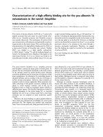
Fourth, V
•
A
/Q
•
inequality is generally a cause of greater
physiological dead space than shunt is (Figure 1) (calcula-
Commentary
Causes of a high physiological dead space in critically ill patients
Peter D Wagner
Division of Physiology, Department of Medicine, University of California at San Diego, 9500 Gilman Drive, 0623A, La Jolla, CA 92093-0623, USA
Corresponding author: Peter D Wagner,
Published: 14 May 2008 Critical Care 2008, 12:148 (doi:10.1186/cc6888)
This article is online at />© 2008 BioMed Central Ltd
See related research by Niklason et al., />CO
2
= carbon dioxide; log SDQ = second moment (dispersion) of the ventilation/perfusiondistribution on a log scale; pCO
2
= partial pressure of
carbon dioxide; V
•
A
/Q
•
= ventilation/perfusion ratio.
Page 2 of 2
(page number not for citation purposes)
Critical Care Vol 12 No 3 Wagner
tions using algorithms from [2]). For example, it takes a very
large, 60% shunt to increase dead space by 20% but a log-
normal pattern of only moderate V
•
A
/Q
•
inequality (log SDQ =
1.3) does the same. Normal log SDQ is less than 0.6 [5], and
the highest log SDQ values seen are about 2 to 2.5. Log
SDQ is a parameter defined for quantifying V
•
A
/Q
•
inequality in
the multiple inert gas elimination technique [6,7] and is the
second moment (dispersion) of the V
•
A
/Q
•
distribution on a log
scale.
Fifth, Niklason and colleagues show that, for any given value
of shunt, additional perturbations commonly seen in the
intensive care unit influence arterial pCO
2
and therefore will
increase calculated dead space. These include a reduction in
cardiac output and, separately, acidosis. This also means that
a high cardiac output will reduce the dead space effect of
shunt, as will alkalosis. The clinical message is that observed
changes in dead space may reflect changes in cardiac output
or acid/base state rather than changes in the shunt itself.
In summary, the calculations of Niklason and colleagues
serve to point out the complexity of gas exchange in critical
illness and the challenges we face in trying to interpret
apparently simple measurements as indicators of the lung’s
ability to carry out its primary responsibility—gas exchange.
Competing interests
The author declares that he has no competing interests.
References
1. Niklason L, Eckerstrom J, Jonson B: The influence of venous
admixture on alveolar dead space and CO
2
exchange in
ARDS: computer modelling. Crit Care 2008, 12:R53.
2. West JB: Ventilation-perfusion inequality and overall gas
exchange in computer models of the lung. Respir Physiol
1969, 7:88-110.
3. Mecikalski MB, Cutillo AG, Renzetti AD Jr.: Effect of right-to-left
shunting on alveolar dead space. Bull Eur Physiopathol Respir
1984, 20:513-519.
4. Riley RL, Cournand A: ‘Ideal’ alveolar air and the analysis of
ventilation/perfusion relationships in the lung. J Appl Physiol
1949, 1:825-847.
5. Wagner PD, Hedenstierna G, Bylin G: Ventilation/perfusion
inequality in chronic asthma. Am Rev Respir Dis 1987, 136:
605-612.
6. Wagner PD, Saltzman HA, West JB: Measurement of continu-
ous distributions of ventilation/perfusion ratios: theory. J Appl
Physiol 1974, 36:588-599.
7. Evans JW, Wagner PD: Limits on V
•
A
/Q
•
distributions from
analysis of experimental inert gas elimination. J Appl Physiol
1977, 42:889-898.
Figure 1
Comparison of effects of shunt (top) and ventilation/perfusion ratio
(V
•
A
/Q
•
) inequality (bottom) on calculated physiological dead space. In
general, V
•
A
/Q
•
inequality leads to greater dead space than shunt does.
Log SDQ, second moment (dispersion) of the ventilation/perfusion
distribution on a log scale