Báo cáo y học: "Norepinephrine to increase blood pressure in endotoxaemic pigs is associated with improved hepatic mitochondrial respiration" doc
Bạn đang xem bản rút gọn của tài liệu. Xem và tải ngay bản đầy đủ của tài liệu tại đây (300.9 KB, 10 trang )
Open Access
Available online />Page 1 of 10
(page number not for citation purposes)
Vol 12 No 4
Research
Norepinephrine to increase blood pressure in endotoxaemic pigs
is associated with improved hepatic mitochondrial respiration
Tomas Regueira
1
, Bertram Bänziger
2
, Siamak Djafarzadeh
1
, Sebastian Brandt
2
, Jose Gorrasi
1
,
Jukka Takala
1
, Philipp M Lepper
1
and Stephan M Jakob
1
1
Department of Intensive Care Medicine, Bern University Hospital (Inselspital) and University of Bern, Freiburgstrasse, CH-3010 Bern, Switzerland
2
Department of Anesthesiology and Pain Therapy, Bern University Hospital (Inselspital) and University of Bern, Freiburgstrasse, CH-3010 Bern,
Switzerland
Corresponding author: Stephan M Jakob,
Received: 22 Apr 2008 Revisions requested: 15 May 2008 Revisions received: 30 May 2008 Accepted: 14 Jul 2008 Published: 14 Jul 2008
Critical Care 2008, 12:R88 (doi:10.1186/cc6956)
This article is online at: />© 2008 Regueira et al.; licensee BioMed Central Ltd.
This is an open access article distributed under the terms of the Creative Commons Attribution License ( />),
which permits unrestricted use, distribution, and reproduction in any medium, provided the original work is properly cited.
Abstract
Introduction Low blood pressure, inadequate tissue oxygen
delivery and mitochondrial dysfunction have all been implicated
in the development of sepsis-induced organ failure. This study
evaluated the effect on liver mitochondrial function of using
norepinephrine to increase blood pressure in experimental
sepsis.
Methods Thirteen anaesthetized pigs received endotoxin
(Escherichia coli lipopolysaccharide B0111:B4; 0.4 μg/kg per
hour) and were subsequently randomly assigned to
norepinephrine treatment or placebo for 10 hours.
Norepinephrine dose was adjusted at 2-hour intervals to achieve
15 mmHg increases in mean arterial blood pressure up to 95
mmHg. Systemic (thermodilution) and hepatosplanchnic
(ultrasound Doppler) blood flow were measured at each step. At
the end of the experiment, hepatic mitochondrial oxygen
consumption (high-resolution respirometry) and citrate synthase
activity (spectrophotometry) were assessed.
Results Mean arterial pressure (mmHg) increased only in
norepinephrine-treated animals (from 73 [median; range 69 to
81] to 63 [60 to 68] in controls [P = 0.09] and from 83 [69 to
93] to 96 [86 to 108] in norepinephrine-treated animals [P =
0.019]). Cardiac index and systemic oxygen delivery (D
O
2
)
increased in both groups, but significantly more in the
norepinephrine group (P < 0.03 for both). Cardiac index (ml/min
per·kg) increased from 99 (range: 72 to 112) to 117 (110 to
232) in controls (P = 0.002), and from 107 (84 to 132) to 161
(147 to 340) in norepinephrine-treated animals (P = 0.001).
D
O
2
(ml/min per·kg) increased from 13 (range: 11 to 15) to 16
(15 to 24) in controls (P = 0.028), and from 16 (12 to 19) to 29
(25 to 52) in norepinephrine-treated animals (P = 0.018).
Systemic oxygen consumption (systemic V
O
2
) increased in both
groups (P < 0.05), whereas hepatosplanchnic flows, D
O
2
and
V
O
2
remained stable. The hepatic lactate extraction ratio
decreased in both groups (P = 0.05). Liver mitochondria
complex I-dependent and II-dependent respiratory control ratios
were increased in the norepinephrine group (complex I: 3.5
[range: 2.1 to 5.7] in controls versus 5.8 [4.8 to 6.4] in
norepinephrine-treated animals [P = 0.015]; complex II: 3.1 [2.3
to 3.8] in controls versus 3.7 [3.3 to 4.6] in norepinephrine-
treated animals [P = 0.09]). No differences were observed in
citrate synthase activity.
Conclusion Norepinephrine treatment during endotoxaemia
does not increase hepatosplanchnic flow, oxygen delivery or
consumption, and does not improve the hepatic lactate
extraction ratio. However, norepinephrine increases the liver
mitochondria complex I-dependent and II-dependent respiratory
control ratios. This effect was probably mediated by a direct
effect of norepinephrine on liver cells.
Introduction
Septic shock is associated with high mortality, especially
when antibiotic treatment and fluid resuscitation are delayed
and oxygen delivery remains insufficient [1]. Despite recent
recommendations on blood pressure targets in septic shock
[2,3], the goals in clinical trials vary substantially [4]. Recently,
Varpula and coworkers [1] showed that mean arterial pressure
(MAP) is the most powerful predictor of mortality in septic
shock, emphasizing the importance of global perfusion
MAP = mean arterial pressure; NADH = nicotinamide adenine dinucleotide; RCR = respiratory control ratio.
Critical Care Vol 12 No 4 Regueira et al.
Page 2 of 10
(page number not for citation purposes)
pressure for survival. Also, according to a recent systematic
review of randomized clinical trials that used MAP as the goal
of resuscitation in septic patients [4], the minimum and maxi-
mum targets for MAP ranged from 60 to 100 mmHg [5-13].
The authors of the review concluded that there was wide vari-
ation in the goals chosen for the studies and that this variation
may lead to bias in the interpretation of study results. More
intriguingly, the achieved blood pressure in these trials was
substantially higher than the target blood pressure.
Vasoactive drugs such as norepinephrine (noradrenaline) are
commonly used to maintain a certain MAP level in septic
(shock) patients. However, the regional haemodynamic and
metabolic effects of norepinephrine during sepsis are not fully
understood and are controversial [14-19]. Systemically, nore-
pinephrine increases cardiac output and oxygen delivery and
consumption [18], and at the regional level it has been
reported to increase portal vein flow [20], total splanchnic
blood flow and oxygen delivery during sepsis [14].
Other studies have identified unchanged mesenteric flows
[19], total splanchnic blood flow and oxygen uptake [17-19].
In a crossover study, Guerin and colleagues [16] compared
norepinephrine with dopamine in septic patients, with the MAP
goal being 80 mmHg. They reported that the drugs were asso-
ciated with similar splanchnic blood flow and hepatic oxygen
consumption, but that patients treated with norepinephrine
exhibited higher levels of hepatic lactate uptake and lower val-
ues of the lactate-pyruvate ratio, suggesting improved hepatic
energy balance with norepinephrine. Also, Revelly and cow-
orkers [21] reported that, during distributive shock induced by
endotoxaemia in pigs, norepinephrine prevented the decrease
in intestinal mucosal ATP content, which was observed only in
fluid-resuscitated animals. This also suggests improved
energy balance with norepinephrine. Only one study evaluated
the effects of increasing MAP from 65 to 85 mmHg using
norepinephrine [22]. In this study, conducted in 10 patients
with septic shock, increasing MAP with norepinephrine was
associated with a significant increase in cardiac index, left ven-
tricular stroke work index, heart rate and systemic oxygen
delivery. Because systemic oxygen consumption, lactate con-
centrations, capillary blood flow, urine output and gastric
mucosal partial carbon dioxide tension were not altered, the
benefit of increasing blood pressure was questioned.
Growing evidence suggests that mitochondrial damage and
dysfunction play an important role in the pathogenesis of sep-
sis-induced organ failure [23,24]. Mitochondrial dysfunction
can be characterized by inability of the mitochondria to couple
oxygen consumption with energy production completely. In a
long-term, fluid-resuscitated, faecal peritonitis model in mice,
Breadley and colleagues [25] demonstrated that severity of
and mortality from sepsis were related to nitric oxide over-pro-
duction and consequent complex I inhibition and ATP
depletion.
Because norepinephrine affects at least systemic haemody-
namics, it may also affect liver mitochondrial function, either
through an indirect regional haemodynamic mechanism (for
instance, an increase in perfusion pressure/flow relationship
or an increase in regional oxygen delivery) or through a direct
cellular mechanism. A direct cellular effect of norepinephrine
on isolated hepatocytes has been described and includes
stimulation of α-adrenergic receptor, increasing cytosol and
intra-mitochondrial calcium levels, and activation of dehydro-
genases of the citrate cycle [26,27]. Therefore, in the present
study we tested whether a stepwise increase in blood pres-
sure with norepinephrine, in the setting of a clinically relevant
range of pressures between 60 and 95 mmHg in a model of
endotoxic sepsis, is associated with a beneficial effect on liver
blood flow, oxygen delivery and consumption, and mitochon-
drial function.
Materials and methods
The study was performed in accordance with the National
Institutes of Health guidelines for the care and use of experi-
mental animals and with the approval of the Animal Care Com-
mittee of the Canton of Bern, Switzerland.
Animal preparation and experimental setting
Thirteen pigs (weight 37 to 44 kg) were fasted overnight and
premedicated with ketamine (20 mg/kg) and xylazine (2 mg/kg
intramuscularly), followed by intravenous administration of
midazolam (0.5 mg/kg) and atropine (0.02 mg) for endotra-
cheal intubation. The animals were ventilated with a volume-
controlled ventilator (Servo ventilator 900 C; Siemens, Elema,
Sweden) with 5 cmH
2
O end-expiratory pressure. The fraction
of inspired oxygen and positive end-expiratory pressure were
adjusted to keep arterial oxygen tension levels between 13.3
kPa (100 mmHg) and 20 kPa (150 mmHg), and the minute
ventilation was adjusted to maintain arterial carbon dioxide
tension levels between 4.5 and 5.5 kPa (34 to 41 mmHg).
Anaesthesia was maintained with propofol (4 mg/kg per hour)
and fentanyl (45 μg/kg per hour during surgery and 30 μg/kg
per hour afterward). After canulation of jugular and femoral
veins and the femoral artery for the placement of hepatic
venous, and pulmonary arterial and femoral arterial catheters,
a laparatomy was performed and a urinary bladder catheter
was inserted. Afterward, ultrasound Doppler flow probes
(Transonic
®
System Inc., Ithaca, NY, USA), which had previ-
ously been calibrated in vitro, were positioned around the
carotid and hepatic arteries and the portal vein. Finally, a cath-
eter was inserted into the portal vein for blood sampling. After
the procedure the abdominal wall was sewn closed.
Regional blood flows and portal vein pressure were monitored
continuously during the experiment and stored in a computer
(WinDaq
®
; DATAQ instruments, Akron, OH, USA). Electro-
cardiogram, heart rate, and carotid and pulmonary arterial and
central venous pressures were monitored continuously, and
pulmonary artery occlusion pressure intermittently. Cardiac
Available online />Page 3 of 10
(page number not for citation purposes)
output was assessed hourly with three measurements using
the thermodilution technique (S/5 Compact Critical Care
monitor; Datex-Ohmeda, Helsinki, Finland). Central tempera-
ture was recorded from the thermistor in the pulmonary artery
catheter (cardiac output/mixed venous oxygen saturation cath-
eter; Edwards Lifesciences, Munich, Germany) and peripheral
temperature from the tip of a toe. All of these variables were
recorded using an electronic patient data management system
(Clinisoft, GE Healthcare, Helsinki, Finland). The system auto-
matically stores median values in a database every 2 minutes.
Once the experiment was started, manipulation was avoided
to minimize the possibility of flow probe displacement. At the
end of the experiment, the correct position of each probe and
catheter was controlled visually.
Experimental protocol
After surgery, a 1-hour period was allowed for haemodynamic
stabilization. After this period, pigs were randomized for a 10-
hour experiment into two groups: endotoxin plus progressively
increasing goals of MAP, achieved by increasing doses of
norepinephrine (norepinephrine group; n = 7); or endotoxin
alone (control group; n = 6). Initially, six pigs were randomized
per group, but because one mitochondrial respiration experi-
ment was missed in the norepinephrine group, one more pig
was included in this group.
Endotoxin (Escherichia coli lipopolysaccharide B0111:B4, 20
mg/l in 5% dextrose; Difco Laboratories, Detroit, MI, USA) was
infused into the right atrium in all animals. The initial infusion
rate was 0.4 μg/kg per hour until the mean pulmonary arterial
pressure reached 35 mmHg. The infusion was then stopped
and subsequently adjusted to maintain moderate pulmonary
artery hypertension (mean pulmonary artery pressure 25 to 30
mmHg). If systemic hypotension persisted below 50 mmHg,
then the endotoxin infusion was temporarily stopped and 50
ml of hydroxyethyl starch (Voluven 6%; Fresenius, Stans, Swit-
zerland) was administered. All pigs received Ringer's lactate
solution 5 ml/kg per hour, and glucose 50% solution was
administered in order to maintain a blood glucose concentra-
tion between 3.5 and 6 mmol/l.
In the norepinephrine group, norepinephrine was continuously
infused to reach the following pre-defined MAP goals: from 0
to 2 hours there was no specific goal; from 2 to 4 hours the
goal was 50 mmHg; from 4 to 6 hours the goal was 65 mmHg;
from 6 to 8 hours the goal was 80 mmHg; and from 8 to 10
hours the goal was 95 mm Hg. The MAP goal for the control
group was between 60 and 70 mmHg for the whole experi-
ment. If the MAP of the control group was below 60 mmHg,
then additional boluses of 50 to 100 ml of colloids (Voluven
6%; Fresenius) were given. If the MAP of the animals in the
norepinephrine group was found to be above the target for the
corresponding time point, then no norepinephrine was added.
Blood sampling
Blood samples for the measurement of haemoglobin, lactate
and blood gases were taken at baseline and every 2 hours
afterward from pulmonary and femoral arteries and from portal
and hepatic veins (ABL 520 and OSM 3 [pig module; Radiom-
eter, Copenhagen, Denmark], and YSI 2300 Stat Plus [Yellow
Springs Instruments, Yellow Springs, OH, USA]).
Liver mitochondrial isolation
At the end of the experiment, liver tissue samples of approxi-
mately 15 g were taken from the living animal for isolation of
mitochondria. Afterward, the animals were killed with an over-
dose of intravenous potassium chloride. Isolation of liver mito-
chondria was performed immediately at 4°C using a standard
procedure based on differential centrifugation [28]. The liver
samples were rapidly immersed in ice-cold isolation buffer
(220 mmol/l mannitol, 70 mmol/l sucrose, 5 mmol/l mor-
pholinopropane sulfonic acid [pH 7.4]), minced with scissors
and homogenized in 10 ml of homogenization medium per
gram of tissue (isolation buffer plus 2 mmol/l ethyleneglycol
tetra-acetate) in a Potter Elvehjem homogenizer with a loose-
fitting Teflon pestle. The homogenate was then centrifuged for
10 minutes at 700 g. The supernatant was collected and cen-
trifuged again for 10 minutes at 7,000 g. The supernatant was
discarded at this time; the pellet was then resuspended in iso-
lation buffer and centrifuged twice for 10 minutes at 7,000 g
for further purification of the mitochondria. The pellets were
then suspended in buffer at a final concentration of 50 to 100
mg mitochondrial protein per milliliter.
Determination of mitochondrial oxygen consumption by
high-resolution respirometry
Protein concentration was determined spectrophotometrically
with the Biuret method using bovine serum albumin as a stand-
ard. Respiratory rates were determined at a final mitochondrial
protein concentration of 0.4 mg/ml. Respiration was measured
at 37°C in 2 ml glass chambers using the High Resolution
Oxygraph (OROBOROS; Oxygraph-2k, Graz, Austria). The
medium used for respiration measurements consisted of 25
mmol/l KCL, 12.5 mmol/l morpholinopropane sulfonic acid, 1
mmol/l ethylene glycol-bis N,N,N',N'-tetra-acetic acid and 5
mmol/l potassium phosphate buffer (pH 7.4). The medium was
equilibrated for 30 to 40 minutes with air in the oxygraph
chambers and stirred at 750 rpm until a stable signal was
obtained for calibration at air saturation. The corresponding
oxygen concentration was calculated from the digitally
recorded barometric pressure and the oxygen solubility at
37°C. The amplified signal was recorded in a computer with
online display of the calibrated oxygen concentration and oxy-
gen flux (negative time derivative of oxygen concentration; Dat-
Lab software for data acquisition and analysis; OROBOROS).
Oxygen consumption was expressed as pmol/second per mg
mitochondrial protein. Oxygen levels were always maintained
above 40 nmol/ml.
Critical Care Vol 12 No 4 Regueira et al.
Page 4 of 10
(page number not for citation purposes)
Maximal oxidative capacities were determined in the presence
of saturating concentrations of oxygen, ADP (0.25 mmol/l) and
specific mitochondrial substrates. For complex I-dependent
respiration, substrates were glutamate (10 mmol/l) plus malate
(5 mmol/l), which provide nicotinamide adenine dinucleotide
(NADH) to the respiratory chain (complex I activation). For
measurement of complex II dependent respiration, first com-
plex I was inhibited with rotenone (0.5 μmol/l), and then succi-
nate (10 mmol/l) was added, which provides flavin adenine
dinucleotide to the respiratory chain (complex II activation).
The coupling of phosphorylation to oxidation was determined
by calculating the respiratory control ratio (RCR) as the ratio
between ADP-stimulated respiration (state 3) and respiration
after ADP depletion (state 4).
Calculations
The equations used I the present study are summarized in
Table 1.
Statistical analysis
The SPSS 13.0 software package (SPSS Inc., Chicago, IL,
USA) was used for statistical analysis. Because of the small
numbers of animals in each group, nonparametric test were
used: Mann Whitney U-test for single measurements, and non-
parametric analysis of variance for repeated measurements
(Friedman test). In the latter case, differences between groups
at certain time points were analyzed with the Mann Whitney U-
test. For lactate and oxygen transport variables only baseline
and end values were used. The two values were compared
with the Wilcoxon test. Data are expressed as median and
range. P < 0.05 was considered statistically significant.
Results
The two groups received comparable total doses of endotoxin
(7.3 [5.9 to 8.2] μg/kg in the control group and 7.9 [5.6 to
10.0] μg/kg in the norepinephrine group; P = 0.62) and total
fluid input (5.9 [5.2 to 9.5] ml/kg per hour in the control group
and 5.7 [5.1 to 7.5] ml/kg per hour in the norepinephrine
group; P = 0.32).
Systemic haemodynamics
At baseline, before randomization, there were no significant
differences between groups in terms of systemic haemody-
namics and respiratory parameters (Table 2). After endotoxin
administration, both groups exhibited a progressive increase
in their mean pulmonary arterial pressures (P = 0.018 for con-
trols and P = 0.003 for norepinephrine-treated animals) and in
their pulmonary capillary wedge pressures (P = 0.016 for con-
trols and P = 0.004 for norepinephrine-treated animals). After-
ward, both parameters remained elevated until the end of the
experiment, without differences between groups. Until hour 5
of the experiments, only one animal required norepinephrine in
the norepinephrine group to reach the pressure goal specified
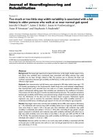
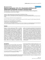
in the protocol (Figure 1). From 6 to 8 hours, six animals
required norepinephrine (average dose 0.09 [0.00 to 0.21]
μg/kg per minute), and from 8 to 10 hours all animals needed
norepinephrine to meet the specified goal (average dose 0.23
[0.11 to 0.70] μg/kg per minute; Figure 1).
As a result of the intervention, there was a significant increase
in MAP only in the norepinephrine-treated animals (in controls:
from 73 [69 to 81] to 63 [60 to 68] mmHg [P = 0.09]; in nore-
pinephrine-treated animals: from 83 [69 to 93] to 96 [86 to
108] mmHg [P = 0.019]). Accordingly, MAP levels at 8 and
10 hours were higher than in controls (P < 0.05 for both time
points; Figure 1). Cardiac index increased in both groups (P <
0.003 for both groups), but end values were significantly
higher in the norepinephrine-treated group (41% in controls
versus 81% in norepinephrine-treated animals; P = 0.022).
The observed increase in cardiac index was mainly the result
of an increase in the heart rate only in the norepinephrine-
treated animals (P = 0.006; Table 2).
Regional blood flows
The two groups exhibited a similar decrease in hepatic and
portal flows until 4 to 6 hours after endotoxin infusion; both
hepatic and portal flows recovered to baseline values at the
end of the experiment in both groups (P < 0.002 for both ves-
sels; Table 2). There was no consistent relationship between
Table 1
Equations used in the present study
Parameter Equation
Systemic oxygen delivery (ml/kg·minute) Cardiac index (ml/kg·minute) × arterial oxygen content (ml/l)
Hepatosplanchnic oxygen delivery (ml/kg·minute) Hepatic arterial blood flow (ml/kg·minute) + portal venous blood flow (ml/kg·min) × arterial
oxygen content (ml/l)
Systemic oxygen consumption (ml/kg·min) Cardiac index (ml/kg·min) × (arterial oxygen content [ml/l] – pulmonary artery oxygen content
[ml/l])
Hepatic lactate uptake (μmol/kg·minute) ([Arterial lactate × hepatic arterial blood flow] + [portal vein lactate × portal vein blood flow]) –
(hepatic vein lactate × [portal vein blood flow + hepatic arterial blood flow])
Hepatic lactate influx (μmol/kg·minute) (Arterial lactate × hepatic arterial blood flow) + (portal vein lactate × portal vein blood flow)
Hepatic lactate extraction ratio Hepatic lactate uptake/hepatic lactate influx
Available online />Page 5 of 10
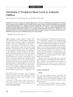
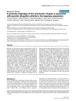
(page number not for citation purposes)
changes in MAP and changes in liver total blood flow in both
groups (Figure 2).
Oxygen delivery and consumption
Systemic oxygen delivery significantly increased from baseline
to the end of the experiment in both groups (P < 0.03 for both
groups), but end values were significantly higher in the nore-
pinephrine-treated animals than in the control group (P =
0.001). Systemic oxygen consumption increased in both
groups (P < 0.05 for both groups) but with no differences
between them. Accordingly, systemic oxygen extraction
decreased over time only in the norepinephrine-treated group
(P = 0.013). Hepatosplanchnic oxygen delivery and consump-
tion did not change over time and were not different between
groups (Table 3).
Lactate handling
For lactate exchange calculations, in three pigs (two control
pigs and one norepinephrine-treated animal) the portal vein
was not sampled. Lactate values were similar in all measured
vessels at baseline (Table 4). Afterward, arterial and hepatic
vein lactate levels increased over time in both groups (P =
0.028 for both vessels; Table 4). Norepinephrine-treated ani-
mals exhibited a significant increase in the hepatic lactate
influx (P = 0.028), whereas the hepatic lactate uptake exhib-
ited a tendency to decrease only in the control group (P =
0.07). Accordingly, the hepatic lactate extraction ratio
decreased over time in both groups (Table 4).
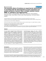
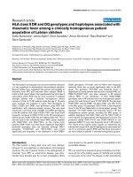
Liver mitochondrial function
At the end of the experiment, norepinephrine-treated animals
exhibited higher levels of liver mitochondria respiration than
did control animals, as indicated by higher values of their RCR
for complex I (3.5 [2.1 to 5.7] for controls animals versus 5.8
[4.8 to 6.4] for norepinephrine-treated animals; P = 0.015)
and a tendency toward higher RCR for complex II (3.1 [2.3 to
3.8] for controls versus 3.7 [3.3 to 4.6] for norepinephrine-
treated animals; P = 0.09). Also, the maximal liver mitochon-
drial respiration (state 3) for complex I was significantly higher
in the norepinephrine-treated group (343 [181 to 422] pmol/
Table 2
Time evolution of systemic hemodynamics and regional blood flows
Parameter n Baseline 2 hours 4 hours 6 hours 8 hours 10 hours P
a
Cardiac index (ml/
minute per kg)
Control 6 99 (72–112) 104
(79–172)
71 (55–103) 72 (51–167) 88 (82–232) 117
(110–232)
0.002
NE 7 107
(84–132)
96 (72–150) 70 (57–195) 103
(52–157)
100
(88–190)
161
(147–340)
b
0.001
Heart rate (beats/min) Control 6 122
(84–134)
120
(102–200)
126
(98–171)
124
(95–187)
122
(94–176)
128 (96–176) 0.7
NE 7 105
(89–139)
122
(92–174)
120
(102–197)
123
(103–171)
157
(113–185)
202
(170–223)
b
0.006
SVI (cardiac index/
heart rate)
Control 6 0.77
(0.6–1.3)
0.88
(0.7–0.9)
0.57
(0.4–0.7)
0.58
(0.4–0.8)
0.81
(0.6–1.3)
0.94
(0.8–1.3)
0.003
NE 7 0.95
(0.8–1.3)
0.86
(0.5–1.1)
0.66 (0.5–1) 0.86
(0.4–0.9)
0.81
(0.6–1.1)
0.76
(0.7–1.7)
0.045
MPAP (mmHg) Control 6 12.5 (11–14) 25.5 (16–34) 29.5 (24–42) 29 (22–37) 30.5 (19–36) 25.5 (21–36) 0.018
NE 7 12 (9–18) 29 (18–41) 30 (24–38) 24 (21–36) 30 (19–56) 25 (19–49) 0.003
PCWP (mmHg) Control 6 2 (1–3) 6 (4–6) 5 (4–8) 4 (3–6) 5 (3–12) 3 (3–5) 0.016
NE 7 3 (0–7) 7 (3–11) 6 (4–9) 5 (4–7) 6 (4–9) 5 (2–7) 0.004
Oxygenation index
(mmHg/%)
Control 6 475
(445–512)
432
(354–496)
380
(316–507)
372
(313–413)
369
(272–410)
349
(326–398)
0.002
NE 7 488
(407–548)
420
(367–445)
388
(294–453)
382
(317–496)
353
(195–478)
325
(222–390)
0.001
Hepatic artery flow
(ml/minute per kg)
Control 5 3.7
(1.7–11.5)
2.6 (2–7.6) 1.1 (0.3–1.9) 2.1 (0.5–2.9) 4.5 (0.6–5.5) 3.5 (1.7–5.8) 0.037
NE 7 3.9 (0.5–7.9) 1.4 (0.9–6.5) 1.3 (0.1–2.7) 1.2 (0.2–7.2) 2.8 (0.6–7.3) 3 (0.3–8.9) 0.002
Portal vein flow (ml/
minute per kg)
Control 6 20 (16–22) 16.9 (12–23) 11.5 (6–14) 11.9 (9–16) 16.2 (10–32) 18.6 (13–32) <0.001
NE 7 19.6 (12–26) 13.8 (10–20) 11.6 (8–16) 14 (10–22) 15.8 (12–27) 19.1 (12–31) 0.001
Data are presented as median (range).
a
Friedman test for each group and variable in time.
b
Mann Whitney U-test between groups at the
corresponding time point. MPAP, mean pulmonary artery pressure; NE, norepinephrine group; oxygenation index, arterial oxygen tension/fractional
of inspired oxygen; PCWP, pulmonary capillary wedge pressure; SVI, stroke volume index.
Critical Care Vol 12 No 4 Regueira et al.
Page 6 of 10
(page number not for citation purposes)
second per mg for controls versus 539 [340 to 879] pmol/
second per mg for norepinephrine-treated animals; P = 0.026;
Figure 3). Resting respiration (state 4; complex I: 86 [60 to
138] pmol/second per mg for controls versus 96 [62 to 151]
pmol/second per mg for norepinephrine-treated animals [P =
0.31]; and complex II: 231 [193 to 623] pmol/second per mg
for controls versus 276 [174 to 283] pmol/second per mg for
norepinephrine-treated animals [P = 0.39]; Figure 3) and cit-
rate synthase activity (12.6 [11.1 to 17.0] versus 16.6 [11.3 to
20.5], respectively [P = 0.3]) were similar between groups.
Previous results in similar control and endotoxin-treated ani-
mals are compared with the current results in Table 5.
Discussion
In our model, endotoxaemia was associated with increasing
levels of cardiac index and mean pulmonary artery pressure,
whereas addition of norepinephrine was associated with
higher levels of MAP and further increases in cardiac index,
heart rate and systemic oxygen delivery. Despite this, nore-
pinephrine treatment was not associated with changes in
stroke volume, total liver blood flow, or hepatosplanchnic oxy-
gen consumption, or with an improvement in the hepatic lac-
tate exchange. Surprisingly, with unaltered hepatic oxygen
transport, the animals treated with norepinephrine exhibited an
increase in efficiency of their liver mitochondrial respiration
when compared with septic animals not treated with
norepinephrine.
The haemodynamic and metabolic effects of norepinephrine
during sepsis are controversial, several studies have been per-
formed to provide insight. LeDoux and coworkers [22] studied
10 patients with septic shock and found that increasing MAP
from 65 to 85 mmHg with norepinephrine was related to sig-
nificant increases in cardiac index, left ventricular stroke work
index, heart rate and systemic oxygen delivery. However, it was
not associated with changes in systemic oxygen consumption,
lactate concentrations, capillary blood flow, urine output, or
gastric mucosal partial carbon dioxide tension. This suggests
that higher blood pressure levels are not associated with bet-
ter organ perfusion during sepsis. Similarly, our data reveal no
consistent relationship between MAP levels and liver blood
flow, and norepinephrine was not associated with a greater
increase in systemic oxygen consumption in comparison with
septic animals not receiving norepinephrine.
Other studies, however, have identified some beneficial
effects of norepinephrine during sepsis. For example, Treggiari
and coworkers [20], in an animal model, showed that increas-
ing MAP with norepinephrine by 10 mmHg above the baseline
level of 50 mmHg was associated with an increase in portal
vein flow and almost restoration of renal and mucosal flows to
pre-shock levels. Further increases in MAP were not associ-
ated with more benefit. Also, one crossover study [16]
conducted in septic patients compared replacement of
dopamine with norepinephrine to achieve a MAP goal of 80
mmHg. This study revealed that the drugs were associated
with similar splanchnic blood flow and hepatic oxygen con-
sumption, but that patients treated with norepinephrine exhib-
ited higher levels of hepatic lactate uptake. This suggests that
norepinephrine may improve liver metabolic function during
sepsis independent of its regional haemodynamic effects. This
is in contrast to our findings in septic pigs, in which norepine-
phrine was associated with an increase in the hepatic lactate
influx with no concomitant increase in the hepatic lactate
uptake, suggesting that the capacity of the liver to increase the
Figure 1
Evolution of MAPEvolution of MAP. Presented is the evolution of mean arterial pressure
(MAP) during the experiment in the control group (black dotted line)
and in the norepinephrine-treated group (black line). The table shows
the number of pigs receiving norepinephrine and the average dose for
each time point. Only the norepinephrine group exhibited a significant
increase in MAP (
&
Friedman test; P = 0.019). Accordingly, values at 8
and 10 hours were higher in this group (*Mann Whitney U-test; P <
0.05 for both).
Figure 2
Relation between MAP and liver total flowRelation between MAP and liver total flow. Each line shows the evolu-
tion for each pig during the experiment, from baseline to end values. No
consistent relationship between changes in mean arterial pressure
(MAP) and changes in liver total blood flow was observed in either
group.
Available online />Page 7 of 10
(page number not for citation purposes)
lactate uptake was exhausted and not improved by
norepinephrine.
Our study shows that both complex I and II respiratory effi-
ciency was increased by the use of norepinephrine during
endotoxaemia in pigs. Septic pigs not treated with norepine-
phrine exhibited decreased RCR (an index of respiratory effi-
ciency), similar to those in septic pigs from previous studies
[24,29,30] (Table 5). In the present study the addition of nore-
pinephrine was associated with liver mitochondria RCRs for
complexes I and II similar to those reproted previously by us
and others in nonseptic control pigs, suggesting that nore-
pinephrine may restore respiratory efficiency (Table 5). Respi-
ratory control depends on the presence of a chemiosmotic
gradient generated by the coupled passage of protons from
the mitochondrial matrix to the mitochondrial inter-membrane
space during the electron flux through the electron transport
chain. The presence and magnitude of this chemiosmotic gra-
dient regulates and limits the flux of electrons through the elec-
tron transport chain, and secondarily mitochondrial oxygen
consumption. In our study, both groups exhibited similar
increased values for their state 4 respiration (resting respira-
tion, which is mitochondrial oxygen consumption by isolated
mitochondria induced by a particular substrate, in the absence
of ADP) [29]. This result confirms that during endotoxaemia
the mitochondrial membrane is damaged, with a secondary
increase in the loss of protons back to the matrix or to the cyto-
plasmatic space, which is not coupled with ATP production.
This reduction in the chemiosmotic gradient is coupled with an
increase in the electron flux and the oxygen consumption,
Table 3
Time evolution of oxygen transport variables
Parameter n Baseline End P
a
DO
2
systemic (ml/minute per kg) Control 6 12.8 (11–15) 16.3 (15–24) 0.028
NE 7 16.4 (12–19) 29.3 (25–52)
b
0.018
V
O
2
systemic (ml/minute per kg) Control 5 4.5 (3–5) 5.7 (4–11) 0.043
NE 7 5.2 (3–7) 6.9 (6–10) 0.028
D
O
2
hepatosplanchnic (ml/minute per kg) Control 6 3.1 (2.6–4.1) 3.2 (1.8–3.9) 0.46
NE 7 3.7 (2.3–4.2) 3.3 (2.7–5.7) 0.4
V
O
2
hepatosplanchnic (ml/minute per kg) Control 6 1.5 (0.6–2.1) 1.8 (0.6–2.2) 0.46
NE 7 1.2 (0.8–1.7) 1.4 (0.5–2.1) 0.4
Data are presented as median (range).
a
Wilcoxon test for each group and variable in time.
b
Mann Whitney U-test between groups at the
corresponding time point. NE, norepinephrine; D
O
2
, oxygen delivery; VO
2
, oxygen consumption.
Table 4
Time evolution of lactate concentrations and hepatic lactate exchange
Parameter n Baseline End P
a
Arterial lactate (mmol/l) Control 6 0.67 (0.3–0.7) 0.92 (0.6–1.1) 0.028
NE 7 0.66 (0.4–0.8) 1.1 (0.7–2.1) 0.028
Hepatic vein lactate (mmol/l) Control 6 0.45 (0.4–0.6) 0.71 (0.5–1.1) 0.028
NE 6 0.41 (0.3–0.5) 0.89 (0.7–1.7) 0.028
Portal vein lactate (mmol/l) Control 4 0.74 (0.6–0.9) 1 (0.6–1.23) 0.14
NE 7 0.68 (0.5–0.9) 1.1 (0.9–1.9) 0.028
Hepatic lactate influx (μmol/minute per kg) Control 4 18.1 (15–21) 21.8 (15–24) 0.3
NE 6 18.3 (8–24) 38.5 (14–42) 0.028
Hepatic lactate uptake (μmol/minute per kg) Control 4 7.4 (7–12) 4.1 (0.3–10) 0.07
NE 6 6.6 (4–11) 5.7 1.7–13) 0.6
Hepatic lactate extraction ratio (%) Control 4 44 (38–60) 22 (2–41) 0.05
NE 6 40 (29–51) 17 (9–34) 0.046
Data are presented as median (range).
a
Wilcoxon test for each group and variable in time. NE, norepinephrine.
Critical Care Vol 12 No 4 Regueira et al.
Page 8 of 10
(page number not for citation purposes)
which was not prevented by norepinephrine (Table 5).
The observed improvement in both complex I-dependent and
complex II-dependent RCRs in septic animals treated with
norepinephrine, into the normal range, in spite of their
increased resting respiration (decreased chemiosmotic gradi-
ent), was mainly due to an increase in their mitochondrial state
3 respiration (maximal respiration, which is mitochondrial oxy-
gen consumption under saturating ADP concentrations stimu-
lated by a particular combination of substrates).
Norepinephrine-treated septic pigs exhibited complex I-
dependent state 3 respiration values that were higher than
those of septic pigs that did not receive norepinephrine, and
higher than those of control pigs from our previous experi-
ments [29] (Table 5). This suggests that norepinephrine, by
some mechanism, improves the rate of oxygen consumption in
the presence of an excess amount of ADP. The RCR is con-
sidered to indicate the degree of coupling, especially when
basal state 4 respiration changes at different conditions. In our
study, state 4 respiration was not different between groups,
indicating that proton leak was unaffected by the different
treatment. Therefore, state 3 respiration and RCR represent
almost exactly the same phenomena, and no further informa-
tion is gained by the concomitant change in RCR.
Table 5
Liver complex I-dependent mitochondrial respiration
Previous controls
a
Previous septic
a
Current septic Current septic + NE
State 3 343 (324–371) 247 (204–296) 343 (181–422) 539 (340–879)
State 4 61 (55–74) 90 (74–101) 86 (60–138) 96 (62–151)
RCR (state 3/state 4) 5.6 (5–6) 2.8 (2.4–3.2) 3.5 (2.1–5.7) 5.8 (4.8–6.4)
Shown are is a comparison of liver complex I-dependent mitochondrial respiration with previous results from similarly instrumented animals with
and without endotoxin exposure. Data are presented as median (range).
a
Data from previous study [29]: pigs were randomized for 12 hours to
control or endotoxaemia (0.4 μg/kg per hour) and were instrumented similar to the present study.
Figure 3
Complex I-dependent and complex II-dependent liver mitochondrial respirationComplex I-dependent and complex II-dependent liver mitochondrial respiration. State 3: equivalent to maximal mitochondrial respiration. P values
from unpaired t-test comparison between groups. Data obtained by high-resolution Oxygraph (Oroboros, DatLab software for data acquisition and
analysis, Graz, Austria).
Available online />Page 9 of 10
(page number not for citation purposes)
Previous studies conducted in isolated or perfused livers from
nonseptic small animals have shown that norepinephrine
increases cellular respiration in a dose-dependent manner,
and that this effect is blocked by the addition of an α-antago-
nist (for example, phenoxybenzamine), but not by the addition
of a β-antagonist (for example, propanolol). It has also been
shown that the increase in cellular respiration depends on the
extracellular calcium concentration [27]. Even more, exposure
of isolated hepatocytes to physiological concentrations of
norepinephrine is related to an increase in cytosolic calcium
levels and to an active transport of calcium into the mitochon-
drial matrix [31]. In liver cells, calcium can stimulate three dif-
ferent dehydrogenases of the citrate cycle, increasing the
substrate availability of NADH to the respiratory chain and thus
increasing mitochondrial respiration [26]. However, it has also
been suggested that an increase in mitochondrial oxygen con-
sumption associated with the increase in calcium concentra-
tions may be related to an increase in mitochondrial membrane
potential and a secondary increase in reactive oxygen species
production [32,33].
A limitation of our study was the normotensive haemodynamic
situation of the groups, although in many ICUs norepinephrine
would be used in this clinical situation. A further limitation is
the lack of assessment of the liver microcirculation, which can
be influenced by norepinephrine, improving cellular oxygen
delivery, although mitochondrial dysfunction may occur even in
the absence of tissue hypoxia [23,34]. A third imitation is the
lack of liver ATP levels, which should be addressed in future
studies. A final limitation is the fact that it is difficult to establish
whether the observed changes in liver mitochondrial respira-
tion are a result of the increased blood pressure or a direct
effect of the compound norephinephrine. In our opinion, a
direct effect of blood pressure is unlikely; rather, an effect of
concomitantly increased oxygen delivery may be expected.
However, neither hepatic oxygen delivery nor consumption
was altered by the addition of norepinephrine. We recently
showed that adding more volume in our model of sepsis leads
to similar pressures [35] and that mitochondrial function is
also impaired by volume [36].
Conclusion
Our study shows that norepinephrine treatment, using clini-
cally relevant doses that are commonly applied in patients with
sepsis [11,37], during endotoxaemia to control blood pres-
sure improves liver mitochondria complex I-dependent and
complex II-dependent efficiency of respiration. This effect was
mainly explained by an increase in liver mitochondria maximal
respiration and was probably mediated by a direct effect of
norepinephrine on liver cells.
Competing interests
The authors declare that they have no competing interests.
Authors' contributions
SMJ and JT devised the study protocol. TR, BB, SMJ, SB, SD
and JG initiated and performed all animal experiments. TR, SD
and PL performed mitochondria-related experiments. TR and
SMJ analyzed the data. All the authors contributed to and
approved the final manuscript.
Acknowledgements
We thank Timo Nannen, Daniel Mettler, Daniel Zalokar and Olgica
Beslac for their assistance during the experiments and Jeannie Wurz for
English editing.
This study was supported by grant 3200BO/102268 from the Swiss
National Fund and a grant from the 'Stiftung für die Forschung in
Anästhesiologie und Intensivmedizin' awarded to PL.
The study was performed at the Experimental Surgical Unit of the
Department of Clinical Research, University of Bern.
References
1. Varpula M, Tallgren M, Saukkonen K, Voipio-Pulkki LM, Pettila V:
Hemodynamic variables related to outcome in septic shock.
Intensive Care Med 2005, 31:1066-1071.
2. Dellinger RP, Levy MM, Carlet JM, Bion J, Parker MM, Jaeschke R,
Reinhart K, Angus DC, Brun-Buisson C, Beale R, Calandra T, Dhai-
naut JF, Gerlach H, Harvey M, Marini JJ, Marshall J, Ranieri M, Ram-
say G, Sevransky J, Thompson BT, Townsend S, Vender JS,
Zimmerman JL, Vincent JL: Surviving Sepsis Campaign: Interna-
tional guidelines for management of severe sepsis and septic
shock: 2008. Crit Care Med 2008, 36:296-327.
3. Beale RJ, Hollenberg SM, Vincent JL, Parrillo JE: Vasopressor
and inotropic support in septic shock: an evidence-based
review. Crit Care Med 2004, 32:S455-S465.
4. Sevransky JE, Nour S, Susla GM, Needham DM, Hollenberg S,
Pronovost P: Hemodynamic goals in randomized clinical trials
in patients with sepsis: a systematic review of the literature.
Crit Care 2007, 11:R67.
Key messages
• Endotoxaemia was associated with increased levels of
cardiac index and mean pulmonary arterial pressure,
whereas the addition of norepinephrine was associated
with higher levels of MAP and further increases in car-
diac index and systemic oxygen delivery.
• Norepinephrine treatment was not associated with
changes in the stroke volume, in hepatosplanchnic oxy-
gen consumption, or an improvement in the hepatic lac-
tate exchange.
• No consistent relationship between changes in MAP
and changes in liver total blood flow were observed.
• Norepinephrine-treated animals exhibited increased effi-
ciency in liver mitochondrial respiration when compared
with septic animals with no norepinephrine.
• The improvement in liver mitochondrial respiration was
mainly accounted for by an increase in their maximal
respiration and was probably mediated by a direct
effect of norepinephrine on liver cells.
Critical Care Vol 12 No 4 Regueira et al.
Page 10 of 10
(page number not for citation purposes)
5. Cole L, Bellomo R, Hart G, Journois D, Davenport P, Tipping P,
Ronco C: A phase II randomized, controlled trial of continuous
hemofiltration in sepsis. Crit Care Med 2002, 30:100-106.
6. Rivers E, Nguyen B, Havstad S, Ressler J, Muzzin A, Knoblich B,
Peterson E, Tomlanovich M: Early goal-directed therapy in the
treatment of severe sepsis and septic shock. N Engl J Med
2001, 345:1368-1377.
7. Boldt J, Papsdorf M, Piper SN, Rothe A, Hempelmann G: Contin-
uous heparinization and circulating adhesion molecules in the
critically ill. Shock 1999, 11:13-18.
8. Briegel J, Forst H, Haller M, Schelling G, Kilger E, Kuprat G, Hem-
mer B, Hummel T, Lenhart A, Heyduck M, Stoll C, Peter K: Stress
doses of hydrocortisone reverse hyperdynamic septic shock:
a prospective, randomized, double-blind, single-center study.
Crit Care Med 1999, 27:723-732.
9. Alia I, Esteban A, Gordo F, Lorente JA, Diaz C, Rodriguez JA, Fru-
tos F: A randomized and controlled trial of the effect of treat-
ment aimed at maximizing oxygen delivery in patients with
severe sepsis or septic shock. Chest 1999, 115:453-461.
10. Spapen H, Zhang H, Demanet C, Vleminckx W, Vincent JL, Huygh-
ens L: Does N-acetyl-L-cysteine influence cytokine response
during early human septic shock? Chest 1998,
113:1616-1624.
11. Bakker J, Grover R, McLuckie A, Holzapfel L, Andersson J, Lodato
R, Watson D, Grossman S, Donaldson J, Takala J: Administration
of the nitric oxide synthase inhibitor NG-methyl-L-arginine
hydrochloride (546C88) by intravenous infusion for up to 72
hours can promote the resolution of shock in patients with
severe sepsis: results of a randomized, double-blind, placebo-
controlled multicenter study (study no. 144-002). Crit Care
Med 2004, 32:1-12.
12. Lopez A, Lorente JA, Steingrub J, Bakker J, McLuckie A, Willatts S,
Brockway M, Anzueto A, Holzapfel L, Breen D, Silverman MS,
Takala J, Donaldson J, Arneson C, Grove G, Grossman S, Grover
R: Multiple-center, randomized, placebo-controlled, double-
blind study of the nitric oxide synthase inhibitor 546C88: effect
on survival in patients with septic shock. Crit Care Med 2004,
32:21-30.
13. Clark MA, Plank LD, Connolly AB, Streat SJ, Hill AA, Gupta R,
Monk DN, Shenkin A, Hill GL: Effect of a chimeric antibody to
tumor necrosis factor-alpha on cytokine and physiologic
responses in patients with severe sepsis: a randomized, clini-
cal trial. Crit Care Med 1998, 26:1650-1659.
14. Meier-Hellmann A, Specht M, Hannemann L, Hassel H, Bredle DL,
Reinhart K: Splanchnic blood flow is greater in septic shock
treated with norepinephrine than in severe sepsis.
Intensive
Care Med 1996, 22:1354-1359.
15. Di Giantomasso D, May CN, Bellomo R: Norepinephrine and vital
organ blood flow. Intensive Care Med 2002, 28:1804-1809.
16. Guerin JP, Levraut J, Samat-Long C, Leverve X, Grimaud D, Ichai
C: Effects of dopamine and norepinephrine on systemic and
hepatosplanchnic hemodynamics, oxygen exchange, and
energy balance in vasoplegic septic patients. Shock 2005,
23:18-24.
17. Ruokonen E, Takala J, Kari A, Saxen H, Mertsola J, Hansen EJ:
Regional blood flow and oxygen transport in septic shock. Crit
Care Med 1993, 21:1296-1303.
18. Ruokonen E, Takala J, Uusaro A: Effect of vasoactive treatment
on the relationship between mixed venous and regional oxy-
gen saturation. Crit Care Med 1991, 19:1365-1369.
19. Sautner T, Wessely C, Riegler M, Sedivy R, Gotzinger P, Losert U,
Roth E, Jakesz R, Fugger R: Early effects of catecholamine ther-
apy on mucosal integrity, intestinal blood flow, and oxygen
metabolism in porcine endotoxin shock. Ann Surg 1998,
228:239-248.
20. Treggiari MM, Romand JA, Burgener D, Suter PM, Aneman A:
Effect of increasing norepinephrine dosage on regional blood
flow in a porcine model of endotoxin shock. Crit Care Med
2002, 30:1334-1339.
21. Revelly JP, Liaudet L, Frascarolo P, Joseph JM, Martinet O, Markert
M: Effects of norepinephrine on the distribution of intestinal
blood flow and tissue adenosine triphosphate content in
endotoxic shock. Crit Care Med 2000, 28:2500-2506.
22. LeDoux D, Astiz ME, Carpati CM, Rackow EC: Effects of per-
fusion pressure on tissue perfusion in septic shock. Crit Care
Med 2000, 28:2729-2732.
23. Crouser ED, Julian MW, Blaho DV, Pfeiffer DR: Endotoxin-
induced mitochondrial damage correlates with impaired respi-
ratory activity. Crit Care Med 2002, 30:276-284.
24. Porta F, Takala J, Weikert C, Bracht H, Kolarova A, Lauterburg BH,
Borotto E, Jakob SM: Effects of prolonged endotoxemia on
liver, skeletal muscle and kidney mitochondrial function. Crit
Care 2006, 10:R118.
25. Brealey D, Brand M, Hargreaves I, Heales S, Land J, Smolenski R,
Davies NA, Cooper CE, Singer M: Association between mito-
chondrial dysfunction and severity and outcome of septic
shock. Lancet 2002, 360:219-223.
26. Brown GC: Control of respiration and ATP synthesis in mam-
malian mitochondria and cells. Biochem J 1992, 284:1-13.
27. Binet A, Claret M: alpha-adrenergic stimulation of respiration in
isolated rat hepatocytes. Biochem J 1983, 210:867-873.
28. Johnson D: Isolation of liver and kidney mitochondria. In Meth-
ods in Enzymology Edited by: Estabrook R. New York, NY: Aca-
demic Press; 1967:94-96.
29. Porta F, Takala J, Kolarova A, Ma Y, Redaelli CA, Brander L, Bracht
H, Jakob SM: Oxygen extraction in pigs subjected to low-dose
infusion of endotoxin after major abdominal surgery. Acta
Anaesthesiol Scand 2005, 49:627-634.
30. Katoh H, Ohkohchi N, Hirano T, Sakurada M, Orii T, Koyamada N,
Fujimori K, Takemura M, Endoh T, Satomi S, Taguchi Y, Mori S:
Viability of partial liver graft from living donor in pigs. Tohoku
J Exp Med 1995, 175:179-184.
31. Poggioli J, Berthon B, Claret M: Calcium movements in in situ
mitochondria following activation of alpha-adrenergic recep-
tors in rat liver cells. FEBS Lett 1980, 115:243-246.
32. Lee I, Bender E, Kadenbach B: Control of mitochondrial mem-
brane potential and ROS formation by reversible phosphoryla-
tion of cytochrome c oxidase. Mol Cell Biochem 2002, 234-
235:63-70.
33. Kozlov A, Staniek K, Haindl S, Piskernik C, Ohlinger W, Gille L,
Nohl H, Bahrami S, Redl H: Different effects of endotoxic shock
on the respiratory function of liver and heart mitochondria in
rats. Am J Physiol Gastrointest Liver Physiol 2006,
290:G543-G549.
34. Rosser DM, Stidwill RP, Jacobson D, Singer M: Oxygen tension
in the bladder epithelium rises in both high and low cardiac
output endotoxemic sepsis. J Appl Physiol 1995,
79:1878-1882.
35. Brandt S, Eleftheriadis A, Regueira T, Bracht H, Gorrasi J, Takala
J, Jakob S: Aggressive and moderate fluid resuscitation in sep-
tic pigs: consequences on morbidity.
Crit Care 2007, 11:P28.
36. Regueira T, Borotto E, Brandt S, Bracht H, Gorrasi J, Lepper P,
Takala J, Jakob S: Effects of volume resuscitation on hepat-
osplanchnic oxygen consumption, liver mitochondrial function
and mortality in endotoxemia [abstract]. Crit Care 2007,
11:P29.
37. Albanese J, Leone M, Garnier F, Bourgoin A, Antonini F, Martin C:
Renal effects of norepinephrine in septic and nonseptic
patients. Chest 2004, 126:534-539.