Báo cáo y học: "Induction of Bim and Bid gene expression during accelerated apoptosis in severe sepsis" potx
Bạn đang xem bản rút gọn của tài liệu. Xem và tải ngay bản đầy đủ của tài liệu tại đây (353.14 KB, 10 trang )
Open Access
Available online />Page 1 of 10
(page number not for citation purposes)
Vol 12 No 5
Research
Induction of Bim and Bid gene expression during accelerated
apoptosis in severe sepsis
Stefan U Weber
1
*, Jens-Christian Schewe
1
*, Lutz E Lehmann
1
, Stefan Müller
1
, Malte Book
1
,
Sven Klaschik
2
, Andreas Hoeft
1
and Frank Stüber
3
1
Department of Anesthesiology and Intensive Care Medicine, University Bonn Medical Center, Sigmund-Freud-Straße 25, 53129 Bonn, Germany
2
Center for Biologics Evaluation and Research (CBER), Food and Drug Administration, 1401 Rockville Pike, Suite 200N, Rockville, MD 20852-1448,
USA
3
Department of Anaesthesiology and Pain Therapy, University Hospital Bern "Inselspital", 3010 Bern, Switzerland
* Contributed equally
Corresponding author: Stefan U Weber,
Received: 19 Jun 2008 Revisions requested: 14 Jul 2008 Revisions received: 29 Aug 2008 Accepted: 16 Oct 2008 Published: 16 Oct 2008
Critical Care 2008, 12:R128 (doi:10.1186/cc7088)
This article is online at: />© 2008 Weber et al.; licensee BioMed Central Ltd.
This is an open access article distributed under the terms of the Creative Commons Attribution License ( />),
which permits unrestricted use, distribution, and reproduction in any medium, provided the original work is properly cited.
Abstract
Introduction In transgenic animal models of sepsis, members of
the Bcl-2 family of proteins regulate lymphocyte apoptosis and
survival of sepsis. This study investigates the gene regulation of
pro-apoptotic and anti-apoptotic members of the Bcl-2 family of
proteins in patients with early stage severe sepsis.
Methods In this prospective case-control study, patients were
recruited from three intensive care units (ICUs) in a university
hospital. Sixteen patients were enrolled when they fulfilled the
criteria of severe sepsis. Ten critically ill but non-septic patients
and 11 healthy volunteers served as controls. Blood samples
were immediately obtained at inclusion. To confirm the presence
of accelerated apoptosis in the patient groups, caspase-3
activation and phosphatidylserine externalisation in CD4+,
CD8+ and CD19+ lymphocyte subsets were assessed using
flow cytometry. Specific mRNAs of Bcl-2 family members were
quantified from whole blood by real-time PCR. To test for
statistical significance, Kruskal-Wallis testing with Dunn's
multiple comparison test for post hoc analysis was performed.
Results In all lymphocyte populations caspase-3 (p < 0.05) was
activated, which was reflected in an increased
phosphatidylserine externalisation (p < 0.05). Accordingly,
lymphocyte counts were decreased in early severe sepsis. In
CD4+ T-cells (p < 0.05) and B-cells (p < 0.001) the Bcl-2
protein was decreased in severe sepsis. Gene expression of the
BH3-only Bim was massively upregulated as compared with
critically ill patients (p < 0.001) and 51.6-fold as compared with
healthy controls (p < 0.05). Bid was increased 12.9-fold
compared with critically ill patients (p < 0.001). In the group of
mitochondrial apoptosis inducers, Bak was upregulated 5.6-
fold, while the expression of Bax showed no significant
variations. By contrast, the pro-survival members Bcl-2 and Bcl-
xl were both downregulated in severe sepsis (p < 0.001 and p
< 0.05, respectively).
Conclusions In early severe sepsis a gene expression pattern
with induction of the pro-apoptotic Bcl-2 family members Bim,
Bid and Bak and a downregulation of the anti-apoptotic Bcl-2
and Bcl-xl proteins was observed in peripheral blood. This
constellation may affect cellular susceptibility to apoptosis and
complex immune dysfunction in sepsis.
Introduction
There has been accumulating evidence both in animals and
humans, that apoptosis of lymphocytes [1,2], monocytes [3],
dendritic cells [4] and gut epithelial cells [5] is accelerated in
severe sepsis and contributes to temporary immunosuppres-
sion [6]. Sepsis is a systemic disorder driven by a dysregula-
tion of the immune system, that clearly displays pro-
inflammatory as well as anti-inflammatory signatures [7]. The
7-AAD: 7-actinoaminomycin; APACHE II:Acute Physiology and Chronic Health Evaluation II; bp: base pair; BSA: boveine serum albumin; dNTP: deox-
yribonucleotide triphosphate; EDTA: ethylenediaminetetraacetic acid; ICU: intensive care unit; IL: interleukin; PBS: phosphate buffered saline; PCR:
polymerase chain reaction; SAPS II: Simplified Acute Physiology Score II; SOFA: Sepsis related Organ Failure Assessment.
Critical Care Vol 12 No 5 Weber et al.
Page 2 of 10
(page number not for citation purposes)
temporary immunosuppression during sepsis is still consid-
ered an unsolved problem in the treatment of sepsis because
during this phase, patients often succumb to secondary infec-
tions, caused by pathogens that are not necessarily virulent in
healthy individuals [6]. In multiple animal models of sepsis,
inhibition of lymphocyte apoptosis has been shown to dramat-
ically improve survival [5,8-10].
Apoptosis is a type of cell death that follows a tightly regulated
program. It results in the disposal of the apoptotic cell without
spilling toxic intracellular components and is the central mech-
anism for tissue homeostasis during development [11]. The
cell death program may be activated via a membrane bound
pathway, where the signal is initiated by so called 'death
receptors' [12] or via the mitochondrion [13]. Pro-apoptotic
compounds are usually sequestered in the intermembrane
space [14]. On permeabilisation of the outer mitochondrial
membrane, these proteins are released into the cytosol form-
ing the apoptosome and subsequently activating caspase-9.
Ultimately, both pathways converge in the activation of cas-
pase-3, the caspase that is the main executioner, [15] which
has multiple targets and degrades structural and functional
proteins [11].
The integrity of the mitochondrial membrane is controlled by
members of the Bcl-2 protein family [16]. Bcl-2 is the proto-
type of a large family of related proteins displaying pro-apop-
totic or anti-apoptotic properties. The related proteins are
divided into three groups: anti-apoptotic Bcl-2-like proteins
such as Bcl-2 and Bcl-xl, which share four regions of homol-
ogy, the BH1-4 domains; pro-apoptotic Bax-like proteins shar-
ing only domains 1–3 [17]; and the group of BH3-only
proteins, which contain only domain 3 and function as sensors
and transducers of apoptotic signals (eg, Bim and Bid) [16].
The proteins Bak and Bax form oligomers that perturb the
outer mitochondrial membrane. To keep them inactive they are
bound by pro-survival guards [18]. According to the displace-
ment model, binding of the mobile BH3-only protein Bim to
pro-survival members of the Bcl-2 family liberates Bak/Bax
from their guards, thereby rendering them active [16].
Several lines of evidence point to the involvement of the mito-
chondrion and the Bcl-family of proteins in the induction of
lymphocyte apoptosis during sepsis. Overexpression of Bcl-2
or adoptive transfer of Bcl-2 overexpressing lymphocytes in
murine models reduces mortality from caecal ligation punc-
ture-induced sepsis [19]. In patients, Bcl-2 expression was
found to be decreased during sepsis [1,20,21]. Recently, it
was shown in animal models, that BH3-only proteins are
involved in the induction of apoptosis during sepsis: Bim -/-
and Bid -/- mice exhibited both reduced apoptosis and
improved survival in murine sepsis as compared with respec-
tive wild-type animals [22].
Taking into account the importance of Bcl-2 family members
for the transduction of apoptosis signals in animal models, we
hypothesised that diverse members of the Bcl-2 family under-
lie transcriptional regulation in humans during sepsis. There-
fore, we studied the gene expression of important members of
Bcl-2-like, Bax-like and BH3-only proteins of the Bcl-2 family
during a phase of accelerated lymphocyte apoptosis in human
patients experiencing severe sepsis. The presence of acceler-
ated apoptosis in the study collective was confirmed by phos-
phatidylserine externalisation and caspase-3 activation in
lymphocyte subpopulations.
Materials and methods
Patients and controls
This study was conducted with approval from the ethics board
of the University of Bonn, Germany. Patients were included
after written informed consent was given by them or their next-
of-kin. The study included 16 patients who had been treated in
three surgical intensive care units (ICUs) at the university hos-
pital of Bonn after a diagnosis of severe sepsis according to
the American College of Chest Physicians/Society of Critical
Care Medicine consensus criteria [23]. A blood sample was
obtained from each patient on the day of inclusion. Ten
patients treated in the ICU without sepsis or signs of infection
served as controls. For both groups exclusion criteria were a
lack of informed consent, age under 18 years, and preexisting
haematological or immunological disease. The basic clinical
characteristics of the critically ill patients and patients with
severe sepsis (Table 1) have been published previously in part
[24]. As a second control group, 11 healthy subjects (six
males, five females, median age = 34 years) were tested. All
patients were Caucasian.
Sample acquisition and storage
Blood samples were obtained within four hours after severe
sepsis was first diagnosed to sample early stage severe sep-
sis. For flow cytometry, samples were anticoagulated with eth-
ylenediaminetetraacetic acid (EDTA) and processed within 30
minutes. For RNA quantification, 2.5 ml whole blood was col-
lected using blood RNA tubes (Paxgene system, PreAnalytiX,
Qiagen, Hilden, Germany) and stored at -80°C until extraction.
This method stabilised the RNA until further analysis.
Phosphatidylserine externalisation
Phosphatidylserine externalisation was measured by annexin V
binding. Samples of 100 μl fresh whole blood were subjected
to lysis with an ammonium chloride solution (Becton Dickin-
son, Heidelberg, Germany) for 15 minutes at room tempera-
ture to substantially lyse red blood cells without affecting the
leucocyte populations. After washing, cells were phenotyped
with phycoerythrin-labelled antibodies directed against CD4,
CD8 and CD19 (Becton Dickinson, Heidelberg, Germany).
They were then incubated with annexin-V-fluorescein-isothio-
cyanate (Becton Dickinson, Heidelberg, Germany) for 10 min-
utes at room temperature in Hepes buffer containing calcium
Available online />Page 3 of 10
(page number not for citation purposes)
(2.5 mM). Before flow cytometric analysis, cells were labelled
with the DNA-dye 7-actinoaminomycin (7-AAD; Becton Dick-
inson, Heidelberg, Germany) for the detection of membrane
leaks and subjected to data acquisition within 10 minutes.
Cells that were phosphatidylserine positive but 7-AAD nega-
tive were selected by gating.
Caspase-3 activation and Bcl-2 expression
For caspase-3 activation and Bcl-2 expression measurement,
samples of 100 μl whole blood were analysed. Erythrocytes
were lysed with 1.5 ml PharmLyse, an ammonium chloride-
based lysing reagent. (Pharmingen, Heidelberg, Germany) at
room temperature. All subsequent steps were carried out at
4°C. Cells were washed with PBS (Sigma-Aldrich, Steinheim,
Germany) and fixed with 750 μl PBS containing 4% parafor-
maldehyde (Sigma-Aldrich, Steinheim, Germany). After fixa-
tion, cells were permeabilised with 1ml PBS containing 0.5%
saponin (Sigma-Aldrich, Steinheim, Germany) and 1% BSA
(Sigma-Aldrich, Steinheim, Germany). Cells were washed
twice with PBS (0.5% saponin/1% BSA). Active caspase-3
was detected by a phycoerythrin-labelled antibody directed
against the active fragment (Becton Dickinson, Heidelberg,
Germany). Bcl-2 was detected with a specific phycoerythrin-
conjugated monoclonal antibody (Becton Dickinson, Heidel-
berg, Germany). Isotype control antibodies (Becton Dickin-
son, Heidelberg, Germany) were used to determine unspecific
binding. After washing cells were phenotyped antibodies
against CD4, CD8 and CD19 that were labelled with perid-
inin-chlorophyll-protein (PerCP; Becton Dickinson, Heidel-
berg, Germany). Cells were washed twice and resuspended in
250 μl PBS (0.5% saponin/1% BSA) for flow cytometric
analysis.
Flow cytometry
Stained and fixed cells were measured by flow cytometry. Data
were acquired in a flow cytometer (FACSCalibur, Becton
Dickinson, Heidelberg, Germany) and analysed via CellQuest
Pro software (CellQuest Pro, Version 4.0.2, Becton Dickinson,
Heidelberg, Germany). Ten-thousand cells of interest were
acquired. Populations of interest were selected by multiple
gating using forward (cell size) and sideward (cell granularity)
scatter and green or red regions on fluorescence channels.
Antibody-binding was expressed as mean fluorescence inten-
sity corrected for nonspecific binding using matched isotype
control antibodies. Results of caspase-3 staining were given
as percentages of positive cells. In case of Bcl-2 staining, daily
calibration curves were generated using latex beads coupled
to defined quantities of phycoerythrin (Quantibrite, Becton
Dickinson, Heidelberg, Germany). Fluorescence intensities
were calculated accordingly to compensate for potential devi-
ations from linearity of the cytometer and to reduce day to day
variability. Since the Bcl-2 antibody used, like most conjugated
antibodies, is not guaranteed to be coupled to phycoerythrin
in a one-to-one ratio, results were not given as molecules per
cell but as linear fluorescence units.
Table 1
Clinical data of patient groups
Critically ill non-septic n = 10 Severe sepsis n = 16 p
Age 61 +/- 5 56 +/- 4 ns
APACHE II score at inclusion 12 +/- 1 26 +/- 2 < 0.05
SAPS II score at inclusion 23 +/- 2 57 +/- 4 < 0.01
SOFA score at inclusion 3 +/- 1 15 +/- 1 < 0.001
Mechanically ventilated at inclusion (n) 6 16
Vasopressor treatment at inclusion (n) 0 13
Antibiotic treatment at inclusion (n) 8 16
Steroid dose* 3 mg/kg/24hours (n) 0 8
Serum interleukin-6 at inclusion (pg/ml) 20.77 +/- 13.37 4304 +/- 2466 < 0.001
Serum procalcitonin at inclusion (μg/l) 0.25 +/- 0.18 39.38 +/- 23.56 < 0.001
White blood cell count at inclusion (G/l) 11.94 +/- 1.17 14.94 +/- 3.09 ns
Lymphocyte count at inclusion (G/l] 1.47 +/- 0.19 0.88 +/- 0.24 < 0.05
Mean +/- standard error of the mean. APACHE II = Acute Physiology and Chronic Health Evaluation II; ns = not significant; SAPS II = Simplified
Acute Physiology Score II; SOFA = Sepsis related Organ Failure Assessment.
* hydrocortisone; before or at the time of sample acquisition.
Critical Care Vol 12 No 5 Weber et al.
Page 4 of 10
(page number not for citation purposes)
RNA isolation and cDNA preparation
Total RNA was extracted from whole blood using a PAXgene
Blood RNA kit (Qiagen, Hilden, Germany) according to the
manufacturer's instructions. cDNA was synthesised with a 1
st
Strand cDNA Synthesis Kit for RT-PCR (Roche Diagnostics,
Mannheim, Germany). The reaction mixture contained 8.2 μl of
total RNA (equivalent to about 500 ng), 5 mM magnesium
chloride, 2.1 mM deoxyribonucleotide triphosphate (dNTP),
3.2 μg of random primer p(dN)
6
(0.04 A
260
units/μl), 6.50 units
of RNase inhibitor, 20 units of avian myeloblastosis virus
reverse transcriptase, and one sample of reaction buffer to a
total volume of 20 μl. The reaction was incubated as follows:
25°C for 10 minutes, 42°C for 60 minutes, 99°C for five min-
utes and 4°C for five minutes.
Real-time PCR
Real-time PCR with cDNA from blood samples was performed
according to the manufactuer's manual in a total volume of 20
μl on a LightCycler instruments using the Light-Cycler Fast-
Start DNA
Plus
SYBR Green I (both from Roche Diagnostics,
Mannheim, Germany). For each sample, reactions were per-
formed in duplicates for target genes. Fluorescence was mon-
itored at the end of the second segment of each cycle. PCR
reactions were performed under the following conditions: the
reactions started with an initial denaturation at 95°C for 10
minutes followed by 45 amplification cycles; Bcl-2: 95°C for
15 seconds, 65°C for five seconds, 72°C for five seconds fol-
lowed by an additional heating to 85°C for melting curve
detection.
The conditions for Bcl-xl and Bax differed only in annealing
temperatures of 64°C and 70°C, respectively. Bid and Bak
PCR reactions were performed at 95°C for 10 seconds, 60°C
for 30 seconds and 72°C for one second. Bim PCR reaction
was performed at 95°C for 15 seconds, 58°C for 30 seconds
and 72°C for one second. Then, a melting curve was created
for each reaction, and the product was cooled at 40°C for 30
seconds.
To calculate the amounts of transcripts relative to the house-
keeping gene h-HPRT, housekeeping gene PCR was per-
formed using the Light-Cycler-h-HPRT Housekeeping Gene
Set (Roche Diagnostics, Mannheim, Germany) according to
the manufacturer's instructions. For relative quantification
analyses the expression target mRNA in each sample was cal-
culated relative to the housekeeping gene using the LightCy-
cler quantification software (Roche Diagnostics, Mannheim,
Germany). An external calibrator was added in duplicates to
each run to compensate for inter-run variability. PCR products
were cloned into Jm109 plasmids (Promega, Mannheim, Ger-
many) according to the manufacturer's instructions.
For specific genes the following primers were used: HPRT 5'-
TGACCTTGATTTATTTTGCATACC, 5'-CGAGCAAGACGT-
TCAGTCCT (Operon, Cologne, Germany); Bim Refseq No.
NM_006538 Band Size: 123 bp Reference Positions: 15–35
(SuperArray, Frederick, MD, USA), Bid Refseq No.
NM_001196 Band Size: 176 bp Reference Positions:
331–351 (SuperArray, Frederick, MD, USA), Bak Refseq No.
NM_001188, Band Size: 176 bp Reference Positions:
405–424 (SuperArray, Frederick, MD, USA), Bcl-2 5'-GCC
AGC TGC ACC TGA CGC CCT TC, 5'-CCG CAT GCT
GGG GCC GTA CAG TT (271 bp), Bcl-xl 5'-CAC AGT CAT
GCC CGT CAG G, 5'-TGA ATG AAC TCT TCC GGG ATG
(281 bp), Bax 5'-ACC CGG TGC CTC AGG ATG CGT, 5'-
ACC CGG TGC CTC AGG ATG CGT (185 bp).
Dilution curves of PCR quantifications of cloned PCR prod-
ucts were created for initial calibration of the software. For
quantification the crossing point method was employed. Con-
centrations were calculated as "normalised ratio" with Relative
Quantification Software (Roche Diagnostics, Mannheim,
Germany).
The mean expression level of each gene was set at the value
100 for healthy controls and relative changes in critically ill and
sepsis patients were calculated accordingly.
Statistics
To detect differences between clinical characteristics of the
two patient groups a Mann-Whitney test was employed. To
evaluate differences between the three groups, healthy con-
trols, non-septic critically ill patients and patients experiencing
severe sepsis, a non-Gaussian-distribution was assumed.
Consequently, Kruskal-Wallis testing was performed. For
post-hoc testing Dunn's multiple comparison test was
employed when appropriate. Correlations were tested using
the Spearman procedure and a two-tailed p value. Values
were given as mean +/- standard error of the mean if not indi-
cated. p < 0.05 was considered statistically significant.
Results
Patients and clinical parameters
Sixteen patients with severe sepsis (11 males and 5 females)
and 10 critically ill non-septic patients (eight males and two
females) were enrolled. The basic clinical characteristics of
the critically ill patients and patients with severe sepsis are
listed in Table 1 and have been published previously in part
[24]. Overall, 8 of 16 patients in the sepsis group died during
the ICU stay. Patients with severe sepsis and non-critically ill
patients did not differ in age but they did differ in Acute Phys-
iology and Chronic Health Evaluation (APACHE) II score, sim-
plified acute physiology score (SAPS) II and Sequential Organ
Failure Assessment (SOFA) score.
Furthermore, patients with severe sepsis had higher interleukin
(IL) 6 and higher procalcitonin serum concentrations. Briefly,
underlying diseases of the sepsis group were necrotising fas-
normalised ratio
conc t et sample
conc reference sample
.
.arg( )
.()
= ::
.arg( )
.( )
conc t et calibrator
conc reference calibrator
Available online />Page 5 of 10
(page number not for citation purposes)
ciitis (n = 2), faecal peritonitis (n = 8) and pneumonia (n = 6).
Nine of the ten critically ill non-septic patients were included in
their post-operative period after trauma, abdominal or pharyn-
geal cancer, or aortic aneurysm rupture with a delayed recov-
ery. These patients received prophylactic antibiotic treatment
with no signs of infection in the perioperative phase. One
patient had abacterial pancreatitis and did not receive antibiot-
ics. Patients did not receive immunosuppressants or drotrec-
ogin alfa (activated) before or during their treatment. Eight
patients with severe sepsis received 3 mg/kg hydrocortisone
before or at the time of sampling. No difference in white blood
cells counts was found comparing critically ill patients with
patients with sepsis. However, lymphocyte counts were
decreased in severe sepsis as compared with critically ill
patients and dropped below the local reference range of 1–4
G/l (Table 1).
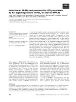
Phosphatidylserine externalisation and caspase-3
activation
Phosphatidylserine externalisation marks cells for phagocyto-
sis as an early event of the apoptotic process. Cells were con-
sidered early apoptotic when phosphatidylserine was
externalised on cells with a still intact membrane, as indicated
by negative staining for 7-AAD. CD4
+
and CD8
+
T-cells and
CD19
+
B-cells exhibited significantly raised portions of phos-
phatidylserine-positive populations in severe sepsis, but not in
critically ill patients (Figure 1a).
Caspase-3 is the central executioner caspase. Activation of
caspase-3 leads to degradation of multiple intracellular sub-
strates and to the typical morphological features of classical
apoptosis. In the current study, activation of caspase-3 was
measured by an antibody specific to the active fragment of
cleaved caspase-3 (Figures 1b and 1c). In patients with
severe sepsis, the subpopulation with active caspase-3 was
elevated in CD4+ T-cells and CD8+ T-cells compared with
critically ill patients or healthy controls. Also, B-cells from sep-
tic patients were found to contain significantly more activated
caspase-3 than B-cells from critically ill patients or healthy
controls.
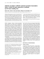
Bcl-2 expression
Since the Bcl-2 family of proteins is known to regulate the
mitochondrial integrity, we analysed the expression of Bcl-2
(Figure 2). The amount of Bcl-2 in CD4
+
T-cells of critically ill
non-septic patients did not differ significantly from healthy con-
trols. However, in patients with sepsis, Bcl-2 protein levels
dropped by about 25%. In CD8
+
T-cells, no significant change
between the three groups could be observed. The decrease in
mitochondrial Bcl-2 was most pronounced in B-cells, where
Bcl-2 dropped by 36% when compared with healthy controls.
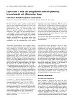
mRNA expression of Bcl-2 family members
When investigating the mRNA expression of mobile pro-apop-
totic BH3-only proteins of the Bcl-2 family, massive induction
was observed in severe sepsis (Figure 3a). When compared
with healthy controls and critically ill patients, mRNA expres-
sion of Bim was upregulated. This corresponds to a 310.5-fold
increase compared with critically ill patients and a 51.7-fold
rise compared with healthy controls. While Bid was decreased
in critically ill patients, it was markedly upregulated in severe
sepsis.
Analysing pro-apoptotic members of the Bcl-2 family located
in the mitochondrial membrane (Figure 3b), a lower expression
of Bak was observed in critically ill patients, while a highly sig-
nificant elevation of Bak mRNA was found in sepsis patients.
Bax mRNA expression was not changed significantly in severe
sepsis as compared with control patients.
The expression of pro-survival molecules was reduced in
severe sepsis (Figure 3c). The mRNA expression of Bcl-2 was
markedly downregulated in patients with severe sepsis as
compared with healthy controls. Bcl-2 expression in critically
ill patients was found at an intermediate level, which showed
no statistical significance. The mRNA of the anti-apoptotic
Bcl-xl decreased in severe sepsis as compared with critically
ill patients.
When comparing survivors versus non-survivors no differ-
ences were observed for Bim (survivors 5548 ± 2510 versus
non-survivors 4784 ± 2307, p > 0.5), Bid (538.3 ± 228.2 ver-
sus 291.8 ± 97.11, p > 0.5), and Bak (409.1 ± 124.6 versus
273.7 ± 79.17, p > 0.5). Corticoid treatment had no signifi-
cant effect on gene expression of Bim (corticoid treatment
5493 ± 2417 versus no corticoid treatment 4839 ± 2406, p
> 0.5), Bid (267.3 ± 102.2 versus 562.8 ± 283.1, p > 0.5) or
Bak (343.9 ± 123.4 versus 338.8 ± 88.79, p = 1).
The degree of organ failure, as expressed by the SOFA score,
correlated with pro-apoptotic Bim, Bid and Bax gene expres-
sion (Table 2). The severity of illness (SAPS II score) corre-
lated with Bim and Bak gene expression (Table 2).
Furthermore, mRNA expression of Bim, Bid and Bak demon-
strated a positive correlation with procalcitonin, although Bim
and Bid correlated with serum IL-6 (Table 2).
Discussion
The current study demonstrates that pro-apoptotic and anti-
apoptotic members of the Bcl-2 family are regulated differen-
tially in a cohort of sepsis patients exhibiting accelerated lym-
phocyte apoptosis as compared with critically ill non-septic
patients. The patients with sepsis in the study exhibited accel-
erated apoptosis of all major lymphocyte subpopulations. The
degree of apoptosis as indicated by phosphatidylserine exter-
nalisation and caspase-3 activation was comparable with find-
ings of previous reports [1,25,26]. Thus the sepsis cohort of
this study exhibited a degree of apoptosis typical of severe
sepsis.
Critical Care Vol 12 No 5 Weber et al.
Page 6 of 10
(page number not for citation purposes)
Besides death receptor mediated mechanisms [9,27,28],
mitochondrial events play a crucial role in the initiation of leu-
cocyte apoptosis during sepsis [1] and their possible involve-
ment in the apoptotic process during sepsis was the focus of
this study. On the side of the pro-death Bcl-2 family, Bim and
Bid were analysed because they are mobile and are sufficient
to activate the mitochondrial apoptosis pathway. For the group
of membrane-associated pro-death members, Bax and Bak
were analysed. As anti-apoptotic members, Bcl-2 and Bcl-xl
gene expression was measured. A typical pattern of gene
expression of the Bcl-2 family was observed in patients with
severe sepsis as compared with the two control groups: on
the one hand the BH-3-only pro-death gene expression of Bim
and Bid was massively upregulated. Furthermore, the mRNA
of the pro-apoptotic Bak was increased. On the other hand,
the genes of the protective Bcl-2 and Bcl-xl were expressed at
lower levels.
The BH3-only Bim is the principal regulator of haematopoietic
homeostasis [17] and a potent inducer of death [29]. Bim can
bind promiscuously to Bcl-2 pro-survival proteins, so it has a
dominant role in apoptosis induction in response to diverse
Figure 1
Confirmation of accelerated apoptosis in patients with severe sepsisConfirmation of accelerated apoptosis in patients with severe sepsis. (a) Phosphatidylserine externalisation on CD4
+
, CD8
+
and CD19
+
lym-
phocytes of patients with severe sepsis. The population of phosphatidylserine-positive lymphocytes was increased as compared with critically ill
patients and healthy controls. *p < 0.05, **p < 0.01. (b) Representative histograms of caspase-3 activation. Intracellular active caspase-3 was
stained with a phycoerythrin-conjugated monoclonal antibody. CD 4
+
T-cells were selected via immunophenotyping in flow cytometry. Region M2
describes the population with activated caspase-3. (c) Detection of active caspase-3 in lymphocyte populations. In patients with severe sepsis the
phosphatidylserine-positive lymphocytes (CD 4
+
T-cells, CD 8
+
T-cells, CD19
+
B-cells) were increased as compared with critically ill patients and
healthy controls. **p < 0.01, ***p < 0.001.
Available online />Page 7 of 10
(page number not for citation purposes)
cytotoxic stimuli [16]. Release of Bim from the dynein motor
complex is regarded as an apoptosis initiating event, because
it does not require caspase activity [30]. Absence of Bim in
Bim -/- mice almost prevented apoptosis after sepsis induction
and improved survival markedly [22]. Also, siRNA-mediated
inhibition of Bim gene expression decreased lymphocyte
apoptosis and improved survival in a mouse model of sepsis
[31].
In the current study, Bim was upregulated massively in
patients with severe sepsis. This event could be a possible ini-
tiator of the accelerated apoptosis observed in the study. On
the transcriptional level, C-Jun N-terminal kinase and the fork-
head transcription factor FKHR-L1 are known to regulate Bim
expression in other models [32]. Our study is limited by the
fact that we did not analyse the activity of relevant transcription
factors and post-transcriptional regulation. These questions
need to be examined in a follow-up study.
Bid transfers apoptosis signals from the extrinsic to the mito-
chondrial pathway of apoptosis [33]. On cleavage by cas-
pase-8, truncated Bid translocates to the mitochondrion [34].
From mouse experiments it is known that lymphocyte apopto-
sis in Bid -/- animals after caecal ligation puncture-induced
sepsis is reduced slightly [22]. Still, survival of sepsis in these
animals is improved significantly [22]. Therefore, it is possible
that the marked upregulation of Bid in our patients with severe
sepsis contributed to a worse outcome.
Bak and Bax belong to the main pro-death members of the
Bcl-2 group. Although Bax, in accordance with Bilbault and
colleagues [20], was not regulated, Bak mRNA expression
was markedly increased in our patients with severe sepsis.
Apoptosis initiates when BH3-only ligands, such as Bim,
engage multiple Bcl-2 homologues, which liberate Bak to exe-
cute mitochondrial membrane opening [35]. Bim and Bak are
known to synergise in the induction of apoptosis of haemat-
opoietic cells [36].
In our study we confirmed the downregulation of Bcl-2 in
severe sepsis. Moreover, we also found Bxl-xl mRNA to be
reduced. The lack of Bxl-xl is likely to be of clinical significance,
because treatment with TAT-Bcl-xl, a cell permeable con-
struct, improved the survival of mice in the model using caecal
ligation puncture-induced sepsis [37]. Interestingly, also IL-6
plays a role in the expression of Bcl-2 and Bcl-xl, because crit-
ical levels of liver Bcl-2 and Bcl-xl could not be maintained in
Figure 2
Expression of Bcl-2 in severe sepsisExpression of Bcl-2 in severe sepsis. The expression of the Bcl-2
protein was measured by flow cytometry in CD4
+
, CD8
+
and CD19
+
lymphocytes. In CD4
+
and CD 19
+
lymphocytes, a significant reduction
in Bcl-2 was observed in severe sepsis. *p < 0.05, **p < 0.01
Figure 3
mRNA expression of Bcl-2 family membersmRNA expression of Bcl-2 family members. mRNA expression was
measured by real-time PCR in patients with severe sepsis, critically ill
patients and healthy controls. (a) The expression of BH3-only members
Bim and Bid was upregulated, (b) the pro death-group Bak and Bax
was regulated heterogeneously and (c) the anti-apoptotic Bcl-2 and
Bcl-xl were downregulated. *p < 0.05, *p < 0.001
Critical Care Vol 12 No 5 Weber et al.
Page 8 of 10
(page number not for citation purposes)
IL-6 deficient mice. The sepsis patients in this study had high
concentrations of IL-6. Still, in our patients IL-6 could not main-
tain the levels of Bcl-2 and Bcl-xl. Another mechanism for the
downregulation of Bcl-2 and Bcl-xl in sepsis has been
described recently by Groesdonk and colleagues [38]. It was
shown that the DNA-binding activity of nuclear factor kB was
reduced in T-cells of septic mice; however, sufficient expres-
sion of these anti-apoptotic proteins is dependent on the
canonical activation of nuclear factor kB.
Although mRNA was extracted from whole blood, possible
contributions to the gene expression pattern by neutrophils
cannot be ruled out. In sepsis, however, apoptosis is delayed
[39] and is associated with the maintenance of mitochondrial
transmembrane potential [40] suggesting an anti-apoptotic
pattern of gene expression of the Bcl-2 family. Accordingly, in
a rat model of sepsis Bim on neutrophils was down-regulated
and Bcl-xl was increased [41]. The opposite was observed in
our study. Thus, it is probable that the massive upregulation of
pro-apoptotic gene expression is generated by mononuclear
cells rather than neutrophils.
In principle, glucocorticoids may induce apoptosis of T-cells,
especially in immunocompromised patients with HIV infection
[42]. On the other hand glucocorticoids may rescue CD4
+
lymphocytes from activation-induced apoptosis triggered by
HIV [43]. In our study, no influence of low-dose corticoid treat-
ment on apoptotic gene expression was found. This is in line
with findings by Hotchkiss and colleagues, who observed no
effect of low-dose glucocorticoid treatment on rates of lym-
phocyte apoptosis in severe sepsis [1].
Gene expression of Bim, and in part of the other pro-apoptotic
Bcl-2 family members, correlated with the severity of disease,
the degree of organ failure and the pro-inflammatory response
(IL-6). It also correlated with serum procalcitonin, which is
regarded as a marker of infection rather than inflammation. Our
study design did not compare septic and non-septic patients
of the same degree of critical illness. Therefore, it cannot be
ruled out that severely ill patients with high SAPS II and SOFA
scores but no indication of infection may also exhibit acceler-
ated lymphocyte apoptosis and pro-apoptotic gene regulation.
The phenomenon of accelerated apoptosis is not unique to
sepsis but is also found in other types of critical illness. For
example, burn injuries preferentially induce CD4
+
T-cell apop-
tosis [44] and haemorrhagic shock induces splenocyte apop-
tosis [45]. Further studies should also investigate Bcl-2 family
expression patterns under these conditions.
Besides mitochondrial events involving the Bcl-2 family sev-
eral other potential inducers of lymphocyte apoptosis are dis-
cussed in sepsis [22,34]. In regular lymphocyte homeostasis,
life and death balance is maintained by activation-induced cell
death, which is induced via death receptors of the tumour
necrosis factor receptor family or T-cell receptor activation
[46]. The death receptor Fas is upregulated [47] on leuco-
cytes in sepsis patients and caspase-8 is active [22] in severe
sepsis in animals. Inhibition of Fas or silencing of caspase-8
inhibits apoptosis during sepsis [9]. Interestingly, cross-talk of
Fas-receptor signalling via truncated Bid may contribute to
apoptosis induction via the mitochondrion [34]. Although acti-
vation-induced cell death is a probable candidate to explain
apoptosis in sepsis, in murine experimental sepsis it was
recently shown that accelerated lymphocyte apoptosis does
not require activation and T-cell receptor engagement [48].
Another member of the tumour necrosis factor receptor family,
CD40 is expressed on B- and T-lymphocytes during sepsis
[49]. Inhibition of CD40 by an antagonising antibody inhibits
lymphocyte apoptosis and improves survival of murine experi-
mental sepsis. Interestingly, at least in part, this protection is
attributed to increased expression of the anti-apoptotic Bcl-2
family member Bcl-xl [49].
Conclusion
A pro-apoptotic pattern of gene expression in the Bcl-2 family
was observed in severe sepsis. The combination of an
increase of Bim, Bid and Bad in conjunction with lowered lev-
els of Bcl-2 and Bcl-xl may render cells more susceptible to
apoptosis during sepsis. This study underlines the potential
importance of the Bcl-2 family and mitochondrial events in the
induction and transduction of apoptosis during sepsis. BH3-
only proteins such as Bim and Bid may be relevant therapeutic
targets in future strategies to inhibit accelerated apoptosis in
patients with severe sepsis.
Competing interests
The authors declare that they have no competing interests.
Authors' contributions
SW and JS conceived the study, participated in its design,
established the flow cytometric methodology, participated in
data acquisition, participated in the data analysis and drafted
the manuscript. LL and MB established the PCR methodology
and designed the primers. SM and SK carried out flow cytom-
Table 2
Correlation of pro-apoptotic gene expression with clinical
parameters.
Bim Bid Bak
r
s
pr
s
pr
s
p
SAPS II 0.49 <0.05 ns 0.40 <0.05
SOFA 0.533 <0.01 0.40 <0.05 0.44 <0.05
Interleukin-6 0.57 <0.01 0.43 <0.05 ns
Procalcitonin 0.56 <0.01 0.44 <0.05 0.41 <0.05
ns = not significant; p = two-tailed p value; r
s
= Spearman correlation
coefficient; SAPS II = Simplified Acute Physiology Score II; SOFA =
Sepsis related Organ Failure Assessment.
Available online />Page 9 of 10
(page number not for citation purposes)
etry analysis. AH and FS participated in the study design and
data analysis. LL, MB, SM, SK, AH and FS critically revised the
manuscript for intellectual content. All authors read and
approved the final manuscript.
Acknowledgements
The authors would like to thank Bettina Bayerer, Makbule Kobilay and
Claudia Baumann for excellent technical assistance with RT-PCR. The
study was supported by a grant from BONFOR (Program for the
advancement of research by the Faculty of Medicine, University of
Bonn).
References
1. Hotchkiss RS, Osmon SB, Chang KC, Wagner TH, Coopersmith
CM, Karl IE: Accelerated lymphocyte death in sepsis occurs by
both the death receptor and mitochondrial pathways. J
Immunol 2005, 174:5110-5118.
2. Schroeder S, Lindemann C, Decker D, Klaschik S, Hering R,
Putensen C, Hoeft A, von Ruecker A, Stuber F: Increased sus-
ceptibility to apoptosis in circulating lymphocytes of critically
ill patients. Langenbecks Arch Surg 2001, 386:42-46.
3. Adrie C, Bachelet M, Vayssier-Taussat M, Russo-Marie F, Boucha-
ert I, Adib-Conquy M, Cavaillon JM, Pinsky MR, Dhainaut JF, Polla
BS: Mitochondrial membrane potential and apoptosis periph-
eral blood monocytes in severe human sepsis. Am J Respir
Crit Care Med 2001, 164:389-395.
4. Efron PA, Martins A, Minnich D, Tinsley K, Ungaro R, Bahjat FR,
Hotchkiss R, Clare-Salzler M, Moldawer LL: Characterization of
the systemic loss of dendritic cells in murine lymph nodes dur-
ing polymicrobial sepsis. J Immunol 2004, 173:3035-3043.
5. Coopersmith CM, Stromberg PE, Dunne WM, Davis CG, Amiot
DM, Buchman TG, Karl IE, Hotchkiss RS: Inhibition of intestinal
epithelial apoptosis and survival in a murine model of pneu-
monia-induced sepsis. JAMA 2002, 287:1716-1721.
6. Hotchkiss RS, Nicholson DW: Apoptosis and caspases regulate
death and inflammation in sepsis. Nat Rev Immunol 2006,
6:813-822.
7. Hotchkiss RS, Karl IE: The pathophysiology and treatment of
sepsis. N Engl J Med 2003, 348:138-150.
8. Hotchkiss RS, Chang KC, Swanson PE, Tinsley KW, Hui JJ,
Klender P, Xanthoudakis S, Roy S, Black C, Grimm E, Aspiotis R,
Han Y, Nicholson DW, Karl IE: Caspase inhibitors improve sur-
vival in sepsis: a critical role of the lymphocyte. Nat Immunol
2000, 1:496-501.
9. Wesche-Soldato DE, Chung CS, Lomas-Neira J, Doughty LA, Gre-
gory SH, Ayala A: In vivo delivery of caspase-8 or Fas siRNA
improves the survival of septic mice. Blood 2005,
106:2295-2301.
10. Oberholzer C, Oberholzer A, Bahjat FR, Minter RM, Tannahill CL,
Abouhamze A, LaFace D, Hutchins B, Clare-Salzler MJ, Moldawer
LL: Targeted adenovirus-induced expression of IL-10
decreases thymic apoptosis and improves survival in murine
sepsis. Proc Natl Acad Sci USA 2001, 98:11503-11508.
11. Hengartner MO: The biochemistry of apoptosis. Nature 2000,
407:770-776.
12. Krammer PH: CD95's deadly mission in the immune system.
Nature 2000, 407:789-795.
13. Zamzami N, Kroemer G: The mitochondrion in apoptosis: how
Pandora's box opens. Nat Rev Mol Cell Biol 2001, 2:67-71.
14. Harris MH, Thompson CB: The role of the Bcl-2 family in the
regulation of outer mitochondrial membrane permeability.
Cell Death Differ 2000, 7:1182-1191.
15. Thornberry NA, Lazebnik Y: Caspases: enemies within. Science
1998, 281:1312-1316.
16. Willis SN, Adams JM: Life in the balance: how BH3-only pro-
teins induce apoptosis. Curr Opin Cell Biol 2005, 17:617-625.
17. Cory S, Adams JM: The Bcl2 family: regulators of the cellular
life-or-death switch. Nat Rev Cancer 2002, 2:647-656.
18. Willis SN, Chen L, Dewson G, Wei A, Naik E, Fletcher JI, Adams
JM, Huang DC: Proapoptotic Bak is sequestered by Mcl-1 and
Bcl-xL, but not Bcl-2, until displaced by BH3-only proteins.
Genes Dev 2005, 19:1294-1305.
19. Hotchkiss RS, Tinsley KW, Swanson PE, Chang KC, Cobb JP,
Buchman TG, Korsmeyer SJ, Karl IE: Prevention of lymphocyte
cell death in sepsis improves survival in mice. Proc Natl Acad
Sci USA 1999, 96:14541-14546.
20. Bilbault P, Lavaux T, Lahlou A, Uring-Lambert B, Gaub MP,
Ratomponirina C, Meyer N, Oudet P, Schneider F: Transient Bcl-
2 gene down-expression in circulating mononuclear cells of
severe sepsis patients who died despite appropriate intensive
care. Intensive Care Med 2004, 30:408-415.
21. Bilbault P, Lavaux T, Launoy A, Gaub MP, Meyer N, Oudet P, Pot-
techer T, Jaeger A, Schneider F: Influence of drotrecogin alpha
(activated) infusion on the variation of Bax/Bcl-2 and Bax/Bcl-
xl ratios in circulating mononuclear cells: a cohort study in
septic shock patients. Crit Care Med 2007, 35:69-75.
22. Chang KC, Unsinger J, Davis CG, Schwulst SJ, Muenzer JT,
Strasser A, Hotchkiss RS: Multiple triggers of cell death in sep-
sis: death receptor and mitochondrial-mediated apoptosis.
Faseb J
2007, 21:708-719.
23. Bone RC, Balk RA, Cerra FB, Dellinger RP, Fein AM, Knaus WA,
Schein RM, Sibbald WJ: Definitions for sepsis and organ failure
and guidelines for the use of innovative therapies in sepsis.
The ACCP/SCCM Consensus Conference Committee. Ameri-
can College of Chest Physicians/Society of Critical Care
Medicine. Chest 1992, 101:1644-1655.
24. Book M, Chen Q, Lehmann LE, Klaschik S, Weber S, Schewe JC,
Luepertz M, Hoeft A, Stuber F: Inducibility of the endogenous
antibiotic peptide beta-defensin 2 is impaired in patients with
severe sepsis. Crit Care 2007, 11:R19.
25. Le Tulzo Y, Pangault C, Gacouin A, Guilloux V, Tribut O, Amiot L,
Tattevin P, Thomas R, Fauchet R, Drenou B: Early circulating lym-
phocyte apoptosis in human septic shock is associated with
poor outcome. Shock 2002, 18:487-494.
26. Roth G, Moser B, Krenn C, Brunner M, Haisjackl M, Almer G, Ger-
litz S, Wolner E, Boltz-Nitulescu G, Ankersmit HJ: Susceptibility
to programmed cell death in T-lymphocytes from septic
patients: a mechanism for lymphopenia and Th2
predominance. Biochem Biophys Res Commun 2003,
308:840-846.
27. Wesche DE, Lomas-Neira JL, Perl M, Chung CS, Ayala A: Leuko-
cyte apoptosis and its significance in sepsis and shock. J Leu-
koc Biol 2005, 78:325-337.
28. Ayala A, Xin XY, Ayala CA, Sonefeld DE, Karr SM, Evans TA,
Chaudry IH: Increased mucosal B-lymphocyte apoptosis dur-
ing polymicrobial sepsis is a Fas ligand but not an endotoxin-
mediated process. Blood 1998, 91:1362-1372.
29. Bouillet P, Metcalf D, Huang DC, Tarlinton DM, Kay TW, Kontgen
F, Adams JM, Strasser A: Proapoptotic Bcl-2 relative Bim
required for certain apoptotic responses, leukocyte homeos-
tasis, and to preclude autoimmunity. Science 1999,
286:1735-1738.
30. Puthalakath H, Huang DC, O'Reilly LA, King SM, Strasser A: The
proapoptotic activity of the Bcl-2 family member Bim is regu-
lated by interaction with the dynein motor complex. Mol Cell
1999, 3:287-296.
31. Schwulst SJ, Muenzer JT, Peck-Palmer OM, Chang KC, Davis CG,
McDonough JS, Osborne DF, Walton AH, Unsinger J, McDunn JE,
Hotchkiss RS: BIM siRNA decreases lymphocyte apoptosis
and improves survival in sepsis. Shock 2008.
32. Puthalakath H, Strasser A: Keeping killers on a tight leash: tran-
scriptional and post-translational control of the pro-apoptotic
Key messages
• Gene expression of the Bcl-2 family during accelerated
apoptosis in severe sepsis displays a pro-apoptotic pat-
tern compared with critically ill non-septic patients and
healthy controls.
• Expression of pro-apoptotic Bim, Bid and Bak mRNA is
markedly upregulated.
• Expression of anti-apoptotic Bcl-xl and Bcl-2 is
downregulated.
Critical Care Vol 12 No 5 Weber et al.
Page 10 of 10
(page number not for citation purposes)
activity of BH3-only proteins. Cell Death Differ 2002,
9:505-512.
33. Gross A: BID as a double agent in cell life and death. Cell
Cycle 2006, 5:582-584.
34. Perl M, Chung CS, Ayala A: Apoptosis. Crit Care Med 2005,
33:S526-S529.
35. Willis SN, Fletcher JI, Kaufmann T, van Delft MF, Chen L, Czabotar
PE, Ierino H, Lee EF, Fairlie WD, Bouillet P, Strasser A, Kluck RM,
Adams JM, Huang DC: Apoptosis initiated when BH3 ligands
engage multiple Bcl-2 homologs, not Bax or Bak. Science
2007, 315:856-859.
36. Hutcheson J, Scatizzi JC, Bickel E, Brown NJ, Bouillet P, Strasser
A, Perlman H: Combined loss of proapoptotic genes Bak or
Bax with Bim synergizes to cause defects in hematopoiesis
and in thymocyte apoptosis. J Exp Med 2005, 201:1949-1960.
37. Hotchkiss RS, McConnell KW, Bullok K, Davis CG, Chang KC,
Schwulst SJ, Dunne JC, Dietz GP, Bahr M, McDunn JE, Karl IE,
Wagner TH, Cobb JP, Coopersmith CM, Piwnica-Worms D: TAT-
BH4 and TAT-Bcl-xL peptides protect against sepsis-induced
lymphocyte apoptosis in vivo. J Immunol 2006,
176:5471-5477.
38. Groesdonk HV, Wagner F, Hoffarth B, Georgieff M, Senftleben U:
Enhancement of NF-kappaB activation in lymphocytes
prevents T cell apoptosis and improves survival in murine
sepsis. J Immunol 2007, 179:8083-8089.
39. Fialkow L, Fochesatto Filho L, Bozzetti MC, Milani AR, Rodrigues
Filho EM, Ladniuk RM, Pierozan P, de Moura RM, Prolla JC,
Vachon E, Downey GP: Neutrophil apoptosis: a marker of dis-
ease severity in sepsis and sepsis-induced acute respiratory
distress syndrome. Crit Care 2006, 10:R155.
40. Taneja R, Parodo J, Jia SH, Kapus A, Rotstein OD, Marshall JC:
Delayed neutrophil apoptosis in sepsis is associated with
maintenance of mitochondrial transmembrane potential and
reduced caspase-9 activity. Crit Care Med 2004,
32:1460-1469.
41. Guo RF, Sun L, Gao H, Shi KX, Rittirsch D, Sarma VJ, Zetoune FS,
Ward PA: In vivo regulation of neutrophil apoptosis by C5a
during sepsis. J Leukoc Biol 2006, 80:1575-1583.
42. Roger PM, Perbost I, Ticchioni M, Fuzibet JG, Breittmayer JP,
Durant J, Pesce A, Bernard A, Dellamonica P:
Apoptosis of naive
CD4+ T-cells from HIV-infected patients with poor immune
response to HAART is enhanced in vitro by steroid. J Infect
2004, 49:216-221.
43. Lu W, Salerno-Goncalves R, Yuan J, Sylvie D, Han DS, Andrieu JM:
Glucocorticoids rescue CD4+ T lymphocytes from activation-
induced apoptosis triggered by HIV-1: implications for patho-
genesis and therapy. Aids 1995, 9:35-42.
44. Patenaude J, D'Elia M, Hamelin C, Garrel D, Bernier J: Burn injury
induces a change in T cell homeostasis affecting preferentially
CD4+ T cells. J Leukoc Biol 2005, 77:141-150.
45. Hostmann A, Jasse K, Schulze-Tanzil G, Robinson Y, Oberholzer
A, Ertel W, Tschoeke SK: Biphasic onset of splenic apoptosis
following hemorrhagic shock: critical implications for Bax, Bcl-
2, and Mcl-1 proteins. Crit Care 2008, 12:R8.
46. Krammer PH, Arnold R, Lavrik IN: Life and death in peripheral T
cells. Nat Rev Immunol 2007, 7:532-542.
47. Papathanassoglou ED, Moynihan JA, McDermott MP, Ackerman
MH: Expression of Fas (CD95) and Fas ligand on peripheral
blood mononuclear cells in critical illness and association with
multiorgan dysfunction severity and survival. Crit Care Med
2001, 29:709-718.
48. Unsinger J, Herndon JM, Davis CG, Muenzer JT, Hotchkiss RS,
Ferguson TA: The role of TCR engagement and activation-
induced cell death in sepsis-induced T cell apoptosis. J
Immunol 2006, 177:7968-7973.
49. Schwulst SJ, Grayson MH, DiPasco PJ, Davis CG, Brahmbhatt TS,
Ferguson TA, Hotchkiss RS: Agonistic monoclonal antibody
against CD40 receptor decreases lymphocyte apoptosis and
improves survival in sepsis. J Immunol 2006, 177:557-565.