Báo cáo y học: " γδ T lymphocytes from cystic fibrosis patients and healthy donors are high TNF-α and IFN-γ-producers in response to Pseudomonas aeruginosa" pptx
Bạn đang xem bản rút gọn của tài liệu. Xem và tải ngay bản đầy đủ của tài liệu tại đây (420.52 KB, 14 trang )
Respiratory Research
BioMed Central
Open Access
Research
γδ T lymphocytes from cystic fibrosis patients and healthy donors
are high TNF-α and IFN-γ-producers in response to Pseudomonas
aeruginosa
Salvador Raga†1, M Rosa Julià*†1, Catalina Crespí1, Joan Figuerola2,
Natalia Martínez1, Joan Milà1 and Núria Matamoros1
Address: 1Immunology Service, Son Dureta Hospital, Palma de Mallorca, Balearic Islands, SPAIN and 2Pediatrics Service, Son Dureta Hospital,
Palma de Mallorca, Balearic Islands, SPAIN
Email: Salvador Raga - ; M Rosa Julià* - ; Catalina Crespí - ;
Joan Figuerola - ; Natalia Martínez - ; Joan Milà - ; Núria Matamoros -
* Corresponding author †Equal contributors
Published: 08 September 2003
Respiratory Research 2003, 4:9
Received: 27 May 2003
Accepted: 08 September 2003
This article is available from: />© 2003 Raga et al; licensee BioMed Central Ltd. This is an Open Access article: verbatim copying and redistribution of this article are permitted in all
media for any purpose, provided this notice is preserved along with the article's original URL.
cystic fibrosiscytokinesPseudomonas aeruginosaγδ T lymphocytes.
Abstract
Background: γδ T cells have an important immunoregulatory and effector function through
cytokine release. They are involved in the responses to Gram-negative bacterium and in protection
of lung epithelium integrity. On the other hand, they have been implicated in airway inflammation.
Methods: The aim of the present work was to study intracytoplasmic IL-2, IL-4, IFN-γ and TNFα production by γδ and αβ T lymphocytes from cystic fibrosis patients and healthy donors in
response to Pseudomonas aeruginosa (PA). Flow cytometric detection was performed after
peripheral blood mononuclear cells (PBMC) culture with a cytosolic extract from PA and
restimulation with phorbol ester plus ionomycine. Proliferative responses, activation markers and
receptor usage of γδ T cells were also evaluated.
Results: The highest production of cytokine was of TNF-α and IFN-γ, γδ being better producers
than αβ. No differences were found between patients and controls. The Vγ9δ2 subset of γδ T cells
was preferentially expanded. CD25 and CD45RO expression by the αβ T subset and PBMC
proliferative response to PA were defective in cystic fibrosis lymphocytes.
Conclusion: Our results support the hypothesis that γδ T lymphocytes play an important role in
the immune response to PA and in the chronic inflammatory lung reaction in cystic fibrosis patients.
They do not confirm the involvement of a supressed Th1 cytokine response in the pathogenesis of
this disease.
Introduction
Cystic Fibrosis (CF) is the most common serious recessively inherited disease among Caucasians [1]. It is associated with impaired mucociliary clearance, abnormally
thick mucus, chronic infections and lung inflammation.
Most of CF patients are chronically colonized by bacteria
such as Pseudomonas aeruginosa (PA) in their respiratory
tract, this microorganism being their main cause of
Page 1 of 14
(page number not for citation purposes)
Respiratory Research 2003, 4
morbidity and mortality [2]. A single major mutation
(∆F508) in the Cystic Fibrosis Transmembrane Conductance Regulator (CFTR) gene is responsible for 70% of CF
cases, but over 800 mutations have been identified. CFTR
is an anion channel and it negatively regulates the amiloride-sensitive epithelial Na channel [3]. Individuals, however, with mutations that affect apical epithelial Na+
channel function do not have infectious lung disease. In
primary ciliary diskinesia, the absence of mucociliary
clearance results in recurrent lung infections, but the diagnosis is often delayed until adulthood and PA colonization is absent. All this evidence suggests that other factors
are involved in CF.
CFTR was found in CD4-positive T cells and a Cl- channel
function, similar to that regulated by CFTR in epithelial
cells, was detected in these lymphocytes. This function
was defective in CF patients [4]. Therefore CFTR mutations could affect immunocompetent or accessory cells. In
fact immunoregulatory defects in CF patients have been
described and include reduced supressor and helper T cell
activity [5].
Most of T cells bear the heterodimeric αβ antigen receptor
but a relatively small subset express two different rearranging genes, γ and δ. About 5% of human peripheral
blood lymphocytes are γδ T lymphocytes, but they are
preferentially localized in epithelial and mucosal tissues.
Protective response to pulmonary injury requires γδ T
lymphocytes [6,7]. In previous reports we described an
increased percentage of γδ T cells in peripheral blood of
CF patients [8] and we showed, in healthy individuals, a
preferential "in vitro" proliferation of these cells in
response to PA cytosolic non-peptidic phosphorilated
antigens [9]. γδ T cells have been described as an immunoregulatory subset [10,11] and their cytotoxic activity
has been associated with some diseases [12,13]. Specific
changes in γδ T receptor genes usage related to function
have been described in patients with pulmonary tuberculosis [14].
In the last few years attention has been focused on the role
of cell-mediated immunity in host defense against bronchopulmonary infection with Gram-negative bacterium.
A correlation between poor lung function and elevated
bacteria-specific antibody level in CF patients and animal
models was demonstrated [15]. Furthermore, protective
immune responses in animal models mimicking CF were
cell-mediated [15–17]. The mechanisms by which T cells
protect the lung are not completely known but it has been
demonstrated that they activate the local phagocytic
response through cytokine release [18]. γδ T cells have
been described as important inducers of TNF-α production by LPS-stimulated macrophages [19]. In addition,
mice depleted of these lymphocytes showed exaggerated
/>
bacterial growth after E. coli infection [20]. Recent "in
vivo" experiments have demonstrated that T lymphocytes
expressing Vγ9 and Vδ2 genes are mediators of resistance
against extracellular gram-negative and positive bacteria
[21].
PA has a biofilm mode of growth that could be regarded
as a protective multicellular survival strategy, resembling
intracellular bacteria such as mycobacteria. Th1-type
cytokines, as IFN-γ and TNF-α, are responsible for a good
immune response to intracellular bacteria; in contrast
Th2-type cytokines, as IL-4, induce antibody production
by B lymphocytes [22]. Production of Th1 cytokines by
CD4-positive lymphocytes has been suggested to be
impaired in CF [17,23].
γδ T cells are a Th1-type cytokines source [24,25] and their
role in the "in vivo" mycobacteria clearance in macaques
has been recently demonstrated [26]. A defect in γδ T lymphocytes activation or a bias in their cytokine secretion in
response to PA could contribute to the chronic colonization by this bacterium. Alternatively chronic γδ T cells activation could determine a local persistent inflammation in
the lung.
The aim of the current work was to assess the "in vitro"
response of αβ and γδ T lymphocytes from peripheral
blood of healthy controls and CF patients to cytosolic
antigens from PA. To estimate this response we evaluated
two surface markers: CD25, that corresponds to the IL-2
receptor α-chain and CD45RO, a putative memory
marker. CD25 expression by T lymphocytes correlates
with proliferative responses to antigens and mitogens and
has been described as more sensitive than others markers,
as CD69, to minor subpopulations responses [27].
CD45RO is expressed by T cells after antigen contact and
further antigenic stimulus, at tissue level, commit
CD45RO-positive cells to an effector function [28]. This
marker is then expressed by both memory and effector T
cells.
To assess the type of cytokines secreted in response to PA,
intracytoplasmic IL-4, IFN-γ, TNF-α and IL-2 production
from αβ and γδ T cells was determined by three color flow
cytometry. Phenotypic characteristics of the expanded γδ T
subpopulation and PBMC proliferative activity were also
evaluated.
Materials and Methods
Donors
14 clinically stable outpatients with Cystic fibrosis (8
males and 6 females), age range: 8–29 years (CFw group)
and 17 sex/age matched healthy controls, were studied.
All CF patients had diagnosis confirmed by pilocarpine
Page 2 of 14
(page number not for citation purposes)
Respiratory Research 2003, 4
/>
Table 1: Characteristics of cystic fibrosis patients included in the study
Patients
Age
Mutations
History of infections
PA culture
Shwachman score
MGV
MGP
AAS
AGH
MCC
LGS
EML
DNP
YML
MVG
VMM
ASA
GCF
MSP
15
14
17
15
14
5
29
12
23
8
8
7
10
21
∆F508/∆F508
∆F508/∆F508
∆F508/?
∆F508/?
∆F508/?
∆F508/∆F508
R347H/∆F508
∆F508/2789+ 563A
R347H/∆F508
∆F508/∆F508
∆F508/G542X
∆F508/L206W
∆F508/∆F508
∆F508/?
PA chronic colonization
PA chronic colonization, occasional SA
PA chronic colonization
PA chronic colonization
SA and PA chronic colonization
PA chronic colonization
PA chronic colonization
PA chronic colonization
PA chronic colonization
occasional SA
#
SA chronic colonization, occasional HI
occasional SA
* PA chronic colonization
Pos
Pos
Pos
Pos
Pos
Pos
Pos
Pos
Pos
Neg
Neg
Neg
Neg
*
95
76
60
80
65
85
55
95
55
90
85
95
85
71
Additional Data
DMID
DMID
PA: Pseudomonas aeruginosa, SA: Staphylococcus aureus, HI: Haemophilus influenzae. Pos: sputum culture positive for PA, Neg: sputum culture negative
for PA, DMID:Diabetes mellitus insulin-dependent. # PA colonization until 2 years before blood extraction, negative from this date until now * PA
negative only for one year, blood extraction was made during this year Schwachman score rates severity based on history, physical examination and
chest roentgenogram
iontophoresis sweat test; 5 patients were homozygous for
the ∆F508 mutation and the remainder were heterozygous for this mutation.
Patients were not receiving systemic corticosteroids and
their treatment included pancreatic enzymes, vitamins
and, in some cases, antibiotic therapy and sympathicomimetics. Patients chronically colonized by PA received
cycles of an antipseudomonal penicillin and an aminoglycoside, oral or intravenous, depending on the antibiogram. Two DMID cases were on insulin therapy.
The clinical severity of the disease was estimated using the
Schwachman score (SC). A SC from 41 to 55 corresponds
to moderate severity, from 56 to 70 to slight severity, from
71 to 85 to good status and from 86 to 100 to excellent
status. In our patients, SC ranged from 55 to 95 (Table 1).
PA was detected, by sputum culture, in 9 patients, all of
them chronically colonized by PA (CFp group). Four
patients were free of PA infection at the moment of the
study (CFn group), 3 of these have always been free of PA
and 1 was colonized by PA until two years before the
study and from this date he has been PA-negative. One
patient has always been PA-positive except for a year that
included the blood extraction data. This last patient was
included in the whole (CFw) group but not in the CFp or
in the CFn group. Genetic, clinical and infectious characteristics of the patients are depicted in Table 1.
The study was approved by the Hospital ethical committee and informed consent was obtained from participants.
Activation kinetics and phenotypic studies
Heparinized peripheral blood was obtained from patients
and controls. Peripheral blood mononuclear cells
(PBMC) were isolated by Ficoll-Hypaque and cultured in
RPMI-1640 supplemented with 10% heat-inactivated
autologous serum, at 37°C, 5% CO2 in air. Heat-treated
cytosolic fraction from PA (PAc) was obtained from a sputum isolated strain as already described [9]. Briefly, we
cultured the isolated bacteria in liquid media and then
washed and heat-treated them at 120°C for 20 min.
Lysates were prepared by passage through a French press
and the supernatants obtained after centrifugation (7840
× g). Cytosolic antigens (PAc), that were the main PA
stimulatory fraction to γδ T cells [9], were obtained by
lysates ultracentrifugation (80000 × g for 45 min). PAc
was added to PBMC cultures at 50 µg/ml.
CD25 and CD45RO expression by αβ and γδ T cells was
evaluated by three-color flow cytometry before and after
4, 6 and 8 days of culture. We chose these days of culture
because T lymphocytes stimulated with antigen or
mitogen show a peak of CD25 and CD45RO expression
between 4 and 8 days of culture [27,29].
Cells were acquired and analysed on a FACScalibur
cytometer, using CellQuest software, Becton-Dickinson,
Mountain View, CA, USA (BD). Anti-CD3-PerCP, antiTCR αβ-FITC and anti-CD25 or anti-CD45RO-PE MAbs
(BD) were used.
In a previous work we demonstrated a preferent γδ T subset expansion between 7 and 10 days of PBMC culture
Page 3 of 14
(page number not for citation purposes)
Respiratory Research 2003, 4
with a PA cytosolic extract [9]. In the current study γδ T
cells phenotype was determined, by three-color flow
cytometry, before and after 8 days of culture. 2500 γδ T
cells were acquired and analysed in dot-plot cytograms.
Anti-TCRγδ-PE from Immunotech (Marseille. France),
anti-CD8-PerCP and anti-CD4-FITC (BD) or either antiVγ9 or anti-Vδ2-FITC from Immunotech were used.
PBMC Proliferation
PBMC (2 × 10⁵ / well) from controls and patients were
cultured with PAc (50 µg/ml) or Phytohemaglutinin
(PHA) at 1 µg/ml (GIBCO BRL. Eragny. France), in RPMI1640 supplemented with 10% heat-inactivated autologous serum, for 6 and 3 days respectively. Cells were
pulsed with 1 µCi [3H]-thymidine (Amersham, UK) for 16
h.
Stimulation index (SI) was calculated by the quotient
between counts per minute (c.p.m.) obtained with and
without stimulus.
Intracellular cytokine production in response to P.
aeruginosa and restimulation with Phorbol 12-Myristate
13-Acetate plus Ionomicine
The activation of γδ and αβ T lymphocytes with PAc produced only low intracytoplasmic cytokine signals. Phorbol 12-Myristate 13-Acetate (PMA) plus Ionomicine (Io)
has been described as non distorting, policlonal stimulus,
for previously expanded antigen-specific T cells [30–32].
In the current work we determined the percentage of γδ
and αβ cytokine-producing cells at day 10 of PBMC culture with PA cytosolic extract, after restimulation with
PMA-Io. On this day, the γδ subset reached a maximal
expansion without excessive cellular death. Measurement
of single cell intracellular cytokines allowed us to determine the kind of produced cytokines and which T subpopulation was their source. This system avoided the
purification of a minor T subset, as the γδ-positive, that
could require an excessive amount of blood sample, not
available from children patients.
PBMC in complete medium (2 ì 106/well) were incubated with PAc (50 àg/ml) as above described. Cells were
harvested on day 10, washed and left 23 h. in culture
medium, without stimulus, at 106 cells/well. BFA (10 µg/
ml), PMA (25 ng/ml) and Io (1 µg/ml) was added for 4 h.
at 37°C, 5% CO2 in air. After washing, cells were incubated for 15 min at room temperature with MAbs: antiTCRγδ-1-FITC (BD) or PE-conjugated (Immunotech) and
anti-CD3-PerCP (BD), fixed with FACS Lysis Solution
(BD) and washed. The pellet was incubated with 200 µl of
FACS Permeabilizing Solution (BD) for 10 min, washed
and incubated for 30 min. at room temperature with
either the anti-cytokine monoclonal antibody: anti-IL-2,
anti-IL-4-PE (BD), anti-TNF-α-PE (CALTAG. Burlingame,
/>
CA) or anti-IFN-γ-FITC from Serotec (Oxford, England).
The cells were then washed and resuspended with 100 µl
of 1% paraformaldehide.
Samples were acquired and analysed using CellQuest software (BD). Ten-thousand T lymphocytes were acquired
according to CD3 expression. The percentage of cytokinepositive cells within each subset, γδ-positive or negative
was calculated. Irrelevant, isotype-matched antibodies
were used in parallel with all experimental samples.
Statistical analysis
For means comparison between two different groups and
between paired samples, the two tailed Student t or the
Mann-Whitney U test was used. For means comparison
between three groups (Controls, CFp and CFn) oneway
ANOVA and LSD PostHoc or the Kruskal-Wallis test were
used. Data are given as mean ± standard error of the mean,
p values are two-sided and considered significant when
<0.05. The calculations were performed using SPSS 6.0
software.
Results
Activation kinetics and phenotypic studies
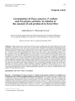
The percentage of γδ-positive T lymphocytes before PBMC
culture was significantly higher in CFw (14.7 ± 2.4) than
in controls (5.8 ± 0.9), p < 0.01. During culture with PAc
γδ percentage first sligthly decreased, but from day 4 it
progressively increased in all groups and there were no
differences between groups (Fig 1).
Most of γδ T lymphocytes were Vγ9Vδ2 before and after
culture. The percentages of Vγ9+ and Vδ2+ cells were significantly higher at the end than at the beginning of culture in CFw, CFp and in controls (Table 2). CFn showed
higher values of Vγ9+ and Vδ2+ at the end than before culture but without statistical significance, probably due to
the low number of individuals. Before and during culture,
few cells expressed CD8 and very few expressed CD4 in all
groups. There were no significant differences in γδ T cells
phenotype between patients and controls.
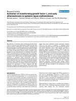
The percentage of CD25-positive γδ T cells was negligible
on day 0 but experienced an important increase after 4
days of culture, prior γδ expansion. From day 6 to 8 it
reached a maximum, over 50% of cells. A higher percentage of CD25-positive αβ than γδ was found at the beginning of culture in controls (p < 0.001), CFp and CFn (p <
0.05), but the increase during the culture was higher in γδ
T cells. In consequence the percentage of CD25-positive
cells was higher in the γδ subset on days 6 (controls: p <
0.001, CFp: p = 0.001, CFn: not significant) and 8 (controls: p < 0.05, CFp: p < 0.001, CFn: not significant) (Fig
2) and corresponded to their preferent expansion.
Page 4 of 14
(page number not for citation purposes)
Respiratory Research 2003, 4
/>
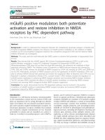
Figure 1 of γδ positive cells(PA)
from Pseudomonas aeruginosa in CD3-positive lymphocytes after 0, 4, 6, 8 and 10 days of PBMC culture with cytosolic extract
Percentage
Percentage of γδ positive cells in CD3-positive lymphocytes after 0, 4, 6, 8 and 10 days of PBMC culture with
cytosolic extract from Pseudomonas aeruginosa (PA). Surface expression of γδ receptor was detected in 2500 gated T
lymphocytes, by three color cytometry, in controls (n = 17), Cystic fibrosis patients infected by PA (CFp, n = 9) and Cystic
fibrosis patients not infected by PA (CFn, n = 4). Bar values correspond to mean ± standard error of the mean. (*) significant
difference between controls and CFp, (**) significant difference between controls and CFn (ANOVA, LSD PostHoc test).
Table 2: Phenotypic characteristics of γδ T cells before and after 8 days of culture with cytosolic extract from P. aeruginosa (PA)
Vγ9 day 0
Controls
CFw
CFp
CFn
Vγ9 day 8
Vδ2 day 0
Vδ2 day 8
CD4 day 0
CD4 day 8
CD8 day 0
CD8 day 8
84.71 ± 3.12**
89.83 ± 2.78*
92.52 ± 1.40**
82.80 ± 8.8
92.85 ± 1.70
94.75 ± 2.06
96.82 ± 1.14
87.45 ± 7.98
77.67 ± 3.8*
84.65 ± 3.89*
85.90 ± 3.88**
79.46 ± 10.32
89.46 ± 3.43
92.39 ± 3.23
94.44 ± 2.72
87.32 ± 8.64
2.03 ± 0.93
0.49 ± 0.26
0.78 ± 0.37
0.1 ± 0.01
0.76 ± 0.65
0.27 ± 0.18
0.54 ± 0.31
0.1 ± 0.01
11.92 ± 2.55
9.21 ± 1.17
9.48 ± 1.80
8.41 ± 1.53
10.13 ± 1.91
10.13 ± 1.91
9.84 ± 2.69
11.28 ± 3.42
Results are expressed as the percentage of positive cells (mean ± s.e.m.) in the γδ T subpopulation. CFw (cystic fibrosis patients, n = 14), CFp (cystic
fibrosis patients infected by PA, n = 8), CFn (cystic fibrosis patients without PA, n = 4). Means comparison between paired samples (days 0 and 8)
were made by the Student's t test: *Significantly lower than the value at day 8: p < 0.01 **Significantly lower than the value at day 8: p < 0.05
The αβ CD25 expression at the end of culture (day 8) was
higher in controls than in CFp (p < 0.05) and CFn (not
significant), but there were no differences in the γδ CD25
expression between groups (Fig 2).
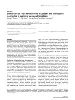
Before culture, the percentage of γδ CD45RO-positive cells
was significantly higher in CFw (78.9 ± 3.2) than in controls (62.7 ± 5.6) (p < 0.05). When we separated patients
into CFn and CFp groups, the difference was not
significant (Fig 3). After day 4 there were no significant
differences between CFw and controls, due to the increase
Page 5 of 14
(page number not for citation purposes)
Respiratory Research 2003, 4
/>
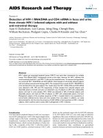
Percentage of CD25-expressing cells from Pseudomonas aeruginosa γδ T lymphocytes (bottom), after 0, 4, 6 and 8 days of
Figure 2
PBMC culture with cytosolic extract in αβ T lymphocytes (top) and
Percentage of CD25-expressing cells in αβ T lymphocytes (top) and γδ T lymphocytes (bottom), after 0, 4, 6
and 8 days of PBMC culture with cytosolic extract from Pseudomonas aeruginosa. Surface expression of CD25 (α
chain of IL-2 receptor) was detected, by three color cytometry, in 2500 gated T lymphocytes from controls (n = 17), Cystic
fibrosis patients infected by PA (CFp, n = 9) and Cystic fibrosis patients not infected by PA (CFn, n = 4). Bar values correspond
to mean ± standard error of the mean. (top) αβ T lymphocytes: (*) significant difference between controls and CFp (ANOVA,
LSD PostHoc test). (bottom) γδ T lymphocytes: significant differences were not found between groups.
Page 6 of 14
(page number not for citation purposes)
Respiratory Research 2003, 4
in the control group. CD45RO expression was always significantly higher in γδ than in αβ, agreeing with other
reports [33], in controls: p < 0.05 on day 0 and p < 0.001
during culture, in CFp: p < 0.001 on day 0 and during culture and in CFn: p < 0.05 on day 0 and during culture (Fig
3).
The percentage of αβ CD45RO-positive before culture was
higher, but not significantly different in controls (45.54 ±
3.9) than in CFw (37.4 ± 2.06). Following stimulation
with PAc it increased only in the former group. At day 8,
controls had significantly higher results than CFp (p <
0.01) and CFn (p < 0.05).
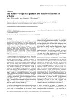
PBMC Proliferation
Statistically significant differences between patients and
controls were only found for SI in response to PA. They
were higher in controls: 67.7 ± 37.8 than in CFw: 14 ± 5.4
(Mann-Whitney U test, p < 0.05). When patients were separated depending on PA infection, the differences were
not significant (Fig 4).
Intracellular cytokine production in response to P.
aeruginosa and PMA/Io restimulation
The frequency of cytokine-producing cells within both Tcell subsets was found in the order of TNF-α>IFN-γ>IL-2
(Fig 5). The percentage of IL-4-producing cells was in all
cases negligible.
There were no significant differences between controls
and CFw, or between controls, CFp and CFn groups, but
γδ T cells from CFp showed higher TNF-α and IFN-γ values
than controls and CFn (Fig 5).
We detected higher IFN-γ production in γδ T cells, in comparison to αβ, in CFw, γδ: 36.9 ± 8.4; αβ: 10.3 ± 2.5 (p <
0.01) and in controls, γδ: 30.6 ± 6.7; αβ: 15.5 ± 5.1 (p =
0.001). The same was found for TNF-α in CFw, γδ: 47.4 ±
7.4; αβ: 25.5 ± 4.8 (p = 0.001) and in controls, γδ: 47.9 ±
8.1; αβ: 20.2 ± 3.7 (p < 0.001). When we considered CFp
and CFn separately, the statistical significance only
remained in CFp, however IFN-γ and TNF-α results were
also higher in γδ than in αβ from CFn (Fig 5).
The percentage of cytokine-producing cells, αβ or γδ-positive, in total T lymphocytic population was calculated
(Fig 6). Both subsets contributed to the same extent in the
percentage of IFN-γ and TNF-α-positive lymphocytes. In
contrast, the highest number of IL-2-producing lymphocytes always proceeded from the αβ subset. Again, we
did not find significant differences between groups.
/>
Correlation with clinical status
No differences were found in cytokine secretion, activation markers and PBMC proliferation in relation to clinical severity of patients (data not shown).
Discussion
The present study is the first, to our knowledge, that determines the "in vitro" intracytoplasmic cytokine response to
Pseudomonas aeruginosa (PA) in αβ and γδ T lymphocytes.
We first expanded PA-reactive T cells for 10 days and then
we restimulated them with PMA-Io. The cytokine-producing frequencies among αβ and γδ T cells were found in this
order: TNF-α>IFN-γ>IL-2, for both subsets. Our results
indicate that despite recognizing different antigens from
PA [9], αβ and γδ T cells have a similar profile of cytokine
secretion, but PAc stimulation induce more γδ than αβ
IFN-γ and TNF-α-positive cells. αβ and γδ contribution to
the percentage of IFN-γ and TNF-α-producing cells in total
T lymphocytic population was the same although the αβ
subset was always predominant.
Cytokine production by αβ and γδ T lymphocytes on stimulation with mycobacteria preparations and its purified
phosphoantigens has been previosly studied. The secretion profile was limited to Th1-type cytokines [24,25,34–
36]. One study compared cytokine levels in supernatants
of αβ and γδ purified cells cultured with live Mycobacterium tuberculosis [25] and concluded that γδ T cells were
more efficient producers of IFN-γ.
These and our present findings reinforce the evidence of
an important role for γδ T lymphocytes in the response to
bacteria through IFN-γ secretion [21]. Furthermore, an "in
vivo" γδ T memory response, already suggested in humans
[37] has been recently described in BCG-vaccinated
macaques [26]. This opens the way to new immunization
strategies.
On the other hand, our findings did not support the
hypothesis that there is a bias in CF T lymphocytes function toward a Th2 response [17,23]. There are
contradictory reports in this field. Agreement exists about
the protective role of the cellular response to PA [15–17]
but the kind of cytokines responsible for a good or a deleterious response in CF is still not elucidated. Some
authors demonstrate a beneficial role of the inflammatory
cytokine IFN-γ [17] and others implicate a defective production of IL-10 in the pulmonary lesions of CF patients
[4]. Moss et al [38], found a lower IFN-γ secretion by CD4positive T cells in CF patients but the stimulus (anti-CD3
plus PMA) and the tested subpopulation differed from
ours.
In a previous work [8] we demonstrated a significant
increase of peripheral blood γδ T lymphocytes in cystic
Page 7 of 14
(page number not for citation purposes)
Respiratory Research 2003, 4
/>
Figure 3 of with cytosolic extract from αβ T lymphocytes (top)
PBMC culture CD45RO-expressing cells in Pseudomonas aeruginosa and γδ T lymphocytes (bottom), after 0, 4, 6, and 8 days of
Percentage
Percentage of CD45RO-expressing cells in αβ T lymphocytes (top) and γδ T lymphocytes (bottom), after 0, 4,
6, and 8 days of PBMC culture with cytosolic extract from Pseudomonas aeruginosa. Surface expression of
CD45RO (memory/activation marker) was detected, by three color cytometry, in 2500 gated T lymphocytes from controls (n
= 17), Cystic fibrosis patients infected by PA (CFp, n = 9) and Cystic fibrosis patients not infected by PA (CFn, n = 4). Bar values correspond to mean ± standard error of the mean. (top) αβ T lymphocytes: (*) significant difference between controls and
CFp, (**) significant difference between controls and CFn (ANOVA, LSD PostHoc test). (bottom) γδ T lymphocytes: significant
differences were not found between groups.
Page 8 of 14
(page number not for citation purposes)
Respiratory Research 2003, 4
/>
PBMC Proliferative response (3H-thymidine incorporation), expressedfor c.p.m. (top) and stimulation index (bottom), after culFigure 4
ture either with cytosolic extract from Pseudomonas aeruginosa (PAc) as 6 days or with Phytohemaglutinin (PHA) for 3 days
PBMC Proliferative response (3H-thymidine incorporation), expressed as c.p.m. (top) and stimulation index
(bottom), after culture either with cytosolic extract from Pseudomonas aeruginosa (PAc) for 6 days or with
Phytohemaglutinin (PHA) for 3 days. Significant differences were not found between controls (n = 10), Cystic fibrosis
patients infected by PA (CFp, n = 9) and Cystic fibrosis patients not infected by PA (CFn, n = 4), (Kruskal-Wallis test) Bar values correspond to mean ± standard error of the mean.
Page 9 of 14
(page number not for citation purposes)
Respiratory Research 2003, 4
/>
Figure 5 of intracytoplasmic cytokine-producing cells withinby 4 T cells (top) and γδ T cells (bottom) after 10 days of culture
with cytosolic extract from Pseudomonas aeruginosa, followed αβ hs restimulacion with PMA-Io
Percentage
Percentage of intracytoplasmic cytokine-producing cells within αβ T cells (top) and γδ T cells (bottom) after 10
days of culture with cytosolic extract from Pseudomonas aeruginosa, followed by 4 hs restimulacion with PMAIo. Intracytoplasmic cytokine expression was detected, by three color cytometry, in 10000 gated T lymphocytes. Column values correspond to mean, bar values correspond to standard error of the mean. Significant differences were not found between
groups (ANOVA). p values were obtained from means comparison between αβ and γδ T lymphocytes within each group: controls (n = 17), Cystic fibrosis patients infected by PA (CFp, n = 9) and Cystic fibrosis patients not infected by PA (CFn, n = 4)
(Student t test for paired samples).
Page 10 of 14
(page number not for citation purposes)
Respiratory Research 2003, 4
/>
Percentage of intracytoplasmicextract from Pseudomonas cells (top) and γδ T cells4(bottom) within total TPMA-Io
Figure 6
days of culture with cytosolic cytokine-producing αβ T aeruginosa, followed by hs restimulation with lymphocytes after 10
Percentage of intracytoplasmic cytokine-producing αβ T cells (top) and γδ T cells (bottom) within total T lymphocytes after 10 days of culture with cytosolic extract from Pseudomonas aeruginosa, followed by 4 hs restimulation with PMA-Io. Intracytoplasmic cytokine expression was detected, by three color cytometry, in 10000 gated T
lymphocytes. Column values correspond to mean, bar values correspond to standard error of the mean. Significant differences
were not found between groups (ANOVA). p values were obtained from means comparison between αβ and γδ T lymphocytes within each group: controls (n = 17), Cystic fibrosis patients infected by PA (CFp, n = 9) and Cystic fibrosis patients
not infected by PA (CFn, n = 4) (Student t test for paired samples).
Page 11 of 14
(page number not for citation purposes)
Respiratory Research 2003, 4
fibrosis patients. In later work we showed an "in vitro"
preferent γδ T cells proliferation in response to small
cytoplasmic phosphorilated non-peptidic compounds
from PA, in healthy individuals [9]. Our present results
show that the percentage of γδ-positive cells in total T lymphocytes increases to the same extent in healthy and CF
individuals after PBMC culture with PAc. In both groups
the expanded subset of γδ cells, was Vγ9+Vδ2+CD4-CD8, already described as predominant in peripheral blood
[39,40] and reactive to ubiquitous bacterial phosphorilated compounds [41,42]. In contrast, this subset was
reported as not responsive to bacterial phosphoantigens
in patients with active pulmonary tuberculosis in comparison to normal PPD+ subjects [14].
In the current study we have not found differences
between controls and patients in CD25 and CD45RO
expression by γδ T cells after stimulation with PA. Before
culture, patients presented a higher percentage of γδ
CD45RO-positive than controls, probably due to "in
vivo" stimulation by PA or other microorganisms.
In contrast, CD25 and CD45RO expression by CF αβ T
lymphocytes was lower than in controls. Selective apoptosis could account for this, but we can not exclude a concomitant low activation-induced expression. Proliferative
response to PA was also lower in patients. Therefore,
patients are more exposed to PA but they could present a
lower percentage of αβ T lymphocytes specific to PA.
/>
Finally, this study has been done in T subsets from peripheral blood and further studies at pulmonary level will be
needed to elucidate if CF epithelial cells are targets of an
excessive inflammatory response mediated by γδ T
lymphocytes.
Conclusion
We have demonstrated that γδ-positive T cells from
peripheral blood are high "in-vitro" producers of IFN-γ
and TNF-α cytokines in response to P. aeruginosa. They
contribute at the same extent as the predominant αβ subset to the percentage of cytokine-positive cells in total T
lymphocyte subpopulation. The only differences found in
cystic fibrosis patients have been in the αβ activation
markers expression and the PBMC proliferative response.
γδ T cells could play a crucial role in airway defense
against P. aeruginosa and their persistent activation could
contribute to the excessive lung inflammation in cystic
fibrosis patients.
Abbreviations
BFA: Brefeldin-A
CF: Cystic Fibrosis
CFTR: Cystic Fibrosis Transmembrane Conductance
Regulator
c.p.m.: counts per minute
Intrinsic and PA-derived T cell defects have already been
described in CF patients and in mouse strains prone to
chronic infection by PA [5,43]. They have been attributed
to inhibitory factors produced by accessory cells or by PA.
In this study no significant differences were found
between PA-positive and PA-negative patients, although
the present work has been mainly focused on intracytoplasmic cytokine production. In addition, patients with
moderate and slight severity did not present differences in
comparison to patients with good or excellent status.
Therefore, Tαβ defects from CF individuals could be
related to intrinsic characteristics or to inhibitory mediators produced by other cells.
γδ T cells have a crucial role in protecting airway function
and integrity [6,7] but they have been also implicated in
some pathogenic cytokine disregulation and development of airway inflammation [11]. Their cytokine production in response to bacterial products is tightly regulated
and this regulation depends on contact with live bacteria
[44]. We postulate that in CF patients the persistent colonization by PA could avoid the IFN-γ and TNF-α switching-off that develops in acute infection, when the amount
of live bacteria decreases.
FITC: Fluorescein isothiocyanate
Io: Ionomycine
MAbs: Monoclonal antibodies
PA: Pseudomonas aeruginosa
PAc: Heat treated cytosolic fraction from PA
PBMC: Peripheral blood mononuclear cells
PE: Phycoerytrin
PerCP: Peridin-chlorophyl-A-protein
PHA: Phytohemaglutinin
PMA: Phorbol 12-Myristate 13-Acetate
SC: Schwachman score
SI: Stimulation index
Page 12 of 14
(page number not for citation purposes)
Respiratory Research 2003, 4
/>
Acknowledgements
We thank Ms. F. Oliver and Ms. C. Serra, who kindly took blood samples
from cystic fibrosis patients. We also thank Dr. X. De Gracia for his collaboration in obtaining patient samples and Dr. M. Bofill for her useful suggestions. This work was financed by a FIS grant (96/1094) (Ministerio de
Sanidad y Consumo. Spain).
References
1.
2.
3.
4.
5.
6.
7.
8.
9.
10.
11.
12.
13.
14.
15.
16.
17.
18.
19.
20.
Moss RB: Cystic fibrosis pathogenesis, pulmonary infection,
and treatment. Clin Infect Dis 1995, 21:839-851.
Gilligan PH: Microbiology of airway disease in patients with
cystic fibrosis. Clin Microbiol Rev 1991, 4:35-51.
Guggino W: Cystic Fibrosis and the salt controversy. Cell 1999,
96:607-610.
Moss RB, Bocian RC, Hsu YP, Dong YJ, Kemna M, Wei T and Gardner
P: Reduced IL-10 secretion by CD4+ T lymphocytes expressing mutant cystic fibrosis transmembrane conductance regulator (CFTR). Clin Exp Immunol 1996, 106:374-388.
Lahat N, Rivlin J and Iancu TC: Functional immunoregulatory Tcell abnormalities in cystic fibrosis patients. J Clin Immunol 1989,
9:287-295.
King D, Hyde D and Jackson KA: Protective response to pulmonary injury requires gamma/delta T lymphocytes. J Immunol
1999, 162:5033-5036.
Born WK, Lahn M, Takeda K, Kanehiro A, O'Brien RL and Gelfand
EW: Role of γδ T cells in protecting normal airway function.
Respir Res 2000, 1:151-158.
Pérez-Payarols J, Julià MR, Matamoros N and Roman J: Increase in
peripheral blood of gamma-delta T cells in patients with
cystic fibrosis. An Esp Pediatr 1994, 44:239-241.
Julià MR, Serra P, Matamoros N, Raga S and Martínez P: Small cytoplasmic antigens from Pseudomonas aeruginosa stimulate
gamma-delta T lymphocytes. Scand J Immunol 1998, 48:672-678.
Ferrick DA, Schrenzel MD, Mulvania T, Hsieh B, Ferlin WG and Lepper H: Differential production of interferon-gamma and
interleukin-4 in response to Th1- and Th2-stimulating pathogens by gamma-delta T cells in vivo. Nature 1995, 373:255-257.
Zuany-Amorim C, Ruffié C, Hilé S, Vargaftig BB, Pereira P and Petrolani M: Requirement for gamma-delta T cells in allergic airway inflammation. Science 1998, 280:1265-1267.
Hohlfeld R, Engel AG and Harper MC: Polymiositis mediated by
T lymphocytes that express the gamma/delta receptor. N Eng
J Med 1991, 324:877-881.
Catalfamo M, Roura-Mir C, Sospedra M, Aparicio P, Costagliola S,
Ludgate M, Pujol-Borrell R and Jaraquemada D: Self-reactive cytotoxic gamma/delta T lymphocytes in Grave's disease specifically recognize thyroid epithelial cells. J Immunol 1996,
156:804-811.
Li B, Rossman MD, Imir T, Oner-Eyuboglu AF, Wa Lee Ch, Biancaniello R and Carding SR: Disease-specific changes in gamma/delta
T cell repertoire and function in patients with pulmonary
tuberculosis. J Immunol 1996, 157:4222-4229.
Johansen HK, Hougen HP, Cryz SJ, Rygaard J and Hoiby N: Vaccination promotes TH1-like inflamation and survival in chronic
Pseudomonas aeruginosa pneumonia in rats. Am J Respir Crit
Care Med 1995, 152:1337-1346.
Markham RB and Powderly WG: Exposure of mice to live Pseudomonas aeruginosa generates protective cell-mediated
immunity in the absence of an antibody response. J Immunol
1988, 140:2039-2045.
Johansen HK, Hougen HP, Rygaard J and Hoiby N: Interferongamma treatment decreases the inflamatory response in
chronic Pseudomonas aeruginosa pneumonia in rats. Clin Exp
Immunol 1996, 103:212-218.
Dunkley M, Pabst R and Cripps A: An important role for intestinally derived T cells in respiratory defence. Immunol Today
1995, 16:231-236.
Nishimura H, Emoto M, Hiromatsu K, Yamamoto S, Matsuura K,
Gomi H, Ikeda T, Itohara S and Yoshikai Y: The role of gamma/
delta T cells in priming macrophages to produce tumor
necrosis factor-alfa. Eur J Immunol 1995, 25:1465-1468.
Takano M, Nishimura H, Kimura Y, Mokuno Y, Washizu J, Itohara S,
Nimura Y and Yoshikai Y: Protective roles of gamma/delta T
21.
22.
23.
24.
25.
26.
27.
28.
29.
30.
31.
32.
33.
34.
35.
36.
37.
38.
39.
40.
cells and interleukin-15 in Escherichia coli infection in mice.
Infect Immun 1998, 66:3270-3278.
Wang L, Kamath A, Das H, Li L and Bukowski JF: Antibacterial
effect of human Vγ2Vδ2 T cells in vivo. J Clin Invest 2001,
108:1349-1357.
Mosmann TR and Sad S: The expanding universe of T-cell subsets. Th1, Th2 and more. Immunol Today 1996, 17:138-146.
Moss RB, Hsu YP and Yssel H: Is CF a Th2 immunoinflammatory
disease? Pediatr Pulmonol 1995, 8:289.
Follows GA, Munk ME, Gatrill AJ, Conradt P and Kaufmann SHE:
Gamma Interferon and interleukin 2, but not interleukin 4,
are detectable in gamma/delta T-cell cultures after activation with bacteria. Infect Immun 1992, 60:1229-1231.
Tsukaguchi K, Balaji KN and Boom WH: CD4+ alfa/beta T cell and
gamma/delta T cell responses to Mycobacterium tuberculosis.
J Immunol 1995, 154:1786-1796.
Shen Y, Zhou D, Qiu L, Lai X, Simon M, Shen L, Kou Z, Wang Q, Jiang
L, Estep J, Hunt R, Clagett M, Sehgal PK, Li Y, Zeng X, Morita CT,
Brenner MB, Letvin NL and Chen ZW: Adaptative immune
response of Vγ2Vδ2+ T cells during mycobacterial infections.
Science 2002, 295:2255-2258.
Caruso A, Licenziati S, Corulli M, Canaris AD, De Francesco MA,
Fiorentini S, Peroni L, Fallacara F, Dima F, Balsari A and Turano A:
Flow cytometric analysis of activation markers on stimulated T cells and their correlation with cell proliferation.
Cytometry 1997, 27:71-76.
Sallusto F, Lenig D, Förster R, Lipp M and Lanzavecchia A: Two subsets of memory T lymphocytes with distinct homing potentials and effector functions. Nature 1999, 401:708-711.
Johannisson A and Festin R: Phenotype transition of CD4+ T
cells from CD45RA to CD45RO is accompanied by cell activation and proliferation. Cytometry 1995, 19:343-352.
Openshaw P, Murphy EE, Hosken NA, Maino V, Davis K, Murphy K
and O'Garra A: Heterogeneity of intracellular cytokine synthesis at the single-cell level in polarized T helper 1 and T
helper 2 populations. J Exp Med 1995, 182:1357-1367.
Prussin C: Cytokine flow cytometry: understanding cytokine
biology at the single-cell level. J Clin Immunol 1997, 17:195-204.
Elson LH, Nutman TB, Metcalfe DD and Prussin C: Flow cytometric analysis for cytokine production identifies T helper 1, T
helper 2, and T helper 0 cells within the human CD4+CD27lymphocyte subpopulation. J Immunol 1995, 154:4294-4301.
Miyawaki T, Kasahara Y, Taga K, Yachie A and Taniguchi N: Differential Expression of CD45RO [UCHL1] and its functional
relevance in two subpopulations of circulating TCR-gamma/
delta+ lymphocytes. J Exp Med 1990, 171:1833-1838.
Munk ME, Gatrill AJ and Kaufmann SHE: Target cell lysis and IL-2
secretion by gamma/delta T lymphocyters after activation
with bacteria. J Immunol 1990, 145:2434-2439.
Poccia F, Cipriani B, Vendetti S, Colizzi V, Poquet Y, Battistini L,
López-Botet M, Fournié JJ and Gougeon ML: CD94/NKG2 inhibitory receptor complex modulates both anti-viral and antitumoral responses of polyclonal phosphoantigen-reactive
Vgamma9/Vdelta2 T lymphocytes. J Immunol 1997,
159:6009-6017.
Garcia VE, Sieling PA, Gong J, Barnes PF, Uyemura K, Tanaka Y,
Bloom BR, Morita CT and Modlin RL: Single-cell cytokine analysis
of gamma delta T cell responses to nonpeptide mycobacterial antigens. J Immunol 1997, 159:1328-1335.
Hoft DF, Brown RM and Roodman ST: Bacille Calmette-Guérin
vaccination enhances human γδ T cell responsiveness to
mycobacteria suggestive of a memory-like phenotype. J
Immunol 1998, 161:1045-1054.
Moss RB, Hsu YP and Olds L: Cytokine dysregulation in activated cystic fibrosis (CF) peripheral lymphocytes. Clin Exp
Immunol 2000, 120:518-525.
Moretta L, Ciccone E, Mingari MC, Bottino C, Ferrini S, Tambussi G,
Melioli G, Grossi CE and Moretta A: Human T lymphocytes
expressing γ/δ T cell antigen receptor. Clin Immunol
Immunopathol 1989, 50:S117-S123.
Groh V, Porcelli S, Fabbi M, Lanier LL, Picker LJ, Anderson T, Warnke
RA, Bhan AK, Strominger JL and Brenner MB: Human lymphocytes
bearing T cell receptor gamma/delta are phenotypically
diverse and evenly distributed throughout the lymphoid
system. J Exp Med 1989, 169:1277-1294.
Page 13 of 14
(page number not for citation purposes)
Respiratory Research 2003, 4
41.
42.
43.
44.
/>
Kabelitz D, Bender A, Prospero T, Wesselborg S, Janssen O and
Pechhold K: The primary response of Human γδ+ T cells to
Mycobacterium tuberculosis is restricted to Vγ9-bearing cells.
J Exp Med 1991, 173:1331-1338.
Tanaka Y, Morita CT, Nieves E, Brenner MB and Bloom BR: Natural
and synthetic non-peptide antigens recognized by human
gamma/delta T cells. Nature 1995, 375:155-158.
Stevenson MM, Kondratieva TK, Apt AS, Tam MF and Skamene E: In
vitro and in vivo responses in mice during bronchopulmonary infection with mucoid Pseudomonas aeruginosa. Clin Exp
Immunol 1995, 99:98-105.
Wang L, Das H, Kamath A and Bukowski JF: Human Vγ2Vδ2 T cells
produce IFN-γ and TNF-α with an on/off/on cycling pattern
in response to live bacterial products. J Immunol 2001,
167:6195-6201.
Publish with Bio Med Central and every
scientist can read your work free of charge
"BioMed Central will be the most significant development for
disseminating the results of biomedical researc h in our lifetime."
Sir Paul Nurse, Cancer Research UK
Your research papers will be:
available free of charge to the entire biomedical community
peer reviewed and published immediately upon acceptance
cited in PubMed and archived on PubMed Central
yours — you keep the copyright
BioMedcentral
Submit your manuscript here:
/>
Page 14 of 14
(page number not for citation purposes)