Báo cáo y học: "Activation of transforming growth factor-β1 and early atherosclerosis in systemic lupus erythematosus" pptx
Bạn đang xem bản rút gọn của tài liệu. Xem và tải ngay bản đầy đủ của tài liệu tại đây (271.44 KB, 7 trang )
Open Access
Available online />Page 1 of 7
(page number not for citation purposes)
Vol 8 No 3
Research article
Activation of transforming growth factor-β
1
and early
atherosclerosis in systemic lupus erythematosus
Michelle Jackson
1
, Yasmeen Ahmad
2
, Ian N Bruce
2
, Beatrice Coupes
1
and Paul EC Brenchley
1
1
Renal Research Laboratories, Manchester Institute of Nephrology and Transplantation, Manchester Royal Infirmary, Manchester, UK
2
University of Manchester Rheumatism Research Centre, Central Manchester and Manchester Children's University NHS Trust, Manchester Royal
Infirmary, Manchester, UK
Corresponding author: Paul EC Brenchley,
Received: 21 Oct 2005 Revisions requested: 21 Dec 2005 Revisions received: 21 Mar 2006 Accepted: 31 Mar 2006 Published: 28 Apr 2006
Arthritis Research & Therapy 2006, 8:R81 (doi:10.1186/ar1951)
This article is online at: />© 2006 Jackson et al.; licensee BioMed Central Ltd.
This is an open access article distributed under the terms of the Creative Commons Attribution License ( />),
which permits unrestricted use, distribution, and reproduction in any medium, provided the original work is properly cited.
Abstract
The efficiency of activating latent transforming growth factor
(TGF)-β
1
in systemic lupus erythematosus (SLE) may control the
balance between inflammation and fibrosis, modulating the
disease phenotype. To test this hypothesis we studied the ability
to activate TGF-β
1
in SLE patients and control individuals within
the context of inflammatory disease activity, cumulative organ
damage and early atherosclerosis. An Activation Index (AI) for
TGF-β
1
was determined for 32 patients with SLE and 33 age-
matched and sex-matched control individuals by quantifying the
increase in active TGF-β
1
under controlled standard conditions.
Apoptosis in peripheral blood mononuclear cells was
determined by fluorescence-activated cell sorting. Carotid artery
intima-media thickness was measured using standard Doppler
ultrasound. These measures were compared between patients
and control individuals. In an analysis conducted in patients, we
assessed the associations of these measures with SLE
phenotype, including early atherosclerosis. Both intima-media
thickness and TGF-β
1
AI for SLE patients were within the normal
range. There was a significant inverse association between
TGF-β
1
AI and levels of apoptosis in peripheral blood
mononuclear cells after 24 hours in culture for both SLE patients
and control individuals. Only in SLE patients was there a
significant negative correlation between TGF-β
1
AI and low-
density lipoprotein cholesterol (r = -0.404; P = 0.022) and
between TGF-β
1
AI and carotid artery intima-media thickness (r
= -0.587; P = 0.0004). A low AI was associated with irreversible
damage (SLICC [Systemic Lupus International Collaborating
Clinics] Damage Index ≥1) and was inversely correlated with
disease duration. Intima-media thickness was significantly linked
to total cholesterol (r = 0.371; P = 0.037). To conclude, in SLE
low normal TGF-β
1
activation was linked with increased
lymphocyte apoptosis, irreversible organ damage, disease
duration, calculated low-density lipoprotein levels and increased
carotid IMT, and may contribute to the development of early
atherosclerosis.
Introduction
Transforming growth factor (TGF)-β
1
is the most potent natu-
rally occurring immunosuppressant [1]; it is produced by all
cells of the immune system and plays a fundamental role in
controlling proliferation and the fate of cells through apoptosis.
In TGF-β
1
knockout mice [2] lack of TGF-β
1
initiates indiscrim-
inate loss of self-tolerant T cells. Consequential dysregulation
of B cell activity leads to production of systemic lupus ery-
thematosus (SLE)-like autoantibodies [3] and development of
a lupus-like illness, resulting in early death at 3–4 weeks [2].
Preliminary human studies suggest that TGF-β
1
expression in
SLE may be dysregulated. Production of TGF-β
1
by lym-
phocytes isolated from SLE patients is reduced compared
with that in control individuals [4]. Spontaneous polyclonal
IgG and autoantibody production can be abrogated by treat-
ment with interleukin-2 and TGF-β
1
[5].
Atherosclerosis is a major cause of mortality and morbidity in
SLE, with 6–10% of patients developing premature clinical
coronary heart disease [6]. The 'protective cytokine
ACL = anticardiolipin; AI = Activation Index; ANA = antinuclear antibody; CCA = common carotid artery; ds = double stranded; ELISA = enzyme-
linked immunosorbent assay; FITC= fluorescein isothiocyanate; HDL = high-density lipoprotein; IMT = intima-media thickness; PBMC = peripheral
blood mononuclear cell; PBS = phosphate-buffered saline; PI = propidium iodide; TGF = transforming growth factor; SDI = SLICC damage index;
SLE = systemic lupus erythematosus; SLEDAI = SLE Disease Activity Index.
Arthritis Research & Therapy Vol 8 No 3 Jackson et al.
Page 2 of 7
(page number not for citation purposes)
hypothesis', recently reviewed [7], proposes that active TGF-
β
1
in the vascular wall is required to maintain the normal vas-
cular wall structure and controls the balance between inflam-
mation and extracellular matrix deposition in atherosclerosis.
TGF-β
1
is an inhibitor of smooth muscle and endothelial cell
proliferation [8]. Mice heterozygous for the deletion of the
TGF-β
1
gene (tgfβ
1
+/-
) have a 50% reduction in levels of TGF-
β
1
in artery walls and, when fed a cholesterol-enriched diet,
such mice exhibit marked deposition of lipid in the artery wall
as compared with wild-type mice [9]. In experimental models
the evidence suggests that lack of TGF-β
1
signalling promotes
the development of atherosclerotic lesions and unstable
plaques [10]. Therefore, because impairment in the TGF-β
1
pathway has been associated with both an SLE-like illness and
enhanced atherogenesis, we hypothesize that this pathway
might represent a link between the inflammatory and athero-
sclerotic processes seen in SLE [11].
The aim of the present study was therefore to measure the effi-
ciency of TGF-β
1
activation in SLE, using a standard assay for
active TGF-β
1
in blood samples that were clotted under con-
trolled conditions. We compared the level of physiological
TGF-β
1
activation during blood clotting in patients and control
individuals, and examined whether TGF-β
1
activation was
associated with clinical phenotype, in particular inflammatory
disease activity, cumulative organ damage and early
atherosclerosis.
Materials and methods
Patients and control individuals
We recruited female Caucasian patients, of British descent,
with SLE (1998 revised criteria) from clinics in the Manchester
Royal Infirmary, North Manchester General Hospital and
Blackburn Royal Infirmary. All studies in patients and control
individuals were conducted with full informed consent of each
participant. The study was approved by the North-West Multi-
center Research Ethics Committee and the Scientific Advisory
Board of the Wellcome Trust Clinical Research Facility.
Patients underwent a full clinical assessment, including meas-
urement of disease activity using the SLE Disease Activity
Index (SLEDAI) [12]. Therapy was recorded, including current
dose of steroids and antimalarial drugs. Damage was
assessed using the American College of Rheumatology
SLICC (Systemic Lupus International Collaborating Clinics)
Damage Index (SDI) [13]. Healthy age-matched and sex-
matched control individuals were recruited from the North
West of England. In addition to the clinical assessment, cur-
rent lipid and autoantibody profiles were noted. Following an
overnight fast and avoidance of alcohol for 48 hours, 50 ml
blood was drawn for laboratory studies. Specifically, antinu-
clear antibodies (ANAs), antibodies to double stranded
(ds)DNA and anticardiolipin (ACL) were measured. ANAs
were measured by indirect immunofluorescence on Hep2
cells. Antibodies to dsDNA (IgG) and cardiolipin (ACL; IgG
and IgM) were detected using commercially available ELISAs
(Aesku Diagnostics, Wendelsheim, Germany), with normal
ranges of <25 units for anti-dsDNA antibodies and <16 units
for ACL. C3 and C4 complement levels and the lupus antico-
agulant were measured using the dilute Russell Viper Venom
Test.
An ultracentrifugation method was used to remove very-low-
density lipoprotein cholesterol from the plasma [14]. High-
density lipoprotein (HDL)-cholesterol was determined follow-
ing precipitation of low-density lipoprotein (LDL) from the
resulting supernatant by heparin/Mn
2+
sulphate [14]. Total
serum cholesterol, HDL-cholesterol and infranatant choles-
terol were determined using the cholesterol oxidase: p-ami-
nophenazone (CHOD-PAP) method. LDL-cholesterol was
calculated as the difference between infranatant cholesterol
and HDL-cholesterol. Serum triglycerides were determined by
the glycerol phosphate oxidase: p-aminophenazone (GPO-
PAP) method. LDL-cholesterol was calculated using the
Friedewald formula (in mmol/l):
Calculated LDL-cholesterol = total cholesterol - HDL-choles-
terol - (triglycerides/2.2)
TGF-β
1
activation assay
A variation of an ELISA format previously reported for detec-
tion of active TGF-β
1
was employed [15,16]. Venous blood
was collected without anticoagulant and 16 × 100 µl samples
were immediately aliquoted into 2 × 8 well ELISA assay strips
(Corning Plastics, Sigma-Aldrich Ltd, Poole, UK). One strip
was immediately frozen at -20°C whereas the other was incu-
bated at 37°C in a humidified incubator for 90 minutes and
then frozen at -20°C. A solid-phase ELISA was carried out on
stored paired samples of clotted (n = 8) and nonclotted blood
(n = 8) arranged on the same plate. Plate lids with individual
probes (TSP; Nunc, Fisher Scientific, Loughborough, UK)
were coated over night with 100 µl/well of 2 µg/ml anti-TGF-
β
1,2,3
antibody (R&D Systems, Abingdon, UK) in coating buffer
at 4°C in a humidified box. Lids were then washed in wash
buffer (phosphate-buffered saline [PBS], 0.01% Tween 20
[Sigma-Aldrich Ltd, Poole, UK]), and blocked for 1 hour at
room temperature with 150 µl/well ELISA buffer (PBS, 0.1%
bovine serum albumen, 0.01% Tween 20). Samples were
incubated at room temperature until just thawed and then the
TSP lids were incubated in the samples for 2 hours at 4°C on
a plate shaker. The lids were then washed and incubated with
100 µl/well of a 2.5 µg/ml anti-TGF-β
1
antibody (R&D Sys-
tems) solution in ELISA buffer for 90 minutes at room temper-
ature on a plate shaker. Lids were washed and incubated with
100 µl/well of a 1:20,000 dilution of donkey anti-chicken IgG
antibody conjugated to peroxidase (Jackson Immunoresearch
Laboratories, Stratech Scientific, Soham, UK) for 1 hour at
room temperature. Freshly prepared 3,3',5,5'-tetramethylben-
zidine substrate was used to develop the plate lids using 100
µl per well, the reaction was stopped by the addition of 50 µl
per well of 2 mol/l H
2
SO
4
, and the plates were read at 450 nm.
Available online />Page 3 of 7
(page number not for citation purposes)
A relative Activation Index (AI) was calculated, by division of
TGF-β
1
levels in blood (A
450
) incubated at 37°C for 90 min-
utes by TGF-β
1
levels in blood (A
450
) immediately frozen. The
samples were collected, processed, stored and assayed in a
standard manner with a between-batch variation (coefficient of
variation) of ± 5%.
Measurements of apoptosis
Apoptosis was measured in cells immediately following isola-
tion of peripheral blood mononuclear cells (PBMCs) from
blood, and after culture for 24 hours in 48-well plates (Nunc)
at 1 × 10
6
cells per ml, 1 ml per well. Staining with annexin-V/
propidium iodide (PI) identified early apoptotic cells. PBMCs
were identified on the basis of light scatter properties. Dual
colour histograms were analyzed for annexin-V/PI labelled
cells and the percentages of apoptotic (fluorescein isothiocy-
anate [FITC]
+
PI
-
) and necrotic (FITC
+
PI
+
) cells determined.
Cells in later stages of apoptosis were analyzed using PI stain-
ing to identify cells containing subdiploid amounts of DNA.
Apoptosis of PI stained cells was defined as the percentage of
cells with a fractional DNA content less than that in intact G
1
cells (subdiploid cells).
For annexin-V/PI staining 1 × 10
6
cells were resuspended in
50 µl binding buffer (Roche Diagnostics, Lewes, UK) and incu-
bated with annexin-V/FITC (Roche) and annexin-V/PI (Roche)
for 15 minutes at room temperature in the dark. Samples were
then washed once in PBS and resuspended in PBS, and
immediate flow cytometric analysis was performed. Cells for PI
staining were resuspended in 1 ml PBS, and 3 ml absolute
ethanol was added whilst vortexing. Cells were fixed for at
least 1 hour at 4°C. Following fixation cells were washed in
PBS and resuspended in 1 ml staining buffer (50 µg/ml PI, 0.5
µg/ml RNase A, PBS). Samples were incubated at 4°C for 2
hours, washed once in PBS, resuspended in PBS and ana-
lyzed by flow cytometry. Flow cytometric analyses were per-
formed on an Epics XL-MCL (Beckman-Coulter, High
Wycombe, UK) flow cytometer. Ten thousand cells were ana-
lyzed for annexin-V/PI staining and 5,000 cells were analyzed
for PI staining.
Carotid artery intima-media thickness
All participants underwent a B-mode Doppler scan of their
carotid arteries using a standard protocol. The common
carotid artery (CCA) was scanned longitudinally and the
intima-media thickness (IMT) was measured in the CCA, 1 cm
proximal to the carotid bulb. IMT was the maximum distance
between the intima-lumen and adventitia-media interfaces in
areas without carotid plaque [17]. IMT was determined as the
average of six measurements, three each from the left and right
CCA. The intraclass correlation coefficient for IMT measure-
ments, assessed in 15 participants on two separate occa-
sions, 2 weeks apart, was 0.92 (95% confidence interval
0.84–1.00).
Statistical analysis
TGF-β
1
activation indices and lymphocyte apoptosis in SLE
patients and control individuals were compared using the
Mann-Whitney U test. Clinical associations were compared
using the Mann-Whitney U test, and correlations were deter-
mined with the Pearson test. P < 0.05 was considered statis-
tically significant.
Results
Clinical data from patients with SLE
We studied 32 patients and 33 control individuals with mean
(± standard deviation) ages of 47.5 ± 9.4 years and 48 ± 10
years, respectively. SLE patients had a mean disease duration
of 13 ± 5.8 years. The mean SLEDAI and SLICC damage
scores were 1.75 ± 1.8 and 1.1 ± 1.2, respectively. Twenty-
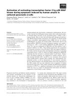
Figure 1
Association of apoptosis and TGF-β
1
Activation IndexAssociation of apoptosis and TGF-β
1
Activation Index. (a) Percentage of PBMCs undergoing apoptosis: control individuals versus SLE patients.
Cells were stained with annexin-V and propidium iodide to differentiate early apoptotic cells from necrosing and late apoptotic cells. The thick hori-
zontal bar denotes the median, and the whiskers show the interquartile range. Significance was calculated using the Mann-Whitney U test. (b)
Degree of apoptosis in PBMCs from patients and TGF-β
1
Activation Index are correlated, using Pearson test. PBMC, peripheral blood mononuclear
cell; SLE, systemic lupus erythematosus; TGF, transforming growth factor.
Arthritis Research & Therapy Vol 8 No 3 Jackson et al.
Page 4 of 7
(page number not for citation purposes)
eight (91%) had a history of arthritis and 24 (75%) had muco-
cutaneous involvement. Three (9%) had a history of renal
involvement. Twenty (60%) patients were receiving pred-
nisolone (mean dose 3.6 ± 3.9 mg/day). Seventeen (53%)
patients were receiving hydroxychloroquine and 18 (56%)
patients were receiving other disease-modifying drugs. Seven
(22%) were taking azathioprine, 5 (16%) methotrexate and
one each were receiving one (3%) monthly pulse of intrave-
nous cyclophosphamide and leflunomide. With regard to anti-
body profile at the time of study, 27 (90%) patients were
positive for ANAs, 21 (69%) had elevated antibodies to
dsDNA, 13 (41%) had ACL antibodies and nine (28%) had
lupus anticoagulant.
TGF-β
1
activation index: an in vitro measure of the in vivo
efficiency of TGF-β
1
activation
To examine the ability of SLE patients and control individuals
to activate TGF-β
1
, a ratio of levels of TGF-β
1
in freshly col-
lected blood and after 90 minutes of clotting at 37°C was
determined. The degranulation of platelets during clotting
results in release of excess latent TGF-β
1
(estimated at >25
ng/ml [18]), some of which is subsequently activated physio-
logically over 90 minutes through protease action and interac-
tion with thrombospondin, as occur in wound healing. That this
initial pool of latent TGF-β
1
was in excess is demonstrated by
the lack of association between platelet count and TGF-β
1
activation index (Pearson r = 0.143, P = 0.435; data not
shown). Comparing levels of TGF-β
1
in fresh unclotted blood
and levels in blood clotted for a limited time (90 minutes) gives
an indication of the overall efficiency of the TGF-β
1
activation
process in an individual. The TGF-β
1
activation indices were
similar between patients and control individuals (median [inter-
quartile range] AI: 1.63 [1.31–1.88] in SLE patients versus
1.50 [1.26–1.73] in control individuals, P = 0.157; data not
shown). The AI was determined once for each patient and con-
trol individuals on entry into the study. A study of the variation
in TGF-β
1
AI over time and with disease activity and treatment
in SLE patients is beyond the scope of the present study.
Apoptosis of peripheral blood mononuclear cells and
TGF-β
1
activation
SLE patients exhibited higher levels of apoptotic cells in the
total PBMC population at 24 hours, as measured by annexin-
V staining (median [interquartile range]: 3.25% [2.25–5.15%]
versus 2.20% [1.7–3.35%], P = 0.012; Figure 1a). The TGF-
βAI was significantly correlated with level of PBMC apoptosis
at 24 hours in both control individuals and patients; Figure 1b
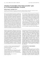
Figure 2
Correlation of LDL cholesterol and carotid IMT score with TGFβ
1
Activation IndexCorrelation of LDL cholesterol and carotid IMT score with TGFβ
1
Activation Index. (a)Correlation of LDL cholesterol and TGF-β
1
Activation Index,
using Pearson test. (b) Carotid artery IMT scores were correlated with TGF-β
1
Activation Index for control patients and SLE patients, using Pearson
correlation. A significant positive correlation was observed for control patients, whereas a significant inverse relationship was identified for SLE
patients, with low TGF-β
1
Activation Index being associated with increased mean carotid IMT score. Patients with SLICC score 0 have a higher TGF-
β
1
Activation Index than those with SLICC score 1–3, analyzed by Mann-Whitney U test. IMT, intima-media thickness; LDL, low-density lipoprotein;
SLICC, Systemic Lupus International Collaborating Clinics; TGF, transforming growth factor.
Available online />Page 5 of 7
(page number not for citation purposes)
shows the relationship for SLE patients (r = -0.504, P =
0.0062).
TGF-β
1
Activation Index and early atherosclerosis in
control individuals and SLE patients
There was no correlation between TGF-β
1
AI and calculated
LDL levels in control patients (Pearson r = 0.209, P = 0.243;
Figure 2a), but there was a significant inverse correlation
between the TGF-β
1
AI and fasting LDL-cholesterol in patients
with SLE (Pearson r = -0.404, P = 0.022; Figure 2a). TGF-β
1
AI also correlated with total cholesterol in patients with SLE
(Pearson r = -0.371, P = 0.037). TGF-β
1
AI did not correlate
with current steroid dose (P = 0.663), total duration of ster-
oids (P = 0.986), dose of antimalarial (P = 0.589), disease-
modifying antirheumatic drug therapy (P = 0.121), or SLEDAI
(P = 0.913; data not shown).
There was no difference in mean carotid IMT between patients
and controls (mean ± standard error: 0.050 ± 0.002 cm ver-
sus 0.050 ± 0.002 cm; not significant). However, the correla-
tion between TGF-β
1
AI and carotid IMT in SLE patients and
control individuals was qualitatively different (Figure 3a,b). In
control individuals there was a significant positive correlation
(r = 0.376, P = 0.031). In contrast, there was a highly signifi-
cant inverse correlation in SLE patients (r = -0.587, P =
0.0004), such that low activation status was linked with higher
IMT score. Analysis of covariance of IMT versus TGF-β
1
activa-
tion and subject group, and testing the interaction term shows
that the slopes in the control and SLE groups are significantly
different (P = 0.0001). IMT exhibited a significant correlation
with total cholesterol (Pearson r = 0.371, P = 0.037) but not
with calculated LDL (Pearson r = 0.246, P = 0.175).
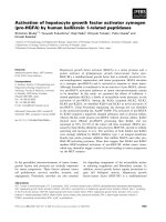
TGF-β
1
activation index, damage and disease duration
TGF-β
1
activation was lower in patients with a SDI of 1 or
greater (n = 18) than in those with a SDI of 0 (n = 12; median
[interquartile range]: 1.43 [1.20–1.59] versus 1.73 [1.43–
1.88], P = 0.034; Figure 3). The TGF-β
1
AI in SLE patients
inversely correlated with disease duration (Pearson r = -0.377,
P = 0.033; data not shown).
Discussion
We investigated the ability of SLE patients and control individ-
uals to activate latent TGF-β
1
in an in vitro assay that utilizes
the physiological activation of latent TGF-β
1
that occurs nor-
mally during blood clotting. The activation of TGF-β
1
during
clotting is complex, being mediated through several mecha-
nisms [19,20] involving protease (plasmin) activation and
interaction of TGF-β
1
with thrombospondin-1. Using an ELISA
assay validated for detection of active TGF-β
1
[15,16], we
determined the increased active TGF-β
1
after clotting a stand-
ard volume of blood at 37°C for 90 minutes relative to the non-
clotted sample. Although no differences in mean values were
observed between AIs of control individuals and SLE patients,
we hypothesized that the level of biological variation in the SLE
group could be used as a surrogate marker of the efficiency of
activating latent TGF-β
1
. This would allow us to establish
whether low or high TGF-β
1
activation efficiency could be
linked with known abnormalities in lymphocyte apoptosis and
markers of early atherosclerosis.
In accordance with other studies [21-23], we found an
increase in apoptosis in the PBMCs of SLE patients compared
with control individuals following 24 hours in culture. We
found a lower rate of apoptosis at 24 hours (median 3.35%)
compared with that described by Emlen and coworkers [21]
(mean 12%). However, our SLE patients have a low disease
activity score (mean SLEDAI score 1.75) and low damage
score (mean SLICC score 1.1). This is consistent with the
finding reported by Emlen and coworkers of a significant pos-
itive correlation between disease severity (SLAM [Systemic
Lupus Activity Measure] index) and rate of apoptosis.
There was no significant difference after 24 hours of culture
between the levels of apoptosis in patients receiving and
those not receiving steroids at the time of study. In both
patients and control individuals we observed a significant
inverse relationship between level of PBMC apoptosis and
TGF-β
1
activation index (low TGF-β
1
AI linked with high level of
apoptosis).
The significance of increased PBMC apoptosis in SLE is pro-
found, possibly reflecting increased levels of cells undergoing
activated induced cell death and/or a defect in non-inflamma-
tory phagocytosis of apoptotic cells. Failure to achieve pro-
grammed cell death and to clear apoptotic cell fragments
could be a key pathogenic factor in the development of
autoimmunity. As demonstrated in TGF-β
1
knockout mouse, a
loss of control in apoptosis affects the development and con-
trol of tolerance. Lack of TGF-β
1
leads to increases in the lev-
els of both the number of activated T cells and the levels of
Figure 3
SLE disease severity and TGFβ
1
Activation IndexSLE disease severity and TGFβ
1
Activation Index. Patients with SLICC
score 1–3 have a significantly lower TGF-β
1
AI than do patients with
mild disease (SLICC score 0). SLICC, Systemic Lupus International
Collaborating Clinics; TGF, transforming growth factor.
Arthritis Research & Therapy Vol 8 No 3 Jackson et al.
Page 6 of 7
(page number not for citation purposes)
apoptosis in activated T cells and self-tolerant T cells – a situ-
ation that may be similar to that found in SLE patients. In the
present study we report, for the first time, a significant associ-
ation between ability to activate TGF-β
1
and the degree of
PBMC apoptosis at 24 hours. In the TGF-β
1
knockout mouse
there is an increase in mitochondrial membrane potential, and
such increases are associated with initiation of apoptosis. It
was recently demonstrated that the mitochondrial membrane
potential in SLE patients is also increased [24]. Those SLE
patients with low TGF-β
1
AI status/increased apoptosis may
be most at risk for the fundamental inflammatory process that
drives SLE autoantibody production.
It is now well established that SLE patients are at fivefold to
ten-fold increased risk for coronary heart disease compared
with the general population. Classic risk factors have been
found to be of importance in promoting the development of
atherosclerosis in SLE [6]. However, after adjusting for Fram-
ingham risk factors, a significant excess risk remains [25]. This
suggests that additional factors contribute to atherogenesis in
SLE. Additional factors at play in SLE may include other meta-
bolic changes such as renal impairment and homocysteine as
well as adverse effects of steroid therapy and factors related
to the underlying disease process, such as endothelial dys-
function and immune complex deposition [26].
The inverse correlation of TGF-β
1
activation status and LDL-
cholesterol levels identified in patients but not in control indi-
viduals is therefore highly relevant to this inflammatory process
in the vascular wall. Two potential mechanisms whereby LDL
might reduce TGF-β
1
function have been described. First, it
has been shown that very-low-density lipoprotein and LDL can
inhibit the binding of active TGF-β
1
to the type II TGF-β recep-
tor and thereby suppress signalling through the receptor [27].
Second, and with particular relevance to this study, oxidized
LDL is reported to interact specifically with thrombospondin-1
and inhibit the thrombospondin-1 dependent activation of
latent TGF-β
1
[28]. In SLE, Nuttal and coworkers [29] noted
that LDL-cholesterol was more likely to exist as small dense
particles that are more prone to oxidation. Although we did not
measure LDL particle size, this difference in the type of LDL
present in patients and control individuals may explain our
observation of an inverse correlation of TGF-β
1
AI and LDL in
SLE patients, which was not seen in control individuals.
In the present study carotid IMT itself was not different
between patients and control individuals; this is consistent
with the findings of other larger series of SLE patients. Indeed,
Roman and coworkers [30] found lower carotid IMT in SLE.
Low TGF-β
1
activation was also strongly associated with
increased carotid IMT, an early marker of atherosclerotic
change. It has been proposed that low levels of active TGF-β
1
in the artery wall, resulting from apolipoprotein(a) inhibition of
plasminogen activation and failure to activate latent TGF-β
1
through plasmin proteolysis, allows endothelial and smooth
muscle cell proliferation, leading to intima-medial expansion
[8,31]. In our study this relationship was observed only in SLE
patients, and the slopes in the SLE and control groups were
significantly different (P = 0.0001), suggesting that the TGF-
β
1
interaction with IMT in SLE patients is different from that in
age-matched/sex-matched control individuals. Although the
TGF-β
1
AI did not differ significantly between patients and
control individuals, in the context of SLE increased oxidized
LDL may promote a low TGF-β
1
milieu, permitting excessive
cellular apoptosis and enhancing the propensity for athero-
genesis. Further studies are now needed to explore this
hypothesis and it may be that several different factors govern
the progression of carotid IMT in SLE. Such prospective stud-
ies will be needed to explore the interaction between inflam-
mation and early atherosclerosis in more detail both in patients
and unaffected individuals.
The association of low TGF-β
1
AI and disease duration sug-
gests that prospective studies of patients might identify
changes in TGF-β
1
activation and lipoprotein subfractions over
time that could influence the development of atherosclerosis in
SLE. Some of the atherogenic risk associated with LDL, in par-
ticular oxidized LDL, could be mediated through modulation of
the availability of active TGF-β
1
in the vasculature.
This observation has wider applications for prospective moni-
toring of TGF-β
1
activation, not only in SLE but generally in
patients developing atherosclerosis and fibrosis (for example,
chronic allograft rejection). Therapeutic manipulation of the
levels of active TGF-β
1
may offer a new perspective in control-
ling the expression of disease in patients with SLE.
Conclusion
Impairment of the TGF-β
1
system in SLE not only may impact
on the autoimmune pathophysiology of the disease but also
may modulate the development of atherosclerosis and the
increased risk for cardiovascular disease. Low activation of
TGF-β
1
is associated with increased apoptosis of PBMCs,
increased carotid IMT, high levels of LDL-cholesterol and more
severe SLE disease score. The factors in blood that modulate
activation of TGF-β
1
remain obscure, but the link with LDL-
cholesterol opens up a novel atherogenic pathway that
requires further study.
Competing interests
The authors declare that they have no competing interests.
Authors' contributions
MJ performed most of the laboratory assays, helped in statisti-
cal analysis of the data and helped to draft the manuscript. YA
obtained consent from patients and collected the samples and
patient data for the study, and participated in the coordination
of the study and writing of the manuscript. IB conceived the
study, selected the patients for study, participated in its design
and coordination, and helped to draft the manuscript. BC
Available online />Page 7 of 7
(page number not for citation purposes)
developed and carried out the TGF-β activation assay, data
analysis and contributed to the writing of the manuscript. PB
conceived the study, participated in its design and coordina-
tion and data analysis and writing of the manuscript. All
authors read and approved the final manuscript
Acknowledgements
The authors acknowledge Drs RM Bernstein, HN Snowden, MG Pat-
trick, LS Teh, P Smith and F Qasim, who identified patients for inclusion
in the study. They also acknowledge the help of Professor PN Dur-
rington and Dr M Mackness, University of Manchester Department of
Medicine, in performing the lipid analysis and Dr Steve Roberts, Univer-
sity of Manchester Biostatistics Group, for statistical advice. The study
was funded by ARC (UK) Grant number B0700 and supported by the
Wellcome Trust Clinical Research Facility, Manchester and the NHS
R&D Levy through CMMC NHS Trust.
References
1. Letterio JJ, Roberts AB: Regulation of immune responses by
TGF-beta. Annu Rev Immunol 1998, 16:137-161.
2. Geiser AG, Letterio JJ, Kulkarni AB, Karlsson S, Roberts AB, Sporn
MB: Transforming growth factor-beta
1
(TGF-beta
1
) controls
expression of major histocompatibility genes in the postnatal
mouse – aberrant histocompatibility antigen expression in the
pathogenesis of the TGF-beta
1
null mouse phenotype. Proc
Natl Acad Sci USA 1993, 90:9944-9948.
3. Dang H, Geiser AG, Letterio JJ, Nakabayashi T, Kong L, Fernandes
G, Talal N: SLE-like autoantibodies and Sjogren's syndrome-
like lymphoproliferation in TGF-beta knockout mice. J
Immunol 1995, 155:3205-3212.
4. Ohtsuka K, Gray JD, Stimmler MM, Toro B, Horwitz DA:
Decreased production of TGF-beta by lymphocytes from
patients with systemic lupus erythematosus. J Immunol 1998,
160:2539-2545.
5. Ohtsuka K, Gray JD, Quismorio FP Jr, Lee W, Horwitz DA:
Cytokine-mediated down-regulation of B cell activity in SLE:
effects of interleukin-2 and transforming growth factor-beta.
Lupus 1999, 8:95-102.
6. Bruce IN, Gladman DD, Urowitz MB: Premature atherosclerosis
in systemic lupus erythematosus. Rheum Dis Clin North Am
2000, 26:257-278.
7. Grainger DJ: Transforming growth factor beta and atheroscle-
rosis: so far, so good for the protective cytokine hypothesis.
Arterioscler Thromb Vasc Biol 2004, 24:399-404.
8. Grainger DJ, Kemp PR, Liu AC, Lawn RM, Metcalfe JC: Activation
of transforming growth factor-beta is inhibited in transgenic
apolipoprotein(a) mice. Nature 1994, 370:460-462.
9. Grainger DJ, Mosedale DE, Metcalfe JC, Bottinger EP: Dietary fat
and reduced levels of TGFbeta1 act synergistically to promote
activation of the vascular endothelium and formation of lipid
lesions. J Cell Sci 2000, 113:2355-2361.
10. Mallat Z, Tedgui A: The role of transforming growth factor beta
in atherosclerosis: novel insights and future perspectives.
Curr Opin Lipidol 2002, 13:523-529.
11. Hahn BH: Systemic lupus erythematosus and accelerated
atherosclerosis. N Engl J Med 2003, 349:2379-2380.
12. Bombardier C, Gladman DD, Urowitz MB, Caron D, Chang CH:
Derivation of the SLEDAI. A disease activity index for lupus
patients. The Committee on Prognosis Studies in SLE. Arthritis
Rheum 1992, 35:630-640.
13. Gladman DD, Urowitz MB, Goldsmith CH, Fortin P, Ginzler E, Gor-
don C, Hanly JG, Isenberg DA, Kalunian K, Nived O, et al.: The reli-
ability of the Systemic Lupus International Collaborating
Clinics/American College of Rheumatology Damage Index in
patients with systemic lupus erythematosus. Arthritis Rheum
1997, 40:809-813.
14. Mackness MI, Durrington PN: Lipoprotein analysis for clinical
studies. In Lipoprotein Analysis: a Practical Approach Edited by:
Converse CA, Skinner ER. Oxford: IRL Press; 1992:1-42.
15. Coupes BM, Newstead CG, Short CD, Brenchley PE: Transform-
ing growth factor beta 1 in renal allograft recipients. Transplan-
tation 1994, 57:1727-1731.
16. Coupes BM, Williams S, Roberts IS, Short CD, Brenchley PE:
Plasma transforming growth factor beta(1) and platelet activa-
tion: implications for studies in transplant recipients. Nephrol
Dial Transplant 2001, 16:361-367.
17. Sidhu PS, Desai SR: A simple and reproducible method for
assessing intimal-medial thickness of the common carotid
artery. Br J Radiol 1997, 70:85-89.
18. Kropf J, Schurek JO, Wollner A, Gressner AM: Immunological
measurement of transforming growth factor-beta I (TGF-beta
1) in blood; assay development and comparison. Clinical
Chemistry 1997, 43:1965-1974.
19. Grainger DJ, Wakefield L, Bethell HW, Farndale RW, Metcalfe JC:
Release and activation of platelet latent TGF-beta in blood
clots during dissolution with plasmin. Nature Medicine 1995,
1:932-937.
20. Lawrence DA: Latent-TGF-beta: an overview. Mol Cell Biochem
2001, 219:163-170.
21. Emlen W, Niebur J, Kadera R: Accelerated in vitro apoptosis of
lymphocytes from patients with systemic lupus
erythematosus. J Immunol 1994, 152:3685-3692.
22. Lorenz HM, Grunke M, Hieronymus T, Herrmann M, Kuhnel A, Man-
ger B, Kalden JR: In vitro apoptosis and expression of apopto-
sis-related molecules in lymphocytes from patients with
systemic lupus erythematosus and other autoimmune
diseases. Arthritis Rheum 1997, 40:306-317.
23. Perniok A, Wedekind F, Herrmann M, Specker C, Schneider M:
High levels of circulating early apoptic peripheral blood mono-
nuclear cells in systemic lupus erythematosus. Lupus 1998,
7:113-118.
24. Gergely P Jr, Niland B, Gonchoroff N, Pullmann R Jr, Phillips PE,
Perl A: Persistent mitochondrial hyperpolarization, increased
reactive oxygen intermediate production, and cytoplasmic
alkalinization characterize altered IL-10 signaling in patients
with systemic lupus erythematosus. J Immunol 2002,
169:1092-1101.
25. Esdaile JM, Abrahamowicz M, Grodzicky T, Li Y, Panaritis C, du
Berger R, Cote R, Grover SA, Fortin PR, Clarke AE, Senecal JL:
Traditional Framingham risk factors fail to fully account for
accelerated atherosclerosis in systemic lupus erythematosus.
Arthritis Rheum 2001, 44:2331-2337.
26. Bruce IN: Cardiovascular disease in lupus patients: should all
patients be treated with statins and aspirin? Best Pract Res
Clin Rheumatol 2005, 19:823-838.
27. Grainger DJ, Byrne CD, Witchell CM, Metcalfe JC: Transforming
growth factor beta is sequestered into an inactive pool by
lipoproteins. J Lipid Res 1997, 38:2344-2352.
28. Sakamoto YI, Miyazaki A, Tamagawa H, Wang GP, Horiuchi S:
Specific interaction of oxidized low-density lipoprotein with
thrombospondin-1 inhibits transforming growth factor-beta
from its activation. Atherosclerosis 2005, 183:85-93.
29. Nuttall SL, Heaton S, Piper MK, Martin U, Gordon C: Cardiovas-
cular risk in systemic lupus erythematosus: evidence of
increased oxidative stress and dyslipidaemia. Rheumatology
(Oxford) 2003, 42:758-762.
30. Roman MJ, Shanker BA, Davis A, Lockshin MD, Sammaritano L,
Simantov R, Crow MK, Schwartz JE, Paget SA, Devereux RB,
Salmon JE: Prevalence and correlates of accelerated athero-
sclerosis in systemic lupus erythematosus. N Engl J Med
2003, 349:2399-2406.
31. Lawn RM, Pearle AD, Kunz LL, Rubin EM, Reckless J, Metcalfe JC,
Grainger DJ: Feedback mechanism of focal vascular lesion for-
mation in transgenic apolipoprotein(a) mice. J Biol Chem
1996, 271:31367-31371.