Báo cáo y học: "Two discrete events, human T-cell leukemia virus type I Tax oncoprotein expression and a separate stress stimulus, are required for induction of apoptosis in T-cells" pptx
Bạn đang xem bản rút gọn của tài liệu. Xem và tải ngay bản đầy đủ của tài liệu tại đây (814.27 KB, 12 trang )
Retrovirology
BioMed Central
Open Access
Research
Two discrete events, human T-cell leukemia virus type I Tax
oncoprotein expression and a separate stress stimulus, are required
for induction of apoptosis in T-cells
Takefumi Kasai and Kuan-Teh Jeang*
Address: Laboratory of Molecular Microbiology, NIAID, National Institutes of Health, Bethesda, Maryland 20892-0460, USA
Email: Takefumi Kasai - ; Kuan-Teh Jeang* -
* Corresponding author
Published: 06 May 2004
Retrovirology 2004, 1:7
Received: 23 April 2004
Accepted: 06 May 2004
This article is available from: />© 2004 Kasai and Jeang; licensee BioMed Central Ltd. This is an Open Access article: verbatim copying and redistribution of this article are permitted in all
media for any purpose, provided this notice is preserved along with the article's original URL.
Abstract
Background: It is poorly understood why many transforming proteins reportedly enhance both
cell growth (transformation) and cell death (apoptosis). At first glance, the ability to transform and
the ability to engender apoptosis seem to be contradictory. Interestingly, both abilities have been
widely reported in the literature for the HTLV-I Tax protein.
Results: To reconcile these apparently divergent findings, we sought to understand how Tax might
cause apoptosis in a Jurkat T-cell line, JPX-9. Tax expression can be induced equally by either
cadmium (Cd) or zinc (Zn) in JPX-9 cells. Surprisingly, when induced by Zn, but not when induced
by Cd, Tax-expression produced significant apoptosis. Under our experimental conditions, Zn but
not Cd, induced SAPK (stress activated protein kinase)/JNK (Jun kinase) activation in cells. We
further showed that transient over-expression of Tax-alone or Jun-alone did not induce cell death.
On the other hand, co-expression of Tax plus Jun did effectively result in apoptosis.
Conclusion: We propose that Tax-expression alone in a T-cell background insufficiently accounts
for apoptosis. On the other hand, Tax plus activation of a stress kinase can induce cell death. Thus,
HTLV-I infection/transformation of cells requires two discrete events (i.e. oncoprotein expression
and stress) to produce apoptosis.
Background
Human T-lymphotropic virus type I (HTLV-I) causes adult
T-cell leukemia (ATL; reviewed in [1-3]). ATL develops in
a minority of HTLV-I infected individuals with a long
latent period. This pathological course suggests a multistage process of immortalization and transformation of Tlymphocyte. HTLV-I encodes a 40 kDa phosphoprotein,
Tax. Tax immortalizes T- lymphocytes [4-6] and transforms rat fibroblasts [7,8]. Tax is also a transcriptional
activator of the HTLV-I LTR [9-11]; reviewed in [12].
While the exact events leading to transformation are
incompletely understood, several important cellular proc-
esses are dysregulated by Tax in parallel (reviewed in
[1,13,14]. This is likely explained by the fact that Tax can
activate NF-κB, SRF-, and CREB/ATF-responsive genes and
can markedly accelerate cell cycle progression [15];
reviewed in [16].
The ability to transform and the ability to engender apoptosis seem to be contradictory functions. Interestingly,
both abilities have been widely reported in the literature
for the HTLV-I Tax protein. Tax has been shown to inhibit
apoptosis [4,5,17-22]. On the other hand, Tax has also
been shown to induce apoptosis [23-32]. Indeed Kao et al.
Page 1 of 12
(page number not for citation purposes)
Retrovirology 2004, 1
showed that Tax sensitized cells to apoptotic cell death
induced by DNA damaging agents [33]. It remains puzzling why Tax like many other oncoproteins seemingly
enhances both cell growth (transformation) and cell
death (apoptosis) ([34]).
To dissect the cell growth/death paradox relevant to
HTLV-I, we sought to examine the requirements for Tax to
cause apoptosis in a T-cell line, Jurkat. We used JPX-9, a
stable transfectant of Jurkat that has incorporated a Taxgene under the inducible control of a metallothionein
promoter [35]. In JPX-9, Tax expression can be induced
equally-well using either Cd or Zn. Intriguingly, we found
that under our induction conditions the latter (Zn) but
not the former (Cd) represented a stress stimulus. Thus,
we observed marked activation of SAPK/JNK in our JPX-9
cells exposed to Zn, but not Cd. We propose that the combined effects of Tax expression and stress kinase activation
perturb the cell growth/cell death equilibrium to favour
the latter.
Results
Zinc, but not cadmium, treatment induces apoptosis in
JPX-9 cells
To examine the effect of Tax on the growth/death of Jurkat
cells, we studied its induction by Zn or Cd in the JPX-9 cell
line. As mentioned above, JPX-9 is a Jurkat derived cell
line in which Tax expression is dictated by a metallothionein promoter. We treated JPX-9 or parental control
Jurkat cells with either Zn or Cd. Interestingly, when
nuclear morphologies were examined by staining with
Hoechst 33258, we saw that JPX-9 cells treated with Zn
showed significant apoptosis, while JPX-9 cells treated
with Cd or parental Jurkat cells treated with either Zn or
Cd were minimally affected (Figure 1A).
We sought to independently confirm the finding of cell
death in JPX-9 cells using a colorimetric MTT assay (see
Materials and Methods) which measures cellular viability
(Figure 1B,1C). To broaden the generality of our experiment, we also examined 6 HTLV-I transformed T-cells
(WT-1, TL-Su, TL-Omi, C8166, WT-4, and ILT-Hod; Figure
1A,1B). Using MTT, we checked cellular viability of these
HTLV-I transformed cells as well as Jurkat and JPX-9
treated with either Zn or Cd. Although there were some
cell to cell variations, the overall trend from the HTLV-I
and JPX-9 cells treated with Zn for 24 (Figure 1B) or 48
(Figure 1C) hours was one of lower viability as compared
to counterparts treated with Cd. We noted that Jurkat was
an exception in exhibiting no difference in viability
between Cd or Zn treatment (Figure 1B,1C).
During apoptosis, the cellular 116 kDa nuclear enzyme
poly(ADP-ribose) polymerase (PARP) is cleaved by caspase-3 to a smaller 85 kDa moiety [36]. To further charac-
/>
terize and confirm that cell death in JPX-9 was due to
apoptosis, we investigated PARP cleavage by Western blotting using anti-PARP (Figure 2). Consistent with apoptotic
death, we observed a higher degree of PARP processing in
JPX-9 cells treated with Zn than cells treated with Cd (Figure 2).
A factor common to all the HTLV-I transformed cells and
JPX-9 (Figure 1B,1C) is the expression of Tax. Because JPX9 cells treated by Zn or Cd should, in principle, induce Tax
equally, we were puzzled by the divergent apoptotic phenotypes. To rule out that the variance in Cd- and Znapoptotic profiles in JPX-9 was trivially due to different
efficiencies of Tax induction by the two cations, we
directly examined the kinetics of Tax protein expression
after Zn- or Cd- treatment. The results indicated essentially no difference in Tax induction by either Zn or Cd
over the 24 to 48 hours treatment period (Figure 3). Thus,
the Zn- vs. Cd- variance in JPX-9 apoptosis is unlikely
explained simply by differences in Tax expression.
Zn and Cd treatments activated SAPK/JNK differently
Activation of SAPK/JNK has been reported to play a role in
the stress induction of cellular apoptosis [14,37,38]. We
next investigated whether Zn and Cd may have different
thresholds for activation of SAPK/JNK. Figure 4 shows
sequential SAPK/JNK activity in Jurkat and JPX-9 cells
upon treatment with Zn or Cd. We monitored the activation of JNK using anti-phospho-c-Jun specific antibody.
Based on Western blotting results, SAPK/JNK was activated by phosphorylation in both Jurkat and JPX-9 cells
after Zn, but not Cd, treatment. Hence, phospho-c-Jun
was detected by 6 hours after treatment with Zn in JPX-9
and Jurkat cells (Figure 4A), but no such phosphorylation
was seen with Cd treatment (Figure 4B). These findings
suggest that JPX-9 cells treated with Zn would contain
both activated SAPK/c-Jun and Tax, while the same cells
treated with Cd would have only Tax.
Different caspase profiles after Zn and Cd treatments
Because caspases are the effector proteases for apoptosis,
we next investigated caspase profiles in Jurkat and JPX-9
cells after Zn and Cd treatments (Figure 5A). Activation of
caspases 3, 8, and 9 requires the processing of pro-protein
precursors to smaller active forms. To monitor the effects
of Zn and Cd, we examined the integrity of pro-caspase 3,
8 and 9 in control and cation-treated Jurkat and JPX-9
cells. When compared to control, there was little reduction in the three pro-capases upon Cd treatment (Figure
5A). On the other hand, the levels of pro-caspase-3, -8 and
-9 in JPX-9 cells treated with Zn were all decreased. Of particular note, pro-caspase 9 was reduced by approximately
50% in JPX-9 cells treated with Zn (i.e. 0.54; Figure 5A).
Page 2 of 12
(page number not for citation purposes)
Retrovirology 2004, 1
/>
P roportion of Apoptotic C ells
(%)
a)
40
Jurkat+Cd
Jurkat +Zn
JPX-9+Cd
JPX-9+Zn
30
20
10
0
0
12
24
36
Hours
t
9
JPX-
Jurka
od
ILT-H
6
WT-4
C816
ml
TL-O
u
Cd treatment
Zn treatment
TL-S
1.4
1.2
1
0.8
0.6
0.4
0.2
0
WT-1
C ell V iability R atio
(treated / untreated)
b)
c)
1
0.8
Cd treatment
Zn treatment
0.6
9
JPX-
t
Jurka
od
ILT-H
WT-4
6
C816
ml
TL-O
TL-S
0
u
0.4
0.2
WT-1
C ell V iability R atio
(treated / untreated)
1.2
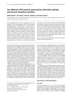
Figure 1
Quantitation of apoptosis and viability in Jurkat and JPX-9 cells treated with ZnCl2 or CdCl2
Quantitation of apoptosis and viability in Jurkat and JPX-9 cells treated with ZnCl2 or CdCl2. A) JPX-9 cell treated
with Zn show higher levels of apoptosis, while JPX-9 cell treated with Cd and Jurkat cell treated with either Zn or Cd showed
lower levels. Y-axis is % apoptosis, and X-axis is hours after treatment. B) and C) Cell viability was quantified using a modified
MTT colorimetric assay. Quantification of viability in HTLV-I transformed cell lines, as indicated, was after treatment with
ZnCl2 or CdCl2 for 24 hours (B) or 48 hours (C). HTLV-I transformed cell lines treated with Zn showed lower cell viability
compared to cells treated with Cd. Y-axes are % viability with 100% set as 1; X-axes indicate the name of the cell line.
Page 3 of 12
(page number not for citation purposes)
Retrovirology 2004, 1
/>
JPX-9
Cd2+
Zn2+
0h
6h
12h
24h 6h
12h
24h
116kD
PARP
85kD
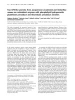
PARP cleavage analysis in JPX-9 by Western blotting with anti-PARP
Figure 2
PARP cleavage analysis in JPX-9 by Western blotting with anti-PARP. PARP proenzyme (116 kD) and cleaved subunit (85 kD) are indicated on the left by arrows. JPX-9 cell treated with Zn showed higher cleaved/uncleaved PARP.
Bcl-2 [39], survivin [40], and XIAP [41] are three cellular
anti-apoptotic factors. We also asked whether these three
factors contribute to Zn-induced apoptosis of JPX-9 cells.
Using specific antisera, we compared the levels of these
three factors in untreated Jurkat/JPX-9 to their Zn or Cdtreated counterparts (Figure 5B). We saw no difference in
Bcl-2 and survivin levels between JPX-9/Zn and JPX-9/Cd
cells. However, XIAP level was more significantly reduced
in JPX-9/Zn (0.62) than in JPX-9/Cd (0.96) cells, suggesting that this factor may contribute to Zn-induced apoptotic outcome (Figure 5B, right).
To confirm the Western blot results in figure 5A, we further quantified the enzymatic profiles of caspase 3, 8 and
9 using a spectrophotometric peptide cleavage assay.
Compared to controls, we observed that capase 8 activity
was mildly enhanced in JPX-9/Zn cells (Figure 6B) while
caspase 3 (Figure 6A) and caspase 9 (Figure 6C) activities
were more significantly increased.
Over-expression of Jun cooperates with Tax to induce
apoptosis
In other settings, SAPK/Jun activation has been shown to
be involved in cellular apoptosis. Above data suggest that
Tax expression alone is insufficient to cause cell death. On
the other hand, our findings are compatible with Tax
expression plus Jun activation cooperating to induce
apoptosis in JPX-9 cells. We next performed transient
transfection experiments in order to address the cooperativity between Tax and Jun. Because suspension cells are
notoriously difficult to transfect with efficiency, in figure
7, we transiently transfected diploid Hct116 colon carcinoma cell line separately with vector control, Tax, inactive
Tax mutant ∆2-58, pCMV-HA-JNK, Tax + pCMV-HA-JNK
Page 4 of 12
(page number not for citation purposes)
Retrovirology 2004, 1
/>
JPX-9
Cd2+
Zn2+
0h
6h
1
2
12h
18h
24h
48h
6h
12h
3
4
5
6
7
8
18h
24h
48h
Tax
Actin
9
10
11
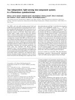
Figure 3
Western blot analysis of Tax expression in the JPX-9 cells
Western blot analysis of Tax expression in the JPX-9 cells. Tax expression was equally induced by either Zn or Cd.
Tax was detected with polyclonal anti-Tax [63]. Equal sample loading was verified with anti-actin (bottom).
or Tax∆2-58 + pCMV-HA-JNK. 48 hours after transfection,
cells were examined morphologically for signs of apoptosis. At transfection efficiency where 50% of cells received
DNA (data not shown), we observed that approximately
40% of Tax + JNK cells became apoptotic (Figure 7). Normalized to transfection efficiency, this suggested that 80%
of all cells that received Tax + JNK succumbed to apoptosis. By contrast, no significant apoptosis was observed for
either Tax-alone or JNK-alone suggesting that under the
conditions employed neither is sufficient to elicit significant cell death (Figure 7).
Discussion
Why oncoproteins seemingly enhance both cell growth
(tranformation) and cell death (apoptosis) remain
incompletely elucidated. Here, using HTLV-I Tax as a
model we asked whether expression of this oncoprotein
alone is sufficient to damage/stress the cell such as to provoke demise. Our findings suggest that Tax cannot singularly induce apoptosis efficiently in a T-cell line.
In an attempt to better understand HTLV-I biology, we
sought to define the requirements for Tax to cause apoptosis in a Jurkat T-cell line. We used JPX-9, a stable transfectant of Jurkat in which Tax expression is controlled by
a metallothionein promoter which can be equally activated by Zn or Cd. In this experimental background, we
found that Tax-expression when induced by Zn, but not
when induced by Cd, provoked highly significant apoptotic death at otherwise non-cytotoxic concentrations for
each divalent cation-alone (Figure 1). Tax + Zn-induced
apoptosis was most strongly associated with enhanced
caspase 9 activity, although smaller increases in caspase 3
Page 5 of 12
(page number not for citation purposes)
Retrovirology 2004, 1
/>
a) Zn2+ treatment
Jurkat
0h
6h
JPX-9
12h
0h
6h
12h
Anti-phospho-c-Jun
Anti-c-Jun
b) Cd2+ treatment
Jurkat
0h
6h
JPX-9
12h
0h
6h
12h
Anti-phospho-c-Jun
Anti-c-Jun
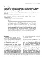
Figure 4
Zn activated phosphorylated SAPK/JNK in Jurkat and JPX-9 cells
Zn activated phosphorylated SAPK/JNK in Jurkat and JPX-9 cells. Western blotting detected phosphorylated c-Jun
within 6 hours after Zn treatment (A), but was not seen after Cd treatment (B). Anti-phospho-c-Jun was used to detect phosphorylated c-Jun while anti-c-Jun detected total c-Jun protein.
Page 6 of 12
(page number not for citation purposes)
Retrovirology 2004, 1
/>
Jurkat
a)
Control
JPX-9
Zn2+
Cd2+
0.94
1.03
0.88
1.13
1.02
1.04
0.85
1.06
0.83
0.81
0.54
1.21
Control
Zn2+
Cd2+
Pro-caspase-3
Ratio
(treated/untreated)
Pro-caspase-8
Ratio
(treated/untreated)
Pro-caspase-9
Ratio
(treated/untreated)
b)
Bcl-2
Ratio
(treated/untreated)
1.11
1.03
1.14
1.03
1.03
1.03
1.08
1.06
0.66
0.68
0.62
0.96
Survivin
Ratio
(treated/untreated)
XIAP
Ratio
(treated/untreated)
Actin
Western blotting analyses of caspase-3, -8, -9, Bcl-2, survivin and XIAP in Jurkat and JPX-9 cells after Zn or Cd treatment
Figure 5
Western blotting analyses of caspase-3, -8, -9, Bcl-2, survivin and XIAP in Jurkat and JPX-9 cells after Zn or Cd
treatment. A) Enhanced processing of pro-caspase 9 in JPX-9 cells after Zn treatment. Expression of pro-caspase-3, -8, and 9 in Jurkat and JPX-9 cells were checked by Western blotting. Jurkat and JPX-9 cells were treated with ZnCl2 or CdCl2 for 24
hours, and the indicated proteins were detected using specific anti-sera. Ratio is the band intensity in treated sample versus
untreated control. B) Expression of Bcl-2, survivin and XIAP in Jurkat and JPX-9 cells. Jurkat and JPX-9 cells were treated with
ZnCl2 or CdCl2 for 24 hours. Note that XIAP expression in ZnCl2 treated JPX-9 cells was reduced while its expression in
CdCl2-treated JPX-9 cells was maintained.
Page 7 of 12
(page number not for citation purposes)
Retrovirology 2004, 1
/>
a)
1.6
C as pas e-3 activity
1.4
1.2
1
0.8
0.6
J urkat+Zn
J P X9+Zn
J urkat C d
J P X9+C d
0.4
0.2
0
b)
1.6
C as pas e-8 activity
1.4
1.2
1
0.8
0.6
J urkat+Zn
J P X9+Zn
J urkat C d
J P X9+C d
0.4
0.2
0
C as pas e-9 activity
c)
2.5
2
1.5
1
J urkat+Zn
J P X9+Zn
J urkat C d
J P X9+C d
0.5
0
Figure 6
Enzymatic assays of caspases in JPX-9 cell treated with Zn or Cd
Enzymatic assays of caspases in JPX-9 cell treated with Zn or Cd. Spectrophotometric assays of caspase activities are
as described in Methods. Caspase 3 (A), caspase 8 (B), and caspase 9 activities were measured in cells 24 hours after treatment.
Caspase 9 activity was especially enhanced in JPX-9 cells treated with Zn (C).
Page 8 of 12
(page number not for citation purposes)
Retrovirology 2004, 1
/>
ment is Tax plus SAPK/JNK activation; and it matters not
whether this occurs via Tax plus Zn or Tax plus Cd. We
also noted with interest that similar to our findings (Figure 6A,6C), Zn activation of a death pathway in a human
Burkitt lymphoma B cell line was associated with activation of caspase-9 and caspase-3 [44].
B eta-gal Activity
Hct 116
120
100
80
60
40
20
0
V ector
J NK
T ax
T ax+J NK
T ax∆2-58
T ax∆2-58+J NK
Figure 7
Induction of cell death in Hct116 cells by Tax + Jun kinase
Induction of cell death in Hct116 cells by Tax + Jun
kinase. Hct116 cells were transfected with pCMV-beta-gal
and control plasmid pUC19 or the indicated plasmids.
Reduction in beta-gal values reflects cell death. Cells transfected with CMV IE-driven JNK-expression plasmid (pCMVHA-JNK) + CMV-Tax showed significantly lower level of
beta-gal values, while cells with other combinations of transfected DNA showed higher levels. Tax∆2-58 is an inactive
Tax mutant. Transfection efficiencies achieved in the experiments were approximately 50%. Values represent averages
from three independent experiments.
and caspase 8 were also observed (Figures 5A, 6). Currently, we do not know whether the caspase 9 findings
reflect yet characterized mitochondrial toxicity of Tax.
How can one explain the different presentations for Zn
and Cd in Tax-induced apoptosis? First, using phosphospecific antibody, we observed increased activation SAPK/
JNK in cells exposed to Zn, while Cd exposure conducted
in parallel did not activate SAPK/JNK. At the low dose (20
µM) used in our study, Cd has been shown not be perturb
SAPK/JNK [42]. However, we caution that higher doses of
Cd (i.e. >30 µM) can also activate SAPK/JNK. On the other
hand, consistent with our results, acute exposure to Zn, as
performed here, has also been reported to enhance SAPK/
JNK activity in human bronchial epithelial cells [43].
While we used a higher concentration of Zn than Cd to
induce JPX-9 cells, the salient point is that under conditions of equal induction of Tax, the former activated
SAPK/JNK while the latter did not. With higher
concentrations of Cd which did induce SAPK/JNK, Tax
expression plus Cd treatment also produced apoptosis
(data not shown). Hence the critical apoptosis require-
Consistent with our observations, several studies support
that SAPK/JNK plays an important role in apoptosis
[37,45-48]. A requirement for SAPK/JNK in apoptotic
induction by UV irradiation was demonstrated using
embryonic fibroblasts derived from a double-knockout
mouse which lacked expression of both JNK1 and JNK2
[49]. Moreover, it was shown that ionizing radiation
induced the translocation of JNK/SAPK to the mitochondria and the association of JNK/SAPK with Bcl-xL protein
[50]. Additional factors required for UV and SAPK/JNK
induced apoptosis include the cytochrome C effectors
Apaf-1, caspase-9, and caspase-3 [51,52].
In a parallel oncoprotein system, Evan et al. had previously demonstrated that expression of c-Myc engendered
apoptosis in serum-deprived rodent fibroblasts [53,54].
Related to these findings, Yu et al. found that Mycdependent apoptosis was also associated with activation
of JNK/SAPK [55]. Accordingly, Tax resembles Myc in that
both proteins are transforming entities which share
conditional apoptotic properties when expressed in the
context of activated SAPK/JNK. One interpretation which
emerges plausibly from our current work is that Tax primarily enforces changes in cellular metabolism for accelerated growth and transformation; however, these driving
impulses may unwittingly dysregulate normal physiological balance to an extent that sensitizes cells to various
pro-apoptotic insults. A similar interpretation has also
been suggested for c-Myc [56].
Our work provides added insight into the various reports
that Tax is both pro- and anti-apoptotic. We believe that
Tax can provoke a pro-apoptotic phenotype in a setting
when the cell is faced with an additional stress stimulus
manifested through the JNK/SAPK cascade. On the other
hand absent additional stress, Tax is primarily pro-survival through its effects on the NF-κB cascade [12].
Indeed, NF-κB has been clearly shown to serve a protective pro-survival role through its upregulation of antiapoptotic genes [57-59]. Finally, the clinical presentation
of ATL does argue that in contesting opposing effects the
pro-transforming/pro-survival function of Tax ultimately
prevails. Nevertheless, the extremely long latency (20 to
30 years) after HTLV-I infection required for ATL emergence suggests that most virally infected cells suffer apoptotic fates and that clonal escape from apoptosis to
transformation is an exceedingly rare event.
Page 9 of 12
(page number not for citation purposes)
Retrovirology 2004, 1
Conclusions
Because Tax is a transforming protein, it seems unlikely
that this oncoprotein's primary function is to induce
apoptosis. Here, we show that Tax-alone, consistent with
its oncogenic role, is insufficient to induce cell death in a
Jurkat T-cell line. On the other hand, Tax plus a stress
stimulus which activates SAPK/JNK can collectively cause
apoptosis. Our work helps to reconcile the divergent
reports that Tax is both apoptosis inducing and anti-apoptotic (i.e. transforming).
Methods
Cell culture
Jurkat cells (ATCC), and Tax-inducible JPX-9 and control
JPX/M cells [35] were cultured in RPMI 1640
supplemented with 10% fetal calf serum (RPMI-FCS).
Expression of Tax was induced by addition of ZnCl2 to
120 µM or CdCl2 to 20 µM, respectively. MT-I, TL-OmI,
TL-Su, C8166, MT-4, and ILT-Hod are human HTLV-1transformed T-cell lines (MT-I,TL-OmI, TL-Su, C8166,
and MT-4 are IL-2 independent. ILT-Hod is IL-2 dependent.). ILT cell line was cultured in RPMI-FCS with 10 U/ml
IL-2.
Apoptosis assay
Analysis of apoptotic cells was by Hoechst dye staining to
characterize nuclear morphology. Cells were harvested at
designated intervals up to 48 h. After harvesting, the cells
were pelleted by centrifugation (1500 rpm, 5 minutes)
and washed with PBS. The cell pellets were resuspended
into 50 µl of 1% formaldehyde-0.2% glutaraldehyde. 20
µl of the cell suspension was dried on a poly-L-lysine
coated slide. After wash with PBS, slides were stained with
PBS containing 10 µg/ml of Hoechst 33258 (Sigma) for
10 minutes at room temperature. Fluorescence microscopy was used to assess the percentage of apoptotic cells.
To measure the proportion of apoptotic cells, at least 300
cells were counted.
Cell survival assay
T-cells (5 × 10⁴ cells/ml) in 96-well flat-bottom plates
were preincubated for 24 h and then treated with ZnCl2
(120 µM) or CdCl2 (20 µM) at 37°C for 48 hours. Cells
were harvested at 12 hour time intervals up to 48 hours.
The number of viable cells in each clone was measured by
a dye-reduction assay using WST-8 (2-(2-methoxy-4nitrophenyl)-3-(4-nitrophenyl)-5-(2,4-disulfophenyl)2H-tetrazolium, monosodium salt) (Dojindo Molecular
Technologies, Gaithersburg, MD, USA). Cell viability
represented the ratio of WST-8 activity of cells treated with
these drugs relative to that of untreated cells.
/>
extraction buffer and sonicated. Protein concentrations
were determined using the Bio-Rad protein assay system
(Bio-Rad, Richmond, CA, USA). Polyclonal anti- caspase3, polyclonal anti-caspase-9, and monoclonal anti-caspase-8 were purchased from Pharmingen. Monoclonal
anti-XIAP was purchased from Panvera. Polyclonal antisurvivin, -cIAP-1, and -cIAP-2, and monoclonal anti-PARP
and anti-Bcl-2 were purchased from Santa Cruz Biotechnology. Mouse monoclonal anti-actin (clone AC-15) was
purchased from Sigma. Cell lysates were fractionated in
10% SDS-polyacrylamide gels prior to transfer to membrane (Immobilon-P; Millipore, Bedford, MA, USA) by
standard protocol. Blots were visualized by chemiluminescence (Tropix, Bedford, MA, USA). c-Jun phosphorylation was selectively measured using a phospho-c-Jun
antibody.
Caspase assays
Cells were grown in RPMI 1640 supplemented with 10%
fetal calf serum (RPMI-FCS) and treated with ZnCl2 or
CdCl2 for 24 hours. Cells (2 × 10⁶) were collected by centrifugation at 200 × g for 10 minutes. Pellets were resuspended into 50 µl of cold cell lysis buffer provided in
ApoAlert caspase colorimetric assay kits (Clontech, Palo
Alto, CA) or caspase-9 colorimetric protease assay kit
(Panvera/Takara). Cell lysates were microcentrifuged at
12,000 rpm for 3 min at 4°C and the supernatants were
transferred to 96-well plates for detection of caspase-3 or
caspase-8 activities. Caspase-3 and caspase-8 activities
were measured using spectrophotometric detection of the
chromophore p-nitroanilide (pNA) after cleavage from
the labeled substrate DEVD-pNA and IETD-pNA, respectively. Caspase-9 activity was measured using spectrophotometric detection of the chromophore pNA after cleavage
from the labeled substrate LEHD-pNA.
Transfection
For assay of cooperativity between JNK and Tax in the
induction of apoptosis, we used colon cancer cell lines
(Hct116) [60]. CMV IE-driven JNK-expression plasmid
(pcDNA-HA-JNK) [61] and CMV-Tax and CMV-Tax
mutant (∆2-58) plasmids have been previously described
[62]. Cells were transfected with CMV-beta-gal and either
control plasmid pUC19 or the indicated combination of
plasmids. Beta-gal activities were measured 24 hours after
transfection. Individual beta-gal values are expressed relative to the value from cells transfected with CMV-beta-gal
and control pUC19 plasmid. Reduction in beta-gal values
was quantitated as a reflection of cell death.
Competing interests
None declared.
Western blotting
Cells were collected by centrifugation at 1,500 rpm, after
washing in PBS. Then cells were lysed by the addition of
Page 10 of 12
(page number not for citation purposes)
Retrovirology 2004, 1
/>
Authors' contributions
TKperformed most of the experiments. Both TK and KTJ
participated in experimental design, data interpretation
and writing of manuscript.
19.
20.
Acknowledgements
We thank Lan Lin for help with preparation of manuscript and figure, and
RK Yedavalli for assistance with reference formatting.
21.
References
1.
2.
3.
4.
5.
6.
7.
8.
9.
10.
11.
12.
13.
14.
15.
16.
17.
18.
Yoshida M: Multiple viral strategies of HTLV-1 for dysregulation of cell growth control. Annu Rev Immunol 2001, 19:475-496.
Poiesz BJ, Poiesz MJ, Choi D: The human T-cell lymphoma/
leukemia viruses. Cancer Invest 2003, 21:253-277.
Matsuoka M: Human T-cell leukemia virus type I and adult Tcell leukemia. Oncogene 2003, 22:5131-5140.
Grassmann R, Berchtold S, Radant I, Alt M, Fleckenstein B, Sodroski
JG: Role of human T-cell leukemia virus type 1 X region proteins in immortalization of primary human lymphocytes in
culture. J Virol 1992, 66:4570-4575.
Grassmann R, Dengler C, Muller-Fleckenstein I, Fleckenstein B,
McGuire K, Dokhelar MC: Transformation to continuous
growth of primary human T lymphocytes by human T-cell
leukemia virus type I X-region genes transduced by a Herpesvirus saimiri vector. Proc Natl Acad Sci U S A 1989,
86:3351-3355.
Rosin O, Koch C, Schmitt I, Semmes OJ, Jeang KT, Grassmann R: A
human T-cell leukemia virus Tax variant incapable of activating NF-kappaB retains its immortalizing potential for primary T-lymphocytes. J Biol Chem 1998, 273:6698-6703.
Tanaka A, Takahashi C, Yamaoka S, Nosaka T, Maki M, Hatanaka M:
Oncogenic transformation by the tax gene of human T-cell
leukemia virus type I in vitro. Proc Natl Acad Sci U S A 1990,
87:1071-1075.
Matsumoto K, Shibata H, Fujisawa JI, Inoue H, Hakura A, Tsukahara
T, Fujii M: Human T-cell leukemia virus type 1 Tax protein
transforms rat fibroblasts via two distinct pathways. J Virol
1997, 71:4445-4451.
Brady J, Jeang KT, Duvall J, Khoury G: Identification of p40xresponsive regulatory sequences within the human T-cell
leukemia virus type I long terminal repeat. J Virol 1987,
61:2175-2181.
Seiki M, Inoue J, Takeda T, Hikikoshi A, Sato M, Yoshida M: The p40x
of human T-cell leukemia virus type I is a trans-acting activator of viral gene transcription. Jpn J Cancer Res 1985,
76:1127-1131.
Jeang KT, Boros I, Brady J, Radonovich M, Khoury G: Characterization of cellular factors that interact with the human T-cell
leukemia virus type I p40x-responsive 21-base-pair
sequence. J Virol 1988, 62:4499-4509.
Jeang KT: Functional activities of the human T-cell leukemia
virus type I Tax oncoprotein: cellular signaling through NFkappa B. Cytokine Growth Factor Rev 2001, 12:207-217.
Marriott SJ, Lemoine FJ, Jeang KT: Damaged DNA and miscounted chromosomes: human T cell leukemia virus type I
tax oncoprotein and genetic lesions in transformed cells. J
Biomed Sci 2002, 9:292-298.
Jeang KT, Giam CZ, Majone F, Aboud M: Life, Death and Tax: role
of HTLV-I oncoprotein in genetic instability and cellular
transformation. J Biol Chem 2004 in press.
Neuveut C, Low KG, Maldarelli F, Schmitt I, Majone F, Grassmann R,
Jeang KT: Human T-cell leukemia virus type 1 Tax and cell
cycle progression: role of cyclin D-cdk and p110Rb. Mol Cell
Biol 1998, 18:3620-3632.
Neuveut C, Jeang KT: HTLV-I Tax and cell cycle progression.
Prog Cell Cycle Res 2000, 4:157-162.
Brauweiler A, Garrus JE, Reed JC, Nyborg JK: Repression of bax
gene expression by the HTLV-1 Tax protein: implications for
suppression of apoptosis in virally infected cells. Virology 1997,
231:135-140.
Mulloy JC, Kislyakova T, Cereseto A, Casareto L, LoMonico A, Fullen
J: Human T-cell lymphotropic/leukemia virus type 1 Tax
abrogates p53-induced cell cycle arrest and apoptosis
22.
23.
24.
25.
26.
27.
28.
29.
30.
31.
32.
33.
34.
35.
36.
37.
38.
39.
40.
through its CREB/ATF functional domain. J Virol 1998,
72:8852-8860.
Tsukahara T, Kannagi M, Ohashi T, Kato H, Arai M, Nunez G.: Induction of Bcl-x(L) expression by human T-cell leukemia virus
type 1 Tax through NF-kappaB in apoptosis-resistant T-cell
transfectants with Tax. J Virol 1999, 73:7981-7987.
Kawakami A, Nakashima T, Sakai H, Urayama S, Yamasaki S, Hida A:
Inhibition of caspase cascade by HTLV-I tax through induction of NF-kappaB nuclear translocation. Blood 1999,
94:3847-3854.
Nakashima K, Kawakami A, Hida A, Yamasaki S, Nakamura H,
Kamachi M: Protection of mitochondrial perturbation by
human T-lymphotropic virus type 1 tax through induction of
Bcl-xL expression. J Lab Clin Med 2003, 142:341-347.
Saggioro D, Acquasaliente L, Daprai L, Chieco-Bianchi L: Inhibition
of apoptosis by human T-lymphotropic virus type-1 tax
protein. Ann N Y Acad Sci 2003, 1010:591-597.
Chen X, Zachar V, Zdravkovic M, Guo M, Ebbesen P, Liu X: Role of
the Fas/Fas ligand pathway in apoptotic cell death induced by
the human T cell lymphotropic virus type I Tax
transactivator. J Gen Virol 1997, 78(Pt 12):3277-3285.
Chlichlia K, Los M, Schulze-Osthoff K, Gazzolo L, Schirrmacher V,
Khazaie K: Redox events in HTLV-1 Tax-induced apoptotic Tcell death. Antioxid Redox Signal 2002, 4:471-477.
Chlichlia K, Busslinger M, Peter ME, Walczak H, Krammer PH, Schirrmacher V: ICE-proteases mediate HTLV-I Tax-induced
apoptotic T-cell death. Oncogene 1997, 14:2265-2272.
Fujita M, Shiku H: Differences in sensitivity to induction of
apoptosis among rat fibroblast cells transformed by HTLV-I
tax gene or cellular nuclear oncogenes. Oncogene 1995,
11:15-20.
Hall AP, Irvine J, Blyth K, Cameron ER, Onions DE, Campbell ME:
Tumours derived from HTLV-I tax transgenic mice are characterized by enhanced levels of apoptosis and oncogene
expression. J Pathol 1998, 186:209-214.
Kitajima I, Nakajima T, Imamura T, Takasaki I, Kawahara K, Okano T:
Induction of apoptosis in murine clonal osteoblasts
expressed by human T-cell leukemia virus type I tax by NFkappa B and TNF-alpha. J Bone Miner Res 1996, 11:200-210.
Los M, Khazaie K, Schulze-Osthoff K, Baeuerle PA, Schirrmacher V,
Chlichlia K: Human T cell leukemia virus-I (HTLV-I) Taxmediated apoptosis in activated T cells requires an enhanced
intracellular prooxidant state. J Immunol 1998, 161:3050-3055.
Kao SY, Lemoine FJ, Mariott SJ: HTLV-1 Tax protein sensitizes
cells to apoptotic cell death induced by DNA damaging
agents. Oncogene 2000, 19:2240-2248.
Nicot C, Harrod R: Distinct p300-responsive mechanisms promote caspase-dependent apoptosis by human T-cell lymphotropic virus type 1 Tax protein. Mol Cell Biol 2000, 20:8580-8589.
Yamada T, Yamaoka S, Goto T, Nakai M, Tsujimoto Y, Hatanaka M:
The human T-cell leukemia virus type I Tax protein induces
apoptosis which is blocked by the Bcl-2 protein. J Virol 1994,
68:3374-3379.
Kao SY, Lemoine FJ, Marriott SJ: p53-independent induction of
apoptosis by the HTLV-I tax protein following UV
irradiation. Virology 2001, 291:292-298.
Nilsson JA, Cleveland JL: Myc pathways provoking cell suicide
and cancer. Oncogene 2003, 22:9007-9021.
Nagata K, Ohtani K, Nakamura M, Sugamura K: Activation of
endogenous c-fos proto-oncogene expression by human Tcell leukemia virus type I-encoded p40tax protein in the
human T-cell line, Jurkat. J Virol 1989, 63:3220-3226.
Lazebnik YA, Kaufmann SH, Desnoyers S, Poirier GG, Earnshaw WC:
Cleavage of poly(ADP-ribose) polymerase by a proteinase
with properties like ICE. Nature 1994, 371:346-347.
Chen SL, Tsao YP, Chen YL, Huang SJ, Chang JL, Wu SF: The induction of apoptosis by SV40 T antigen correlates with c-jun
overexpression. Virology 1998, 244:521-529.
Sanchez-Perez I, Benitah SA, Martinez-Gomariz M, Lacal JC, Perona R:
Cell stress and MEKK1-mediated c-Jun activation modulate
NFkappaB activity and cell viability. Mol Biol Cell 2002,
13:2933-2945.
Liston P, Fong WG, Korneluk RG: The inhibitors of apoptosis:
there is more to life than Bcl2. Oncogene 2003, 22:8568-8580.
Altieri DC: Survivin, versatile modulation of cell division and
apoptosis in cancer. Oncogene 2003, 22:8581-8589.
Page 11 of 12
(page number not for citation purposes)
Retrovirology 2004, 1
41.
42.
43.
44.
45.
46.
47.
48.
49.
50.
51.
52.
53.
54.
55.
56.
57.
58.
59.
60.
61.
Bratton SB, Cohen GM: Death receptors leave a caspase footprint that Smacs of XIAP. Cell Death Differ 2003, 10:4-6.
Chuang SM, Wang IC, Yang JL: Roles of JNK, p38 and ERK
mitogen-activated protein kinases in the growth inhibition
and apoptosis induced by cadmium. Carcinogenesis 2000,
21:1423-1432.
Samet JM, Graves LM, Quay J, Dailey LA, Devlin RB, Ghio AJ: Activation of MAPKs in human bronchial epithelial cells exposed
to metals. Am J Physiol 1998, 275:L551-L558.
Schrantz N, Auffredou MT, Bourgeade MF, Besnault L, Leca G,
Vazquez A: Zinc-mediated regulation of caspases activity:
dose-dependent inhibition or activation of caspase-3 in the
human Burkitt lymphoma B cells (Ramos). Cell Death Differ
2001, 8:152-161.
Chen YR, Wang X, Templeton D, Davis RJ, Tan TH: The role of cJun N-terminal kinase (JNK) in apoptosis induced by ultraviolet C and gamma radiation. Duration of JNK activation may
determine cell death and proliferation. J Biol Chem 1996,
271:31929-31936.
Verheij M, Bose R, Lin XH, Yao B, Jarvis WD, Grant S: Requirement
for ceramide-initiated SAPK/JNK signalling in stress-induced
apoptosis. Nature 1996, 380:75-79.
Eilers A, Whitfield J, Babij C, Rubin LL, Ham J: Role of the Jun
kinase pathway in the regulation of c-Jun expression and
apoptosis in sympathetic neurons. J Neurosci 1998,
18:1713-1724.
Luo Y, Umegaki H, Wang X, Abe R, Roth GS: Dopamine induces
apoptosis through an oxidation-involved SAPK/JNK activation pathway. J Biol Chem 1998, 273:3756-3764.
Tournier C, Hess P, Yang DD, Xu J, Turner TK, Nimnual A: Requirement of JNK for stress-induced activation of the cytochrome
c-mediated death pathway. Science 2000, 288:870-874.
Kharbanda S, Saxena S, Yoshida K, Pandey P, Kaneki M, Wang Q:
Translocation of SAPK/JNK to mitochondria and interaction
with Bcl-x(L) in response to DNA damage. J Biol Chem 2000,
275:322-327.
Kuida K, Haydar TF, Kuan CY, Gu Y, Taya C, Karasuyama H:
Reduced apoptosis and cytochrome c-mediated caspase
activation in mice lacking caspase 9. Cell 1998, 94:325-337.
Yoshida H, Kong YY, Yoshida R, Elia AJ, Hakem A, Hakem R: Apaf1
is required for mitochondrial pathways of apoptosis and
brain development. Cell 1998, 94:739-750.
Evan GI, Wyllie AH, Gilbert CS, Littlewood TD, Land H, Brooks M:
Induction of apoptosis in fibroblasts by c-myc protein. Cell
1992, 69:119-128.
Harrington EA, Bennett MR, Fanidi A, Evan GI: c-Myc-induced
apoptosis in fibroblasts is inhibited by specific cytokines.
EMBO J 1994, 13:3286-3295.
Yu K, Ravera CP, Chen YN, McMahon G: Regulation of Mycdependent apoptosis by p53, c-Jun N-terminal kinases/stressactivated protein kinases, and Mdm-2. Cell Growth Differ 1997,
8:731-742.
Juin P, Hueber AO, Littlewood T, Evan G: c-Myc-induced sensitization to apoptosis is mediated through cytochrome c
release. Genes Dev 1999, 13:1367-1381.
Stehlik C, de Martin R, Kumabashiri I, Schmid JA, Binder BR, Lipp J:
Nuclear factor (NF)-kappaB-regulated X-chromosomelinked iap gene expression protects endothelial cells from
tumor necrosis factor alpha-induced apoptosis. J Exp Med
1998, 188:211-216.
Chu ZL, McKinsey TA, Liu L, Gentry JJ, Malim MH, Ballard DW: Suppression of tumor necrosis factor-induced cell death by
inhibitor of apoptosis c-IAP2 is under NF-kappaB control.
Proc Natl Acad Sci U S A 1997, 94:10057-10062.
Hofer-Warbinek R, Schmid JA, Stehlik C, Binder BR, Lipp J, de Martin
R: Activation of NF-kappa B by XIAP, the X chromosomelinked inhibitor of apoptosis, in endothelial cells involves
TAK1. J Biol Chem 2000, 275:22064-22068.
Bunz F, Fauth C, Speicher MR, Dutriaux A, Sedivy JM, Kinzler KW:
Targeted inactivation of p53 in human cells does not result
in aneuploidy. Cancer Res 2002, 62:1129-1133.
Teramoto H, Coso OA, Miyata H, Igishi T, Miki T, Gutkind JS: Signaling from the small GTP-binding proteins Rac1 and Cdc42
to the c-Jun N-terminal kinase/stress-activated protein
kinase pathway. A role for mixed lineage kinase 3/protein-
/>
62.
tyrosine kinase 1, a novel member of the mixed lineage
kinase family. J Biol Chem 1996, 271:27225-27228.
Semmes OJ, Jeang KT: Mutational analysis of human T-cell
leukemia virus type I Tax: regions necessary for function
determined with 47 mutant proteins. J Virol 1992,
66:7183-7192.
Publish with Bio Med Central and every
scientist can read your work free of charge
"BioMed Central will be the most significant development for
disseminating the results of biomedical researc h in our lifetime."
Sir Paul Nurse, Cancer Research UK
Your research papers will be:
available free of charge to the entire biomedical community
peer reviewed and published immediately upon acceptance
cited in PubMed and archived on PubMed Central
yours — you keep the copyright
BioMedcentral
Submit your manuscript here:
/>
Page 12 of 12
(page number not for citation purposes)