Báo cáo y học: " Kaposi''''s sarcoma associated herpes virus-encoded viral FLICE inhibitory protein activates transcription from HIV-1 Long Terminal Repeat via the classical NF-κB pathway and functionally cooperates with Tat" pptx
Bạn đang xem bản rút gọn của tài liệu. Xem và tải ngay bản đầy đủ của tài liệu tại đây (967.99 KB, 14 trang )
BioMed Central
Page 1 of 14
(page number not for citation purposes)
Retrovirology
Open Access
Research
Kaposi's sarcoma associated herpes virus-encoded viral FLICE
inhibitory protein activates transcription from HIV-1 Long
Terminal Repeat via the classical NF-κB pathway and functionally
cooperates with Tat
Qinmiao Sun
1
, Hittu Matta
1,2
and Preet M Chaudhary*
1,2
Address:
1
Hamon Center for Therapeutic Oncology Research, UT Southwestern Medical Center, Dallas TX 75390-8593, USA and
2
Department of
Medicine, Division of Hematology-Oncology and the Hillman Cancer Center, University of Pittsburgh, PA 15213, USA
Email: Qinmiao Sun - ; Hittu Matta - ; Preet M Chaudhary* -
* Corresponding author
Abstract
Background: The nuclear transcription factor NF-κB binds to the HIV-1 long terminal repeat
(LTR) and is a key regulator of HIV-1 gene expression in cells latently infected with this virus. In
this report, we have analyzed the ability of Kaposi's sarcoma associate herpes virus (KSHV, also
known as Human Herpes virus 8)-encoded viral FLIP (Fas-associated death domain-like IL-1 beta-
converting enzyme inhibitory protein) K13 to activate the HIV-1 LTR.
Results: We present evidence that vFLIP K13 activates HIV-1 LTR via the activation of the classical
NF-κB pathway involving c-Rel, p65 and p50 subunits. K13-induced HIV-1 LTR transcriptional
activation requires the cooperative interaction of all three components of the IKK complex and
can be effectively blocked by inhibitors of the classical NF-κB pathway. K13 mutants that lacked the
ability to activate the NF-κB pathway also failed to activate the HIV-1 LTR. K13 could effectively
activate a HIV-1 LTR reporter construct lacking the Tat binding site but failed to activate a
construct lacking the NF-κB binding sites. However, coexpression of HIV-1 Tat with K13 led to
synergistic activation of HIV-1 LTR. Finally, K13 differentially activated HIV-1 LTRs derived from
different strains of HIV-1, which correlated with their responsiveness to NF-κB pathway.
Conclusions: Our results suggest that concomitant infection with KSHV/HHV8 may stimulate
HIV-1 LTR via vFLIP K13-induced classical NF-κB pathway which cooperates with HIV-1 Tat
protein.
Background
The human immunodeficiency virus type 1 (HIV-1) estab-
lishes latent infection following integration into the host
genome [1]. The expression of integrated HIV-1 provirus
in cells latently infected with this virus is controlled at the
level of transcription by an interplay between distinct cel-
lular and viral transcription factors which bind to the HIV-
1 long terminal repeat (LTR) [1-4]. The HIV-1 LTR is
divided into three regions: U3, R and U5, which contain
four functional elements: transactivation response ele-
ment (TAR), a basal or core promoter, a core enhancer,
and a modulatory element [1,4]. The viral transactivator
Tat is a key activator of HIV-1 LTR via its binding to the
TAR region, while the core region contains three binding
Published: 15 February 2005
Retrovirology 2005, 2:9 doi:10.1186/1742-4690-2-9
Received: 24 September 2004
Accepted: 15 February 2005
This article is available from: />© 2005 Sun et al; licensee BioMed Central Ltd.
This is an Open Access article distributed under the terms of the Creative Commons Attribution License ( />),
which permits unrestricted use, distribution, and reproduction in any medium, provided the original work is properly cited.
Retrovirology 2005, 2:9 />Page 2 of 14
(page number not for citation purposes)
sites for Sp1 transcription factor and a TATA box [1]. The
enhancer region of HIV-1 LTR contains two highly con-
served consecutive copies of κB elements at nucleotides -
104 to -81 that are critical for HIV-1 replication in T cells
[1]. Finally, the modulatory region harbors binding sites
for numerous transcription factors, such as c-Myb, NF-AT,
USF and AP1. Among the various signaling pathways
known to activate HIV-1 LTR, the NF-κB pathway is partic-
ularly important as it is activated by several cytokines
involved in immune and inflammatory response [1].
However, all pathways that stimulate NF-κB do not reac-
tivate latent HIV and HIV-1 gene expression is also known
to be regulated by NF-κB-independent mechanisms, for
example via Tat [2,3].
There are five known members of the NF-κB family in
mammalian cells including p50/p105 (NF-κB1), p52/
p100 (NF-κB2), p65 (RelA), c-Rel, and RelB [5,6].
Although many dimeric forms of NF-κB have been
described, the classical NF-κB complex is a heterodimer of
the p65/RelA and p50 subunits. The activity of NF-κB is
tightly regulated by their association with a family of
inhibitory proteins, called IκBs [5-7]. The best character-
ized Rel-IκB interaction is between IκBα and p65-p50
dimer, which blocks the ability of NF-κB to enter the
nucleus. Stimulation by a number of stimuli results in the
activation of a multi-subunit IκB kinase (IKK) complex,
which contains two catalytic subunits, IKK1/IKKα and
IKK2/IKKβ, and a regulatory subunit, NEMO/IKKγ [7].
The IKK complex leads to the inducible phosphorylation
of IκB proteins at two conserved serine residues located
within their N-terminal region [5]. Phosphorylation of
IκB proteins lead to their ubiquitination and subsequent
proteasome-mediated degradation, thereby releasing NF-
κB from their inhibitory influence [7]. Once released, NF-
κB is free to migrate to the nucleus and bind to the pro-
moter of specific genes possessing its cognate binding site.
In addition to the above classical NF-κB pathway, an alter-
native (or noncanonical) pathway of NF-κB activation
that involves proteasome-mediated processing of p100/
NF-κB2 into p52 subunit, has been described recently [8].
Unlike the classical NF-κB pathway, which involves IKK2
and NEMO, activation of the alternative NF-κB pathway
by TNF family receptors is critically dependent on NIK
and IKK1 [9,10].
Kaposi's sarcoma associated herpes virus (KSHV), also
known as Human herpes virus 8 (HHV8), is a γ-2 herpes
virus which is frequently associated with malignancy
among AIDS patients [11-13]. In addition to Kaposi's sar-
coma (KS), KSHV genome has been consistently found in
primary effusion lymphoma (PEL) or body cavity lym-
phoma and multicentric Castleman's disease. KSHV
genome is known to encode for homologs of several
cytokines, chemokines and their receptors [11-13]. How-
ever, none of the above proteins is expressed in cells
latently-infected with KSHV [11]. KSHV also encodes for a
protein called K13 (or orf71), which is one of the few viral
proteins known to be expressed in cells latently infected
with KSHV [11,14-16].
The K13 protein contains two homologous copies of a
Death Effector Domain (DED) that is also present in the
prodomains of caspase 8 (also known as FLICE), caspase
10 and cellular FLICE Inhibitory Protein (cFLIP, also
known as MRIT) [17]. Proteins with two DEDs have been
discovered in other viruses as well, including MC159L and
MC160 from the molluscum contagiosum virus and E8
from the equine herpes virus 2 [18-20]. These virally
encoded DED-containing proteins are collectively referred
to as vFLIPs (viral FLICE Inhibitory Proteins) [18-20].
We recently demonstrated that KSHV vFLIP K13 possesses
the unique ability to activate both the classical and the
alternate NF-κB pathways [21-24]. Several recent studies
suggest that binding of NF-κB to HIV-1 LTR may not be
sufficient and interaction with additional viral and cellu-
lar factors may be required to induce its transcriptional
activation [25,26]. As such, in this report we have carried
out a detailed analysis of the ability of K13 to activate the
HIV-1 LTR and analyzed the contribution of the canonical
vs alternate NF-κB signaling pathways, various subunits of
the IKK complex and the HIV-1 Tat to this process.
Results
vFLIP K13 activates the HIV-1 LTR
We used a luciferase reporter construct to test the effect of
vFLIP K13 on HIV-1 LTR transcriptional activation. This
reporter construct expresses the firefly luciferase gene
downstream of the HIV-1 LTR. As shown in Fig. 1A–C
transient transfection of vFLIP K13 in 293T and Cos7 cells
led to significant (3 and 5 fold, respectively) activation of
the HIV-1 LTR where as expression of the vFLIP E8 from
the equine herpes virus 2 failed to do so. As HIV-1 LTR is
known to be responsive to proinflammatory cytokines, we
also carried out a comparative analysis of the HIV-1 LTR
activation by K13, TNF-α and IL-1β in 293T cells. As
shown in Fig 1C, while K13-induced approximately 3-fold
increase in HIV-1 LTR transcriptional activation, treat-
ment with TNF-α(50 ng/ml) and IL-1β (50 ng/ml)
resulted in 5–6 fold increase. A possible explanation for
this difference lies in the fact that unlike TNF-α and IL-1β,
K13 lacks the ability to induce the transcription factor
AP1, which is known to activate HIV-1 LTR. We also tested
whether vFLIP K13 possesses the ability to activate the
HIV-1 LTR in cells naturally infected with HIV-1. As
shown in Fig. 1D, transient transfection of K13 in Jurkat
cells (human T cell lymphoma cell line) led to modest (2-
fold) activation of HIV-1 LTR transcription activity.
Retrovirology 2005, 2:9 />Page 3 of 14
(page number not for citation purposes)
K13 mutants defective in NF-
κ
B activation fail to activate
HIV-1 LTR
We have recently generated point mutants of the vFLIP
K13 which differ in their ability to activate the NF-κB
pathway [27]. In order to test the hypothesis that vFLIP
K13 activates the HIV-1 LTR via NF-κB pathway, we car-
ried out a comparative analysis of the ability of wild-type
and mutant K13 constructs to activate the HIV-1 LTR
reporter construct. In a parallel experiment, we also tested
the effect of different K13 constructs on an NF-κB luci-
ferase reporter construct to serve as a positive control. The
luciferase expression in the latter construct is driven by
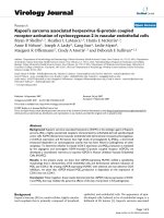
K13 activates HIV-1 LTR promoterFigure 1
K13 activates HIV-1 LTR promoter. A. 293T cells were transfected with an empty vector or the indicated constructs (100
ng/well) along with an HIV-1 LTR/luciferase reporter construct (10 ng/well) and a pRSV/LacZ (β-galactosidase) reporter con-
struct (75 ng/well), and the experiment was performed as described under "Materials and Methods." The values shown are
averages (Mean ± S.E.) of one representative experiment out of three in which each transfection was performed in duplicate. B.
A dose-response analysis of HIV-1 LTR activation by K13 and pro-inflammatory cytokines. 293T cells were transfected with
the indicated amounts of a K13 expression plasmid and luciferase assay performed 36 h post-transfection as described for (A).
The total amount of transfected DNA was kept constant by adding an empty vector. For experiments involving TNF-α and IL-
1β, cells were treated with the indicated concentration of cytokines 12 h after transfection of the reporter plasmids and
assayed for reporter activity after 24 h of stimulation. C. K13 activates HIV-1 LTR in Cos-7 cells. The experiment was per-
formed as described in 1A except LIPOFECTAMINE 2000 Reagent (Invitrogen, Carlsbad, CA) was used for transfection and
Renilla luciferase was used for normalization. D. K13 activates HIV-1 LTR in Jurkat cells. The experiment was performed as
described for 1C by using LIPOFECTAMINE 2000 Reagent (Invitrogen, Carlsbad, CA).
Retrovirology 2005, 2:9 />Page 4 of 14
(page number not for citation purposes)
four copies of a consensus NF-κB binding-site [28]. Con-
sistent with our published results [27], the triple mutant
58AAA demonstrated a complete lack of NF-κB reporter
activation while the mutant 67AAA retained partial ability
to do so (Fig 2A). Importantly, essentially a similar pat-
tern of reporter activation was obtained when the wild-
type and mutant K13 constructs were tested on the HIV-1
LTR reporter construct (Fig 2B). Collectively, the above
results suggested the involvement of the NF-κB pathway
in vFLIP K13-induced HIV-1 LTR activation.
vFLIP K13 induces binding of specific transcription factors
to HIV-1 LTR
In order to test the hypothesis that vFLIP K13 activates
HIV-1 LTR by inducing the binding of specific transcrip-
tion factors to the NF-κB binding sites present in the HIV-
1 LTR, we used an electrophoretic mobility shift assay
(EMSA). As shown in Fig. 3A, nuclear extracts from Jurkat
cells expressing vFLIP K13 demonstrated significant DNA-
binding activity on radiolabelled oligonucleotides-
derived from the NF-κB binding sites present in HIV-1
LTR. In contrast, no HIV-1 LTR DNA-binding activity was
observed in nuclear extracts of empty vector-expressing
cells (Fig. 3A, compare lanes 1 and 2). The specificity of
the complex was demonstrated by its disappearance upon
competition with excess cold HIV-1 LTR oligonucleotide
duplex and lack of effect upon competition with a non-
specific oligonucleotide duplex (Fig. 3A, lanes 3 and 4).
Nature and subunit composition of K13-induced
transcription factors bound to HIV-1 LTR
In addition to the classical NF-κB pathway, an alternative
(or non-canonical) pathway of NF-κB activation, which
involves proteasome-mediated processing of p100/NF-
κB2 into p52 subunit, has been described [8]. We have
recently demonstrated that vFLIP K13 can activate the
alternate NF-κB pathway via an IKK1-dependent and NIK-
and IKK2-independent process [24]. In order to deter-
mine the contribution of the classical vs alternate NF-κB
pathway to vFLIP K13-induced HIV-1 LTR activation, we
used a supershift assay to analyze the nature of the protein
complexes bound to HIV-1 LTR from nuclear extracts of
vFLIP K13-expressing cells. This assay demonstrated that
p50 and c-Rel subunits are the major components of the
HIV-1 LTR-bound NF-κB complexes induced by vFLIP
K13 with modest contribution from the p65 subunit (Fig.
3B). As the p50, c-Rel and p65 subunits are primarily acti-
vated by the classical NF-κB pathway, the above results
support the hypothesis that K13 activates the HIV-1 LTR
via the classical NF-κB pathway.
Role of classical NF-
κ
B activation in K13-induced HIV-1
LTR reporter activity
We have previously demonstrated that vFLIP K13 activates
the classical NF-κB pathway via phosphorylation of IκBα,
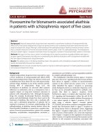
Activation of HIV- LTR by K13 mutants correlates with their ability to activate the NF-κB pathwayFigure 2
Activation of HIV- LTR by K13 mutants correlates
with their ability to activate the NF-κB pathway. A.
NF-κB activation by mutants of K13. 293T cells were trans-
fected with an empty vector (pCDNA3) or the indicated K13
expression constructs (100 ng/well) along with an NF-κB/
luciferase reporter construct (75 ng/well) and an pRSV/LacZ
(β-galactosidase) reporter construct (75 ng/well) and luci-
ferase reporter assay performed as described in Fig. 1A. The
values shown are averages (mean ± SEM) of one representa-
tive experiment out of three in which each transfection was
performed in duplicate. B. HIV-1 LTR activation by wild-type
and mutant K13 constructs. The experiment was performed
as described for Fig. 1A.
Retrovirology 2005, 2:9 />Page 5 of 14
(page number not for citation purposes)
which leads to its ubiquitination and subsequent degrada-
tion via proteasome [22]. We used a phosphorylation-
resistant mutant of IκBα to test the involvement of the
classical NF-κB pathway in vFLIP K13-induced HIV-1 LTR
reporter activity. As shown in Fig. 4A, a phosphorylation-
resistant mutant of IκBα (IκBαSS32/36AA), in which the
two critical N-terminal serine residues have been mutated
to alanine, completely blocked vFLIP K13-induced HIV-1
LTR reporter activity.
We used siRNA-mediated downregulation of key subunits
of the classical and alternate NF-κB pathways to test their
involvement in K13-induced HIV-1 LTR activation. As
shown in Fig. 4B, we achieved effective silencing of c-Rel
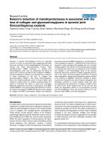
Electrophoretic mobility shift assayFigure 3
Electrophoretic mobility shift assay. A. The nuclear extract from Jurkat cells stably expressing an empty vector (lane 1)
and K13 (lanes 2–4) were used for EMSA. The position of the induced HIV-1 LTR complex is marked with an asterisk. The spe-
cificity of the complex is demonstrated by competition with excess cold HIV-1 LTR probe (lane 3) and a nonspecific (N.S.)
probe (lane 4), respectively. B. A supershift assay showing the subunit composition of K13-induced NF-κB subunits bound to
HIV-1 LTR. The supershift assay was performed using a control rabbit antisera (lane 3), control mouse antisera (lane 4), or
antisera against p50 (lane 5), p65 (lane 6), p52 (lane 7), Rel B (lane 8) and c-Rel (lane 9) subunits of NF-κB, respectively. The
position of the induced HIV-1 LTR complex is marked with an asterisk, while the super-shifted bands are marked by
arrowheads.
Retrovirology 2005, 2:9 />Page 6 of 14
(page number not for citation purposes)
and RelA/p65 expression by siRNA-mediated silencing.
Consistent with our supershift assay (Fig. 3B), siRNA-
mediated silencing of c-Rel expression led to almost com-
plete suppression of K13-induced HIV-1 LTR activation
(Fig. 4C). Similarly, silencing of p65 expression led to sig-
nificant suppression of HIV-1 LTR activity, although some
residual activity was still evident (Fig. 4C). Although p100
acts as a precursor of p52, another important function of
p100 is to retain the RelB/p50 and RelB/p52 complexes in
the cytoplasm. As such, in order to shut-off the alternate
NF-κB pathway, we chose to silence the expression of
RelB. As shown in Fig. 4B–C, siRNA-mediated downregu-
lation of RelB, had no significant effect on K13-induced
HIV-1 LTR activity. We also failed to observe any effect of
p100/p52 silencing on HIV-1 LTR activation (data not
shown). Taken together, the above results demonstrate a
key role of the c-Rel and p65 subunits of the classical NF-
κB pathway in K13-induced HIV-1 LTR reporter
activation.
Role of individual subunits of the IKK complex in K13-
induced HIV-1 LTR activation
K13 is known to associate with a 700 kDa multi-subunit
IKK complex, which consists of two catalytic subunits,
IKK1/IKKα and IKK2/IKKβ and a regulatory subunit,
NEMO/IKKγ [22]. We tested the involvement of the indi-
vidual components of the IKK complex in vFLIP K13-
induced HIV-1 LTR reporter activity by using mouse
fibroblast (MEF) cells deficient in IKK1, IKK2 and NEMO,
respectively. As shown in Fig. 5A, we observed significant
HIV-1 LTR reporter activity by the expression of vFLIP K13
in the wild type MEF cells. In contrast, almost no HIV-1
LTR reporter activity was observed in NEMO-deficient
cells. However, some residual HIV-1 LTR reporter activity
was observed in IKK1- and IKK2-deficient MEF cells. Col-
lectively, the above results suggest that synergistic action
of IKK1, IKK2 and NEMO is required for maximal activa-
tion of HIV-1 LTR by K13.
Next we sought to determine whether pharmacological
inhibitors of the NF-κB pathway may be used to block
vFLIP K13-induced HIV-1 LTR reporter activation. Lacta-
cystin and MG132 are inhibitors of proteasome and block
the NF-κB pathway by preventing the degradation of IκB.
On the other hand, arsenic acid is believed to block the
NF-κB pathway by inhibiting the IKK complex [29]. As
shown in Fig. 5B, vFLIP K13-induced HIV-1 LTR reporter
activation was effectively blocked by MG132, lactacystin
and arsenic acid. These results suggest that inhibitors of
the NF-κB pathway might have a role in preventing K13-
induced HIV-1 LTR reporter activation.
Effect of Murr1 on K13-induced HIV-1 LTR activation
Murr1 is a gene product that has been previously impli-
cated in copper regulation [30,31]. A recent study
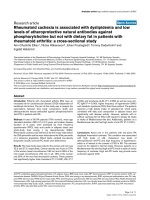
K13 activates HIV LTR through the classical NF-κB pathwayFigure 4
K13 activates HIV LTR through the classical NF-κB
pathway. A. 293T cells were transfected with an empty vec-
tor or K13 along with an HIV LTR/luciferase reporter con-
struct and a β-galactosidase reporter construct as described
in Fig. 1A. The amount of IκBαSS32/36AA inhibitor plasmid
(500 ng/well) was five times the amount of vector (pcDNA3)
or K13 (100 ng/well) plasmid. The values shown are averages
(Mean ± S.E.) of one representative experiment out of three
in which each transfection was performed in duplicate. B.
Western blot analysis showing siRNA-mediated knock-down
of p65, c-Rel and RelB expression. The blot was re-probed
with a monoclonal antibody against actin (bottom panel) to
show equal loading of all lanes and specificity of gene silenc-
ing. C. 293T cells were transfected with an empty vector or a
K13 expression plasmid along with a control siRNA oligo-
duplexes or siRNA duplexes against c-Rel, p65 and RelB,
respectively. The luciferase reporter assay was performed as
described in Fig 1A.
Retrovirology 2005, 2:9 />Page 7 of 14
(page number not for citation purposes)
Mechanism of K13-induced HIV-1 LTR activationFigure 5
Mechanism of K13-induced HIV-1 LTR activation. A. Role of IKK complex in K13-induced HIV-1 LTR reporter activity.
Wild-type and IKK-deficient cells were transiently transfected with an empty vector or K13 expression plasmid (500 ng/well)
along with an HIV/luciferase reporter construct (100 ng/well) and a synthetic Renilla luciferase (phRL-TK) reporter vector (75
ng/well) by using LIPOFECTAMINE 2000 Reagent (Invitrogen, Carlsbad, CA) according to manufacturer's instruction. Thirty-
six hours after transfection, cell lysates were used for reporter assays. Luciferase activity was normalized relative to the Renilla
luciferase activity to control for the difference in the transfection efficiency. The values shown are averages (Mean ± S.E.) of
one representative experiment out of three in which each transfection was performed in duplicate. B. Inhibitors of the NF-κB
pathway significantly block K13-actived HIV LTR promoter. 293T cells were transfected with the empty vector or K13 along
with an HIV LTR/luciferase reporter construct and a β-galactosidase reporter construct as described in Fig.1A. Eight hours
after transfection, cells were treated with DMSO or different inhibitors for 24 h and then lysed for the reporter assay. The val-
ues shown are averages (Mean ± S.E.) of one representative experiment out of three in which each transfection was performed
in duplicate. C. Effect of Murr1 on K13-induced HIV-1 LTR activation. 293T cells were transfected with an empty vector
(pCDNA3) or K13 along with an HIV LTR/luciferase reporter construct and a β-galactosidase reporter construct as described
in Fig. 1A. The amount of Murr1 plasmid (500ng/well) was five times the amount of vector or K13 (100ng/well). The values
shown are averages (Mean ± S.E.) of one representative experiment out of three in which each transfection was performed in
duplicate.
Retrovirology 2005, 2:9 />Page 8 of 14
(page number not for citation purposes)
demonstrated that Murr1 is highly expressed in CD4+ T
cells and serve as a genetic inhibitor factor for HIV-1 rep-
lication in the resting lymphocytes [32]. Murr1 was
shown to block HIV-1 LTR activation and HIV-1 replica-
tion by inhibiting the proteasomal degradation of IκB and
blocking basal and cytokine-stimulated NF-κB activation
[32]. Based on the above study demonstrating the impor-
tance of Murr1 as an endogenous regulator of HIV-1 LTR
activation, we tested its effect on K13-induced HIV-1 LTR
activation. As shown in Figure 5C, co-expression of Murr1
led to significant block in K13-induced HIV-1 LTR
reporter activity, thereby suggesting that K13-induced
activation of HIV-1 replication in resting lymphoid cells
may be regulated by Murr1 and K13 may selectively acti-
vate HIV-1 replication in activated cells in which expres-
sion of Murr1 is known to be down-regulated [32].
Synergistic activation of HIV-1 LTR by vFLIP K13 and HIV
Tat protein
HIV-1 Tat is a viral nuclear protein that plays an essential
role in HIV-1 gene expression at the transcriptional level
[2,3]. Tat has been shown to associate with p300/CBP and
P/CAF histone acetyltransferases (HAT) and efficient acti-
vation of the integrated HIV-1 LTR is largely dependent on
Tat-dependent rearrangement of the nucleosome posi-
tioned at the transcription start site [2]. HIV-1 LTR is
known to bind and respond to HIV Tat protein via a spe-
cific Tat-binding site [2]. We used deletion mutagenesis of
the HIV-1 LTR to test whether vFLIP induced transcrip-
tional activation is dependent on this Tat-binding site. As
shown in Figures 6A and 6B, a bulge mutant (containing
deletion of nucleotides +23/+25) of HIV-1 LTR, which is
defective in Tat activation [33], had no significant effect
on vFLIP K13-induced reporter activity. In contrast, vFLIP
K13 failed to activate a luciferase report construct contain-
ing an HIV-1 LTR in which the NF-κB binding sites had
been mutated (Fig. 6C). The above results confirm that
vFLIP activates the HIV-1 LTR via the NF-κB binding sites
and can do so independent of the Tat-binding site.
Transcriptional activation of genes is usually regulated by
multiple transcription factors acting in concert. Thus,
while NF-κB has been shown to play a major role in the
activation of the HIV-1 LTR, it fails to do so when acting
alone [25,34-36]. Along the same lines, the transactivat-
ing function of Tat protein requires the presence of NF-κB
sites in the HIV-1 LTR and Tat protein is known to
cooperate with NF-κB to activate the HIV-1 LTR [1,34,36].
We hypothesized that a functional interaction between
K13-induced NF-κB and Tat may be particularly impor-
tant in the early stages of HIV-1 infection when the
amount of Tat is limited. To test this hypothesis, we began
by performing a dose-response analysis of Tat and
selected a dose of Tat (20 ng/ml) which led to sub-maxi-
mal activation of HIV-1 LTR activation in 293T cells (Fig.
6D). Next, we analyzed the effect of co-expression of Tat
on K13-induced HIV-1 LTR activation. As shown in Figure
6E, while transfection of K13 (250–500 ng/well) led to
approximately 2.5–3.5 fold increase in HIV-1 LTR activa-
tion, transfection of Tat (20 ng/well) induced 4-fold
increase in HIV-1 LTR activity. However, co-expression of
K13 with Tat led to a synergistic 12-fold activation of the
HIV-1 LTR. These results suggest that K13-induced NF-κB
functions synergistically with the Tat protein to activate
the HIV-1 LTR.
Effect of vFLIP K13 on LTRs-Derived from different strains
of HIV
There is considerable sequence diversity among the HIV-1
isolates that comprise the current global pandemic and
these can be grouped into several distinct subtypes or
clades [37]. In particular, the LTRs of different subtypes
show distinct enhancer-promoter configuration and vary
in the sequence and number of binding sites for different
transcription factor, including NF-κB [38,39]. Although
different HIV-1 LTRs are transcriptionally active, they dif-
fer in the level of basal reporter activity [38,39]. In addi-
tion, different HIV-1 LTRs are known to show differential
response to TNF-α treatment, which correlates with the
number of NF-κB binding sites [38,39]. Therefore, we
sought to determine whether vFLIP K13 will differentially
activate luciferase reporter constructs driven by LTRs
derived from different HIV strains. Consistent with the
published studies [38,39], we observed considerable dif-
ference in the basal activities of different HIV-1 LTRs pro-
moters when transfected into 293T cells along with an
empty vector (Fig. 7A). More importantly, coexpression of
vFLIP K13 led to differential activation of luciferase
reporter constructs containing LTRs from different sub-
types of HIV-1 (Fig. 7A). Thus, subtype C, which possesses
three NF-κB binding sites showed the maximum increase
in vFLIP-induced HIV-1 LTR reporter activity while sub-
type E, which possesses only one NF-κB binding site
showed the lowest level of basal and vFLIP-induced HIV-
1 LTR transcriptional activation (Fig. 7A,B). These results
demonstrate that, similar to situation with TNFα, K13
may differentially activate LTRs derived from different
strains of HIV-1, which correlate with their NF-κB binding
sites.
Discussion
Although co-infection with HHV-8 and HIV-1 is known to
synergistically increase the incidence of KS, until recently
intracellular interaction between HHV8 and HIV-1 has
not received adequate attention under the assumption
that these viruses infect distinct cell types. Thus, HHV8 is
typically believed to infect B lymphocytes, epithelial cells,
keratinocytes, KS tumor cells, and endothelial cells
[40,41], while the predominant host cells for HIV-1 are
CD4
+
T lymphocytes, dendritic cells, and mononuclear
Retrovirology 2005, 2:9 />Page 9 of 14
(page number not for citation purposes)
Effect of HIV Tat protein on K13-induced HIV-1 LTR activationFigure 6
Effect of HIV Tat protein on K13-induced HIV-1 LTR activation. A-C. 293T cells were transfected with an empty vec-
tor (pCDNA3) or K13 along with different HIV LTR/luciferase reporter constructs and β-galactosidase as described in Fig. 1A.
The values shown are averages (Mean ± S.E.) of one representative experiment out of three in which each transfection was
performed in duplicate. A, Wild type HIV-1 LTR reporter; B, HIV-1 LTR reporter with deletion of Tat-binding site (nucleotides
+23 to +25); C. HIV-1 LTR reporter lacking the NF-κB binding-sites. D. Dose-response analysis of Tat-induced HIV-1 LTR acti-
vation. E. K13 and Tat synergistically activate HIV-1 LTR. The experiment was performed as described for Fig. 6 A. The total
amount of transfected DNA was kept constant by adding empty vector.
Retrovirology 2005, 2:9 />Page 10 of 14
(page number not for citation purposes)
phagocytes [41,42]. However, as recently pointed out by
Huang et al, several lines of evidence suggest that the
above assumption may not be completely true and HHV8
and HIV-1 may, in fact, interact in vivo [41]. First, both
HHV8 and HIV-1 can efficiently infect cells of monocyte/
macrophage lineage, including dendritic cells [43,44].
Second, Moir et al have shown that induction of CD4 and
CXCR4 on B cells by CD40 stimulation leads to an
increased susceptibility of these cells to T-trophic HIV
infection [45]. Third, HHV8-infected B cells can be
infected by HIV-1 via a cell-cell pathway and such infected
B cells can support productive HIV-1 replication [46].
Finally, the range of HHV8-susceptible cells in vivo is
unclear at the present. Therefore, it stands to reason that
HHV8 and HIV-1 genomes may co-exist in the same cells
in vivo and reciprocally regulate the gene expression of
each other. Support for the above hypothesis is provided
by a recent study which demonstrated that co-culture of
HIV-1-infected CD4+ T cells with HHV8-infected B cell
lines resulted in increased HIV-1 replication [47].
Differential activation of HIV-1 subtype LTRs by K13Figure 7
Differential activation of HIV-1 subtype LTRs by K13. A. 293T cells were transfected with an empty vector (pCDNA3)
or K13 along with luciferase reporter constructs containing LTRs derived from the indicated strains of HIV and a β-galactosi-
dase reporter construct as described in Fig. 1A. The values shown are averages (Mean ± S.E.) of one representative experiment
out of three in which each transfection was performed in duplicate. B. Partial sequence of LTRs of HIV-1 subtypes A through F.
The LTR region spanning positions -129 to -77 of subtype A is shown at the top. The NF-κB binding motifs are shaded gray.
Retrovirology 2005, 2:9 />Page 11 of 14
(page number not for citation purposes)
With the goal of elucidating intracellular signaling interac-
tions which could be potentially involved in the
induction of HIV-1 replication by HHV-8, we carried out
a detailed analysis of the effect of HHV8 vFLIP on HIV-1
LTR activation. Consistent with an earlier report, we
observed that HHV8 vFLIP strongly activates HIV-1 LTR in
an NF-κB-dependent fashion [48]. We further demon-
strate that vFLIP K13 could activate HIV-1 LTR in both epi-
thelial and human lymphoma cell lines, although the
magnitude of stimulatory effect was more pronounced in
the epithelial cells. A possible explanation for this differ-
ence may lie in the differential expression of proteins that
could modulate the effect of K13 on NF-κB and/or HIV-1
LTR activation. As an example, we demonstrate that K13-
induced HIV-1 LTR activation can be effectively blocked
by Murr1, a recently identified inhibitor of the NF-κB
pathway which is highly expressed in T cells [32]. How-
ever, alternative explanation, including difference in the
transfection efficiency between different cell lines, could
apply as well.
We have recently reported that vFLIP K13 can activate
both the classical and alternate NF-κB pathways and, as
such, we were interested in determining the relative con-
tribution of these pathways to K13-induced HIV-1 LTR
activation. Based on the following data, we believe that
activation of HIV-1 LTR is mainly through the classical
pathway. First, our gel super-shift assay demonstrated that
NF-κB complexes formed by vFLIP expression were prima-
rily composed of c-Rel, p50 and p65 subunits. Second,
siRNA-mediated downregulation of c-Rel and p65 led to
near complete inhibition of K13-induced HIV-1 LTR
activation whereas silencing of RelB expression was with-
out significant effect. Third, K13-induced HIV-1 LTR
activation was completely inhibited by super-repressor
form of IκBα, which primarily blocks the classical NF-κB
pathway. Finally, while K13 activates the alternate NF-κB
pathway independent of IKK2, it failed to activate the
HIV-1 LTR in IKK2-deficient MEFs.
Based on some early gene-knockout studies, IKK1 was
believed to be not involved in cytokine-induced activa-
tion of the classical NF-κB pathway [49-51]. In the present
study, we have observed that, in addition to IKK2- and
NEMO-deficient MEFs, K13-induced HIV-1 LTR activa-
tion was markedly reduced in IKK1-deficient MEFs as
well. We believe that the above results with IKK1-deficient
cells do not necessarily support the involvement of the
alternate NF-κB pathway in K13-induced HIV-1 LTR acti-
vation for the following reasons. First, we have recently
reported that K13-induced p65/50 DNA binding and NF-
κB transcriptional activation is markedly reduced in IKK1-
deficient MEFs [23] Thus, the reduced HIV-1 LTR activa-
tion in the IKK1-deficient cells observed in the current
study is consistent with requirement for IKK1 in K13-
induced classical NF-κB activation. Second, recent studies
suggest that IKK1 may be involved in transcriptional acti-
vation of classical NF-κB responsive genes through its
ability to phosphorylate histones and p65 [52-54]. Thus,
taken together, our results demonstrate that K13 activates
HIV-1 LTR through the activation of the classical NF-κB
pathway, in which IKK1 plays a major role. Thus, selective
inhibitors of IKK1 may have a role in blocking K13-
induced HIV-1 LTR transcriptional activation. However, it
is important to point out that while IKK1 may be uniquely
important for K13-induced classical NF-κB activation
pathway, maximal activation of this pathway via K13
relies on cooperative interaction between IKK1, IKK2 and
NEMO.
The transcription of cellular and viral genes is regulated by
structural and functional interactions among a number of
transcriptional factors that act in concert. This is also
known to be the case with HIV-1 LTR. Thus, while NF-κB
plays a major role in the transcriptional activation of HIV-
1, it requires synergistic interaction with a number of cel-
lular and viral proteins for maximal stimulation of this
activity [1]. Although NF-κB is known to interact with
Sp1, Ets and NF-AT to activate HIV-1 LTR, cooperative
interaction between NF-κB and Tat has received the most
interaction in the literature [1,55]. Tat has been shown to
act synergistically with PMA, PHA and Tax-induced NF-κB
to activate the HIV-1 LTR [1,34,36,55]. Consistent with
these previous studies, we demonstrate that although K13
can activate the HIV-1 LTR by itself, it functionally coop-
erates with Tat to synergistically activate transcription
from HIV-1 LTR. HIV-1 infection itself is known to induce
persistent NF-κB activation, which is probably mediated
via Tat and Nef [56,57], and interacts in a positive-feed-
back manner with Tat to enhance HIV-1 replication.
However, in the immediate post-integration period of the
HIV-1 life-cycle, Tat is expressed at very low levels which
may not be enough to effectively stimulate HIV-1 LTR acti-
vation. Therefore, it is conceivable that vFLIP K13 could
amplify the activity of Tat via NF-κB activation and thus
support enhanced HIV-1 replication during the early
stages of HIV-1 infection or in cells which express Tat at
suboptimal levels.
The human immunodeficiency virus has considerably
diversified during its worldwide spread in the current pan-
demic and can be classified into several distinct subtypes
[37]. Subtype B is predominant in North America and
Europe, subtype E in Southeast Asia and subtype C in sub-
Saharan Africa, respectively [58]. Previous studies have
demonstrated that LTRs from HIV-1 subtypes B, C and E
vary in number and binding sites for NF-κB in their
enhancer elements [59]. Thus, subtype C isolates are
known to contain three functional NF-κB binding sites, as
compared to two such sites in the enhancer of the more
Retrovirology 2005, 2:9 />Page 12 of 14
(page number not for citation purposes)
commonly studied subtype B [59]. On the other hand, in
the subtype E, one of the NF-κB-binding sites has been
switched to a GABP site, resulting only one functional NF-
κB site and gain of a new specificity [60]. Consistent with
the above results, in the present study we demonstrate
that vFLIP-induced HIV-1 LTR activation is strongest in
subtype C and weakest in subtype E. Thus, the differential
response of different HIV-1 LTRs to K13-induced tran-
scriptional activation may be explained on the basis of
number of functional NF-κB sites in their enhancer ele-
ments. Future studies should address the question
whether co-infection with HHV8 has a differential effect
on the replication and natural history of different HIV-1
subtypes.
Methods
Plasmids, cell lines and reagents
Plasmids containing pcDNA3-K13-Flag and pcDNA3-E8-
Flag, pRSV/LacZ and 293T cells have been described pre-
viously [21]. An expression construct encoding Murr-1
was generated by RT-PCR using cDNA prepared from
H460 cells as a template and subsequently cloned in
pcDNA3 vector with a C-terminal HA tag. Luciferase
reporter constructs containing LTRs derived from different
strains of HIV-1 (pBlue3'LTR-Luc-A-F) were obtained
from AIDS Research and Reference Reagent Program,
Division of AIDS, NIAID, NIH from Drs. Reink Jeeninga
and Ben Berkhout. Wild-type and mutant HIV-1 LTR
reporter constructs [61] and expression constructs for HIV
Tat were obtained from Dr. Richard Gaynor. The IKKα-/-
and IKKβ-/- mouse embryonic fibroblast cells were gener-
ated in Dr. Inder Verma's laboratory [62,63] and IKKγ/
NEMO-/- cells were generated in Dr. Michael Karin's lab-
oratory [64]. These cells were kindly provided by Dr. Rich-
ard Gaynor and were maintained in DMEM
supplemented with 10% FBS. Jurkat cells were cultured in
RPMI medium supplemented with 10% FBS and selected
in the presence of 1500 µg/ml of G418 (Invitrogen). 293T
cells were grown in DMEM with 10% FBS. Arsenic Acid
was purchased from Sigma. MG132 and lactacystin were
purchased from Calbiochem and Biomol, respectively.
Retrovirus constructs containing C-terminal Flag epitope
tagged HHV8 vFLIP (K13-Flag) was generated in MSCV
neo-based retroviral vector and amphotropic viruses gen-
erated and used for infection as described previously [22].
Electrophoretic mobility shift assay
Electrophoretic mobility shift assay was performed essen-
tially as described previously [22], except an HIV LTR oli-
gonucleotide duplex (sense strand, 5' TGC TAC AAG GGA
CTT TCC GCT GGG GAC TTT CCA GG 3') was used
instead of κB binding oligonucleotide. Nuclear extracts
were prepared from Jurkat cells stably expressing an
empty vector or vFLIP K13, which have been described
previously [23]. Antibodies against p50, p65, RelB and c-
Rel were purchased from Santa Cruz Biotechnology. An
antibody against p52 was purchased from Upstate
biotechnology
Luciferase reporter assay
293 T cells were transfected in duplicate in a 24-well plate
with the various test plasmids along with an HIV LTR/luci-
ferase reporter construction (10 ng/well) and a pRSV/LacZ
(β-galactosidase) reporter construct (75 ng/well) using
calcium phosphate transfection protocol as described pre-
viously [21]. Cells were lysed 36–48 hours later and
extracts were used for the measurement of firefly luciferase
and galactosidase activity. Luciferase activity was normal-
ized relative to the galactosidase activity to control for the
difference in the transfection efficiency. Cos-7, Jurkat and
MEF cells were transiently transfected with empty vector
(pCDNA3) or K13 (500 ng/well) along with a HIV/luci-
ferase reporter construct (100 ng/well) and a synthetic
Renilla luciferase (phRL-TK; Promega) reporter vector (75
ng/well) by using LIPOFECTAMINE 2000 Reagent (Invit-
rogen, Carlsbad, CA) according to manufacturer's instruc-
tion. Thirty-six hours after transfection, cells lysates used
for reporter assays. Luciferase activity was normalized rel-
ative to the Renilla luciferase activity to control for the dif-
ference in the transfection efficiency. The values shown
are averages (Mean ± S.E.) of one representative experi-
ment out of three in which each transfection was per-
formed in duplicate.
siRNA Oligonucleotides
siRNA oligonucleotides with two thymidine residues
(dTdT) at the 3'-end of the sequence were designed to p65
(sense, 5'-GCCCUAUCCCUUUACGUCAdTdT-3'), c-Rel
(sense, 5'-CAACCGUGCUCCAAAUACU dTdT-3'), RelB
(sense, AGAUCAUCGACGAGUACAUdTdT-3') and
control (sense, 5' GCGCGCUUUGUAGGAUUCGdTdT-
3'), along with their corresponding antisense oligonucle-
otides. The RNA oligonucleotides were synthesized at
RNA Oligonucleotide Synthesis Core facility, UT South-
western Medical center. siRNA oligonucleotides (80 nM)
were transfected using calcium phosphate as described
previously [65].
Western Blot
Western blot analysis was performed essentially as
described previously [22]. Primary antibodies used in
these experiments were: p65, c-Rel, Rel-B (rabbit polyclo-
nal, Santa Cruz biotechnology) and actin (mouse mono-
clonal, Sigma).
List of Abbreviations Used
DED, death effector domain; EMSA, electrophoretic
mobility shift assay; FLICE, Fas-associated death domain-
like IL-1 beta-converting enzyme; FLIP, FLICE inhibitory
protein; HHV8, Human herpes virus 8; HIV-1, human
Retrovirology 2005, 2:9 />Page 13 of 14
(page number not for citation purposes)
immunodeficiency virus 1; KS, Kaposi's sarcoma; NEMO,
NF-κB essential modulator; NIK, NF-κB-inducing kinase;
NF-κB, Nuclear factor kappa B; MEF, murine embryonic
fibroblast; PEL, primary effusion lymphoma, TNFR,
Tumor necrosis factor receptor; IκB, inhibitor of NF-κB,
IKK, IκB kinase; vFLIP, viral FLICE inhibitory protein;
LTR, long terminal repeat.
Competing Interests
The author(s) declare that they have no competing
interests.
Authors' contributions
QS and HM carried out most of the experiments described
in this manuscript. PMC conceived of the study and par-
ticipated in its design and coordination and helped to
draft the manuscript. All authors read and approved the
final manuscript.
Acknowledgement
We will like to thank Drs. Inder Verma, Michael Karin and Richard Gaynor
for MEF cells and various expression and reporter plasmids. The following
reagents were obtained through the AIDS Research and Reference Reagent
Program, Division of AIDS, NIAID, NIH: pBlue3'LTR-Luc-A-F from Drs.
Reink Jeeninga and Ben Berkhout. This work was supported by a grant from
the National Institutes of Health (CA85177).
References
1. Rohr O, Marban C, Aunis D, Schaeffer E: Regulation of HIV-1
gene transcription: from lymphocytes to microglial cells. Jour-
nal of Leukocyte Biology 2003, 74:736-49.
2. Wu Y: HIV-1 gene expression: lessons from provirus and non-
integrated DNA. Retrovirology 2004, 1:13.
3. Jeang KT, Xiao H, Rich EA: Multifaceted activities of the HIV-1
transactivator of transcription, Tat. J Biol Chem 1999,
274:28837-40.
4. Pereira LA, Bentley K, Peeters A, Churchill MJ, Deacon NJ: A com-
pilation of cellular transcription factor interactions with the
HIV-1 LTR promoter. Nucleic Acids Res 2000, 28:663-8.
5. Ghosh S, May MJ, Kopp EB: NF-kappa B and Rel proteins: evolu-
tionarily conserved mediators of immune responses. Annu Rev
Immunol 1998, 16:225-60.
6. Baldwin AS Jr: The NF-kappa B and I kappa B proteins: new
discoveries and insights. Annu Rev Immunol 1996, 14:649-83.
7. Karin M, Ben-Neriah Y: Phosphorylation meets ubiquitination:
the control of NF-[kappa]B activity. Annu Rev Immunol 2000,
18:621-63.
8. Pomerantz JL, Baltimore D: Two pathways to NF-kappaB. Molec-
ular Cell 2002, 10:693-5.
9. Xiao G, Harhaj EW, Sun SC: NF-kappaB-inducing kinase regu-
lates the processing of NF-kappaB2 p100. Mol Cell 2001,
7:401-9.
10. Senftleben U, Cao Y, Xiao G, Greten FR, Krahn G, Bonizzi G, Chen
Y, Hu Y, Fong A, Sun SC, et al.: Activation by IKKalpha of a sec-
ond, evolutionary conserved, NF-kappa B signaling pathway.
Science 2001, 293:1495-9.
11. Schulz TF: Kaposi's sarcoma-associated herpesvirus (human
herpesvirus-8). J Gen Virol 1998, 79:1573-91.
12. Verma SC, Robertson ES: Molecular biology and pathogenesis of
Kaposi sarcoma-associated herpesvirus. FEMS Microbiol Lett
2003, 222:155-63.
13. Dourmishev LA, Dourmishev AL, Palmeri D, Schwartz RA, Lukac DM:
Molecular Genetics of Kaposi's Sarcoma-Associated Herpes-
virus (Human Herpesvirus 8) Epidemiology and
Pathogenesis. Microbiol Mol Biol Rev 2003, 67:175-212.
14. Sturzl M, Hohenadl C, Zietz C, Castanos-Velez E, Wunderlich A,
Ascherl G, Biberfeld P, Monini P, Browning PJ, Ensoli B: Expression
of K13/v-FLIP gene of human herpesvirus 8 and apoptosis in
Kaposi's sarcoma spindle cells. J Natl Cancer Inst 1999,
91:1725-33.
15. Rainbow L, Platt GM, Simpson GR, Sarid R, Gao SJ, Stoiber H, Her-
rington CS, Moore PS, Schulz TF: The 222- to 234-kilodalton
latent nuclear protein (LNA) of Kaposi's sarcoma-associated
herpesvirus (human herpesvirus 8) is encoded by orf73 and
is a component of the latency-associated nuclear antigen. J
Virol 1997, 71:5915-21.
16. Sarid R, Flore O, Bohenzky RA, Chang Y, Moore PS: Transcription
mapping of the Kaposi's sarcoma-associated herpesvirus
(human herpesvirus 8) genome in a body cavity-based lym-
phoma cell line (BC-1). J Virol 1998, 72:1005-12.
17. Tibbetts MD, Zheng L, Lenardo MJ: The death effector domain
protein family: regulators of cellular homeostasis. Nature
Immunology 2003, 4:404-9.
18. Thome M, Schneider P, Hofmann K, Fickenscher H, Meinl E, Neipel F,
Mattmann C, Burns K, Bodmer JL, Schroter M, et al.: Viral FLICE-
inhibitory proteins (FLIPs) prevent apoptosis induced by
death receptors. Nature 1997, 386:517-21.
19. Bertin J, Armstrong RC, Ottilie S, Martin DA, Wang Y, Banks S, Wang
GH, Senkevich TG, Alnemri ES, Moss B, et al.: Death effector
domain-containing herpesvirus and poxvirus proteins inhibit
both Fas- and TNFR1-induced apoptosis. Proc Natl Acad Sci U S
A 1997, 94:1172-6.
20. Hu S, Vincenz C, Buller M, Dixit VM: A novel family of viral death
effector domain-containing molecules that inhibit both CD-
95- and tumor necrosis factor receptor-1-induced apoptosis.
J Biol Chem 1997, 272:9621-4.
21. Chaudhary PM, Jasmin A, Eby MT, Hood L: Modulation of the NF-
kappa B pathway by virally encoded death effector domains-
containing proteins. Oncogene 1999, 18:5738-46.
22. Liu L, Eby MT, Rathore N, Sinha SK, Kumar A, Chaudhary PM: The
Human Herpes Virus 8-encoded Viral FLICE Inhibitory Pro-
tein Physically Associates with and Persistently Activates
the Ikappa B Kinase Complex. J Biol Chem 2002, 277:13745-51.
23. Matta H, Sun Q, Moses G, Chaudhary PM: Molecular genetic anal-
ysis of human herpes virus 8-encoded viral FLICE inhibitory
protein-induced NF-kappaB activation. Journal of Biological
Chemistry 2003, 278:52406-11.
24. Matta H, Chaudhary PM: Activation of alternative NF-kappa B
pathway by human herpes virus 8-encoded Fas-associated
death domain-like IL-1 beta-converting enzyme inhibitory
protein (vFLIP). Proc Natl Acad Sci U S A 2004, 101:9399-404.
25. Doppler C, Schalasta G, Amtmann E, Sauer G: Binding of NF-kB to
the HIV-1 LTR is not sufficient to induce HIV-1 LTR activity.
AIDS Res Hum Retroviruses 1992, 8:245-52.
26. Conant K, Atwood WJ, Traub R, Tornatore C, Major EO: An
increase in p50/p65 NF-kB binding to the HIV-1 LTR is not
sufficient to increase viral expression in the primary human
astrocyte. Virology 1994, 205:586-90.
27. Sun Q, Zachariah S, Chaudhary PM: The human herpes virus 8-
encoded viral FLICE-inhibitory protein induces cellular
transformation via NF-kappaB activation. Journal of Biological
Chemistry 2003, 278:52437-45.
28. Berberich I, Shu GL, Clark EA: Cross-linking CD40 on B cells rap-
idly activates nuclear factor-kappa B. J Immunol 1994,
153:4357-66.
29. Roussel RR, Barchowsky A: Arsenic inhibits NF-kappaB-medi-
ated gene transcription by blocking IkappaB kinase activity
and IkappaBalpha phosphorylation and degradation. Arch Bio-
chem Biophys 2000, 377:204-12.
30. van De Sluis B, Rothuizen J, Pearson PL, van Oost BA, Wijmenga C:
Identification of a new copper metabolism gene by positional
cloning in a purebred dog population. Hum Mol Genet 2002,
11:165-73.
31. Klomp AE, van de Sluis B, Klomp LW, Wijmenga C: The ubiqui-
tously expressed MURR1 protein is absent in canine copper
toxicosis. J Hepatol 2003, 39:703-9.
32. Ganesh L, Burstein E, Guha-Niyogi A, Louder MK, Mascola JR, Klomp
LW, Wijmenga C, Duckett CS, Nabel GJ: The gene product Murr1
restricts HIV-1 replication in resting CD4+ lymphocytes.
Nature 2003, 426:853-7.
33. Wu-Baer F, Sigman D, Gaynor RB: Specific binding of RNA
polymerase II to the human immunodeficiency virus trans-
Publish with Bio Med Central and every
scientist can read your work free of charge
"BioMed Central will be the most significant development for
disseminating the results of biomedical research in our lifetime."
Sir Paul Nurse, Cancer Research UK
Your research papers will be:
available free of charge to the entire biomedical community
peer reviewed and published immediately upon acceptance
cited in PubMed and archived on PubMed Central
yours — you keep the copyright
Submit your manuscript here:
/>BioMedcentral
Retrovirology 2005, 2:9 />Page 14 of 14
(page number not for citation purposes)
activating region RNA is regulated by cellular cofactors and
Tat. Proc Natl Acad Sci U S A 1995, 92:7153-7.
34. Cheng H, Tarnok J, Parks WP: Human immunodeficiency virus
type 1 genome activation induced by human T-cell leukemia
virus type 1 Tax protein is through cooperation of NF-kap-
paB and Tat. J Virol 1998, 72:6911-6.
35. Zack JA, Cann AJ, Lugo JP, Chen IS: HIV-1 production from
infected peripheral blood T cells after HTLV-I induced
mitogenic stimulation. Science 1988, 240:1026-9.
36. Siekevitz M, Josephs SF, Dukovich M, N Peffer, Wong-Staal F, Greene
WC: Activation of the HIV-1 LTR by T cell mitogens and the
trans-activator protein of HTLV-I. Science 1987, 238:1575-8.
37. Korber BT, Allen EE, Farmer AD, Myers GL: Heterogeneity of
HIV-1 and HIV-2. Aids 1995, 9(Suppl A):S5-18.
38. Jeeninga RE, Hoogenkamp M, Armand-Ugon M, de Baar M, Verhoef
K, Berkhout B: Functional differences between the long termi-
nal repeat transcriptional promoters of human immunodefi-
ciency virus type 1 subtypes A through G. Journal of Virology
2000, 74:3740-51.
39. Roof P, Ricci M, Genin P, Montano MA, Essex M, Wainberg MA,
Gatignol A, Hiscott J: Differential regulation of HIV-1 clade-spe-
cific B, C, and E long terminal repeats by NF-kappaB and the
Tat transactivator. Virology 2002, 296:77-83.
40. Blauvelt A: The role of human herpesvirus 8 in the pathogen-
esis of Kaposi's sarcoma. Adv Dermatol 1999, 14:167-206. discus-
sion 207
41. Huang LM, Chao MF, Chen MY, Shih H, Chiang YP, Chuang CY, Lee
CY: Reciprocal regulatory interaction between human her-
pesvirus 8 and human immunodeficiency virus type 1. J Biol
Chem 2001, 276:13427-32.
42. Martin JC, Bandres JC: Cells of the monocyte-macrophage line-
age and pathogenesis of HIV-1 infection. J Acquir Immune Defic
Syndr 1999, 22:413-29.
43. Rettig MB, Ma HJ, Vescio RA, Pold M, Schiller G, Belson D, Savage A,
Nishikubo C, Wu C, Fraser J, et al.: Kaposi's sarcoma-associated
herpesvirus infection of bone marrow dendritic cells from
multiple myeloma patients. Science 1997, 276:1851-4.
44. Blasig C, Zietz C, Haar B, Neipel F, Esser S, Brockmeyer NH, Tsch-
achler E, Colombini S, Ensoli B, Sturzl M: Monocytes in Kaposi's
sarcoma lesions are productively infected by human herpes-
virus 8. Journal of Virology 1997, 71:7963-8.
45. Moir S, Lapointe R, Malaspina A, Ostrowski M, Cole CE, Chun TW,
Adelsberger J, Baseler M, Hwu P, Fauci AS: CD40-Mediated induc-
tion of CD4 and CXCR4 on B lymphocytes correlates with
restricted susceptibility to human immunodeficiency virus
type 1 infection: potential role of B lymphocytes as a viral
reservoir. J Virol 1999, 73:7972-80.
46. Varthakavi V, Browning PJ, Spearman P: Human immunodefi-
ciency virus replication in a primary effusion lymphoma cell
line stimulates lytic-phase replication of Kaposi's sarcoma-
associated herpesvirus. J Virol 1999, 73:10329-38.
47. Mercader M, Nickoloff BJ, Foreman KE: Induction of human
immunodeficiency virus 1 replication by human herpesvirus
8. Archives of Pathology & Laboratory Medicine 2001, 125:785-9.
48. Belanger C, Gravel A, Tomoiu A, Janelle ME, Gosselin J, Tremblay MJ,
Flamand L: Human herpesvirus 8 viral FLICE-inhibitory pro-
tein inhibits Fas-mediated apoptosis through binding and
prevention of procaspase-8 maturation. J Hum Virol 2001,
4:62-73.
49. Takeda K, Takeuchi O, Tsujimura T, Itami S, Adachi O, Kawai T, Sanjo
H, Yoshikawa K, Terada N, Akira S: Limb and skin abnormalities
in mice lacking IKKalpha. Science 1999, 284:313-6.
50. Hu Y, Baud V, Delhase M, Zhang P, Deerinck T, Ellisman M, Johnson
R, Karin M: Abnormal morphogenesis but intact IKK activa-
tion in mice lacking the IKKalpha subunit of IkappaB kinase
[see comments]. Science 1999, 284:316-20.
51. Li Q, Estepa G, Memet S, Israel A, Verma IM: Complete lack of NF-
kappaB activity in IKK1 and IKK2 double-deficient mice:
additional defect in neurulation. Genes & Development 2000,
14:1729-33.
52. Yamamoto Y, Verma UN, Prajapati S, Kwak YT, Gaynor RB: Histone
H3 phosphorylation by IKK-alpha is critical for cytokine-
induced gene expression. Nature 2003, 423:655-9.
53. Anest V, Hanson JL, Cogswell PC, Steinbrecher KA, Strahl BD, Bald-
win AS: A nucleosomal function for IkappaB kinase-alpha in
NF-kappaB-dependent gene expression. Nature 2003,
423:659-63.
54. O'Mahony AM, Montano M, Van Beneden K, Chen LF, Greene WC:
HTLV-I tax induction of biologically active NF-kappa B
requires IKK1-mediated phosphorylation of RelA/p65. J Biol
Chem 2004, 12:12.
55. Nabel G, Baltimore D: An inducible transcription factor acti-
vates expression of human immunodeficiency virus in T
cells. Nature 1987, 326:711-3.
56. Demarchi F, d'Adda di Fagagna F, Falaschi A, Giacca M: Activation of
transcription factor NF-kappaB by the Tat protein of human
immunodeficiency virus type 1. J Virol 1996, 70:4427-37.
57. Baur AS, Sawai ET, Dazin P, Fantl WJ, Cheng-Mayer C, Peterlin BM:
HIV-1 Nef leads to inhibition or activation of T cells depend-
ing on its intracellular localization. Immunity 1994, 1:373-84.
58. Zimmerman PA, Buckler-White A, Alkhatib G, Spalding T, Kubofcik J,
Combadiere C, Weissman D, Cohen O, Rubbert A, Lam G, et al.:
Inherited resistance to HIV-1 conferred by an inactivating
mutation in CC chemokine receptor 5: studies in popula-
tions with contrasting clinical phenotypes, defined racial
background, and quantified risk. Molecular Medicine 1997,
3:23-36.
59. Montano MA, Nixon CP, Essex M: Dysregulation through the
NF-kappaB enhancer and TATA box of the human immun-
odeficiency virus type 1 subtype E promoter. J Virol 1998,
72:8446-52.
60. Verhoef K, Sanders RW, Fontaine V, Kitajima S, Berkhout B: Evolu-
tion of the human immunodeficiency virus type 1 long termi-
nal repeat promoter by conversion of an NF-kappaB
enhancer element into a GABP binding site. J Virol 1999,
73:1331-40.
61. Yamamoto Y, Yin MJ, Gaynor RB: IkappaB kinase alpha (IKKa-
lpha) regulation of IKKbeta kinase activity. Mol Cell Biol 2000,
20:3655-66.
62. Li Q, Lu Q, Hwang JY, Buscher D, Lee KF, Izpisua-Belmonte JC,
Verma IM: IKK1-deficient mice exhibit abnormal develop-
ment of skin and skeleton. Genes Dev 1999, 13:1322-8.
63. Li Q, Van Antwerp D, Mercurio F, Lee KF, Verma IM: Severe liver
degeneration in mice lacking the IkappaB kinase 2 gene. Sci-
ence 1999, 284:321-5.
64. Makris C, Godfrey VL, Krahn-Senftleben G, Takahashi T, Roberts JL,
Schwarz T, Feng L, Johnson RS, Karin M: Female mice hetero-
zygous for IKK gamma/NEMO deficiencies develop a der-
matopathy similar to the human X-linked disorder
incontinentia pigmenti. Mol Cell 2000, 5:969-79.
65. Sinha SK, Chaudhary PM: Induction of apoptosis by X-linked
ectodermal dysplasia receptor via a caspase 8-dependent
mechanism. J Biol Chem 2004, 279:41873-81.