Báo cáo y học: " The Vpr protein from HIV-1: distinct roles along the viral life cycle" potx
Bạn đang xem bản rút gọn của tài liệu. Xem và tải ngay bản đầy đủ của tài liệu tại đây (668.04 KB, 14 trang )
BioMed Central
Page 1 of 14
(page number not for citation purposes)
Retrovirology
Open Access
Review
The Vpr protein from HIV-1: distinct roles along the viral life cycle
Erwann Le Rouzic and Serge Benichou*
Address: Institut Cochin, Department of Infectious Diseases, INSERM U567, CNRS UMR8104, Université Paris 5, Paris, France
Email: Erwann Le Rouzic - ; Serge Benichou* -
* Corresponding author
Abstract
The genomes of human and simian immunodeficiency viruses (HIV and SIV) encode the gag, pol and
env genes and contain at least six supplementary open reading frames termed tat, rev, nef, vif, vpr,
vpx and vpu. While the tat and rev genes encode regulatory proteins absolutely required for virus
replication, nef, vif, vpr, vpx and vpu encode for small proteins referred to "auxiliary" (or
"accessory"), since their expression is usually dispensable for virus growth in many in vitro systems.
However, these auxiliary proteins are essential for viral replication and pathogenesis in vivo. The
two vpr- and vpx-related genes are found only in members of the HIV-2/SIVsm/SIVmac group,
whereas primate lentiviruses from other lineages (HIV-1, SIVcpz, SIVagm, SIVmnd and SIVsyk)
contain a single vpr gene. In this review, we will mainly focus on vpr from HIV-1 and discuss the
most recent developments in our understanding of Vpr functions and its role during the virus
replication cycle.
Introduction
The viral protein R (Vpr) of HIV-1 is a small basic protein
(14 kDa) of 96 amino acids, and is well conserved in HIV-
1, HIV-2 and SIV [1]. The role of Vpr in the pathogenesis
of AIDS is undeniable, but its real functions during the
natural course of infection are still subject to debate. The
Vpr role in the pathophysiology of AIDS has been investi-
gated in rhesus monkeys experimentally infected with
SIVmac, and it was initially shown that monkeys infected
with a vpr null SIV mutant decreased virus replication and
delayed disease progression [2,3]. Moreover, monkeys
infected with a SIV that did not express the vpr and vpx
genes displayed a very low virus burden and did not
develop immunodeficiency disease [4,5]. Regarding these
in vivo phenotypic effects, numerous laboratories have
dissected the role of Vpr in various in vitro, in vivo and ex
vivo systems to explore the contribution of this protein in
the different steps of the virus life cycle. Despite its small
size, Vpr has been shown to play multiple functions dur-
ing virus replication, including an effect on the accuracy of
the reverse-transcription process, the nuclear import of
the viral DNA as a component of the pre-integration com-
plex (PIC), cell cycle progression, regulation of apoptosis,
and the transactivation of the HIV-LTR as well as host cell
genes (Fig. 1). Furthermore, Vpr is found in virions, in
cells, and exists as free molecules found in the sera and the
cerebrospinal fluid of AIDS patients, indicating that it
may exert its biological functions through different
manners.
Structure of the HIV-1 Vpr protein
Because the full length protein aggregated in aqueous
solution, the overall structure of Vpr has been difficult to
access [6], and preliminary strategies used two distinct
synthetic peptides corresponding to Vpr (1–51) and (52–
96) fragments for NMR and circular dichroism studies [6-
9]. As previously predicted [10], the structure of the
Vpr(1–51) fragment has a long motif of α helix turn-α
Published: 22 February 2005
Retrovirology 2005, 2:11 doi:10.1186/1742-4690-2-11
Received: 17 January 2005
Accepted: 22 February 2005
This article is available from: />© 2005 Le Rouzic and Benichou; licensee BioMed Central Ltd.
This is an Open Access article distributed under the terms of the Creative Commons Attribution License ( />),
which permits unrestricted use, distribution, and reproduction in any medium, provided the original work is properly cited.
Retrovirology 2005, 2:11 />Page 2 of 14
(page number not for citation purposes)
helix type encompassing the Asp17-Ile46 region, and ends
with a γ turn [8]. The Vpr(52–96) fragment contains an α-
helix encompassing the 53–78 region that is rich in leu-
cine residues [7]. One side of the helix offers a stretch of
hydrophobic residues that can form a leucine-zipper like
motif [11]. This structure may account for the formation
of Vpr dimers [7,12,13] and/or for the interaction with
cellular partners [14]. Finally, NMR analysis of a soluble
full length Vpr (1–96) polypeptide was recently per-
formed, and gave access to the tertiary structure of the pro-
tein (Fig. 2), confirming the amphipathic nature of the
three α-helices of HIV-1 Vpr. The helices are connected by
loops and are folded around a hydrophobic core [15] sur-
rounded by a flexible N-terminal domain and a C-termi-
nal arginine-rich region that are negatively and positively
charged, respectively. Four conserved prolines (positions
5, 10, 14 and 35) which present cis/trans isomerization are
found in the N-terminal domain [16]. It was reported that
the cellular peptidyl-propyl isomerase cyclophilin A was
able to interact with Vpr via prolines in position 14 and
35, which insured the correct folding of the viral protein
[17]. The carboxy-terminus of Vpr contains six arginines
between residues 73 and 96. This domain shows similar-
ity with those of arginine-rich protein transduction
Schematic view of the early steps of the HIV-1 infection of a target cellFigure 1
Schematic view of the early steps of the HIV-1 infection of a target cell. The functional events in which the Vpr protein is
involved are highlighted. Vpr has been shown to play multiple functions during the virus life cycle, including an effect on the
accuracy of the reverse-transcription process, the nuclear import of the viral DNA as a component of the pre-integration com-
plex, cell cycle progression, regulation of apoptosis, and the transactivation of the HIV-LTR as well as host cell genes.
Chemokine
coreceptor
2. Fusion
3. Uncoating
Pre-integration
complex
5. Nuclear import
Plasma membrane
Vpr
Nuclear pore
complex
6. Integration
Nuclear envelope
NUCLEUS
HIV provirus
G2 arrest
CD4 receptor
CYTOPLASM
Envelope protein
Matrix (MA)
Integrase (IN)
Vpr
Reverse transcriptase (RT)
RNA genome
Protease
Nucleocaspid (NCp7)
Caspid
Mitochondrion
Apoptosis
4. Retrotranscription
Transactivation of the
LTR and/or targets genes
Microtubule
network
1. Receptor binding
Retrovirology 2005, 2:11 />Page 3 of 14
(page number not for citation purposes)
domains (PTD), and may explain the transducing proper-
ties of Vpr, including its ability to cross the cell membrane
lipid bilayer [6,18-20].
Vpr is packaged into virus particles
Vpr is expressed at a late stage of the virus life cycle, but it
is present during the early steps of infection of target cells
since it is packaged into virions released from the produc-
ing cells. The incorporation of Vpr occurs through a direct
interaction with the carboxy-terminal p6
Gag
region of the
gag-encoded Pr55
Gag
precursor [21-24]. While the integ-
rity of the α-helices of Vpr is required for efficient packag-
ing into virions [25], a leucine-rich motif found in the
p6
Gag
region of the Pr55
Gag
precursor is directly involved
in the interaction with Vpr [23,26]. After assembly and
proteolytic cleavage of Pr55
Gag
in matrix, capsid, nucleo-
capsid (NCp7), and p6 mature proteins, Vpr is recruited
into the conical core of the virus particle [27,28] where it
is tightly associated with the viral RNA [29,30]. Interest-
ingly, Vpr displays a higher avidity for NCp7 than for the
mature p6 protein [23,24,31]. Since p6 is excluded from
the virion core [27,28], Vpr could switch from the p6
Gag
region of the precursor to the mature NCp7 protein to
gain access to the core of the infectious virus particle bud-
ding at the cell surface. It seems that Vpr is less avid for the
fully processed p6 protein than for the p6
Gag
region in the
context of the p55
Gag
precursor. Because of this differential
avidity, Vpr is recruited into to the core of the particle
where it could interact with nucleic acids, NCp7 [24,31]
and/or the matrix protein [32]. Since it was estimated that
Vpr is efficiently incorporated with a Vpr/Gag ratio of ~1:7
[33], that may represent 275 molecules of Vpr per virion.
The incorporation of Vpr has been also used as a unique
tool to target cargoes (i.e., cellular and viral proteins,
drugs) into viral particles [34,35]. This property was
extensively used to study the respective functions of inte-
grase (IN) and reverse transcriptase (RT) during virus rep-
lication by expressing Vpr-IN and Vpr-RT fusions in trans
in virus-producing cells [36-38]. This strategy of trans-
Three-dimensional structure of the HIV-1 Vpr protein (from [15])Figure 2
Three-dimensional structure of the HIV-1 Vpr protein (from [15]). The three α-helices (17–33, 38–50, 55–77) are colored in
pink, blue and orange, respectively; the loops and flexible domains are in green. We can the Trp54 residue localized between
the second and the third a-helix, and that is likely accessible for protein-protein interaction with UNG2 [54].
Retrovirology 2005, 2:11 />Page 4 of 14
(page number not for citation purposes)
complementation also allowed the analysis of mutant of
IN without altering assembly, maturation and other sub-
sequent viral events [37,39].
Furthermore, Vpr fused to the green fluorescence protein
(GFP) has been recently used to tag HIV particles in order
to follow intracellular virus behavior during the early
steps of infection of target cells [40,41].
Vpr influences the fidelity of the reverse transcription
process
Following virus entry, the viral core is released into the
cytoplasm of the target cell and the reverse transcription of
the viral RNA takes place in the cytoplasm within a large
nucleoprotein complex termed the reverse transcription
complex (RTC) containing the two copies of viral RNA
and the viral proteins: RT, IN, NCp7, Vpr and a few mole-
cules of the matrix protein [42-46]. It is generally believed
that the reverse transcription process is initiated in virus
particles and is then completed, after virus entry, in the
cystosol of the target cell. This process is likely concomi-
tant of both virus uncoating and trafficking through the
cytosol (for reviews, see [47,48]). Recent studies con-
firmed that Vpr co-localizes with viral nucleic acids and IN
within purified HIV-1 RTCs [41,45,49], and remains asso-
ciated with the viral DNA within 4 to 16 h after acute
infection [43].
In addition to a potential role in the initiation step of the
reverse transcription process [50], it has been shown that
Vpr modulates the in vivo mutation rate of HIV-1 by influ-
encing the accuracy of the reverse transcription. The HIV-
1 RT is an error-prone RNA dependant DNA polymerase,
and quantification of the in vivo rate of forward virus
mutation per replication cycle revealed that the mutation
rate was as much as fourfold higher in the absence of Vpr
expression when measured in actively dividing cells using
a genetically engineered system [51,52]. Furthermore,
recent analysis in non-dividing cells shows that this phe-
notype is exacerbated in primary monocyte-derived mac-
rophages (MDM) leading to a 18-fold increase of the HIV-
1 mutation frequency [53]. This activity strikingly corre-
lates with the interaction of Vpr with the nuclear form of
uracil DNA glycosylase (UNG2) [54], an enzyme involved
in the base excision repair pathway that specifically
removes the RNA base uracil from DNA. Uracil can occur
in DNA either by misincorporation of dUTP or by cyto-
sine deamination. Initially identified from a yeast two-
hybrid screening using Vpr as a bait, the interaction with
UNG was confirmed both in vitro and ex vivo in Vpr-
expressing cells. While the Trp residue in position 54
located in the exposed loop connecting the second and
the third α-helix of HIV-1 Vpr has been shown critical to
maintain the interaction with UNG, the Vpr-binding site
was mapped within the C-terminal part of UNG2 and
occurs through a TrpXXPhe motif. Currently, three dis-
tinct cellular partners of Vpr contain a WXXF motif includ-
ing the TFIIB transcription factor, the adenosine-
nucleotide translocator (ANT) and UNG2 [55,56].
The association of Vpr with UNG2 in virus-producing
cells allows the incorporation of a catalytically active
enzyme into HIV-1 particles where UNG2 may directly
influence the reverse transcription accuracy [54], and this
plays a specific role in the modulation of the virus muta-
tion rate. The model supporting the direct contribution of
incorporated UNG2 in the reverse transcription process
was recently demonstrated by using an experimental sys-
tem in which UNG2 was recruited into virions independ-
ently of Vpr. UNG2 was expressed as a chimeric protein
fused to the C-terminal extremity of the VprW54R mutant,
a Vpr variant that fails to recruit UNG2 into virions and to
influence the virus mutation rate, even though it is incor-
porated as efficiently as the wild type (wt) Vpr protein.
The VprW54R-UNG fusion is also efficiently packaged
into HIV-1 virions and restores a mutation rate equivalent
to that observed with the wt Vpr, both in actively dividing
cells and in MDMs. In agreement with this phenotype on
the virus mutation frequency, it was finally documented
that the Vpr-mediated incorporation of UNG2 into virus
particles contributes to the ability of HIV-1 to replicate in
primary macrophages. When the VprW54R variant was
introduced into an infectious HIV-1 molecular clone,
virus replication in MDMs was both reduced and delayed
whereas replication in PBMC was not altered by the lack
of UNG2 incorporation into virus particles. Although it
was proposed that the viral integrase was also able to
mediate interaction with UNG2, Vpr seems the main viral
determinant that allows for the incorporation of cellular
UNG2 into virus particles. However, preliminary results
obtained from in vitro binding assays suggest that both
Vpr and IN associate with UNG to form a trimeric com-
plex (ELR and SB, unpublished results), but further analy-
ses are required to document the nature of the interactions
between UNG2, Vpr, IN as well as RT both in virus-pro-
ducing cells and then in target cells.
HIV-1 and other lentiviruses are unusual among retrovi-
ruses in their ability to infect resting or terminally differ-
entiated cells. While Vpr has been shown to facilitate the
nuclear import of viral DNA in non-dividing cells, the vir-
ion incorporation of UNG2 via Vpr also contributes to the
ability of HIV-1 to replicate in primary macrophages. This
implies that UNG2 is a cellular factor that plays an impor-
tant role in the early steps of the HIV-1 replication cycle (i.
e. viral DNA synthesis). This observation is in good agree-
ment with a recent report showing that the misincorpora-
tion of uracil into minus strand viral DNA affects the
initiation of the plus strand DNA synthesis in vitro [57].
This observation suggests that UNG is likely recruited into
Retrovirology 2005, 2:11 />Page 5 of 14
(page number not for citation purposes)
HIV-1 particles to subsequently minimize the detrimental
accumulation of uracil into the newly synthesized proviral
DNA. While further work is needed to explain the precise
mechanism for how UNG catalytic activity may specifi-
cally influence HIV-1 replication in macrophages, it is
worth noting that nondividing cells express low levels of
UNG and contain relatively high levels of dUTP [58]. Sim-
ilarly, most non-primate lentiviruses, such as feline
immunodeficiency virus (FIV), caprine-arthritis-encepha-
litis virus (CAEV) and equine infectious anemia (EIAV),
have also developed an efficient strategy to reduce accu-
mulation of uracil into viral DNA. These lentiviruses
encode and package a dUTP pyropshophatase (dUTPase)
into virus particles, an enzyme that hydrolyzed dUTP to
dUMP, and thus maintains a low level of dUTP. Interest-
ingly, replication of FIV, CAEV or EIAV that lack func-
tional dUTPase activity is severely affected in nondividing
host cells (e.g., primary macrophages). Taken together,
these results indicate that uracil misincorporation in viral
DNA strands during reverse transcription is deleterious for
the ongoing steps of the virus life cycle. The presence of a
viral dUTPase or a cellular UNG will prevent these detri-
mental effects for replication of non-primate and primate
lentiviruses in macrophages, respectively.
In addition, it is intriguing to note that two viral auxiliary
proteins from HIV-1, Vpr and Vif, can both influence the
fidelity of viral DNA synthesis. The Vif protein forms a
complex with the cellular deaminase APOBEC-3G
(CEM15) preventing its encapsidation into virions [59-
63], while Vpr binds the DNA repair enzyme, UNG, to
recruit it into the particles. It is tempting to speculate that
the action of both viral proteins may influence the muta-
tion rate during the course of HIV-1 infection, and their
balance may play a key role during disease progression in
infected individuals.
Vpr and the nuclear import of the viral pre-integration
complex
Nondividing cells, such as resting T cells and terminally-
differentiated macrophages, are important targets for viral
replication during the initial stages of infection, since pri-
mary infection of these cell populations contributes to the
establishment of virus reservoirs, crucial for subsequent
virus spread to lymphoid organs and T-helper lym-
phocytes [64]. Infection of lymphoid histoculture using
human tonsil or splenic tissue showed that Vpr greatly
enhances HIV replication in macrophages but did not
influence productive infection of proliferating or resting T
cells [65]. After virus entry into the cell, the viral capsid is
rapidly uncoated and the reverse transcription of the
genomic HIV-1 RNA leading to the full length double-
strand DNA is completed. This viral DNA associates with
viral and host cell proteins into the so-called pre-integra-
tion complex (PIC). In contrast to oncoretroviruses which
require nuclear envelope disintegration during mitosis to
integrate their viral genome into host chromosomes, len-
tiviruses, such HIV and SIV, have evolved a strategy to
import their own genome through the envelope of the
interphasic nucleus via an active mechanism 4–6 h after
infection (for review, see [66]). Vpr has been reported to
enhance the transport of the viral DNA into the nucleus of
nondividing cells [67-69], by promoting direct or indirect
interactions with the cellular machinery regulating the
nucleo-cytoplasmic shuttling [70-74].
PIC en route to the NE
The exact composition of the PIC is still an area of debate
but it contains the viral DNA at least associated with inte-
grase, and many recent studies have confirmed that Vpr is
also an integral component of this complex (for reviews,
see [75-77]). Of course, the PIC likely contains cellular
factors that participate in both intra-cytoplasmic routing
and nuclear translocation of the viral DNA. While actin
microfilaments seem to play a role in the early events of
infection by acting as a scaffold for the appropriate local-
ization and activation of the RTC [78], the PIC is tightly
associated with microtubular structures in the cytoplasm.
An elegant system using Vpr fused to GFP as a probe was
developed to follow the movement of the PIC soon after
virus entry in living cells [40]. It has been shown that the
GFP-Vpr labeled-PIC progresses throughout the cyto-
plasm along cytoskeletal filaments and then accumulates
in the perinuclear region close to centrosomes. More pre-
cisely, it was observed that the viral complex uses the cyto-
plasmic dynein motor to travel along the microtubule
network to migrate towards the nucleus. It is not yet
known whether Vpr plays an active role during this move-
ment of the PIC along microtubules or whether it is only
associated with the complex and then actively participates
in the subsequent steps, including the anchoring of the
PIC to the nuclear envelope (NE) and the nuclear translo-
cation of the viral DNA.
Vpr docks at the NE
Indeed, Vpr displays evident karyophilic properties and
localizes in the nucleus, but a significant fraction is
anchored at the NE and can be visualized as a nuclear rim
staining in fluorescence microscopy experiments [73,79-
81]. The NE consists of two concentric inner and outer
membranes studded with nuclear pore complexes (NPC)
that form a conduit with a central aqueous channel which
allows selective trafficking between the nucleus and cyto-
plasm and creates a permeability barrier to free diffusion
of macromolecules or complexes. NPC corresponds to a
125-MDa structure consisting of 30 distinct nuclear pore
proteins, named nucleoporins (Nups) [82]. A specific sub-
set of Nups contain FG- or FxFG peptide repeats that con-
stitute most of the filamentous structures emanating from
both sides of the NPC and that provide docking sites for
Retrovirology 2005, 2:11 />Page 6 of 14
(page number not for citation purposes)
various transport factors [83]. Initial studies revealed that
HIV-1 Vpr bound to the FG-rich region of several nucleop-
orins including the human p54 and p58 Nups, the rodent
POM121, and the yeast NUP1P [71,73,74], but a direct
interaction with the human CG1 nucleoporin was more
recently reported [70]. This interaction is not mediated by
the FG-repeat region of this Nup but rather via a region
without consensus motif located in the N-terminus of the
protein. Using an in vitro nuclear import assay, it has been
demonstrated that the association with the N-terminal
region of hCG1 is required for the docking of Vpr to the
NE, whereas the FG-repeat region does not participate in
this process [70]. The role of Vpr at the NE is not clear but
two explanations can be proposed. First, this localization
may account for the targeting of the PIC to the NPC before
its translocation into the nuclear compartment. In this
model, the virion-associated Vpr would be primarily
involved, after virus entry and uncoating, in the initial
docking step of the viral DNA to the NPC, while other
karyophilic determinants of the PIC, such as IN, would
then allow for the second step of nuclear translocation to
proceed [81,84-86]. Alternatively, another explanation
may come from the observation that Vpr was able to pro-
voke herniations and transient ruptures of the NE [87].
The molecular mechanism supporting the local bursting
induced by Vpr is not known but the interaction of Vpr
with nucleoporins may cause initial misassembly of the
NPC leading to alterations of the NE architecture. Conse-
quently, these transient ruptures may provide an uncon-
ventional route for nuclear entry of the viral PIC [87,88].
Translocation of Vpr into the nucleus
Despite the lack of any identifiable canonical nuclear
localization signal (NLS), Vpr displays evident kary-
ophilic properties and is rapidly targeted to the host cell
nucleus after infection [89]. Even though the small size of
Vpr does not strictly require an NLS-dependent process,
experiments performed both in vitro or in transfected cells
have shown that Vpr is able to actively promote nuclear
import of a reporter protein, such as BSA, β-galastosidase
or GFP [10,13,90-94]. Like proteins containing a basic-
type NLS, it was initially proposed that Vpr uses an impor-
tin α-dependant pathway to access the nuclear compart-
ment [72,73]. In addition, Vpr may enhance the
inherently low affinity of the viral MA for importin α to
allow nuclear import of MA [95,96], but conflicting data
exists on the nuclear localization of this viral protein
[81,85]. Finally, it was reported that Vpr nuclear import
was mediated by an unidentified pathway, distinct from
the classical NLS- and M9-dependant pathways [92]. Two
independent nuclear targeting signals have been charac-
terized within the HIV-1 Vpr sequence, one spanning the
α-helical domains in the N-terminal part of the protein
and the other within the arginine-rich C-terminal region
[92,94]. These results are consistent with data showing
that the structure of the α-helical domains of Vpr must be
maintained both for its nuclear localization and for Vpr
binding with nucleoporins [25,70,80].
In conclusion, the nucleophilic property of Vpr and its
high affinity for the NPC, associated with its presence in
the viral PIC, at least support a role during the docking
step of the PIC at the NE, a prerequisite before the trans-
location of viral DNA into the nucleus. Even though there
is no evidence that Vpr directly participates in the translo-
cation process, it is worth noting that purified PICs also
dock at the NE before nuclear translocation using a path-
way also distinct from the NLS and M9 nuclear import
pathways [49]. One can suggest that among the redun-
dancy of nuclear localization signals characterized within
the PIC, both in associated viral proteins (i.e. IN, MA, Vpr)
and also in the viral DNA [97], Vpr primarily serves to
dock the PIC at the NE, while IN and MA act in coopera-
tion with the central DNA flap to target the viral DNA to
the nucleus (for review, see [98]).
Vpr, a nucleocytoplasmic protein
In addition to its nonconventional NLS for targeting into
the nucleus, Vpr is a dynamic mobile protein able to shut-
tle between the nucleus and cytoplasmic compartments
[23,99,100]. Photobleaching experiments on living cells
expressing a Vpr-GFP fusion confirmed that Vpr displays
nucleocytoplasmic shuttling properties [70]. This shut-
tling activity has been related to the distal leucine-rich
helix which could form a classical CRM1-dependant
nuclear export signal (NES) [99]. The exact role of this
NES in the function of Vpr is not known but since Vpr is
rapidly imported into the nucleus after biosynthesis, the
NES could redirect it into the cytoplasm for subsequent
incorporation into virions through direct binding to the
viral p55
Gag
precursor during the late budding step of the
virus life cycle [23,100].
Vpr and the cell cycle
A further important biological activity of SIV and HIV Vpr
proteins is related to their ability to induce an arrest in the
G2 phase of the cell cycle of infected proliferating human
and simian T cells [91,101-105]. Cell cycle arrest does not
require de novo synthesis of Vpr, but is induced by Vpr
molecules packaged into infecting virions [87,106]. This
indicates that induction of the G2 cell cycle arrest might
happen before the integration step of the viral DNA
genome. It is noteworthy that the S. pombe fission yeast as
well as S. cerevisiae overexpressing HIV-1 Vpr are also
blocked in the G2 phase of the cell cycle [107-109], sup-
porting the idea that the cellular pathway altered by Vpr is
well conserved in all eukaryotic cells. Moreover, infection
of caprine cells with a caprine arthritis encephalitis virus
(CAEV) expressing the vpr gene from SIV similarly pro-
voked a G2 arrest [110]. The biological significance of this
Retrovirology 2005, 2:11 />Page 7 of 14
(page number not for citation purposes)
arrest during the natural infection is not well understood,
but the HIV-1 LTR seems to be more active in the G2
phase, implying that the G2 arrest may confer a favorable
cellular environment for efficient transcription of HIV-1
[111]. In agreement, the Vpr-induced G2 arrest correlates
with high level of viral replication in primary human T
cells.
The determinants of the G2 arrest activity are mainly
located in the C-terminal unstructured basic region of
HIV-1 Vpr and phosphorylation of the protein is required
[112,113]. Regulators of the cell cycle, such as cyclin-
dependant kinases (CDKs), control progression through
the cell cycle by reversible phosphorylation [114]. The
p34/cdc2 CDK associates with cyclin B1 in the G2 phase
(for review, see [115]) to regulate the G2 to M transition.
Accumulation of the cells expressing Vpr in the G2 phase
has been correlated to the inactivation of the p34/cdc2-
cyclinB kinase [102,103]. The activity of cdc2 is controlled
by opposite effects of the Wee-1 and Myt1 kinases and the
cdc25 phosphatase. Wee1 inhibits cdc2 activity through
tyrosine phosphorylation, while dephosphorylation of
cdc2 by the phosphatase cdc25 promotes cdc2-cyclinB
activation that drives cells into mitosis. The activities of
both cdc25 and Wee-1 are also regulated by
phosphorylation/dephosphorylation. It was initially
described that Vpr-expressing cells contained both hyper-
phosphorylated cdc2 and hypophosphorylated cdc25,
their inactive status [101-103]. Consequently, these two
regulators of the G2/M switch are blocked preventing any
cell cycle progression. The molecular mechanism leading
to this inhibition is not yet clear, but different cellular
partners interacting with Vpr which could play a role in
cell cycle regulation have been proposed as potential
mediators of the Vpr-induced G2 arrest. hVIP/MOV34, a
member of the eIF3 complex, was identified as a Vpr-part-
ner in a yeast two-hybrid assay [116], and was associated
with the cell cycle arrest activity of Vpr [117]. eIF3 is a
large multimeric complex that regulates transcriptional
events and is essential for both G1/S and G2/M progres-
sion. Intracellular localization studies revealed that
expression of Vpr induces a relocalization of MOV34 that
shifts from a cytoplasmic to a nuclear localization pattern
[116,117]. Two other cellular partners of Vpr, UNG and
HHR23A (i.e., the human homologue of the yeast rad23
protein), are implicated cellular DNA repair processes.
Since a clear relationship exists between the DNA damage
response pathway and the progression of the cell cycle, it
was initially suggested that Vpr binding to these DNA
repair proteins could account for the observed G2 arrest
[118-120], but subsequent analyses indicated that there
was no correlation between the association of Vpr with
HHR23A and/or UNG and the block in G2 [121,122].
These analyses are in agreement with a previous report
showing that the Vpr-mediated arrest is distinct from the
cell cycle arrest in G2 related to DNA damage. However, it
has also been reported that Vpr induces cell cycle arrest via
a DNA damage-sensitive pathway [123]. The G2 DNA
damage checkpoint is under the control of the phosphati-
dylinositol 3-kinase-like proteins, ATR and ATM [124],
which lead to the inactivation of the cdc2-cyclinB com-
plex. The ATR protein has been recently linked to the G2-
arrest induced by Vpr [125]. Inhibition of ATR either by
drugs, a dominant-negative form of ATR or by siRNA
reverts the Vpr-induced cell cycle arrest while activation of
ATR by Vpr results in Chk1 phosphorylation, the kinase
regulating cdc25c activity. These authors suggested that
the G2 arrest induced by Vpr parallels the ATR-DNA dam-
age pathway, but additional work is needed to demon-
strate that Vpr causes DNA damage or mimics a signal
activating one of the DNA damage sensors.
The protein phosphatase 2A (PP2A) has been shown to be
directly associated with Vpr via its B55α subunit [126].
PP2A is a serine/threonine phosphatase involved in a
broad range of cellular processes, including cell cycle pro-
gression. PP2A inactivates cdc2 indirectly both by the
inactivation of the Wee1 kinase and by activation of cdc25
(for review, see [127]). Genetic studies performed in S.
pombe suggest the involvement of PP2A and Wee1 in the
Vpr-induced cell cycle arrest [128]. Intriguingly, expres-
sion of Vpr and B55α results in the nuclear localization of
B55α subunit while it remains cytoplasmic in normal
condition. Together, these studies emphasized the fact
that Vpr might play a role in the subcellular redistribution
of several regulatory protein complexes involved in the
progression of the cell cycle. Indeed, the mitotic function
of cdc2-cyclinB complex is triggered not only by the turn
of phosphorylation/desphorylation of both subunits on
specific residues, but also by spatio-temporal control of
their intracellular distribution. For example, cyclinB is
predominantly cytoplasmic throughout the G2 phase
until it translocates rapidly into the nucleus 10 min before
nuclear envelope breakdown [129]. As mentioned earlier,
Vpr induces herniations and local bursting of the nuclear
envelope leading to redistribution of key cell cycle regula-
tors, including Wee1, cdc25, and cyclin B into the cyto-
plasm of the host cell [87]. It seems evident that
alterations of the subcellular localization of segregated
cell cycle regulators could explain the G2 arrest induced
by Vpr; this may also explain the overall variety of cellular
factors that have been involved in this process. Alterna-
tively, nuclear herniations induced by Vpr could also
affect chromatin structure leading to the activation of
ATR. However, it not known if the Vpr-induced alteration
of the NE architecture could cause DNA damage such as
double-strand breaks, but disruption of the nuclear lamin
structure is sufficient to block DNA replication, another
abnormality recognized by the ATR protein (for reviews,
see [130,131]).
Retrovirology 2005, 2:11 />Page 8 of 14
(page number not for citation purposes)
Vpr and apoptosis
HIV infection causes a depletion of CD4
+
T cells in AIDS
patients, which results in a weakened immune system,
impairing its ability to fight infections. The major mecha-
nism for CD4
+
T cell depletion is programmed cell death,
or apoptosis, that can be induced by HIV through multi-
ple pathways of both infected cells and non-infected
"bystander" cells (for review, see [132]). Even though the
exact contribution of Vpr as a pro-apoptotic factor respon-
sible for the T cell depletion observed in the natural course
of HIV infection is still unknown, it was repeatedly evi-
denced that Vpr has cytotoxic potential and is able to
induce apoptosis in many in vitro systems. In addition,
transgenic mice expressing Vpr under the control of the
CD4 promoter show both CD4 and CD8 T cell depletion
associated with thymic atrophy [133]. However, contro-
versial results indicating that Vpr can also act as negative
regulator of T cell apoptosis have been reported
[134,135].
Initially proposed as a consequence of the prolonged cell
cycle arrest [136-140], other investigations have then
revealed that the Vpr-mediated G2 arrest was not a prereq-
uisite for induction of apoptosis, suggesting that both
functions are separated [79,87,141,142]. However, the
recent observation that the activity of the cell cycle regula-
tory Wee-1 kinase is decreased in Vpr-induced apoptotic
cells led to the hypothesis of a direct correlation between
the G2 arrest and apoptotic properties of Vpr [143].
Hence, reduction of Wee-1 activity, probably related to its
delocalization provoked by Vpr [87], results in an inap-
propriate activation of cdc2 leading to cell death with phe-
notypical aberrant mitotic features, a process known as
mitotic catastrophe [144,145]. Using an established cell
line expressing Vpr, it was observed that after the long G2
phase, cell rounded up with aberrant M-phase spindle
with multiple poles resulting from abnormal centrosome
duplication [138,146]. The cells stopped prematurely in
pro-metaphase and died by subsequent apoptosis.
However, works from the G. Kroemer's group have then
well established that synthetic Vpr, as well as truncated
polypeptides, are able to induce apoptosis by directly act-
ing on mitochondria leading to the permeabilization of
the mitochondrial membrane and subsequent dissipation
of the mitochondrial transmembrane potential (∆Ψm)
[56]. This direct effect of Vpr was related to its ability to
interact physically with the adenine nucleotide transloca-
tor (ANT), a component of the permeability transition
pore of mitochondria localized in the inner mitochon-
drial membrane. Since ANT is a transmembrane protein
and presents a WxxF motif on the inner membrane face
which is recognized by Vpr [56,147], this interaction
implies that Vpr must first cross the outer mitochondria
membrane to access ANT. The interaction between Vpr
and ANT triggers permeabilization of the inner membrane
followed by permeabilization of the outer mitochondrial
membrane with consequent release of soluble intermem-
brane proteins, such as cytochrome c and apoptosis
inducing factors, in the cytosol. Cytochrome c then asso-
ciates with Apaf-1 in a complex with caspase-9 to create
the apoptosome, allowing activation of effector caspases,
such as caspase-3, and subsequently the final execution of
the apoptotic process (for review, see [148]). While
numerous reports have shown that Vpr mediated-apopto-
sis was associated with activation of caspase-9 and capase-
3 [56,79,137,140,147,149], it is intriguing that Vpr was
still able to induce cell death in embryonic stem cells lack-
ing Apaf-1, caspase-9 and IAF [150]. These results suggest
a model in which the direct action of Vpr on mitochon-
dria may be sufficient to cause cell death in HIV-1 infected
cells [149].
Although the causal role of Vpr in the induction of apop-
tosis is evident both in vitro and ex vivo, its real contribu-
tion with other viral determinants, such as gp120
envelope, Tat, Nef and the viral protease, in the physiopa-
thology of AIDS needs to be further documented during
the course of HIV infection [151]. However, it was
recently revealed that long term non-progressor HIV-1
infected patients show a highest frequency of mutation at
the position Arg77 of the Vpr protein than patients with
progressive AIDS disease. Interestingly, this residue seems
crucial for the capacity of the protein to induce apoptosis
through permeabilization of the mitochondrial mem-
brane [152]. Conversely, it was reported that mutation of
the Leu64 residue enhanced the pro-apoptopic activity of
Vpr [153], indicating that mutations affecting the C-termi-
nal region of the protein may generate Vpr molecules with
different pro-apoptotic potentials during the course of
natural HIV-1 infection.
In addition, soluble Vpr protein is found in the sera as
well as in the cerebrospinal fluid of HIV-infected patients,
and was proposed to play a role related to its pro-apop-
totic activity in AIDS-associated dementia [154,155]. The
involvement of Vpr in these neurological disorders has
been suggested, since recombinant Vpr has neurocyto-
pathic effects on both rat and human neuronal cells [156-
158]. Neurons killed by extracellular Vpr display typical
features of apoptosis evidenced by direct activation of the
initiator caspase-8 that will lead to subsequent activation
of effector caspases. These effects have been linked to the
property of the first amphipathic α-helix of Vpr to form
cation-selective ion channels in planar lipid bilayers, caus-
ing a depolarization of the plasma membrane
[6,157,159,160]. These observations indicate that Vpr can
trigger apoptotic processes by different alternative path-
ways depending of the target cells.
Retrovirology 2005, 2:11 />Page 9 of 14
(page number not for citation purposes)
Nuclear role(s) of Vpr
The first reported function of Vpr was a modest transcrip-
tional activity on the viral LTR promotor as well as on
heterologous cellular promotors [161,162]. While the
connection between cell cycle arrest and LTR-transactiva-
tion by Vpr is not well understood, it was concluded that
activation of the Vpr-induced viral transcription is second-
ary to its G2/M arrest function [111,163]. An increase
transcriptional activity is indeed observed from the viral
LTR in arrested cells expressing Vpr [164-166]. The trans-
activation of HIV-1 induced by Vpr is mediated through
cis-acting elements, including NF-κB, Sp1, C/EBP and the
GRE enhancer sequences found in the LTR promotor
[167-170]. Also related to this activity, Vpr regulates the
expression of host cell genes such as NF-κB, NF-IL-6,
p21
Waf1
and survivin [171-173]. Finally, Vpr seems also
able to interact directly with the ubiquitous cellular tran-
scription factor Sp1 [168], the glucocorticoid receptor
[174,175], the p300 coactivator [163,176], and with the
transcription factor TFIIB, a component of the basal tran-
scriptional machinery [177]. This latter interaction is also
mediated by a WxxF motif found within the TFIIB primary
sequence [55].
Vpr displays high affinity for nucleic acids but no specific
DNA sequence targeted by Vpr has been yet identified
[19,29]. Interestingly, Vpr does not bind to the Sp1 factor
or cis-acting elements alone but it associates with Sp1 in
the context of the G/C box array [168], as well as in a ter-
nary complex with p53 [178], indicating that Vpr might
bind specific DNA sequence once associated with cellular
partners to subsequently drive expression of both host cell
and viral genes. Consistently, it has been reported that Vpr
can directly bind to p300 via a LXXLL motif present in the
C-terminal α-helix of the protein [179], suggesting that
Vpr may act by recruiting the p300/CBP co-activators to
the HIV-1 LTR promotor and thus enhance viral expres-
sion. Since p300 is a co-activator of NF-κB, Vpr can also
mediate up-regulation of promotors containing NF-κB
and NF-IL-6 enhancer sequences in primary T cells and
macrophages. In addition, Vpr markedly potentiates glu-
cocorticoid receptor (GR) action on its responsive promo-
tors [174,175]. The Vpr-mediated LTR transcription was
inhibited by the addition of the GR antagonist, RU486, in
cultured macrophages [175]. That Vpr-mediated co-acti-
vation of the GR is distinct from the G2 arrest and
required both LLEEL
26
and LQQLL
68
motifs contained
within the first and third α-helical domains of HIV-1 Vpr
[174,180].
Vpr may also function as an adaptor molecule for an effi-
cient recruitment of transcriptional co-activators (GRE,
p300/CBP ) to the HIV-1 LTR promotor and thus
enhances viral replication. Additionally, it may be
involved in the activation of host cell genes inducing cel-
lular pathways in relation with the AIDS pathogenesis.
Indeed, cDNA microarray analysis using isogenic HIV-1
either with or without vpr expression revealed that Vpr
induces up and down regulation of various cell genes
[181].
Conclusion
By interfering with many distinct cellular pathways all
along the virus life cycle, it is now evident that Vpr's con-
tribution to the overall pathogenesis of HIV-1 infection in
vivo is likely crucial. While major efforts have been made
during the last years to define the molecular mechanisms
and cellular targets of Vpr, additional work is needed for
the complete understanding of its wide range of activities.
An important issue now is to define the precise contribu-
tion of each activity to the viral replication and pathogen-
esis during the natural course of HIV infection. The
involvement of Vpr in key processes of the early steps the
viral life cycle (i.e., reverse transcription and nuclear
import of the viral DNA) represents a good target for
developing novel therapeutic strategies for AIDS therapy.
In addition, this viral factor represents a valuable tool to
elucidate many fundamental cellular processes.
List of abbreviations
HIV, human immunodeficiency virus; SIV, simian immu-
nodeficiency virus; CypA, cyclophilin A; nup, nucleop-
orin; PIC, pre-integration complex; RTC, reverse
transcription complex.
Acknowledgements
We thank Louis Mansky for critical review of the manuscript, Guillaume
Jacquot, Serge Bouaziz and Nelly Morellet for the kind gift of the figures.
E.L.R. is supported by "Ensemble contre le SIDA/SIDACTION" and the French
Agency for AIDS Research ("ANRS").
References
1. Tristem M, Marshall C, Karpas A, Hill F: Evolution of the primate
lentiviruses: evidence from vpx and vpr. Embo J 1992,
11:3405-3412.
2. Hoch J, Lang SM, Weeger M, Stahl-Hennig C, Coulibaly C, Dittmer U,
Hunsmann G, Fuchs D, Muller J, Sopper S, et al.: vpr deletion
mutant of simian immunodeficiency virus induces AIDS in
rhesus monkeys. J Virol 1995, 69:4807-4813.
3. Lang SM, Weeger M, Stahl-Hennig C, Coulibaly C, Hunsmann G,
Muller J, Muller-Hermelink H, Fuchs D, Wachter H, Daniel MM, et al.:
Importance of vpr for infection of rhesus monkeys with sim-
ian immunodeficiency virus. J Virol 1993, 67:902-912.
4. Gibbs JS, Lackner AA, Lang SM, Simon MA, Sehgal PK, Daniel MD,
Desrosiers RC: Progression to AIDS in the absence of a gene
for vpr or vpx. J Virol 1995, 69:2378-2383.
5. Hirsch VM, Sharkey ME, Brown CR, Brichacek B, Goldstein S, Wake-
field J, Byrum R, Elkins WR, Hahn BH, Lifson JD, Stevenson M: Vpx
is required for dissemination and pathogenesis of SIV(SM)
PBj: evidence of macrophage-dependent viral amplification.
Nat Med 1998, 4:1401-1408.
6. Henklein P, Bruns K, Sherman MP, Tessmer U, Licha K, Kopp J, de
Noronha CM, Greene WC, Wray V, Schubert U: Functional and
structural characterization of synthetic HIV-1 Vpr that
transduces cells, localizes to the nucleus, and induces G2 cell
cycle arrest. J Biol Chem 2000, 275:32016-32026.
7. Schuler W, Wecker K, de Rocquigny H, Baudat Y, Sire J, Roques BP:
NMR structure of the (52–96) C-terminal domain of the HIV-
Retrovirology 2005, 2:11 />Page 10 of 14
(page number not for citation purposes)
1 regulatory protein Vpr: molecular insights into its biologi-
cal functions. J Mol Biol 1999, 285:2105-2117.
8. Wecker K, Roques BP: NMR structure of the (1–51) N-terminal
domain of the HIV-1 regulatory protein Vpr. Eur J Biochem
1999, 266:359-369.
9. Wecker K, Morellet N, Bouaziz S, Roques BP: NMR structure of
the HIV-1 regulatory protein Vpr in H2O/trifluoroethanol.
Comparison with the Vpr N-terminal (1–51) and C-terminal
(52–96) domains. Eur J Biochem 2002, 269:3779-3788.
10. Yao XJ, Subbramanian RA, Rougeau N, Boisvert F, Bergeron D,
Cohen EA: Mutagenic analysis of human immunodeficiency
virus type 1 Vpr: role of a predicted N-terminal alpha-helical
structure in Vpr nuclear localization and virion
incorporation. J Virol 1995, 69:7032-7044.
11. Bourbigot S, Beltz H, Denis J, Morellet N, Roques BP, Mely Y, Bouaziz
S: The C-terminal domain of VPR adopts an antiparallel
dimeric structure in solution via its leucine-zipper-like
domain. Biochem J 2004 in press.
12. Wang L, Mukherjee S, Narayan O, Zhao LJ: Characterization of a
leucine-zipper-like domain in Vpr protein of human immun-
odeficiency virus type 1. Gene 1996, 178:7-13.
13. Mahalingam S, Ayyavoo V, Patel M, Kieber-Emmons T, Weiner DB:
Nuclear import, virion incorporation, and cell cycle arrest/
differentiation are mediated by distinct functional domains
of human immunodeficiency virus type 1 Vpr. J Virol 1997,
71:6339-6347.
14. Zhao LJ, Wang L, Mukherjee S, Narayan O: Biochemical mecha-
nism of HIV-1 Vpr function. Oligomerization mediated by
the N-terminal domain. J Biol Chem 1994, 269:32131-32137.
15. Morellet N, Bouaziz S, Petitjean P, Roques BP: NMR structure of
the HIV-1 regulatory protein VPR. J Mol Biol 2003, 327:215-227.
16. Bruns K, Fossen T, Wray V, Henklein P, Tessmer U, Schubert U:
Structural characterization of the HIV-1 Vpr N terminus:
evidence of cis/trans-proline isomerism. J Biol Chem 2003,
278:43188-43201.
17. Zander K, Sherman MP, Tessmer U, Bruns K, Wray V, Prechtel AT,
Schubert E, Henklein P, Luban J, Neidleman J, et al.: Cyclophilin A
interacts with HIV-1 Vpr and is required for its functional
expression. J Biol Chem 2003, 278:43202-43213.
18. Sherman MP, Schubert U, Williams SA, de Noronha CM, Kreisberg JF,
Henklein P, Greene WC: HIV-1 Vpr displays natural protein-
transducing properties: implications for viral pathogenesis.
Virology 2002, 302:95-105.
19. Kichler A, Pages JC, Leborgne C, Druillennec S, Lenoir C, Coulaud D,
Delain E, Le Cam E, Roques BP, Danos O: Efficient DNA transfec-
tion mediated by the C-terminal domain of human immun-
odeficiency virus type 1 viral protein R. J Virol 2000,
74:5424-5431.
20. Coeytaux E, Coulaud D, Le Cam E, Danos O, Kichler A: The cati-
onic amphipathic alpha-helix of HIV-1 viral protein R (Vpr)
binds to nucleic acids, permeabilizes membranes, and effi-
ciently transfects cells. J Biol Chem 2003, 278:18110-18116.
21. Accola MA, Bukovsky AA, Jones MS, Gottlinger HG: A conserved
dileucine-containing motif in p6(gag) governs the particle
association of Vpx and Vpr of simian immunodeficiency
viruses SIV(mac) and SIV(agm). J Virol 1999, 73:9992-9999.
22. Bachand F, Yao XJ, Hrimech M, Rougeau N, Cohen EA: Incorpora-
tion of Vpr into human immunodeficiency virus type 1
requires a direct interaction with the p6 domain of the p55
gag precursor. J Biol Chem 1999, 274:9083-9091.
23. Jenkins Y, Sanchez PV, Meyer BE, Malim MH: Nuclear export of
human immunodeficiency virus type 1 Vpr is not required
for virion packaging. J Virol 2001, 75:8348-8352.
24. Selig L, Pages JC, Tanchou V, Preveral S, Berlioz-Torrent C, Liu LX,
Erdtmann L, Darlix J, Benarous R, Benichou S: Interaction with the
p6 domain of the gag precursor mediates incorporation into
virions of Vpr and Vpx proteins from primate lentiviruses. J
Virol 1999, 73:592-600.
25. Singh SP, Tomkowicz B, Lai D, Cartas M, Mahalingam S, Kalyanaraman
VS, Murali R, Srinivasan A: Functional role of residues corre-
sponding to helical domain II (amino acids 35 to 46) of
human immunodeficiency virus type 1 Vpr. J Virol 2000,
74:10650-10657.
26. Kondo E, Gottlinger HG: A conserved LXXLF sequence is the
major determinant in p6gag required for the incorporation
of human immunodeficiency virus type 1 Vpr. J Virol 1996,
70:159-164.
27. Accola MA, Ohagen A, Gottlinger HG: Isolation of human immu-
nodeficiency virus type 1 cores: retention of vpr in the
absence of p6(gag) [In Process Citation]. J Virol 2000,
74:6198-6202.
28. Welker R, Hohenberg H, Tessmer U, Huckhagel C, Krausslich HG:
Biochemical and structural analysis of isolated mature cores
of human immunodeficiency virus type 1. J Virol 2000,
74:1168-1177.
29. Zhang S, Pointer D, Singer G, Feng Y, Park K, Zhao LJ: Direct bind-
ing to nucleic acids by Vpr of human immunodeficiency virus
type 1. Gene 1998, 212:157-166.
30. de Rocquigny H, Caneparo A, Delaunay T, Bischerour J, Mouscadet
JF, Roques BP: Interactions of the C-terminus of viral protein
R with nucleic acids are modulated by its N-terminus. Eur J
Biochem 2000, 267:3654-3660.
31. de Rocquigny H, Petitjean P, Tanchou V, Decimo D, Drouot L, Delau-
nay T, Darlix JL, Roques BP: The zinc fingers of HIV nucleocapsid
protein NCp7 direct interactions with the viral regulatory
protein Vpr. J Biol Chem 1997, 272:30753-30759.
32. Sato A, Yoshimoto J, Isaka Y, Miki S, Suyama A, Adachi A, Hayami M,
Fujiwara T, Yoshie O: Evidence for direct association of Vpr and
matrix protein p17 within the HIV-1 virion. Virology 1996,
220:208-212.
33. Muller B, Tessmer U, Schubert U, Krausslich HG: Human immun-
odeficiency virus type 1 Vpr protein is incorporated into the
virion in significantly smaller amounts than gag and is phos-
phorylated in infected cells. J Virol 2000, 74:9727-9731.
34. Wu X, Liu H, Xiao H, Kim J, Seshaiah P, Natsoulis G, Boeke JD, Hahn
BH, Kappes JC: Targeting foreign proteins to human immuno-
deficiency virus particles via fusion with Vpr and Vpx. J Virol
1995, 69:3389-3398.
35. Yao XJ, Kobinger G, Dandache S, Rougeau N, Cohen E: HIV-1 Vpr-
chloramphenicol acetyltransferase fusion proteins: sequence
requirement for virion incorporation and analysis of antiviral
effect. Gene Ther 1999, 6:1590-1599.
36. Wu X, Liu H, Xiao H, Conway JA, Hunter E, Kappes JC: Functional
RT and IN incorporated into HIV-1 particles independently
of the Gag/Pol precursor protein. Embo J 1997, 16:5113-5122.
37. Liu H, Wu X, Xiao H, Kappes JC: Targeting human immunode-
ficiency virus (HIV) type 2 integrase protein into HIV type 1.
J Virol 1999, 73:8831-8836.
38. Wu X, Liu H, Xiao H, Conway JA, Hehl E, Kalpana GV, Prasad V, Kap-
pes JC: Human immunodeficiency virus type 1 integrase pro-
tein promotes reverse transcription through specific
interactions with the nucleoprotein reverse transcription
complex. J Virol 1999, 73:2126-2135.
39. Padow M, Lai L, Deivanayagam C, DeLucas LJ, Weiss RB, Dunn DM,
Wu X, Kappes JC: Replication of chimeric human immunode-
ficiency virus type 1 (HIV-1) containing HIV-2 integrase (IN):
naturally selected mutations in IN augment DNA synthesis.
J Virol 2003, 77:11050-11059.
40. McDonald D, Vodicka MA, Lucero G, Svitkina TM, Borisy GG, Emer-
man M, Hope TJ: Visualization of the intracellular behavior of
HIV in living cells. J Cell Biol 2002, 159:441-452.
41. McDonald D, Wu L, Bohks SM, KewalRamani VN, Unutmaz D, Hope
TJ: Recruitment of HIV and its receptors to dendritic cell-T
cell junctions. Science 2003, 300:1295-1297.
42. Farnet CM, Haseltine WA: Determination of viral proteins
present in the human immunodeficiency virus type 1 pre-
integration complex. J Virol 1991, 65:1910-1915.
43. Fassati A, Goff SP: Characterization of intracellular reverse
transcription complexes of human immunodeficiency virus
type 1. J Virol 2001, 75:3626-3635.
44. Miller MD, Farnet CM, Bushman FD: Human immunodeficiency
virus type 1 preintegration complexes: studies of organiza-
tion and composition. J Virol 1997, 71:5382-5390.
45. Nermut MV, Fassati A: Structural analyses of purified human
immunodeficiency virus type 1 intracellular reverse tran-
scription complexes. J Virol 2003, 77:8196-8206.
46. Bukrinsky MI, Haggerty S, Dempsey MP, Sharova N, Adzhubel A, Spitz
L, Lewis P, Goldfarb D, Emerman M, Stevenson M: A nuclear local-
ization signal within HIV-1 matrix protein that governs infec-
tion of non-dividing cells [see comments]. Nature 1993,
365:666-669.
Retrovirology 2005, 2:11 />Page 11 of 14
(page number not for citation purposes)
47. Goff SP: Intracellular trafficking of retroviral genomes during
the early phase of infection: viral exploitation of cellular
pathways. J Gene Med 2001, 3:517-528.
48. Zhang H, Dornadula G, Orenstein J, Pomerantz RJ: Morphologic
changes in human immunodeficiency virus type 1 virions sec-
ondary to intravirion reverse transcription: evidence indicat-
ing that reverse transcription may not take place within the
intact viral core. J Hum Virol 2000, 3:165-172.
49. Fassati A, Gorlich D, Harrison I, Zaytseva L, Mingot JM: Nuclear
import of HIV-1 intracellular reverse transcription com-
plexes is mediated by importin 7. Embo J 2003, 22:3675-3685.
50. Stark LA, Hay RT: Human immunodeficiency virus type 1 (HIV-
1) viral protein R (Vpr) interacts with Lys-tRNA synthetase:
implications for priming of HIV-1 reverse transcription. J Virol
1998, 72:3037-3044.
51. Mansky LM, Temin HM: Lower in vivo mutation rate of human
immunodeficiency virus type 1 than that predicted from the
fidelity of purified reverse transcriptase. J Virol 1995,
69:5087-5094.
52. Mansky LM: The mutation rate of human immunodeficiency
virus type 1 is influenced by the vpr gene. Virology 1996,
222:391-400.
53. Chen R, Le Rouzic E, Kearney JA, Mansky LM, Benichou S: Vpr-
mediated incorporation of UNG2 into HIV-1 particles is
required to modulate the virus mutation rate and for repli-
cation in macrophages. J Biol Chem 2004, 279:28419-28425.
54. Mansky LM, Preveral S, Selig L, Benarous R, Benichou S: The inter-
action of Vpr with uracil DNA glycosylase modulates the
human immunodeficienty virus type 1 in vivo mutation rates.
J Virol 2000, 74:7039-7047.
55. Agostini I, Navarro JM, Bouhamdan M, Willetts K, Rey F, Spire B,
Vigne R, Pomerantz R, Sire J: The HIV-1 Vpr co-activator induces
a conformational change in TFIIB. FEBS Lett 1999, 450:235-239.
56. Jacotot E, Ravagnan L, Loeffler M, Ferri KF, Vieira HL, Zamzami N,
Costantini P, Druillennec S, Hoebeke J, Briand JP, et al.: The HIV-1
viral protein R induces apoptosis via a direct effect on the
mitochondrial permeability transition pore. J Exp Med 2000,
191:33-46.
57. Klarmann GJ, Chen X, North TW, Preston BD: Incorporation of
uracil into minus strand DNA affects the specificity of plus
strand synthesis initiation during lentiviral reverse
transcription. J Biol Chem 2003, 278:7902-7909.
58. Chen R, Wang H, Mansky LM: Roles of uracil-DNA glycosylase
and dUTPase in virus replication. J Gen Virol 2002, 83:2339-2345.
59. Sheehy AM, Gaddis NC, Choi JD, Malim MH: Isolation of a human
gene that inhibits HIV-1 infection and is suppressed by the
viral Vif protein. Nature 2002, 418:646-650.
60. Zhang H, Yang B, Pomerantz RJ, Zhang C, Arunachalam SC, Gao L:
The cytidine deaminase CEM15 induces hypermutation in
newly synthesized HIV-1 DNA. Nature 2003, 424:94-98.
61. Mariani R, Chen D, Schrofelbauer B, Navarro F, Konig R, Bollman B,
Munk C, Nymark-McMahon H, Landau NR: Species-specific exclu-
sion of APOBEC3G from HIV-1 virions by Vif. Cell 2003,
114:21-31.
62. Mangeat B, Turelli P, Caron G, Friedli M, Perrin L, Trono D: Broad
antiretroviral defence by human APOBEC3G through lethal
editing of nascent reverse transcripts. Nature 2003, 424:99-103.
63. Lecossier D, Bouchonnet F, Clavel F, Hance AJ: Hypermutation of
HIV-1 DNA in the absence of the Vif protein. Science 2003,
300:1112.
64. Cohen OJ, Fauci AS: Current strategies in the treatment of
HIV infection. Adv Intern Med 2001, 46:207-246.
65. Eckstein DA, Sherman MP, Penn ML, Chin PS, De Noronha CM,
Greene WC, Goldsmith MA: HIV-1 Vpr enhances viral burden
by facilitating infection of tissue macrophages but not nondi-
viding CD4+ T cells. J Exp Med 2001, 194:1407-1419.
66. Greber UF, Fassati A: Nuclear import of viral DNA genomes.
Traffic 2003, 4:136-143.
67. Connor RI, Chen BK, Choe S, Landau NR: Vpr is required for effi-
cient replication of human immunodeficiency virus type-1 in
mononuclear phagocytes. Virology 1995, 206:935-944.
68. Gallay P, Stitt V, Mundy C, Oettinger M, Trono D: Role of the kary-
opherin pathway in human immunodeficiency virus type 1
nuclear import. J Virol 1996, 70:1027-1032.
69. Heinzinger NK, Bukinsky MI, Haggerty SA, Ragland AM, Kewalramani
V, Lee MA, Gendelman HE, Ratner L, Stevenson M, Emerman M: The
Vpr protein of human immunodeficiency virus type 1 influ-
ences nuclear localization of viral nucleic acids in nondividing
host cells. Proc Natl Acad Sci U S A 1994, 91:7311-7315.
70. Le Rouzic E, Mousnier A, Rustum C, Stutz F, Hallberg E, Dargemont
C, Benichou S: Docking of HIV-1 Vpr to the nuclear envelope
is mediated by the interaction with the nucleoporin hCG1. J
Biol Chem 2002, 277:45091-45098.
71. Popov S, Rexach M, Ratner L, Blobel G, Bukrinsky M: Viral protein
R regulates docking of the HIV-1 preintegration complex to
the nuclear pore complex. J Biol Chem 1998, 273:13347-13352.
72. Popov S, Rexach M, Zybarth G, Reiling N, Lee MA, Ratner L, Lane
CM, Moore MS, Blobel G, Bukrinsky M: Viral protein R regulates
nuclear import of the HIV-1 pre-integration complex. Embo J
1998, 17:909-917.
73. Vodicka MA, Koepp DM, Silver PA, Emerman M: HIV-1 Vpr inter-
acts with the nuclear transport pathway to promote macro-
phage infection. Genes Dev 1998, 12:175-185.
74. Fouchier RA, Meyer BE, Simon JH, Fischer U, Albright AV, Gonzalez-
Scarano F, Malim MH: Interaction of the human immunodefi-
ciency virus type 1 Vpr protein with the nuclear pore
complex. J Virol 1998, 72:6004-6013.
75. Fouchier RA, Malim MH: Nuclear import of human immunode-
ficiency virus type-1 preintegration complexes. Adv Virus Res
1999, 52:275-299.
76. Cullen BR: Journey to the center of the cell. Cell 2001,
105:697-700.
77. Bukrinsky M, Adzhubei A: Viral protein R of HIV-1. Rev Med Virol
1999, 9:39-49.
78. Bukrinskaya A, Brichacek B, Mann A, Stevenson M: Establishment
of a functional human immunodeficiency virus type 1 (HIV-
1) reverse transcription complex involves the cytoskeleton.
J Exp Med 1998, 188:2113-2125.
79. Waldhuber MG, Bateson M, Tan J, Greenway AL, McPhee DA: Stud-
ies with GFP-Vpr fusion proteins: induction of apoptosis but
ablation of cell-cycle arrest despite nuclear membrane or
nuclear localization. Virology 2003, 313:91-104.
80. Kamata M, Aida Y: Two putative alpha-helical domains of
human immunodeficiency virus type 1 Vpr mediate nuclear
localization by at least two mechanisms. J Virol 2000,
74:7179-7186.
81. Depienne C, Roques P, Creminon C, Fritsch L, Casseron R, Dormont
D, Dargemont C, Benichou S: Cellular Distribution and Kary-
ophilic Properties of Matrix, Integrase, and Vpr Proteins
from the Human and Simian Immunodeficiency Viruses. Exp
Cell Res 2000, 260:387-395.
82. Cronshaw JM, Krutchinsky AN, Zhang W, Chait BT, Matunis MJ: Pro-
teomic analysis of the mammalian nuclear pore complex. J
Cell Biol 2002, 26:915-927.
83. Rout MP, Aitchison JD: The nuclear pore complex as a trans-
port machine. J Biol Chem 2001, 276:16593-16596.
84. Reil H, Bukovsky AA, Gelderblom HR, Gottlinger HG: Efficient
HIV-1 replication can occur in the absence of the viral matrix
protein. Embo J 1998, 17:2699-2708.
85. Haffar OK, Popov S, Dubrovsky L, Agostini I, Tang H, Pushkarsky T,
Nadler SG, Bukrinsky M: Two nuclear localization signals in the
HIV-1 matrix protein regulate nuclear import of the HIV-1
pre-integration complex. J Mol Biol 2000, 299:359-368.
86. Dupont S, Sharova N, DeHoratius C, Virbasius CM, Zhu X, Bukrin-
skaya AG, Stevenson M, Green MR: A novel nuclear export activ-
ity in HIV-1 matrix protein required for viral replication.
Nature 1999, 402:681-685.
87. de Noronha CM, Sherman MP, Lin HW, Cavrois MV, Moir RD, Gold-
man RD, Greene WC: Dynamic disruptions in nuclear envelope
architecture and integrity induced by HIV-1 Vpr. Science 2001,
294:1105-1108.
88. Segura-Totten M, Wilson KL: Virology. HIV – breaking the rules
for nuclear entry. Science 2001, 294:1016-1017.
89. Lu YL, Spearman P, Ratner L: Human immunodeficiency virus
type 1 viral protein R localization in infected cells and virions.
J Virol 1993, 67:6542-6550.
90. Zhou Y, Lu Y, Ratner L: Arginine residues in the C-terminus of
HIV-1 Vpr are important for nuclear localization and cell
cycle arrest. Virology 1998, 242:414-424.
91. Di Marzio P, Choe S, Ebright M, Knoblauch R, Landau NR: Muta-
tional analysis of cell cycle arrest, nuclear localization and
Retrovirology 2005, 2:11 />Page 12 of 14
(page number not for citation purposes)
virion packaging of human immunodeficiency virus type 1
Vpr. J Virol 1995, 69:7909-7916.
92. Jenkins Y, McEntee M, Weis K, Greene WC: Characterization of
HIV-1 vpr nuclear import: analysis of signals and pathways. J
Cell Biol 1998, 143:875-885.
93. Subbramanian RA, Yao XJ, Dilhuydy H, Rougeau N, Bergeron D,
Robitaille Y, Cohen EA: Human immunodeficiency virus type 1
Vpr localization: nuclear transport of a viral protein modu-
lated by a putative amphipathic helical structure and its rel-
evance to biological activity. J Mol Biol 1998, 278:13-30.
94. Karni O, Friedler A, Zakai N, Gilon C, Loyter A: A peptide derived
from the N-terminal region of HIV-1 Vpr promotes nuclear
import in permeabilized cells: elucidation of the NLS region
of the Vpr. FEBS Lett 1998, 429:421-425.
95. Bukrinsky MI, Haffar OK: HIV-1 nuclear import: matrix protein
is back on center stage, this time together with Vpr. Mol Med
1998, 4:138-143.
96. Agostini I, Popov S, Li J, Dubrovsky L, Hao T, Bukrinsky M: Heat-
Shock Protein 70 Can Replace Viral Protein R of HIV-1 dur-
ing Nuclear Import of the Viral Preintegration Complex. Exp
Cell Res 2000, 259:398-403.
97. Zennou V, Petit C, Guetard D, Nerhbass U, Montagnier L, Charneau
P: HIV-1 genome nuclear import is mediated by a central
DNA flap. Cell 2000, 101:173-185.
98. Sherman MP, Greene WC: Slipping through the door: HIV entry
into the nucleus. Microbes Infect 2002, 4:67-73.
99. Sherman MP, de Noronha CM, Heusch MI, Greene S, Greene WC:
Nucleocytoplasmic shuttling by human immunodeficiency
virus type 1 Vpr. J Virol 2001, 75:1522-1532.
100. Sherman MP, de Noronha CM, Eckstein LA, Hataye J, Mundt P, Wil-
liams SA, Neidleman JA, Goldsmith MA, Greene WC: Nuclear
export of Vpr is required for efficient replication of human
immunodeficiency virus type 1 in tissue macrophages. J Virol
2003, 77:7582-7589.
101. Jowett JB, Planelles V, Poon B, Shah NP, Chen ML, Chen IS: The
human immunodeficiency virus type 1 vpr gene arrests
infected T cells in the G2 + M phase of the cell cycle. J Virol
1995, 69:6304-6313.
102. He J, Choe S, Walker R, Di Marzio P, Morgan DO, Landau NR:
Human immunodeficiency virus type 1 viral protein R (Vpr)
arrests cells in the G2 phase of the cell cycle by inhibiting
p34cdc2 activity. J Virol 1995, 69:6705-6711.
103. Re F, Braaten D, Franke EK, Luban J: Human immunodeficiency
virus type 1 Vpr arrests the cell cycle in G2 by inhibiting the
activation of p34cdc2-cyclin B. J Virol 1995, 69:6859-6864.
104. Bartz SR, Rogel ME, Emerman M: Human immunodeficiency
virus type 1 cell cycle control: Vpr is cytostatic and mediates
G2 accumulation by a mechanism which differs from DNA
damage checkpoint control. J Virol 1996, 70:2324-2331.
105. Planelles V, Jowett JB, Li QX, Xie Y, Hahn B, Chen IS: Vpr-induced
cell cycle arrest is conserved among primate lentiviruses. J
Virol 1996, 70:2516-2524.
106. Poon B, Grovit-Ferbas K, Stewart SA, Chen ISY: Cell cycle arrest
by Vpr in HIV-1 virions and insensitivity to antiretroviral
agents. Science 1998, 281:266-269.
107. Zhang C, Rasmussen C, Chang LJ: Cell cycle inhibitory effects of
HIV and SIV Vpr and Vpx in the yeast Schizosaccharomyces
pombe. Virology 1997, 230:103-112.
108. Zhao Y, Cao J, O'Gorman MR, Yu M, Yogev R: Effect of human
immunodeficiency virus type 1 protein R (vpr) gene expres-
sion on basic cellular function of fission yeast Schizosaccha-
romyces pombe. J Virol 1996, 70:5821-5826.
109. Yao XJ, Lemay J, Rougeau N, Clement M, Kurtz S, Belhumeur P,
Cohen EA: Genetic selection of peptide inhibitors of human
immunodeficiency virus type 1 Vpr. J Biol Chem 2002,
277:48816-48826.
110. Bouzar AB, Guiguen F, Morin T, Villet S, Fornazero C, Garnier C, Gal-
lay K, Gounel F, Favier C, Durand J, et al.: Specific G2 arrest of
caprine cells infected with a caprine arthritis encephalitis
virus expressing vpr and vpx genes from simian immunode-
ficiency virus. Virology 2003, 309:41-52.
111. Goh WC, Rogel ME, Kinsey CM, Michael SF, Fultz PN, Nowak MA,
Hahn BH, Emerman M: HIV-1 Vpr increases viral expression by
manipulation of the cell cycle: a mechanism for selection of
Vpr in vivo. Nat Med 1998, 4:65-71.
112. Zhou Y, Ratner L: Phosphorylation of Human Immunodefi-
ciency Virus Type 1 Vpr Regulates Cell Cycle Arrest. J Virol
2000, 74:6520-6527.
113. Agostini I, Popov S, Hao T, Li JH, Dubrovsky L, Chaika O, Chaika N,
Lewis R, Bukrinsky M: Phosphorylation of Vpr regulates HIV
type 1 nuclear import and macrophage infection. AIDS Res
Hum Retroviruses 2002, 18:283-288.
114. Nurse P: Checkpoint pathways come of age. Cell 1997,
91:865-867.
115. Smits VA, Medema RH: Checking out the G(2)/M transition. Bio-
chim Biophys Acta 2001, 1519:1-12.
116. Mahalingam S, Ayyavoo V, Patel M, Kieber-Emmons T, Kao GD,
Muschel RJ, Weiner DB: HIV-1 Vpr interacts with a human 34-
kDa mov34 homologue, a cellular factor linked to the G2/M
phase transition of the mammalian cell cycle. Proc Natl Acad Sci
U S A 1998, 95:3419-3424.
117. Ramanathan MP, Curley E 3rd, Su M, Chambers JA, Weiner DB: Car-
boxyl terminus of hVIP/mov34 is critical for HIV-1-Vpr inter-
action and glucocorticoid-mediated signaling. J Biol Chem 2002,
277:47854-47860.
118. Gragerov A, Kino T, Ilyina-Gragerova G, Chrousos GP, Pavlakis GN:
HHR23A, the human homologue of the yeast repair protein
RAD23, interacts specifically with Vpr protein and prevents
cell cycle arrest but not the transcriptional effects of Vpr.
Virology 1998, 245:323-330.
119. Withers-Ward ES, Jowett JB, Stewart SA, Xie YM, Garfinkel A,
Shibagaki Y, Chow SA, Shah N, Hanaoka F, Sawitz DG, et al.: Human
immunodeficiency virus type 1 Vpr interacts with HHR23A,
a cellular protein implicated in nucleotide excision DNA
repair. J Virol 1997, 71:9732-9742.
120. Bouhamdan M, Benichou S, Rey F, Navarro JM, Agostini I, Spire B,
Camonis J, Slupphaug G, Vigne R, Benarous R, Sire J: Human immu-
nodeficiency virus type 1 Vpr protein binds to the uracil
DNA glycosylase DNA repair enzyme. J Virol 1996, 70:697-704.
121. Mansky LM, Preveral S, Le Rouzic E, Bernard LC, Selig L, Depienne C,
Benarous R, Benichou S: Interaction of human immunodefi-
ciency virus type 1 Vpr with the HHR23A DNA repair pro-
tein does not correlate with multiple biological functions of
Vpr. Virology 2001, 282:176-185.
122. Selig L, Benichou S, Rogel ME, Wu LI, Vodicka MA, Sire J, Benarous R,
Emerman M: Uracil DNA glycosylase specifically interacts with
Vpr of both human immunodeficiency virus type 1 and sim-
ian immunodeficiency virus of sooty mangabeys, but binding
does not correlate with cell cycle arrest. J Virol 1997,
71:4842-4846.
123. Poon B, Jowett JB, Stewart SA, Armstrong RW, Rishton GM, Chen IS:
Human immunodeficiency virus type 1 vpr gene induces
phenotypic effects similar to those of the DNA alkylating
agent, nitrogen mustard. J Virol 1997, 71:3961-3971.
124. Cliby WA, Lewis KA, Lilly KK, Kaufmann SH: S phase and G2
arrests induced by topoisomerase I poisons are dependent
on ATR kinase function. J Biol Chem 2002, 277:1599-1606.
125. Roshal M, Kim B, Zhu Y, Nghiem P, Planelles V: Activation of the
ATR-mediated DNA damage response by the HIV-1 viral
protein R. J Biol Chem 2003, 278:25879-25886.
126. Hrimech M, Yao XJ, Branton PE, Cohen EA: Human immunodefi-
ciency virus type 1 Vpr-mediated G(2) cell cycle arrest: Vpr
interferes with cell cycle signaling cascades by interacting
with the B subunit of serine/threonine protein phosphatase
2A. Embo J 2000, 19:3956-3967.
127. Zolnierowicz S: Type 2A protein phosphatase, the complex
regulator of numerous signaling pathways. Biochem Pharmacol
2000, 60:1225-1235.
128. Masuda M, Nagai Y, Oshima N, Tanaka K, Murakami H, Igarashi H,
Okayama H: Genetic studies with the fission yeast Schizosac-
charomyces pombe suggest involvement of wee1, ppa2, and
rad24 in induction of cell cycle arrest by human immunode-
ficiency virus type 1 Vpr. J Virol 2000, 74:2636-2646.
129. Hagting A, Jackman M, Simpson K, Pines J: Translocation of cyclin
B1 to the nucleus at prophase requires a phosphorylation-
dependent nuclear import signal. Curr Biol 1999, 9:680-689.
130. Carr AM: Molecular biology. Beginning at the end. Science 2003,
300:1512-1513.
131. Hutchison CJ: Lamins: building blocks or regulators of gene
expression? Nat Rev Mol Cell Biol 2002, 3:848-858.
Retrovirology 2005, 2:11 />Page 13 of 14
(page number not for citation purposes)
132. Alimonti JB, Ball TB, Fowke KR: Mechanisms of CD4+ T lym-
phocyte cell death in human immunodeficiency virus infec-
tion and AIDS. J Gen Virol 2003, 84:1649-1661.
133. Yasuda J, Miyao T, Kamata M, Aida Y, Iwakura Y: T cell apoptosis
causes peripheral T cell depletion in mice transgenic for the
HIV-1 vpr gene. Virology 2001, 285:181-192.
134. Ayyavoo V, Mahboubi A, Mahalingam S, Ramalingam R, Kudchodkar S,
Williams WV, Green DR, Weiner DB: HIV-1 Vpr suppresses
immune activation and apoptosis through regulation of
nuclear factor kappa B [see comments]. Nat Med 1997,
3:1117-1123.
135. Conti L, Rainaldi G, Matarrese P, Varano B, Rivabene R, Columba S,
Sato A, Belardelli F, Malorni W, Gessani S: The HIV-1 vpr protein
acts as a negative regulator of apoptosis in a human lym-
phoblastoid T cell line: possible implications for the patho-
genesis of AIDS. J Exp Med 1998, 187:403-413.
136. Stewart SA, Poon B, Jowett JB, Chen IS: Human immunodefi-
ciency virus type 1 Vpr induces apoptosis following cell cycle
arrest. J Virol 1997, 71:5579-5592.
137. Stewart SA, Poon B, Song JY, Chen IS: Human immunodeficiency
virus type 1 vpr induces apoptosis through caspase
activation. J Virol 2000, 74:3105-3111.
138. Watanabe N, Yamaguchi T, Akimoto Y, Rattner JB, Hirano H,
Nakauchi H: Induction of M-phase arrest and apoptosis after
HIV-1 Vpr expression through uncoupling of nuclear and
centrosomal cycle in HeLa cells. Exp Cell Res 2000, 258:261-269.
139. Yao XJ, Mouland AJ, Subbramanian RA, Forget J, Rougeau N,
Bergeron D, Cohen EA: Vpr stimulates viral expression and
induces cell killing in human immunodeficiency virus type 1-
infected dividing Jurkat T cells. J Virol 1998, 72:4686-4693.
140. Muthumani K, Hwang DS, Desai BM, Zhang D, Dayes N, Green DR,
Weiner DB: HIV-1 Vpr induces apoptosis through caspase 9 in
T cells and peripheral blood mononuclear cells. J Biol Chem
2002, 277:37820-37831.
141. Nishizawa M, Kamata M, Katsumata R, Aida Y: A carboxy-termi-
nally truncated form of the human immunodeficiency virus
type 1 Vpr protein induces apoptosis via G(1) cell cycle
arrest. J Virol 2000, 74:6058-6067.
142. Nishizawa M, Kamata M, Mojin T, Nakai Y, Aida Y: Induction of
Apoptosis by the Vpr Protein of Human Immunodeficiency
Virus Type 1 Occurs Independently of G(2) Arrest of the Cell
Cycle. Virology 2000, 276:16-26.
143. Yuan H, Xie YM, Chen IS: Depletion of Wee-1 kinase is neces-
sary for both human immunodeficiency virus type 1 Vpr- and
gamma irradiation-induced apoptosis. J Virol 2003,
77:2063-2070.
144. Krek W, Nigg EA: Mutations of p34cdc2 phosphorylation sites
induce premature mitotic events in HeLa cells: evidence for
a double block to p34cdc2 kinase activation in vertebrates.
Embo J 1991, 10:3331-3341.
145. Heald R, McLoughlin M, McKeon F: Human wee1 maintains
mitotic timing by protecting the nucleus from cytoplasmi-
cally activated Cdc2 kinase. Cell 1993, 74:463-474.
146. Chang F, Re F, Sebastian S, Sazer S, Luban J: HIV-1 Vpr Induces
Defects in Mitosis, Cytokinesis, Nuclear Structure, and
Centrosomes. Mol Biol Cell 2004, 15:1793-1801.
147. Jacotot E, Ferri KF, El Hamel C, Brenner C, Druillennec S, Hoebeke
J, Rustin P, Metivier D, Lenoir C, Geuskens M, et al.: Control of
mitochondrial membrane permeabilization by adenine
nucleotide translocator interacting with HIV-1 viral protein
rR and Bcl-2. J Exp Med 2001, 193:509-519.
148. Wang X: The expanding role of mitochondria in apoptosis.
Genes Dev 2001, 15:2922-2933.
149. Roumier T, Vieira HL, Castedo M, Ferri KF, Boya P, Andreau K, Druil-
lennec S, Joza N, Penninger JM, Roques B, Kroemer G: The C-ter-
minal moiety of HIV-1 Vpr induces cell death via a caspase-
independent mitochondrial pathway. Cell Death Differ 2002,
9:1212-1219.
150. Muthumani K, Choo AY, Hwang DS, Chattergoon MA, Dayes NN,
Zhang D, Lee MD, Duvvuri U, Weiner DB: Mechanism of HIV-1
viral protein R-induced apoptosis. Biochem Biophys Res Commun
2003, 304:583-592.
151. Roshal M, Zhu Y, Planelles V: Apoptosis in AIDS. Apoptosis 2001,
6:103-116.
152. Lum JJ, Cohen OJ, Nie Z, Weaver JG, Gomez TS, Yao XJ, Lynch D,
Pilon AA, Hawley N, Kim JE, et al.: Vpr R77Q is associated with
long-term nonprogressive HIV infection and impaired induc-
tion of apoptosis. J Clin Invest 2003, 111:1547-1554.
153. Jian H, Zhao LJ: Pro-apoptotic activity of HIV-1 auxiliary regu-
latory protein Vpr is subtype-dependent and potently
enhanced by nonconservative changes of the leucine residue
at position 64. J Biol Chem 2003, 278:44326-44330.
154. Levy DN, Refaeli Y, Weiner DB: Extracellular Vpr protein
increases cellular permissiveness to human immunodefi-
ciency virus replication and reactivates virus from latency. J
Virol 1995, 69:1243-1252.
155. Levy DN, Refaeli Y, MacGregor RR, Weiner DB: Serum Vpr regu-
lates productive infection and latency of human immunode-
ficiency virus type 1. Proc Natl Acad Sci U S A 1994,
91:10873-10877.
156. Patel CA, Mukhtar M, Harley S, Kulkosky J, Pomerantz RJ: Lentiviral
expression of HIV-1 Vpr induces apoptosis in human
neurons. J Neurovirol 2002, 8:86-99.
157. Piller SC, Ewart GD, Jans DA, Gage PW, Cox GB: The amino-ter-
minal region of Vpr from human immunodeficiency virus
type 1 forms ion channels and kills neurons. J Virol 1999,
73:4230-4238.
158. Huang MB, Weeks O, Zhao LJ, Saltarelli M, Bond VC: Effects of
extracellular human immunodeficiency virus type 1 vpr pro-
tein in primary rat cortical cell cultures. J Neurovirol 2000,
6:202-220.
159. Piller SC, Ewart GD, Premkumar A, Cox GB, Gage PW: Vpr protein
of human immunodeficiency virus type 1 forms cation-selec-
tive channels in planar lipid bilayers. Proc Natl Acad Sci U S A
1996, 93:111-115.
160. Patel CA, Mukhtar M, Pomerantz RJ: Human immunodeficiency
virus type 1 Vpr induces apoptosis in human neuronal cells. J
Virol 2000, 74:9717-9726.
161. Cohen EA, Dehni G, Sodroski JG, Haseltine WA: Human immun-
odeficiency virus vpr product is a virion-associated regula-
tory protein. J Virol 1990, 64:3097-3099.
162. Ogawa K, Shibata R, Kiyomasu T, Higuchi I, Kishida Y, Ishimoto A,
Adachi A: Mutational analysis of the human immunodefi-
ciency virus vpr open reading frame. J Virol 1989, 63:4110-4114.
163. Felzien LK, Woffendin C, Hottiger MO, Subbramanian RA, Cohen EA,
Nabel GJ: HIV transcriptional activation by the accessory pro-
tein, VPR, is mediated by the p300 co-activator. Proc Natl Acad
Sci U S A 1998, 95:5281-5286.
164. Gummuluru S, Emerman M: Cell cycle- and Vpr-mediated regu-
lation of human immunodeficiency virus type 1 expression in
primary and transformed T-cell lines. J Virol 1999,
73:5422-5430.
165. Subbramanian RA, Kessous-Elbaz A, Lodge R, Forget J, Yao XJ,
Bergeron D, Cohen EA: Human immunodeficiency virus type 1
Vpr is a positive regulator of viral transcription and infectiv-
ity in primary human macrophages. J Exp Med 1998,
187:1103-1111.
166. Hrimech M, Yao XJ, Bachand F, Rougeau N, Cohen EA: Human
immunodeficiency virus type 1 (HIV-1) Vpr functions as an
immediate-early protein during HIV-1 infection. J Virol 1999,
73:4101-4109.
167. Hogan TH, Nonnemacher MR, Krebs FC, Henderson A, Wigdahl B:
HIV-1 Vpr binding to HIV-1 LTR C/EBP cis-acting elements
and adjacent regions is sequence-specific. Biomed Pharmacother
2003, 57:41-48.
168. Wang L, Mukherjee S, Jia F, Narayan O, Zhao LJ: Interaction of vir-
ion protein Vpr of human immunodeficiency virus type 1
with cellular transcription factor Sp1 and trans-activation of
viral long terminal repeat. J Biol Chem 1995, 270:25564-25569.
169. Vanitharani R, Mahalingam S, Rafaeli Y, Singh SP, Srinivasan A, Weiner
DB, Ayyavoo V: HIV-1 Vpr transactivates LTR-directed
expression through sequences present within -278 to -176
and increases virus replication in vitro. Virology 2001,
289:334-342.
170. Poon B, Chen IS: Human immunodeficiency virus type 1 (HIV-
1) Vpr enhances expression from unintegrated HIV-1 DNA.
J Virol 2003, 77:3962-3972.
171. Zhu Y, Roshal M, Li F, Blackett J, Planelles V: Upregulation of sur-
vivin by HIV-1 Vpr. Apoptosis 2003, 8:71-79.
172. Roux P, Alfieri C, Hrimech M, Cohen EA, Tanner JE: Activation of
transcription factors NF-kappaB and NF-IL-6 by human
Publish with BioMed Central and every
scientist can read your work free of charge
"BioMed Central will be the most significant development for
disseminating the results of biomedical research in our lifetime."
Sir Paul Nurse, Cancer Research UK
Your research papers will be:
available free of charge to the entire biomedical community
peer reviewed and published immediately upon acceptance
cited in PubMed and archived on PubMed Central
yours — you keep the copyright
Submit your manuscript here:
/>BioMedcentral
Retrovirology 2005, 2:11 />Page 14 of 14
(page number not for citation purposes)
immunodeficiency virus type 1 protein R (Vpr) induces inter-
leukin-8 expression. J Virol 2000, 74:4658-4665.
173. Chowdhury IH, Wang XF, Landau NR, Robb ML, Polonis VR, Birx DL,
Kim JH: HIV-1 Vpr activates cell cycle inhibitor p21/Waf1/
Cip1: a potential mechanism of G2/M cell cycle arrest. Virology
2003, 305:371-377.
174. Kino T, Gragerov A, Kopp JB, Stauber RH, Pavlakis GN, Chrousos
GP: The HIV-1 virion-associated protein vpr is a coactivator
of the human glucocorticoid receptor. J Exp Med 1999,
189:51-62.
175. Refaeli Y, Levy DN, Weiner DB: The glucocorticoid receptor
type II complex is a target of the HIV-1 vpr gene product. Proc
Natl Acad Sci U S A 1995, 92:3621-3625.
176. Hottiger MO, Nabel GJ: Viral replication and the coactivators
p300 and CBP. Trends Microbiol 2000, 8:560-565.
177. Agostini I, Navarro JM, Rey F, Bouhamdan M, Spire B, Vigne R, Sire J:
The human immunodeficiency virus type 1 Vpr transactiva-
tor: cooperation with promoter-bound activator domains
and binding to TFIIB. J Mol Biol 1996, 261:599-606.
178. Sawaya BE, Khalili K, Mercer WE, Denisova L, Amini S: Cooperative
actions of HIV-1 Vpr and p53 modulate viral gene
transcription. J Biol Chem 1998, 273:20052-20057.
179. Kino T, Gragerov A, Slobodskaya O, Tsopanomichalou M, Chrousos
GP, Pavlakis GN: Human immunodeficiency virus type 1 (HIV-
1) accessory protein Vpr induces transcription of the HIV-1
and glucocorticoid-responsive promoters by binding directly
to p300/CBP coactivators. J Virol 2002, 76:9724-9734.
180. Sherman MP, de Noronha CM, Pearce D, Greene WC: Human
immunodeficiency virus type 1 vpr contains two leucine-rich
helices that mediate glucocorticoid receptor coactivation
independently of its effects on G(2) cell cycle arrest [In Proc-
ess Citation]. J Virol 2000, 74:8159-8165.
181. Janket ML, Manickam P, Majumder B, Thotala D, Wagner M, Schafer
EA, Collman RG, Srinivasan A, Ayyavoo V: Differential regulation
of host cellular genes by HIV-1 viral protein R (Vpr): cDNA
microarray analysis using isogenic virus. Biochem Biophys Res
Commun 2004, 314:1126-1132.


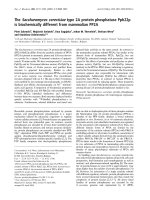


![Báo cáo Y học: The membrane-bound [NiFe]-hydrogenase (Ech) from Methanosarcina barkeri : unusual properties of the iron-sulphur clusters docx](https://media.store123doc.com/images/document/14/rc/ee/medium_eeh1395026426.jpg)
