Báo cáo y học: "The basophil activation test by flow cytometry: recent developments in clinical studies, standardization and emerging perspectives" pot
Bạn đang xem bản rút gọn của tài liệu. Xem và tải ngay bản đầy đủ của tài liệu tại đây (366.98 KB, 8 trang )
BioMed Central
Page 1 of 8
(page number not for citation purposes)
Clinical and Molecular Allergy
Open Access
Review
The basophil activation test by flow cytometry: recent
developments in clinical studies, standardization and emerging
perspectives
Radhia Boumiza
1
, Anne-Lise Debard
1
and Guillaume Monneret*
1,2
Address:
1
Immunology Laboratory, Lyon-Sud University Hospital, Lyon, France and
2
Immunology Laboratory, Hôpital Neurologique, Lyon,
France
Email: Radhia Boumiza - ; Anne-Lise Debard - ;
Guillaume Monneret* -
* Corresponding author
allergybasophilsflow cytometryCD63CD203CCRTH2
Abstract
The diagnosis of immediate allergy is mainly based upon an evocative clinical history, positive skin
tests (gold standard) and, if available, detection of specific IgE. In some complicated cases, functional
in vitro tests are necessary. The general concept of those tests is to mimic in vitro the contact
between allergens and circulating basophils. The first approach to basophil functional responses
was the histamine release test but this has remained controversial due to insufficient sensitivity and
specificity. During recent years an increasing number of studies have demonstrated that flow
cytometry is a reliable tool for monitoring basophil activation upon allergen challenge by detecting
surface expression of degranulation/activation markers (CD63 or CD203c). This article reviews
the recent improvements to the basophil activation test made possible by flow cytometry, focusing
on the use of anti-CRTH2/DP
2
antibodies for basophil recognition. On the basis of a new triple
staining protocol, the basophil activation test has become a standardized tool for in vitro diagnosis
of immediate allergy. It is also suitable for pharmacological studies on non-purified human basophils.
Multicenter studies are now required for its clinical assessment in large patient populations and to
define the cut-off values for clinical decision-making.
Introduction
Anaphylaxis consists of an immediate IgE-dependent
reaction in response to allergens. Clinical symptoms are
caused by an initial systemic histamine release by mast
cells and basophils that may lead to shock with laryngeal
edema, lower-airway obstruction and hypotension. The
most frequent allergens involved in immediate allergy are
found in peanuts, fish, bee and wasp venoms, drugs and
latex [1,2]. The identification of responsible allergens
remains a key step for practicing allergen avoidance and
specific immunotherapy. The diagnosis is mainly based
upon an evocative clinical history (including temporal
association between symptoms and allergen exposure),
positive skin tests, which remain the gold standard in this
context and, if available, detection of specific IgE [3]. In
most patients, these features allow both diagnosis and
identification of the offending allergen. Nevertheless, skin
testing is contraindicated in some patients with histories
Published: 30 June 2005
Clinical and Molecular Allergy 2005, 3:9 doi:10.1186/1476-7961-3-9
Received: 04 May 2005
Accepted: 30 June 2005
This article is available from: />© 2005 Boumiza et al; licensee BioMed Central Ltd.
This is an Open Access article distributed under the terms of the Creative Commons Attribution License ( />),
which permits unrestricted use, distribution, and reproduction in any medium, provided the original work is properly cited.
Clinical and Molecular Allergy 2005, 3:9 />Page 2 of 8
(page number not for citation purposes)
of life-threatening anaphylaxis, and discrepant results
may be found between clinical assessment of the disease
and biological results, especially for drug allergy. In these
cases, functional in vitro tests are necessary. The general
concept of those tests is to mimic in vitro the contact
between allergens and the cells responsible for symptoms
(i.e,. those possessing the ability to release histamine).
Until recently, basophils were neglected and only consid-
ered to be circulating forms of mast cells of minor impor-
tance. Furthermore, basophils represent in peripheral
blood less than 0.5 percent of total leukocytes, making
their purification difficult in clinical laboratories. This
lack of satisfactory in vitro protocols has clearly hampered
research on basophils for many years [4]. Nevertheless,
there is considerable recent evidence that basophils are
clinically relevant. Indeed, they are now considered as
equivalent to tissue mast cells cells since they play, by
themselves, a pivotal role in the immediate allergic reac-
tion [5-8]. Consequently, functional in vitro tests for aller-
gic reactions are focused on circulating basophils. The first
approach to basophil functional studies was the hista-
mine release test. However, its clinical benefit has
remained controversial due to insufficient sensitivity and
specificity [3,9,10]. That is why several groups took advan-
tage of flow cytometry to develop new tools for monitor-
ing basophil activation upon allergen challenge by
detecting surface expression of degranulation markers
[11-13].
Principle of basophil activation test by flow cytometry
As flow cytometry is a valuable tool for the analysis of
many different cell types and can be used to identify spe-
cific populations of cells, even when present in low num-
bers, it seemed to be suitable for the study of allergen-
induced basophil degranulation. Identification of cells
was initially based both on CD45 expression, a common
leukocyte antigen, and on the presence of IgE on the cell
surface, since basophils express the high affinity receptor
for IgE (FcεRI) [9,14]. In this gated population, cell activa-
tion upon allergen challenge was assessed by the expres-
sion of CD63 on the membrane [15,16]. CD63 is
anchored in the basophilic granule membrane (which
contains histamine) and its exposure to the outside of the
cells reflects cell degranulation due to fusion between
granules and plasma membranes (figure 1). Thus, CD63
expression has been proposed as a reliable means to mon-
itor basophil activation [11-13]. Briefly, whole blood was
incubated at 37°C with allergens for 15 minutes. The reac-
tion was stopped on ice, followed by a 30-min staining
with antibodies (figure 1). Finally, samples were lysed to
eliminate red cells. Basophils expressing both CD45 and
surface IgE were then examined for their CD63 expres-
sion. The threshold for positivity was determined with the
use of a negative control (i.e., whole blood and vehicle
without allergen). Results were considered positive when
at least 2 sequential allergen dilutions induced greater
than than 10% increases in CD63-positive basophils
above control values. This kind of protocol has been vali-
dated for common allergens by several groups and has
shown convincing results [17-24]. The technique has
proven to be accessible, rapid (results in less than 1 hour)
and requires small amount of blood (< 5 mL, even for
assessing several allergens in the same experiment). In our
hands, in allergy to muscle relaxants, the results were
quite interesting, since we found the sensitivity of the
CD63 test was similar to that for specific IgE detection and
higher than the one for histamine release test [25]. This
confirmed the value of performing the CD63 test rather
than histamine release, which is furthermore costly in
terms of both reagents and laboratory technician time. In
accord with previous studies focusing on different aller-
gens [17-21], this method showed excellent specificity.
However, with respect to drug allergy, the main indication
for this kind of test, three independent studies reported
similar sensitivities ranging between 50 and 64 %, which
is not sufficient for clinical usefulness [12,25,26]. In fact,
this first approach relied on two important characteristics
of basophils which were problematic: recognition
through the expression of IgE on their surface (which is
known to be highly variable from one patient to another)
and the monitoring of their activation by detecting CD63
(which is also expressed to some extent by other activated
leukocytes and by activated platelets that may adhere to
basophils). This may explain why, when applied to drug
allergy, these tests have remained somewhat disappoint-
ing in terms of sensitivities [12,25,26]. Consequently, we
concluded that an activation marker that is more specific
and/or sensitive than CD63 would be desirable.
CD203c as a specific marker of activated basophils
CD203c corresponds to a surface antigen expressed on
human basophils recently recognized by the monoclonal
antibody 97A6 [27]. This antigen, belonging to the type II
transmembrane protein family, is a multifunctional ecto-
enzyme called ectonucleotide pyrophosphatase pho-
phodiesterase 3 (E-NPP3) [28] that catalyzes the cleavage
of a number of molecules including deoxynucleotides and
nucleotide sugars [29]. In addition, E-NPP3 contains a
somatomedin B-like domain and a cell adhesive motif,
but their potential functions remain totally unknown
with respect to basophil physiology. Among leukocytes
CD203c appears to be selectively expressed on the
basophil/mastocytes lineage [27]. To date, no other cells
from human peripheral blood have been reported to
express this marker. Its expression on basophils is rapidly
upregulated after stimulation with the appropriate aller-
gen in patients sensitized to acarids or hymenoptera or
after crosslinking of FcεRI with anti-IgE antibodies
[28,30]. This suggests that CD203c up-regulation is more
or less specific to the crosslinking of FcεRI (figure 2).
Clinical and Molecular Allergy 2005, 3:9 />Page 3 of 8
(page number not for citation purposes)
Hence, as CD203c is rapidly upregulated after allergen
challenge, it has been proposed as a new tool for allergy
diagnosis [30-33]. We compared basophil activation tests
using either CD63 or CD203c in the diagnosis of latex
allergy [34] and found that the sensitivity was considera-
bly higher with CD203c (75% compared to 50% with
CD63). The improved sensitivity may be due to two fac-
tors. First, the recognition of basophils is better with
CD203c. Indeed, the identification of basophils using
prior protocols relied on a single IgE-labeling, although it
is known that FcεRI expression can vary considerably on
cell surfaces from one patient to another [35]. This may
explain why in some cases basophils were difficult or
impossible to identify. The second reason for the
improved sensitivity with CD203c is due to its higher
expression in activated basophils compared to CD63 in
our experiments. In sensitized patients, basophils
increased their CD203c levels up to 350 % above control
values in response to allergens whereas the increase in
CD63 was below 100 %. Similar results were obtained
when expressing the results as the percentages of
basophils that were CD203c- or CD63-positive. Even with
the highest concentration of latex, the mean percentage of
CD63-positive basophils was below 20 % while that of
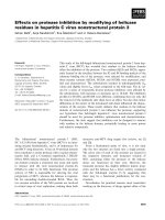
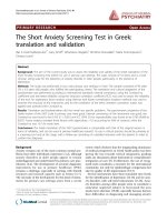
Principle of the basophil activation test by flow cytometry (triple staining)Figure 1
Principle of the basophil activation test by flow cytometry (triple staining). Basophils are identified on the basis of
CD45 expression (fluorescence 3 / Phyco-Cyanine 5) and the presence of IgE or CRTH2/DP2 on their surface (fluorescence 1
/ Fluorescein isothiocyanate). Resting basophils do not express CD63 (anchored in the basophilic granule) and weakly express
CD203c. The cross-linking of two FcεRI (induced by an allergen or anti-IgE antibodies) provokes the histamine release (and as
a consequence the CD63 expression) and the upregulation of CD203c. The rise in CD63 or CD203c expression (measured by
fluorescence 2 / Phycoerythrin) before and after allergen challenge reflects thus the basophil activation / degranulation in
response to an allergen.
CD203c
Histamine
CD63
FcεRI
Allergen
Histamine
release
IgE
PE-mAb
anti-CD203c
Fluo. 2
PE-mAb
anti-CD63
Fluo. 2
FITC-mAb
anti-IgE
CD45
PC5-mAb
anti-CD45
Fluo. 3
hν
hν
hν
hν
Fluo.1
FITC-mAb
Anti-CRTH2/DP2
CRTH2/DP2
Clinical and Molecular Allergy 2005, 3:9 />Page 4 of 8
(page number not for citation purposes)
CD203c-positive basophils was 48 %, allowing a clear dis-
tinction between resting and activated basophils [34]. In
conclusion, both easier gating and higher range of activa-
tion in response to allergen may contribute to an
improvement in the basophil activation test when using
CD203c rather than CD63. However, as very few studies
concomitantly compared CD203c and CD63, this point
remains to be confirmed by additional works dealing with
various allergens. Bühring and colleagues in a recent
report proposed to use both markers in the same test to
increase sensitivity [32]. It is supported by recent evidence
showing that CD63 and CD203c overexpression depend
on different stimulatory pathways [36,37]. It is to note
that some novel basophil-activation markers (CD13,
CD107a, CD164) have been very recently identified [37].
They have to be further investigated in clinical studies
either by their own or in combination with CD63 or
CD203c.
CRTH2/DP
2
as a new marker for basophil recognition
Finally, the last drawback of the previously described pro-
tocols remained the use of an anti-IgE reagent to identify
basophils. Because of its selective expression on cells asso-
ciated with Th2 responses (Th2 lymphocytes, eosinophils
and basophils), CRTH2 (chemoattractant receptor-
homologous molecule expressed on Th2 cells)/DP
2
has
been proposed and validated as the most reliable tool for
the detection of circulating human Th2 cells [38,39].
CRTH2 is also termed DP
2
since it corresponds to the sec-
ond receptor of prostaglandin D
2
[40,41]. As CRTH2 is
highly expressed on basophils, we hypothesized that it
could improve the basophil activation test by facilitating
basophil recognition. Consequently, we developed a new
three-colour flow cytometric protocol (PE-CD203c /
FITC-CRTH2 / PC5-CD3) for monitoring allergen-
induced basophil activation. First results were encourag-
ing: CRTH2 staining allowed CRTH2-expressing cells
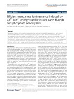
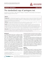
CD203c expression in whole blood before and after basophil activationFigure 2
CD203c expression in whole blood before and after basophil activation. Ungated leukocytes are shown as a bipara-
metric representation on the basis of side scatter characteristics (SSC, y-axis) and CD203c (x-axis). Left histogram depicts
resting cells, basophils express low levels of CD203c (some of them are not distinguishable from lymphocytes and monocytes).
Right histogram depicts cells after anti-IgE challenge, activated basophils are easily recognized on the basis of their high CD203c
expression.
Resting
Basophils
(CD203c dim)
Activated
Basophils
(CD203c bright)
Lymphocytes
Neutrophils
M
o
n
oc
y
t
e
s
SSC
CD203c
Clinical and Molecular Allergy 2005, 3:9 />Page 5 of 8
(page number not for citation purposes)
(eosinophils, basophils and Th2 lymphocytes) to easily
be distinguished from other cells in samples of whole
blood (figure 3). On the basis of light scattering, eosi-
nophils were easily excluded from the analysis (figure 3).
Basophils could then readily be distinguished from Th2
lymphocytes on the basis of CD3, staining, as this marker
is not present on basophils (figure 3). Finally, on this
gated population of basophils (low light scatterings,
CRTH2+ and CD3-), modulation of CD203c after allergen
challenge was monitored as described in the former pro-
tocol (figure 4). To validate this protocol, 18 subjects were
included in a preliminary study [42]. Patients were allergic
to either latex (k82) or Dermatophagoïdes pteronyssinus
(d1), had a suggestive clinical history, positive skin test
and/or specific IgE ≥ class III. Healthy donors, from our
laboratory, were not known to be allergic and presented
total IgE < 100 kU/L. In terms of clinical interpretation,
sensitivity and specificity were 88% and 100%, respec-
tively [40]. CRTH2 staining was an excellent means to
identify basophils and we confirmed our earlier observa-
tions of a wide range of CD203c expression in response to
allergen in tehse cells. In terms of basophil recovery, we
compared our CRTH2-staining protocol with 2 others
protocols using either anti-IgE or anti-CD123 (IL-3 recep-
tor). In all patients and healthy individuals, we found
more basophils (up to 50 % in certain patients) with the
CRTH2-staining protocol, illustrating its superiority with
respect to basophil recovery. To conclude, the easy recog-
nition of basophils and the reliable assessment of their
activation make this protocol the most reliable tool for
investigating basophil activation by flow cytometry. It
may constitute a critical step for the interlab standardiza-
tion of this kind of test. Lastly, since CRTH2 is also a
marker of Th2 cells and eosinophils, it may become a
promising tool for flow cytometry, providing a direct
overview of cells involved in "Th2 diseases" such as
allergy.
Perspectives in pharmacological studies
Until recently, due to the very low number of circulating
basophils in humans, pharmacological studies on these
cells were difficult to perform. This required large amount
of blood and / or lengthy purification procedures that
may induce nonspecific activation. By the use of flow
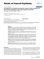
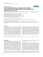
Identification of CRTH2 expressing cells by flow cytometryFigure 3
Identification of CRTH2 expressing cells by flow cytometry. Left histogram : ungated leukocytes biparametric repre-
sentation on the basis of side scatter characteristics (SSC, Y axis) and FITC-CRTH2 (X axis). Two CRTH2 expressing cell pop-
ulations are easily distinguishable: the one with high light scatterings corresponds to the eosinophil population; the second one
(gating region: A) comprises Th2 lymphocytes and basophils. Right histogram: cells from the gating region (A) expressed on the
basis of PE-CD203c (X axis) and PC5-CD3 (Y axis) characteristics. Th2 lymphocytes were readily separated from basophils
based on their positive CD3 expression while activated basophils express high levels of CD203c without expressing CD3.
CD3-PC5
PE-CD203c
SSC
FITC-CRTH2
Ungated
A
Gated on A
Th2 lymphocytes
(CD3+)
Activated
Basophils
(CD3-)
Clinical and Molecular Allergy 2005, 3:9 />Page 6 of 8
(page number not for citation purposes)
cytometry, the effects of different compounds on
basophils may be examined in unfractionated human
blood cells. Recently, we have been able to demonstrate
that among various eicosanoids, prostaglandin D
2
was by
far the most potent activator of basophils, inducing
CD203c and CD11b elevation [37]. This response was
mediated by the DP2 receptor / CRTH2 as it was shared by
selective agonists of this receptor. As previously observed
in eosinophils [39], the interaction of prostaglandin D
2
with the DP
1
receptor limited the activation of basophils
by this prostaglandin. This suggested that the balance
between DP
1
and DP
2
receptors may be crucial in deter-
mining the magnitude of basophil responses during aller-
gic processes since prostaglandin D2 is known to be
involved in allergic diseases and asthma. Using a similar
approach, Heinemann et al. [43] examined the effects of
various chemokines on human basophils and demon-
Representative increased expression of CD203c after allergen challenge in a patient allergic to Dermatophagoïdes pteronyssi-nus (d1)Figure 4
Representative increased expression of CD203c after allergen challenge in a patient allergic to Dermat-
ophagoïdes pteronyssinus (d1). Gated CRTH2-positive basophils (after excluding Th2 lymphocytes as described in figure
3) are presented on the basis of CD203c-CRTH2 staining: before stimulation (negative control, upper left dot-plot), after anti-
IgE challenge (positive control, upper right) and after allergen challenge at 3 different concentrations (dose-effect response,
lower dot-plots). Activated basophils: percentage of basophils expressing CD203c.
Negative control Positive control (anti-IgE)
d1 (/100)
Activated
Basophils : 5 %
Activated
Basophils : 88 %
FITC-CRTH2
d1 (/500) d1 (/1000)
Activated
Basophils : 96 %
Activated
Basophils : 62 %
Activated
Basophils : 47 %
PE-CD203c
Clinical and Molecular Allergy 2005, 3:9 />Page 7 of 8
(page number not for citation purposes)
strated a different pattern of chemokine receptor usage
than those described for eosinophils and monocytes.
These studies illustrate that it is now possible to perform
pharmacological and drug screening studies by flow
cytometry. This approach could be very useful in assessing
the possible risks of inducing anaphylactoid or pseudo-
anaphylactoid reactions when developing new molecules.
To this end, one important task for the future will be to
extend these kinds of protocols to animal models
although, to our knowledge, there is no available infor-
mation on CD203c in animals and monoclonal antibod-
ies directed against human CD203c do not cross-react
with other species [32].
Conclusion
After several improvements, the basophil activation test
(using either CD203c or CD63 as activation marker) has
become a robust and reliable test for in vitro investigations
of immediate allergy, complementary to other existing in
vitro tests. It is suitable for experimental and pharmaco-
logical studies as well as allergy diagnosis in clinical prac-
tice. There is now a crucial need for inter-laboratory
standardization in clinical decision-making. Each allergen
has to be assessed one by one to determine its optimal
concentration (i.e., inducing maximal activation in vitro)
as well as the definition of the threshold for positivity
(using ROC analysis) since the use of an arbitrary cut-off
value is likely not suitable for all allergens. The present
challenge is to take advantage of the availability of
improved methods to perform multicenter studies using a
standardized protocol.
Competing interests
The author(s) declare that they have no competing
interests.
Authors' contributions
RB participated as investigator and is the main author of
the article.
ALD participated in drafting the manuscript.
GM was project leader and participated in the design of
the different studies and drafting the manuscript.
Acknowledgements
We thank the technical staff of the flow cytometry unit – Immunology lab
(J. Baudot, C. Fernandez, MA. Guinand, MC. Gutowski) at the Lyon-Sud
University Hospital, Pr. J. Bienvenu (Immunology lab, Lyon-Sud University
Hospital) for supporting our work on basophil activation over the years, G.
Bouvier and C. Canino (Immunotech, Marseille, France) for kindly providing
anti-CRTH2 antibodies.
References
1. Kay AB: Allergy and allergic diseases (part 1). N Engl J Med
2001, 344:30-7.
2. Kay AB: Allergy and allergic diseases (part 2). N Engl J Med
2001, 344:109-13.
3. Hamilton RG, Adkinson NF: In vitro assays for the diagnosis of
IgE-mediated disorders. J Allergy Clin Immunol 2004, 114:213-225.
4. Falcone FH, Haas H, Gibbs BF: The human basophil: a new
appreciation of its role in immune responses. Blood 2000,
96:4028-38.
5. Gauchat JF, Henchoz S, Mazzei G, Aubry JP, Brunner T, Blasey H, Life
P, Talabot D, Flores-Romo L, Thompson J: Induction of human IgE
synthesis in B cells by mast cells and basophils. Nature 1993,
365:340-43.
6. Kasaian MT, Clay MJ, Happ MP, Garman RD, Hirani S, Luqman M: IL-
4 production by allergen-stimulated primary cultures: Iden-
tification of basophils as the major IL-4 producing cell type.
Int Immunol 1996, 8:1287-97.
7. Devouassoux G, Forster B, Scott LM, Metcalfe DD, Prussin C: Fre-
quency and characterization of antigen-specific IL-4 and IL-
13 producing basophils and Tcells in peripheral blood of
healthy and asmathic subjects. J Allergy Clin Immunol 1999,
104:811-19.
8. Galli SJ: Mast cells and basophils. Curr Opin Hematol 2000, 7:32-9.
9. Gane P, Pecquet C, Lamblin P, Abuaf N, Leynadier F, Rouger P: Flow
cytometric evaluation of human basophils. Cytometry 1993,
14:344-8.
10. Demoly P, Lebel B, Messaad D, Sahla H, Rongier M, Daures JP,
Godard P, Bousquet J: Predictive capacity of histamine release
for the diagnosis of drug allergy. Allergy 1999, 54:500-6.
11. Sainte-Laudy J, Sabbah A, Vallon C, Guerin JC: Analysis of anti-IgE
and allergen induced human basophil activation by flow
Cytometry. Comparison with histamine release. Inflamm Res
1998, 47:401-8.
12. Abuaf N, Rajoely B, Ghazouani E, Levy DA, Pecquet C, Chabane H,
Leynadier F: Validation of a flow cytometric assay detecting in
vitro basophil activation for the diagnosis of muscle relaxant
allergy. J Allergy Clin Immunol 1999, 104:411-8.
13. Monneret G, Gutowski MC, Bienvenu J: Detection of allergen-
induced basophil activation by expression of CD63 antigen
using flow cytometric method. Clin Exp Immunol 1999,
115:393-6.
14. Gane P, Pecquet C, Crespeau H, Lambin P, Leynadier F, Rouger P:
Flow cytometric monitoring of allergen induced basophil
activation. Cytometry 1995, 19:361-365.
15. Knol EF, Mul FP, Jansen H, Calafat J, Roos D: Monitoring human
basophil activation via CD63 monoclonal antibody 435. J
Allergy Clin Immunol 1991, 88:328-38.
16. Fureder W, Agis H, Sperr WR, Lechner K, Valent P: The surface
membrane antigen phenotype of human blood basophils.
Allergy 1994, 49:861-5.
17. Pâris-Köhler A, Demoly P, Persi L, Lebel B, Bousquet J, Arnoux B: In
vitro diagnosis of cypress pollen allergy by using cytofluori-
metric analysis of basophils (Basotest). J Allergy Clin Immunol
2000, 105:339-45.
18. Sainte-Laudy J, Sabbah A, Drouet M, Lauret MG, Loiry M: Diagnosis
of venom allergy by flow cytometry. Correlation with clinical
history, skin tests, specific IgE, Histamine and Leukotriene
C4 release. Clin Exp Allergy 2000, 30:1166-71.
19. Sanz ML, Sanchez G, Gamboa PM, Vila L, Uasuf C, Chazot M, Dieguez
I, De Weck AL: Allergen-induced basophil activation: CD63
cell expression deteced by flow cytometry in patients aller-
gic to Dermatophagoides pteronyssinus and Lolium perenne.
Clin Exp Allergy 2001, 31:1007-13.
20. Saporta M, Kamei S, Persi L, Bousquet J, Arnoux B: Basophil activa-
tion during pollen season in patients monosensitized to grass
pollen. Allergy 2001, 56:442-5.
21. Ebo DG, Hagendorens MM, Bridts CH, Schuerwegh AJ, De Clerck LS,
Stevens WJ: In vitro allergy diagnosis: should we follow the
flow? Clin Exp Allergy 2004, 34:332-339.
22. Torres MJ, Padial A, Mayorga C, Fernandez T, Sanchez-Sabate E, Cor-
nejo-Garcia JA, Antunez C, Blanca M: The diagnostic interpreta-
tion of basophil activation test in immediate allergic
reactions to betalactams. Clin Exp Allergy 2004, 34:1768-75.
23. Erdmann SM, Sachs B, Kwiecien R, Moll-Slodowy S, Sauer I, Merk HF:
The basophil activation test in wasp venom allergy: sensitiv-
Publish with BioMed Central and every
scientist can read your work free of charge
"BioMed Central will be the most significant development for
disseminating the results of biomedical research in our lifetime."
Sir Paul Nurse, Cancer Research UK
Your research papers will be:
available free of charge to the entire biomedical community
peer reviewed and published immediately upon acceptance
cited in PubMed and archived on PubMed Central
yours — you keep the copyright
Submit your manuscript here:
/>BioMedcentral
Clinical and Molecular Allergy 2005, 3:9 />Page 8 of 8
(page number not for citation purposes)
ity, specificity and monitoring specific immunotherapy.
Allergy 2004, 59:1102-9.
24. Ebo DG, Hagendorens MM, Bridts CH, Schuerwegh AJ, De Clerck LS,
Stevens WJ: Flow cytometric analysis of in vitro activated
basophils, specific IgE and skin tests in the diagnosis of pol-
len-associated food allergy. Cytometry 2005, 64:28-33.
25. Monneret G, Benoit Y, Debard AL, Gutowski MC, Topenot I, Bien-
venu J: Monitoring of Basophil activation using CD63 and
CCR3 in allergy to muscle relaxant drugs. Clin Immunol 2002,
102:192-9.
26. Sanz ML, Gamboa PM, Antepara I, Uasuf C, Vila L, Garcia-Aviles C,
Chazot M, De Weck AL: Flow cytometric basophil activation
test by detection of CD63 expression in patients with imme-
diate-type reactions to betalactam antibiotics. Clin Exp Allergy
2002, 32:277-86.
27. Buhring HJ, Simmons PJ, Pudney M, Muller R, Jarrossay D, van Agth-
oven A, Willheim M, Brugger W, Valent P, Kanz L: The monoclonal
antibody 97A6 defines a novel surface antigen expressed on
human basophils and their multipotent progenitors. Blood
1999, 94:2343-56.
28. Buhring HJ, Seiffert M, Giesert C, Marxer A, Kanz L, Valent P, Sano K:
The basophil activation marker defined by antibody 97A6 is
identical to the ectonucleotide pyrophosphatase/phosphodi-
esterase 3. Blood 2001, 97:3303-5.
29. Bollen M, Gijbers R, Ceulemans H, Stalmans W, Stefan C: Nucle-
otide pyrophosphatase/phophodiesterases on the move. Crit
Rev Biochem Mol Biol 2000, 35:393-432.
30. Platz IJ, Binder M, Marxer A, Lischka G, Valent P, Bühring HJ:
Hymenoptera-venom-induced upregulation of the basophil
activation marker ecto-nucleotide pyrophosphatase/phos-
phodieterase 3 in sensitized individuals. Int Arch Allergy Immunol
2001, 126:335-42.
31. Hauswirth AW, Natter S, Ghannadan M, Majlesi Y, Schernthaner GH,
Sperr WR, Buhring HJ, Valenta R, Valent P: Recombinant allergens
promote expression of CD203c on basophils in sensitized
individuals. J Allergy Clin Immunol 2002, 110:102-9.
32. Bühring HJ, Streble A, Valent P: The basophil-specific ectoen-
zyme E-NPP3 (CD203c) as a marker for cell activation and
allergy diagnosis. Int Arch Allergy Immunol 2004, 133:317-329.
33. Kahlert H, Cromwell O, Fiebig H: Measurement of basophil-acti-
vating capacity of grass pollen allergens, allergoids and
hypoallergenic recombinant derivatives by flow cytometry
using anti-CD203c. Clin Exp Allergy 2003, 33:1266-72.
34. Boumiza R, Monneret G, Forissier MF, Savoye J, Gutowski MC, Pow-
ell WS, Bienvenu J: Marked improvement of the basophil acti-
vation test by detecting CD203c instead of CD63. Clin Exp
Allergy 2003, 33:259-265.
35. Kinet JP: The high-affinity IgE receptor (FcεRI): From physiol-
ogy to pathology. Ann Rev Immunol 1999, 17:931-72.
36. Hennersdorf F, Florian S, Jakob A, Baumgartner K, Sonneck K, Nord-
heim A, Biedermann T, Valent P, Buhring HJ: Identification of
CD13, CD107a, and CD164 as novel basophil-activation
markers and dissection of two response patterns in time
kinetics of ige-dependent upregulation. Cell Res 2005,
15:325-35.
37. Monneret G, Boumiza R, Gravel S, Cossette C, Bienvenu J, Rokach J,
Powell WS: Effects of prostaglandin D2 and 5-lipoxygenase
products on the expression of CD203c and CD11b by
basophils. J Pharmacol Exp Ther 2005, 312:627-34.
38. Cosmi L, Annunziato F, Galli MIG, Maggi RME, Nagata K, Romagnani
S: CRTH2 is the most reliable marker for the detection of
circulating human type 2 Th and type 2 T cytotoxic cells in
health and disease. Eur J Immunol 2000, 30:2972-2979.
39. Nagata K, Tanaka K, Ogawa K, Kemmotsu K, Imai T, Yoshie O, Abe
H, Tada K, Nakamura M, Sugamura K, Takano S: Selective expres-
sion of a novel surface molecule by human Th2 cells in vivo.
J Immunol 1999, 162:1278-1286.
40. Hirai H, Tanaka K, Yoshie O, Ogawa K, Kenmotsu K, Takamori Y,
Ichimasa M, Sugamura K, Nakamura M, Takano S, Nagata K: Prostag-
landin D2 selectively induces chemotaxis in T helper type 2
cells, eosinophils, and basophils via seven-transmembrane
receptor CRTH2. J Exp Med 2001, 193:255-261.
41. Monneret G, Gravel S, Diamond M, Rokach J, Powell WS: Prostag-
landin D2 is a potent chemoattractant for human eosinophils
that acts via a novel DP receptor. Blood 2001, 98:1942-1948.
42. Bouvier G, Debard AL, Gendt L, Canino C, Bienvenu J, Monneret G:
A new three-colour flow cytometric combination (CD203C/
CRTH2/CD3) for monitoring allergen-induced basophil
activation. Cytometry 2004, 59A:134.
43. Heinemann A, Hartnell A, Stubbs VE, Murakami K, Soler D, LaRosa
G, Askenase PW, Williams TJ, Sabroe I: Basophil responses to
chemokines are regulated by both sequential and coopera-
tive receptor signalling. J Immunol 2000, 165:7224-7233.