Báo cáo y học: "Patients with allergic rhinitis and allergic asthma share the same pattern of eosinophil and neutrophil degranulation after allergen challenge" pdf
Bạn đang xem bản rút gọn của tài liệu. Xem và tải ngay bản đầy đủ của tài liệu tại đây (367.2 KB, 10 trang )
RESEARCH Open Access
Patients with allergic rhinitis and allergic asthma
share the same pattern of eosinophil and
neutrophil degranulation after allergen challenge
Mary Kämpe
1,2*
, Ingrid Stolt
2,3
, Maria Lampinen
2,3
, Christer Janson
1,2
, Gunnemar Stålenheim
1,2
, Marie Carlson
2,3
Abstract
Background: Patients with allergic rhinitis and allergic asthma demonstrate comparable local and systemic
eosinophil inflammation, and yet they present with differe nt clinical pictures. Less is even known about the
contribution of neutrophil inflammation in allergic diseases. The aim of the study was to examine the propensity
and selectivity of granule release from primed systemic eosinophils and neutrophils in allergic rhinitis and allergic
asthma after seasonal and experimental allergen exposure. We hypothesize that the dissimilar clinical
manifestations are due to diverse eosinophil and neutrophil degranulation.
Methods: Nine birch pollen allergic patients with rhinitis, eight with asthma and four controls were studied during
pollen season and after nasal and bronchial allergen challenge. Eosinophils and neutrophils were incubated in vitro
with assay buffer and opsonized Sephadex particles for spontaneous and C3b-induced granule protein release. The
released amount of eosinophil cationic protein (ECP), eosinophil peroxidase (EPO) and myeloperoxidase (MPO) was
measured by specific radioimmunoassay.
Results: C3b-induced degranulation resulte d in increased release of ECP and MPO from prim ed blood eosinophils
and neutrophils in both allergic rhinitis and allergic asthma during pollen season and after both nasal and
bronchial challenge (p-values 0.008 to 0.043). After bronchial challenge, the ECP release was significantly higher in
the rhinitic group compared to the asthmatic group [19.8 vs. 13.2%, (p = 0.010)]. The propensity for EPO release
was weak in all challenge models but followed the same pattern in both allergic groups.
Conclusions: Systemically activated eosinophils and neutrophils have similar patterns of degranulation after
allergen exposure in allergic rhinitis and allergic asthma. The released amount of ECP, EPO and MPO was similar in
all allergen challenge models in both allergic groups. Our results indicate that other mechanisms than the
magnitude of eosinophil and neutrophil inflammation or the degranulation pattern of the inflammato ry cells
determines whether or not an allergic patient develops asthma.
Introduction
Allergic diseases, such as allergic ast hma, allergic rhinitis
and atopic dermatitis are characterised by an increased
number of eosinophil granulocytes in the circulating blood
and degra nulation in the target tissue is con sidered the
major pathogenic event [1]. The eosinophil is a multifunc-
tional leukocyte playing a central role in Th
2
mediated
allergic diseases [2], parasitic killing and tissue repair [1].
Recent studies have also pointed out eosinophil
involvement in modulating both innate and adaptive
immune responses [3]. The primed eosinophil rapidly
secretes four preformed, highly cytotoxic, cationic granule
proteins at the site of inflammation: eosinophil cationic
protein (ECP), eosinophil peroxidase (EPO), eosinophil
derived neurotoxin (EDN)/former eosinophil protein X
(EPX) and major basic protein (MBP) beside chemokines,
cytokines and growth factors [1,3]. In addition to regulated
exocytosis and cytolysis [4], the e osinophils release their
granule proteins through a process of piecemeal degranu-
lation by transport vesicles allowing selective release of the
eosinophilic granule proteins [5,6].
* Correspondence:
1
Department of Medical Sciences, Respiratory Medicine and Allergology,
Uppsala University, Uppsala, Sweden
Full list of author information is available at the end of the article
Kämpe et al. Clinical and Molecular Allergy 2011, 9:3
/>CMA
© 2011 Kämpe et al; licensee BioMed Central Ltd. This is an Open Access article distributed under the terms of the Creative Commons
Attribution License ( which permits unrestricted use, distribution, and reproduction in
any medium, provided the or iginal work is properly cited.
Jatakanon et al reported more than a decade ago that
neutrophils have an important role in chronic severe
asthma [7], and neutrophil inflammation of the airways
is today considered relevant to the pathogenesis of the
more severe forms of the disease [8,9]. However, in a
novel study, neutrophilia was observed in induced spu-
tum in children with non-atopic asthma [10], but the
role of neutrophils in allergic rhinitis and mild asthma is
uncertain and under debate. It has been speculated that
neutrophils are taking part in both the initiation and
resolution of even mild asthma attacks [8].
The neutrophils house two major granule populations,
primary (azurophil) and secon dary (specific) granules,
formed during the maturation process. The primary
granules contain mainly myeloperoxidase (MPO), several
proteases and the antibiotic defensin peptides, all
released in a potentially active state [11]. The specific
granules store latent pro-forms of mainly metallopro-
teases, activated by the azurophilic proteases first after
the degranulation [11]. The highly cytotoxic myeloper-
oxidase from the primary granules has been used as a
marker of the neutrophil activity [12].
It has been known for long that binding of eosinophils
and neutrophils to a surface by complement receptors
induces a strong signal for degranulation, involving the
receptor for complement factor 3 (C3b receptor)
[13,14]. Using serum-opsonised Sephadex particles
in vitro in exp erimental settings [15,16] enhances this
C3b-induced degranulation of the eosinophils in allergy
as well as in infections [17,18]. Previous studies h ave
reported increased propensit y of granule release in vitro
from primed eosinophils and neutrophils in allergic
asthma compared to controls after Sephadex stimula-
tion, bot h during pollen season as well as out of season
[19,20]. This data indicates priming of both types of
granulocytes in allergic asthmatics.
The link between the upper and lower airways is well-
established [21]. Many studies have reported both blood
eosinophilia and local eosinophilia in nasa l lavage as well
as in induced sputum both during pollen season and
after local allergen challenge in the n ose and bronc hi
respectively [22-24]. The q uestion rem ains why patients
with allergic asthma and allergic rhinitis demonstrate
moreorlessthesamedegreeofsystemiceosinophil
inflammation both during pollen season and after nasal
and b ronchial challenge and yet they prese nt with differ-
ent clinical pictures. The hypothesis of the present study
was that the dissimilar clinical manifestations of asth-
matic and rhinitic patients are due to differences in selec-
tive eosinophil and neutrophil degranulation. The
primary aim of the study was thus to study differences in
allergic rhinitis and allergic asth ma w ith regard to the
degra nulation pattern of allergen primed eosino phils and
neutrophils. A seco ndary aim was to investigate if there
is a differential and selective granule release from primed
eosinophils and neutrophils in the two allergic groups
depending on the allergen challenge model.
Materials and methods
Patients
Seventeen birch pollen allergic patients were sel ected for
the study, all diagnosed with s easonal allergic rhinitis or
allergic asthma by a lung physician and allergologist at
the allergy out-patient clinic at Uppsala University
Hospital All patients were skin prick test positive to
birch pollen and none of the patients had symptoms or
were on any regular treatment outside birch pollen sea-
son. Eight patients had a diagnosis of allergic seasonal
asthma, having respiratory symptoms (wheeze and dys-
pnea) and denying nasal symptoms during birch pollen
season, and thus were categorised as h aving asthma as
the predominant symptom. Nine patients were diagnosed
with allergic rhinitis, having eye and nose symptoms and
denying respiratory symptoms, and consequently cate-
gorised as having rhinitis as the predominant symptom.
Topical steroids were not allowed during pollen season
or outside season, and none of the patients were on any
regular medication during season. None of the patients
had smoked for the past ten years. Forced expiratory
volume in one s econd (F EV
1
) out of season was more
than 75% of predicted and FEV
1
/forced vital capacity
(FVC) more than 70% in all patients (Table 1).
Control group
The control group consisted of five healthy, non-atopic,
never smoking subjects, having allergic symptoms
neither outside nor during the birch pollen season.
They were skin prick test negative to all nine standard
allergens, had no serum IgE antibodies, and had normal
lung fu nction with an FEV
1
>80% of predicted. The
control group only completed investigations during the
pollen season (Table 1).
Study design
The study included altogether five visits to our out-
patient clinic: inclusion, baseline, during birch pollen
season and after bronchial and nasal allergen challenge
respectively. The season visit was made two to three
weeks after the airborne pollen counts had reached
4000grains/m
3
, pollen grains count ed by the Palyno-
logical Laboratory, Swedish Museum of Natural
History, Stockholm, Sweden [23]. The study was per-
formed during the birch pollen seasons in 2000 and
2002; the season 2001 was excluded due to low pollen
counts. After inclusion patients were investigated con-
secutively, thus all patien ts were studied pre-season
and during season in the same year. Bronchia l and
nasal allergen challenges were performed during a four
Kämpe et al. Clinical and Molecular Allergy 2011, 9:3
/>Page 2 of 10
week period in January and February the following
year. The subjects were told to avoid short-acting
bronchodilators and anti-histamines for 24 hours
before the visits and nasal decongestants for four
hours before the visits. When pollen counts reached
4 000 grains/m
3
the subjects were told to start record-
ing their morning and evening PEFR in a diary. The
design of the present study has been described in
detail in previous reports [23,24].
Skin prick tests
Skin prick tests were performed with nine standard
aeroallergen extracts (birch, timothy, mugwort, cat dan-
der, dog dander, horse dander, Dermatophagoides ptero-
nyssinus, Cladosporium herbarum and Alternaria using
Soluprick SQ A LK (Hørsholm, Denmark). The results
were read after 15 minutes, measuring the largest dia-
meter of the wheal and its perpendicular diameter, and
the product was expressed in mm
2
.Skinreactionswere
considered positive when larger than 9 mm
2
.
Spirometry
Lung function tests were performed with a Vitalograph-
Compact spirometer (Vitalograph Ltd., Buckingham,
England). FEV
1
,FVC,FEV
1
/FVC% and PEFR were
recorded. The reference values were those from
European Community for Coal and Steel [25]. Spirome-
try was performed before and after the hypertonic saline
inhalation. The magnitude of the FEV
1
decrease after
the hypertonic saline inhalation was used as a marker of
bronchial responsiveness [23].
Nasal challenge test
The experimental nasal challenge test was performed by
instillation in the same nostril of 0.3 mL d iluent fol-
lowed by birch pollen extract (Aquagen
®
SQ, ALK-
Abelló, Hørsholm, Denmark) every 15 minutes in three
steps: 1 000 SQ-U/mL, 10 000 SQ-U/mL and 100 000
SQ-U/mL. The symptom score w as estimated; if pro-
nounced local symptoms and sneezing occurred, the
challenge test was stopped. The response to the allergen
provocation was categorized into four groups: no
response or response to one or more of the three a ller-
gen doses. Blood samples and nasal lavage were taken
18 hr (±1 hr) after the challenge test was completed.
Bronchial allergen challenge test
The experimental bronchial challenge test was performed
using a DeVilbiss-40 nebulizer (particle size 0.5 to 5.5 μm,
output 0.175 ± 0.3 mL/min, mean ± SD) (Devillbiss Co,
Somerset, PA) [26]. Bronchial challenge with birch pollen
extract (Aquagen
®
SQ, ALK-Abelló, Hørsholm, Denmark)
was performed in three steps with the doses 1 000 SQE,
10 000 SQ-U and 100 000 SQ-U, starting with inhalation
of a diluent. The response to the allergen provocation was
calculated as the cumulative dose that caused at least 20%
decrease in FEV
1
(allergen provocation dose, PD
20
). The
challenge test was stopped if F EV
1
decreased by 20%.
Blood samples were taken after 18 hr (±1 hr).
Isolation of granulocytes
Isolation was performed on heparinized blood. The mono-
nuclear leukocytes were separated by percoll gradient
Table 1 Demographic data of the control group and patients with allergic rhinitis and allergic asthma (mean and
range)
Control group Allergic rhinitis Allergic asthma p-value
n = 5 (n = 9) (n = 8) (AR/AA)***
Gender (male/female) 2/3 8/1 3/8 0.36
Age 38 (27-58) 43 (24 - 66) 41 (19 - 56) 0.85
Ex-smoker (>10 yr) 0 2 1 0.79
SPT birch (in mm
2
) 0 47.1 (20 - 88) 43.6 (26 - 64) 1.0
IgE for birch, Class 0-5 0 3.1 (2 - 4) 3.4 (2 - 5) 0.43
FEV
1
(L) 3.59(3.02-3.95) 4.0 (2.4 - 4.9) 3.5 (2.6 - 4.0) 0.12
FEV
1
% of predicted 105 (88-125) 102 (75 - 139) 97 (83 - 108) 0.44
PEFR (L/min) 571 (348-854) 615 (415 - 826) 504 (347 - 652) 0.18
PEFR % of predicted 117 (84-169) 113 (82 - 140) 101 (73 - 133) 0.25
FEV
1
-decrease in % * 1.1 (-0.5-6.2) 0.42 (-5.6 - 5.8) 6.81 (1.55 - 16.4) 0.02
PD
20
(birch) (SQE)** - 34 500 (3850 - 150 000) 3 700 (2450 - 7700) 0.04
Morning PEFR (L/min) diary during pollen season - 575 (550 - 620) 475 (433 - 551) 0.02
Evening PEFR (L/min) diary during pollen season - 610 (555 - 630) 478 (449 - 551) 0.005
* After inhalation of hypertonic 4.5% saline solution at baseline.
** After bronchial allergen challenge (median and range).
*** AR = allergic rhinitis and AA = allergic asthma.
Kämpe et al. Clinical and Molecular Allergy 2011, 9:3
/>Page 3 of 10
centrifugation [27]. The erythrocytes were lysed by ice-
cold, sterile water and then washed. The granulocyte mix-
ture obtained by this procedure had a purity of 99.8% ±
0.2% (SD). The cell viability after this procedure was 99.0-
99.5%, determined by staining with Trypan blue.
Inflammatory cell counts
Four ml of EDTA blood was collected for routine
laboratory tests of eosinophil and neutrophil counts
(Cell-Dyn 4000, Abbott Laboratories, Abbot Park, Illi-
nois, USA) at the accredited laboratory at the Depart-
ment of Clinical Chemistry, Uppsala University Hospital.
Differential cell co unts were obtain ed using a cytospin
preparation (Cytospin, S handon, Southern Instruments,
Sewickley, PA, USA), stained with May-Grünewald-
Giemsa and examined under light microscope.
Radioimmunoassays (RIA) of ECP, EPO and MPO and
RadioAllergoSorbent Test (RAST)
The released amounts of ECP and MPO from the eosi-
nophils and neutrophils, respectively, were assayed by
means of specific RIA (Pharmacia Diagnostics AB,
Uppsala, Sweden) and EPO with ImmunoCAP FEIA
(Pharmacia Diagnostics AB, Uppsala, Sweden). Specific
IgE was determined with RAST (Pharmacia Diagnostic s
AB, Uppsala, Sweden) at the Department of Clinical
Immunology, Uppsala University Hospital (normal
<0.35 kU/L).
Calculations of released amounts of ECP, EPO and MPO
The amounts of released ECP, EPO and MPO were
expressed as percent of total cellular content, calculated
from a standard curve of serial dilutions of respective
cell extracts. Results were calculated by regression
analysis.
Measurement of eosinophil and neutrophil degranulation
The assay for C3b-mediated granule release by Sepha-
dex-particles, was performed according to Winquist
et al [13] with some minor modifications as previously
described [20]. The final concentration of granulocytes
intheassaywas1.0×10
9
/L. The cells were pre-incu-
bated for 10 min with assay buffer. Incubation was then
performed at 37°C for 0 and 20 min with assay buffer
for spontaneous granule release or washed, with serum-
treated Sephadex G-15 particles for stimulated granule
release (83.5 g/L) [GE Healthcare (formerly Amersham
Biosciences) NJ, USA] for stimulated release. Hanks’
solution supplemented with 0.74 mM Ca
2+
and 0.1%
human serum albumin (HSA) wasusedasassaybuffer.
All incubations were made in duplicate. For measure-
ment of total cell content of granule proteins; 300 mL
of granulocytes (3.0 × 10
9
/L)wasmixedwith1.5mLof
0.5% N-acetyl-N,N,N-trimethylammonium bromide in
0.15 mM NaCl and then incubated for 1 hr at room
temperature followed by centrifugation at 600 g for
10 min at 4°C. The volume of 1.5 mL of supernatant
was removed and stored for later measurement of gran-
ule proteins. The released amounts of granule proteins
were expressed as % of total cell content.
Ethical approval
The study was performed with the approval of the ethics
committee at the Medical Faculty at Uppsala University
and informed consent was obtained from each subject.
Statistical evaluation
The Kruskal-Wallis, ANOVA and Mann-Whitney U test
were used to evaluate statistical differences between
patient groups. For paired analyses, we used Friedman’s
ANOVA and Wilcoxon’s matched pairs test. Correla-
tions were i nvestigated with Spearman’stest(rho).
A p-value of < 0.05 was considered significant. All the
calculations were performed using the statistical soft-
ware package Statistica (Statsoft Inc, Tulsa, Oklahoma,
USA).
Results
Clinical characteristics
No significant differences at baseline concerning gender,
age, smoking, allergy variables and lung function were
recorded between patients with allergic rhinitis and
allergic asthma. However, patients with allergic asthma
were more responsive as measured by FEV
1
-decline to
inhalation of hypertonic 4.5% saline solution at baseline,
had a greater decreas e in both morning and evening
PEFR during pollen season and also had a greater
responsiveness expressed as allergen PD
20
for birch after
bronchial challenge [23,24], (Table 1).
Spontaneous degranulation (0 to 20 min) of ECP, EPO
and MPO in assay buffer
Pollen season
There were no significant increases in degranulation of
ECP, EPO or MPO in patients with allergic rhinitis,
allergic asthma (Table 2, 3 and 4) or in the control
group.
Nasal challenge
The release of ECP increased significantly in both patients
with allergic rhinitis and allergic as thma (Table 2). A sig-
nificant increase of MPO was also demonstrated in
patients with allergic asthma (Table 4). For EPO no signifi-
cant increase in degranulation was presented in either
allergic group (Table 3).
Bronchial challenge
The sponta neous release of MPO significantly increased
in the asthmatic group, but not in patients with allergic
rhinitis (Table 4). For ECP and EPO no significant
increases in degranulation could be recorded (Table 2
and 3).
Kämpe et al. Clinical and Molecular Allergy 2011, 9:3
/>Page 4 of 10
C3b-stimulated degranulation (0 to 20 min) of ECP,
EPO and MPO
Pollen season
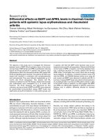
A significant increase of ECP and MPO could be recorded
in both patients with allergic rhinitis and allergic asthma
(Table 2 and 4, Figure 1). However, EPO release increased
significantly only in patients with allergic rhinitis (Table
3). In t he con trol group no increases in ECP, EPO or
MPO were observed.
Nasal challenge
ECP increased significantly in both patients with allergic
rhinitis and allergic asthma (Table 2, Figure 1). A signif-
icant increase of release of MPO was also seen in the
two allergic groups (Table 4, Figure 1). No significant
Table 2 ECP release from eosinophils spontaneously and after C3b-stimulation (at 0 and 20 min) in allergic rhinitis,
allergic asthma and the control group during pollen season and after nasal and bronchial challenge
Spontaneous degranulation of ECP* p-value Stimulated degranulation of ECP* p-value
(median, range) increase 0-20 min (median, range) increase 0-20 min
0 min 20 min 0 min 20 min
Pollen season
Allergic rhinitis 1.78 2.04 0.12 2.42 17.9 0.01
(0.24 - 2.88) (0.38 - 3.17) (0.66 - 3.79) (9.21 - 28.6)
Allergic asthma 1.48 1.70 0.40 2.05 14.0 0.02
(0.99 - 7.10) (0.8 - 7.32) (0.76 - 8.97) (7.13 - 45.1)
Nasal challenge
Allergic rhinitis 1.23 2.02 0.04 1.24 19.5 0.04
(0.17 - 2.03) (0.78 - 2.46) (0.31 - 2.7) (14.7 - 23.5)
Allergic asthma 1.78 2.12 0.02 1.72 14.9 0.02
(0.97 - 3.07) (1.14 - 3.92) (0.88 - 2.49) (8.06 - 26.8)
Bronchial challenge
Allergic rhinitis 1.50 2.08 0.12 2.45 19.8 0.03
(0.99 - 2.38) (1.36 - 3.16) (1.08 - 3.21) (15.5 - 24.3)
Allergic asthma 1.66 1.68 0.4 1.38 13.2 0.02
(0.12 - 1.92) (0.6 - 2.11) (0.08 - 2.02) (9.70 - 17.1)
* Release of ECP in % of total cell content.
Table 3 EPO release from neutrophils spontaneously and after C3b-stimulation (at 0 and 20 min) in allergic rhinitis
and allergic asthma during pollen season and after nasal and bronchial challenge
Spontanous degranulation of EPO Stimulated degranulation of EPO p-value p-value
0 min 20 min 0 min 20 min
Pollen season
Rhinitics 0.42 0.37 0.45 2.05 0.18 0.02*
(0.12 - 0.63) (0.13 - 0.61) (0.35 - 0.92) (1.08 - 3.0)
Asthmatics 0.32 0.30 0.39 1.58 - 0.59 0.11
(0.17 - 0.52) (0.08 - 0.43) (0.38 - 0.74) (1.52 - 3.46)
Nasal challenge
Rhinitics 0.50 0.49 0.61 1.4 0.9 0.07
(0.38 - 0.62) (0.36 - 0.62) (0.51 - 0.71) (1.2 - 1.6)
Asthmatics 0.49 0.44 0.54 1.86 0.07 0.07
(0.45 - 0.69) (0.42 - 0.46) (0.48 - 0.74) (1.54 - 3.22)
Bronchial challenge
Rhinitics 0.28 0.32 0.55 1.99 0.7 0.04*
(0.21 - 0.46) (0.16 - 0.40) (0.26 - 0.93) (1.71 - 2.17)
Astmatics 0.44 0.41 0.52 1.84 0.14 0.07
(0.31 - 0.62) (0.26 - 0.56) (0.35 - 0.64) (1.04 - 2.04)
* Release of EPO in % of total cell content.
Kämpe et al. Clinical and Molecular Allergy 2011, 9:3
/>Page 5 of 10
increase in EPO degranulation was detected in either
the rhinitic or asthmatic patients (Table 3).
Bronchial challenge
Both ECP and MPO increased significantly in both aller-
gicgroups(Table2and4,Figure1).Theincreasein
EPO degranulation was statistically significant only in
patients with allergic rhinitis but not in the asthmatic
group (Table 3).
Degranulation (0 to 20 min) in allergic rhinitis compared
to allergic asthma
No significant differences in the de gree of spontaneous
degranulation of ECP, EPO or MPO could be recorded
between patients with allergic rhinitis and allergic
asthma in either allergen challenge model. After in vitro
stimulation with Sephadex particles, the increased
degranulation of ECP was significantly higher in the rhi-
nitic than the asthmatic group (p = 0.010), (Figure 1).
There was a similar tendency for stimulated MPO
release in allergic rhinitis but this was not significant.
Relationship between the released amount of granule
proteins, clinical data and systemic inflammation
No correlation between degranulation and lung function
(measured as FEV
1
or PEFR) or blood parameters (B-
eosinophils, S-ECP or S-HNL) could be observed.
Discussion
The main finding of our study was that all three allergen
challenge models could prime bot h eosinophils and
neutrophils to an increased propensity of selective
degranulation after stimulation in vitro by opsonised
Sephadex particles. Remarkably, there was no significant
difference in the degranulation response between
patients with allergic rhinitis and allergic asthma except
for a significantly greater release of E CP in the rhinitic
patients after bronchial allergen challenge (p = 0.010).
The three provocation models also primed the granulo-
cytes for degranulation on a comparable level even
though the systemic inflammation was more pro-
nounced during long-term pollen exposure compared to
single-dose allergen challenge [24]. This again hig hlights
the close relationship b etween the upper and lower air-
ways, but it also raises new questions about the cellular
nature of inflammation in atopy.
The eosinophil granulocytes account for 1-2% of the
circulating white blood cells but they are primarily tis-
sue-residi ng cells in the hematopoietic organs as well as
in the airways, the gastrointestinal tract and the skin.
The physiological function of the eosinophils is not
completely understood, but they are known to be
involved in the innate immune response against parasitic
infections, t issue repair and recently it has been discov-
ered that they also have the ability to modulate immune
responses [3]. The activation of the eosinophils is strictly
regulated as an inappropriate activation would be harm-
ful to the subject and in healthy conditions the eosino-
phils are inactivated with a high threshold for release of
their granule proteins [28]. However, after stimulation
the activated eosino phils are primed for extensive degra-
nulation in the different target organs, expressing high-
affinity IgE-receptors (Fcε-receptors), Fcg-receptors and
Table 4 MPO release from neutrophils spontaneously and after C3b-stimulation (at 0 and 20 min) in allergic rhinitis
and allergic asthma during pollen season and after nasal and bronchial challenge
Spontaneous degranulation of MPO* p-value Stimulated degranulation of MPO* p-value
(median, range) increase 0-20 min (median, range) increase 0-20 min
0 min 20 min 0 min 20 min
Pollen season
Allergic rhinitis 2.12 2.45 0.81 2.84 15.9 0.008
(1.18 - 6.86) (1.67 - 5.64) (0.93 - 7.07) (10.6 - 29.2)
Allergic asthma 2.25 2.50 0.74 3.05 17.6 0.018
(0.28 - 5.05) (0.95 - 5.5) (0.37 - 5.20) (7.48 - 24.0)
Nasal challenge
Allergic rhinitis 2.61 3.16 0.68 2.93 14.9 0.043
(1.42 - 5.64) (1.68 - 4.1) (1.63 - 5.55) (12.7 - 21.7)
Allergic asthma 2.21 3.31 0.018 2.35 18.4 0.018
(1.18 - 5.62) (2.08 - 7.40) (1.35 - 5.22) (12.6 - 25.4)
Bronchial challenge
Allergic rhinitis 2.42 3.43 0.075 3.88 21.6 0.028
(2.17 - 2.92) (2.24 - 4.93) (2.14 - 6.00) (16.8 - 27.3)
Allergic asthma 1.50 1.91 0.018 1.58 16.8 0.018
(0.84 - 2.3) (1.36 - 2.48) (0.71 - 2.96) (13.1 - 23.3)
*release of MPO in % of total cell content.
Kämpe et al. Clinical and Molecular Allergy 2011, 9:3
/>Page 6 of 10
complement receptors [3,19]. In vitro studies have
demonstrated selective release of the individual granule
proteins [19], and interestingly, different eosinophilic
diseases are characterized by a m arked heterogeneity in
degranulation levels [29]. Previous studies suggested that
the priming-degree of the blood eosinophils is related to
the degranulation status of the tissue-residing eosino-
phils and so corresponds to the activity of the eosino-
philic disease [30].
Previous analyses of the study population have shown
that the asthmatic group was more responsive to inhala-
tion of hypertonic saline [23], h ad more pronounced
lung function impairment during the pollen season [23],
and was more responsive to allergen PD
20
after bron-
chial challenge than the rhinitic group [24]. Despite
these differences, both patient groups showed a similar
degree of eosinophil inflammation b oth locally and sys-
temically during pollen season as well as after both
nasal and bronchial chall enge [23,24]. Our hypothesis
was therefore that differences in degranulation patterns
contribute to the outcome of different clinical manifes-
tations between the allergic groups. However, the results
in this study did not support this hypothesis.
We found that both in patients with a llergic rhinitis
and allergic asthma, the released amount of ECP after
C3b-induced stimulation was in the same range during
pollen season as after both nasal and bronchial chal-
lenge. Surprisingly, we also recorded the same pattern
for stimulated MPO release in both patient groups. Our
interpretation is that seasonal exposure as well as nasal
and bronchial allergen challenge can activate, prime,
eosinophils and neutrophils more or less to the same
degree. The tendency that patients with allergic rhinitis
and allergic asthma display the same pattern of degranu-
lation of ECP and MPO is in line with previous observa-
tions from our group where we demonstrated an
increased propensity of ECP and EPX/EDN secretion
during pollen season in patients with allergic asthma
[19]. In that study, however, we only recorded a slight
tendency of increase for MPO [19]. This could partly be
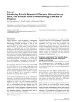
Figure 1 C3b-induced degranulation of eosinophil cationic protein (ECP) and myeloperoxidas (MPO) (at 20 min), in patients with
allergic asthma and allergic rhinitis, during pollen season and after nasal and bronchial challenge, respectively.
Kämpe et al. Clinical and Molecular Allergy 2011, 9:3
/>Page 7 of 10
explained by the fact that the granulocytes in the pre-
sent study were pre-incubated for 10 min with assay
buffer, which was not the case in our previous paper.
The results indicate that priming of gr anulocytes is also
applicable for patients with allergic rhinitis and follows
thesamepatternasforpatients with allergic asthma.
The observation of neutrophi l activation in both allergic
groups is in contrast to data that other groups have
reported in which mild and moderate asthmatics did
not display any neutrophil inflammation [8].
Stimulated EPO degranulation tended to increa se in
the rhinitic patients compared to the asthmatics both
during pollen season and after nasal and bronchial chal-
lenge. However, there was only a minor absolute
increase in EPO release even after C3b-induced stimula-
tion in both allergic patient groups. This discrepancy
between the release of ECP and EPO is an interesting
finding considering that EPO is regarded to be the most
specific eosinophil granule protein [31]. Our data is in
line with previous reports from both our and other
groups where it has been observed that EPO is more
difficult to mobilize than ECP [29,30,32]. This difference
could be explained by selective granule release in
response to different stimuli for degranulation [30], as
EPO is a potent enzyme and perhaps plays a more
important role in the innate defence agains t parasites
and not primarily in allergy.
We were intrigued by the observation that the rhinitic
patients showed a higher release of ECP and MPO after
bronchial allergen challenge than the asthmatic patients.
One interpretation could be that the granulocytes of the
patients with allergic asthma are easier to prime and
activate, particularly after bronchial allergen challe nge,
and therefore already have released their granule pro-
teins in response to the allergen exposure. This hypoth-
esis is suppor ted by a slightly higher amount of ECP per
eosinophil cell prior to the C3b-induced granule release
after bronchial challenge in the rhinitic patients com-
pared to the asthmatics (mean 3.01 vs. 2.73 μgECP/B-
eos 10
6
). This is in accordance with results from other
groups t hat have observed hypodense blood eosinophils
after allergen exposure, implicating degranulation in
response to allergen challenge [5]. On the other hand,
Malm-Erjefält et al evaluated patients with allergic
asthma, allergic rhinitis and atopic dermatitis with
regard to intracellular EPO by transmission electron
microscopy, demonstrating no degranulation of the eosi-
nophils in circulating blood. The degranulation status
was, however, based on the cell content of EPO [33].
This is in line with our results and also with previous
studies where it has been observed that EPO is more
difficult to mobilize from the primed blood eosinophils
[20,30,32].
Eosinophils have been considered as major effector
cells in the pathogenesis of asthma, but the role of the
neutrophils is less understoodintheallergicairway
inflammation except in more severe forms of chronic
asthma [34]. Histologically, the asthmatic lung is charac-
terized by an eosinophil-rich inflammation and by a
variety of chronic changes including remodelling and
deposition of extracellular matrix components [35,36].
Interestingly, Phipps et al recently showed that even in
mild atopic asthma acute allergen-induced remodelling
could occur early [37], and in another study neutrophi ls
were prominently elevated in asthma exacerbations [38].
The novel finding of neutrophils in induced sputum of
non-atopic asthmatic children [10] also points in the
direction of the neutrophils playing an important role,
not just in severe chronic stages of the disease, but also
in mild disease. Additionally, the recent advances using
anti-IL-5 therapy indicate involvement of other inflam-
matory cells than just the eosinophils, as the bronchial
hyp erresponsiveness is not affected by anti-IL-5 therapy
despite depletion of the eosin ophils from circulation by
this treatment [39]. Alto gether, this implies that there
might not be a clear-cut difference between mild and
severe asthma with regard to the neutrophil involve-
ment, and thus eosinophilic and neutrophilic asthma
might not be mutually exclusive subtypes of asthma.
The strength of our study is the simultaneous evalua-
tion of the priming status of the eosinophils and neutro-
phils in blood after b oth long-term natural allergen
exposure during pollen season and a single high -dose
allergen challenge in the nose and bronchi in both aller-
gic rhinitics and aller gic asthmatic patients concurrently.
One drawback of this study is t he relative small number
of subjects in each allergic group which limited the
opportunity to find differences between the two allergic
groups, but the results imply that blood granulocytes of
both allergic rhinitis an d allergi c asthma are more or less
equally primed for chemotaxis and degranulation in their
target tissue. However, there are many questions to be
resolved and further investigations are needed in order to
study the degranulation process at the site of action.
Conclusion
In conclusion, patients with a llergic rhinitis and allergic
asthma display similar patterns of eosinophil and neu-
trophil propensity for degranulation when exposed to
allergen. However, there is a tendency to increased
release i n the rhinitic patients, but this only significant
for ECP release after bronchial challenge. Our results
indicate that othe r mechanisms than the magnitude of
inflammation and degranulation patterns of the inflam-
matory cells determine whether or not an allergic
patient with rhinitis develops asthma.
Kämpe et al. Clinical and Molecular Allergy 2011, 9:3
/>Page 8 of 10
Acknowledgements
The study was supported financially by the Swedish Association against
Asthma and Allergy, the Swedish Heart and Lung Foundation, Bror
Hjerpstedt’s Foundation, the Uppsala County Against Heart and Lung
diseases and the Medical Faculty of Uppsala University.
The study nurses Signe Svedberg Brandt and Katarina Göthberg are
acknowledged for the skilful technical assistance. We also acknowledge
Dominic-Luc Webb, Hepatology and Gastroenterology Group, Dept of
Medical Sciences, Uppsala University, for skilful linguistic review.
Author details
1
Department of Medical Sciences, Respiratory Medicine and Allergology,
Uppsala University, Uppsala, Sweden.
2
Asthma Research Centre, Uppsala
University, Uppsala, Sweden.
3
Department of Medical Sciences,
Gastroenterology Research Group, Uppsala University, Uppsala, Sweden.
Authors’ contributions
MK, MC, CJ and GS designed the study and were responsible for analyzing
and interpreting the results as well as critically revising the manuscript. IS
carried out the assays and degranulation measurements. ML was involved in
drafting the manuscript and the figures. All authors have contributied in
reading an improving the manuscript.
Competing interests
The authors declare that they have no competing interests.
Received: 24 August 2010 Accepted: 21 January 2011
Published: 21 January 2011
References
1. Kariyawasam HH, Robinson DS: The eosinophil: the cell and its weapons,
the cytokines, its locations. Semin Respir Crit Care Med 2006, 27:117-27.
2. Venge P: Review article: Monitoring allergic inflammation. Allergy 2004,
59:26-32.
3. Hogan SP, Rosenberg HF, Moqbel R, Phipps S, Foster PS, Lacy P, Kay AB,
Rothenberg ME: Eosinophils: Biological properties and role in health and
disease. Clin Exp Allergy 2008, 38:709-750.
4. Logan MR, Odemuyiwa SO, Moqbel R: Understanding exocytosis in
immune and inflammatory cell: the molecular basis of mediator
secretion. J Allergy Clin Immunol 2003, 111:923-32.
5. Karawajczyk M, Sevéus L, Garcia R, Björnsson E, Peterson CGB, Roomans GM,
Venge P: Piecemeal degranulation of peripheral blood eosinophils: A
study of allergic subjects during and out of the pollen season. Am J
Respir Cell Mol Biol 2000, 34:521-29.
6. Melo RC, Spencer LA, Dvorak AM, Wleer PF: Mechanisms of eosinophil
secretion: large vesicotubular carriers mediate transport and release of
granule-derived cytokines and other proteins. J Leukoc Biol 2008,
83:229-36.
7. Jatakanon A, Uasuf C, Maziak W, Lim S, Chung KF, Barnes P: Neutrophilic
inflammation in severe persistent asthma. Am J Respir Crit Care Med 1999,
60:1532-39.
8. Fahly JV: Eosinophilic and neutrophilic inflammation in asthma. Proc Am
Thorac Soc 2009, 6:256-59.
9. Holgate ST, Holloway J, Wilson S, Howarth PH, Haitchi HM, Babu S,
Davies DE: Understanding the pathophysiology of severe asthma to
generate new therapeutic opportunities. J Allergy Clin Immunol 2006,
117:496-506.
10. Drews AC, Pizzichini MMM, Pizzichini E, Peireira MU, Pitrez PM, Jones MH,
Sly PD, Stein RT: Neutrophilic inflammation is a main feature of
induced sputum in nonatopic asthmatic children. Allergy 2009,
64:1597-601.
11. Gullberg U, Bengtsson N, Bülow E, Garwicz D, Lindmark A, Olsson I:
Processing and targeting of granule proteins in human neutrophils.
J Immunol Methods 1999, 232:201-10.
12. Hansson M, Olsson I, Nausseef WM: Biosynthesis, processing and sorting
of human myeloperoxidase. Arch Biochem and Biophys 2006, 445:214-224.
13. Winqvist I, Olofsson T, Olsson I: Mechanisms for eosinophil degranulation;
release of the eosinophil cationic protein. Immunology 1984, 51:1-8.
14. Egesten A, Malm J: Eosinophil leukocyte degranulation in response to
serum-opsonized beads: C5a and platelet-activating factor enhance ECP
release, with roles for protein kinases A and C. Allergy 1998, 53:1066-73.
15. Tai PC, Spry CJ:
The effect of recombinant granulocyte-macrophage
colony-stimulating
factor (GM-CSF) and interleukin-3 on the secretory
capacity of human blood eosinophils. Clin Exp Immunol 1990, 80:426-434.
16. Carlson M, Peterson C, Venge P: The influence of IL-3, IL-5, and GM-CSF
on normal human eosinophil and neutrophil C3b-induced
degranulation. Allergy 1993, 8:437-442.
17. Tomassini M, Tsicopoulos A, Tai PC, Gruart V, Tonnel AB, Prin L, Capron A,
Capron M: Release of granule proteins by eosinophils from allergic and
non-allergic patients with eosinophilia on immunoglobulin-dependent
activation. J Allergy Clin Immunol 1991, 88:365-75.
18. Karawajczyk M, Pauksen K, Peterson C, Eklund E, Venge P: The differential
release of eosinophil granule proteins. Studies on patients with acute
bacterial and viral infections. Clin Exp All 1995, 25:713-19.
19. Carlson M, Håkansson L, Kämpe M, Stålenheim G, Peterson C, Venge P:
Degranulation of eosinophils from pollen-atopic patients with asthma is
increased during pollen season. J Allergy Clin Immunol 1992, 89:131-39.
20. Carlson M, Håkansson L, Peterson C, Stålenheim G, Venge P: Secretion of
granule proteins from eosinophils and neutrophils is increased in
asthma. J Allergy Clin Immunol 1991, 87:27-33.
21. Bousquet J, Jacot W, Vignola AM, Bachert C, Van Cauwenberge P: Allergic
rhinitis: A disease remodelling the upper airways? J Allergy Clin Immunol
2004, 113:43-49.
22. Braunstahl GJ: The unified system: Respiratory tract-nasobronchial
interaction mechanisms in allergic disease. J Allergy Clin Immunol 2005,
115:142-48.
23. KämpeM,StålenheimG,JansonC,StoltI,CarlsonM:Systemic and
local eosinophil infla mmation during birch pollen season in allergic
patients with predominant rhinitis or asthma. Clin Mol Allergy 2007,
29:1-8.
24. Kämpe M, Janson C, Stålenheim G, Stolt I, Carlson M: Experimental and
seasonal exposure to birch pollen in allergic rhinitis and allergic asthma
with regard to the inflammatory response. Clin Resp J 2010, 4:37-44.
25. Quanjer PH, Tammeling GJ, Cotes JE, Pedersen OF, Peslin R, Yernault JC:
Lung volumes and forced ventilatory flows. Report Working Party
Standardization of Lung Function Tests, European Community for Steel
and Coal. Official Statement of the European Respiratory Society. Eur
Respir J 1993, , S16: 5-40.
26. Machado L: Increased bronchial hypersensitivity after early and late
bronchial reactions provoked by allergen inhalation. Allergy 1985,
40:580-85.
27. Hansell TT, De Vries IJ, Iff T, Rihs S, Wandzalik M, Betz S, Blaser K, Walker C:
An improved immunomagnetic procedure for the isolation of highly
purified human blood eosinophils. J Immunol Methods 1991, 145:105-110.
28. Kato M, Kephart GM, Talley NJ, Wagner JM, Sarr MG, Bonno M,
Mcgovern TW, Gleich GJ: Eosinophil infiltration and degranulation in
normal human tissue. Anat Rec 1998, 252:418-425.
29. Erjefält JS, Greiff L, Andersson M, Ädelroth E, Jeffery PK, Persson CG:
Degranulation
patterns of eosinophil granulocytes as determinants of
eosinophil driven disease. Thorax 2001, 56:341-44.
30. Carlson M, Öberg G, Peterson CH, Venge P: The releasability of human
hypereosinophilic eosinophils is related to the density of the cells. Br J
Haematol 1994, 1:41.
31. Metso T, Venge P, Haahtela T, Peterson CGB, Sevéus L: Cell specific
markers for eosinophils and neutrophils in sputum and bronchoalveolar
lavage fluid of patients with respiratory conditions and healthy subjects.
Thorax 2002, 57:449-51.
32. Carlson M, Raab Y, Peterson CH, Hällgren R, Venge P: Increased
intraluminal release of eosinophil granule proteins EPO, EPX and
cytokines in ulcerative colitis and proctitis in segmental perfusion. Am J
Gastroenterol 1999, 94:1876.
33. Malm-Erjefält M, Greiff l, Ankers J, Andersson M, Wallengren J, Cardell LO,
Rak S, Persson CGA, Erjefält JS: Circulating eosinophils in asthma, allergic
rhinitis, and atopic dermatitis lack morphological signs of degranulation.
Clin Exp Allergy 2005, 35:1334-40.
34. Wenzel SE, Szefler SJ, Leung DY, Sloan SI, Rex MD, Martin RJ:
Bronchoscopic evaluation of severe asthma: persistent inflammation
associated with high dose glucocorticoids. Am J Respir Crit Care Med 1997,
156:737-43.
35. Laitinen LA, Laitinen A, Virtanen I, Kämpe M, Simonsson BG, Karlsson SE,
Håkansson L, Venge P, Sillastu H: Bronchial biopsy findings in intermittent
or “early” asthma. J Allergy Clin Immunol 1996, 98:S3-6.
Kämpe et al. Clinical and Molecular Allergy 2011, 9:3
/>Page 9 of 10
36. Hogan SP: Recent advances in eosinophil biology. Int Arch Allergy
Immunol 2007, 143(S1):3-14.
37. Phipps S, Benyahia F, Ou TT, Barkans J, Robinson DS, Kay AB: Acute
allergen-induced airway remodelling in atopic asthma. Am J Respir Cell
Mol Biol 2004, 31:626-32.
38. Fahly JV, Kim KW, Liu J, Boushey HA: Prominent neutrophilic inflammation
in sputum from subjects with asthma exacerbation. J Allergy Clin
Immunol 1995, 95:843-52.
39. Flood-Page P, Menzies-Gow A, Phipps S, Ying S, Wangoo A, Ludwig MS,
Barnes N, Robinson D, Kay AB: Anti-IL-5 treatment reduces deposition of
ECM proteins in the bronchial subepithelial basement membrane of
mild atopic asthmatics. J Clin Invest 2003, 112:1029-36.
doi:10.1186/1476-7961-9-3
Cite this article as: Kämpe et al.: Patients with allergic rhinitis and
allergic asthma share the same pattern of eosinophil and neutrophil
degranulation after allergen challenge. Clinical and Molecular Allergy 2011
9:3.
Submit your next manuscript to BioMed Central
and take full advantage of:
• Convenient online submission
• Thorough peer review
• No space constraints or color figure charges
• Immediate publication on acceptance
• Inclusion in PubMed, CAS, Scopus and Google Scholar
• Research which is freely available for redistribution
Submit your manuscript at
www.biomedcentral.com/submit
Kämpe et al. Clinical and Molecular Allergy 2011, 9:3
/>Page 10 of 10