Báo cáo y học: "Diet as prophylaxis and treatment for venous thromboembolism" pot
Bạn đang xem bản rút gọn của tài liệu. Xem và tải ngay bản đầy đủ của tài liệu tại đây (743.48 KB, 24 trang )
REVIEW Open Access
Diet as prophylaxis and treatment for venous
thromboembolism?
David K Cundiff
1
, Paul S Agutter
2*
, P Colm Malone
3
, John C Pezzullo
4
* Correspondence:
2
Theoretical Medicine and Biology
Group, 26 Castle Hill, Glossop,
Derbyshire, SK13 7RR, UK
Abstract
Background: Both prophylaxis and treatment of venous thromboembolism (VTE:
deep venous thrombosis (DVT) and pulmonary emboli (PE)) with anticoagulants are
associated with significant risks of major and fatal hemorrhage. Anticoagul ation
treatment of VTE has been the standard of care in the USA since before 1962 when
the U.S. Food and Drug Administration began requiring randomized controlled clinical
trials (RCTs) showing efficacy, so efficacy trials were never required for FDA approval. In
clinical trials of ‘high VTE risk’ surgical patients before the 1980s, anticoagulant
prophylaxis was clearly beneficial (fatal pulmonary emboli (FPE) without anticoagulants
= 0.99%, FPE with anticoagulants = 0.31%). However, observational studies and RCTs of
‘high VTE risk’ surgical patients from the 1980s until 2010 show that FPE deaths
without anticoagulants are about one-fourth the rate that occurs during prophylaxis
with anticoagulants (FPE without anticoagulants = 0.023%, FPE while receiving
anticoagulant prophylaxis = 0.10%). Additionally, an FPE rate of about 0.012% (35/
28,400) in patients receiving prophylactic anticoagulants can be attributed to ‘rebound
hypercoagulation’ in the two months after stopping anticoagulants. Alternatives to
anticoagulant prophylaxis should be explored.
Methods and Findings: The literature concerning dietary influences on VTE
incidence was reviewed. Hypotheses concerning the etiology of VTE were critiqued
in relationship to the rationale for dietary versus anticoagulant approaches to
prophylaxis and treatment.
Epidemiological evidence suggests that a diet with ample fruits and vegetables and
little meat may substantially reduce the risk of VTE; vegetarian, vegan, or Mediterra-
nean diets favorably affect serum markers of hemostasis and inflammation. The valve
cusp hypoxia hypothesis of DVT/VTE etiology is consistent with the development of
VTE being affected directly or indirectly by diet. However, it is less consistent with the
rationale of using anticoagulants as VTE prophylaxis. For both prophylaxis and treat-
ment of VTE, we propose RCTs comparing standard anticoagulation with low VTE risk
diets, and we discuss the statistical considerations for an example of such a trial.
Conclusions: Because of (a) the risks of biochemical anticoagulation as anti-VTE
prophylaxis or treatment, (b) the lack of placebo-controlled efficacy data supporting
anticoagulant treatment of VTE, (c) dramatically reduced ho spital-acquired FPE
incidence in surgical patients without anticoagulant prophylaxis from 1980 - 2010
relative to the 1960s and 1970s, and (d) evidence that VTE incidence and outcomes
may be influenced by diet, randomized controlled non-inferiority clinical trials are
proposed to compare standard anticoagulant treatment with potentially low VTE risk
diets. We call upon the U. S. National Institutes of Health and the U.K. National
Institute for Health and Clinical Excellence to design and fund those trials.
Cundiff et al. Theoretical Biology and Medical Modelling 2010, 7:31
/>© 2010 Cundiff et al; licensee BioMed Central Ltd. This is an Open Access article distributed under the terms of the Cre ative Commons
Attribution License ( censes/by/2.0), which permits unrestricted use, distribution, and reproduction in
any medium, provide d the original work is properly cited.
Two accounts of the etiology of DVT and VTE
The consensus view that DVT and VTE are hematological disorders arose shortly after
the Second World War and had become the new orthodoxy by the early 1960s. It still
dominates research and practice in the field. E ssentially, this consensus added ‘hyper-
coagulab ility’ to the ‘stasis’ and ‘vessel wall injury’ thesis of Hunterian pathophysiology,
generating a set of loosely-defined terms that was misleadingly ascribed to Virchow
[1,2]. In suppo rt of that consensus, at least some inherited and acquired thrombophi-
lias (’hypercoagulability c onditions’) appear to increase the inciden ce of VTE, though
this may indicate that thrombophilias aggravate rather than cause the disease. More-
over, there is an argument [3] that the so-called ‘Virchow’striad’ constitutes a useful
rule of thumb for managing patients. Strikingly, however, the consensus view arose
when anticoagulant therapy for thrombosis patients was becoming popular [4] and has
developed along with such therapy and with the subsequent deployment of thromboly-
tic agents [1,2]. It seems integral with the pharmaceutical approach to D VT/VTE pro-
phylaxis and treatment.
An alternative a ccount of the etiology of DVT, the valve cusp hypoxia hypothesis
(VCHH), is r ooted in the tradition o f thought and practice initiated by Hunter and
traceable from Harvey through Virchow, Lister, Welch and a number of early 20
th
cen-
tury investigators [1,2]. According to the VCHH, DVT may occur wherever sustained
non-pulsatile (streamline) venous blood flow leads to suffocating hypoxemia in the
valve pockets, resulting in hypoxic injury to and hence death of the inner (parietali s)
endothelium of the cusp leaflets. This injury activates the elk-1/egr-1 pathway, which
initiates many responses of endothelial cells to hypoxia and activates chemoattractant
and procoagulant factors [5]. (Briefly: elk-1 is a receptor tyrosine kinase stimulated by
hypoxia; it phosphorylates the zinc-finger tra nscription factor ear ly growth response-1
(egr-1) , which then activates downstream genes encoding factors directly or indirect ly
involved in blood coagulation.) When normal pulsatile blood flow is restored, however
transiently, leukocytes and platelets are attracted by these factors and inevitably re-
enter the lately-affected valve pockets and marginate and sequestrate at the site of
injury, the inner/parietal surfaces of the valve cusps, w hereupon local blood coagula-
tion (semi-solidification) is likely to be initiated.
Any subsequent period of non-pulsatile flow may kill the accumulated blood cells
marginated on the dying or dead valve pocket. These dead cells may then form the
core of a nascent thrombus. If periods of non-pulsatile and pulsatile flow continue to
alternate, serial deposition of white cells and fibrin may ensue, resulting in the charac-
teristic ‘Lines of Zahn’ morphology of a venous thrombus. Subsequently, only the
blood cells on the outermost layer of a thrombus are living.
The VCHH explains many of the recognized risk factors for DVT and accounts for
the morphology of thrombi. It also predicts that venous thrombi will readily embolize ,
because the area of endothelium to which they are an chored, the valve cusp parietalis,
hasbecomenecroticsoitmaybereadilydetachedbytheflowofbloodpastthe
obstruction.
Compared with the Virchow’s triad hypothesis of DVT etiology, the V CHH better
explains what appears to be a marked reduction in the incidence of hospit al-acquired
VTE (see below) following the introduction of early mobilization of post-operative
Cundiff et al. Theoretical Biology and Medical Modelling 2010, 7:31
/>Page 2 of 24
patients and the widespread use of mechanical methods for maintainin g pulsatile leg
vein blood flow (e.g., flexion and extension of the ankles, support hoses, and intermit-
tent pneumatic pressure leg devices). According to the VCHH, drugs that inhibit or
‘kill’ any part of the coagulation process might slow the progression of established
DVTs but would be ineffective in preventing the initiation of thrombi.
Problems with anticoagulant treatment for VTE
Bleeding
Regarding patients treated for VTE with standard anticoagulants, a rece nt meta-analysis
of published RCTs showed major and fatal bleeding rat es of 1.8% and 0.2%, respectively
[6]. Older cohort studies report up to triple these rates [7-9]. Applying the range of
reported fatal bleeding rates for VTE treatment (0.2% - 0.6%) t o an estimated 300,000-
1.2 million people treated for VTE worldwide per year (about half in the USA [10]), 600-
7,200 people per year suffer fatal bleeds from VTE anticoagulant treatment. There are
many more non-fatal major bleeds, some of which are permanently debilitating.
Anticoagulant prophylaxis for surgical patients in creases the risk of major bleeding
[11]. VTE prevention trials report markedly different rates of major bleeding despite
similar patient populations and doses and durations of anticoagulant prophylaxis. For
instance, major bleeding with enoxaparin reportedly ranged from 0.1% to 3.1% in h ip
arthroplasty trials and from 0.2% to 1.4% in kneearthroplastytrials. If surgical-site
bleeding is included in the definition of major bleeding, the reported rates have been
about 10-fold higher [12]. Major bleeding adversely affects overall mortality. In a meta-
analysis of trials comparing fondaparinux with LMWHs or placebos (major bleeding
incidence overall = 2.4%), the risk of death by 30 days was 7-fold higher among
patients with compared to those without a major bleeding event (8.6% versus 1.7%)
[13]. If the major bleeding is considered the cause of the higher death rate, 6.9% of
deaths in patients with major bleeds may be attributed to the bleeding (8.6% - 1.7% =
6.9%). Consequently, deaths of a bout 0.166% of anticoagulated patients are arguably
attribut able to bleeding (0.069 × 0.024 = 0.00166). Given that at least 12 million medi-
cal and surgical patients worldwide receive prophylac tic anticoagulants per year
[14,15], this means that approximately 20,000 people may die each year from complica-
tions of bleeding from prophylactic anticoagulants (0.00166 × 12 million = 19,872);
many more may suffer the consequences of hypovolemia.
Efficacy
Anticoagulant therapy for VTE became established as the standard of care in the 1940s
and 1950s before randomized trials were considered necessary to prove efficacy and
safety. A very small RCT comparing anticoagulants versus placebo for people with clin-
ical diagnoses of PE published in 1960 [4] has been used to justify anticoagulant ther-
apy. However, by current scientific standards, this study is highly flawed [10,16].
In 1962 when the U.S. Food and Drug Administration began requiring randomized
controlled clinical trials (RCTs) showing efficacy before approving drugs, anticoagula-
tion treatment of VTE was ‘grandfathered in’ with no rigorous efficacy trials ever
required. Only three small methodologically rigorous RCTs of patients with DVTs
[17-19] have compared standard anticoagulants with placebos or non-steroidal anti-
inflammatory drugs. Combining the data from these trials, 6/66 patients receiving
Cundiff et al. Theoretical Biology and Medical Modelling 2010, 7:31
/>Page 3 of 24
standard heparin and vitamin K inhibitors died and 1/60 unanticoagulated patients
died [10]. Consequently, standard anticoagulant treatment for VTE cannot be consid-
ered evidence-based to be effective [10,20].
Anticoagulant prophylaxis of ‘high VTE risk’ patients may increase fatal pulmonary
emboli (FPE) due to ‘rebound hypercoagulation’
Goldhaber and colleagues tracked the incidence of developing DVT or PE during or up
to 30 days after h ospital discharge in about 80,000 patients admitted over a two year
period in Boston’s Brigham and Women’s Hospit al. Out of 384 patients with hospital-
acquired VTE, 318 (82.8%) were potential candidates for prophylaxis (i.e., they had ≥2
VTE risk factors). Of prophylaxis candidates, 170 (53%) of those with hospital-acquired
VTE had received anticoagulants [21]. To estimate the influence of prophylact ic antic-
oagulants in this study, we can use Goldhaber’s USA-wide estimates of hospitalized
patients that are at ‘high VTE risk’–32% [14] or 25,600/80,000 in the Brigham and
Women’s Hospital study – and the proportion of those at VTE risk who receive antic-
oagulant prophylaxis – 50% [14] or 12,800/25,600 in this study. According to these
estimates, ‘high VTE risk’ patients receiving anticoagulants in this population had a
non-significant trend toward a higher incidence of VTE (OR = 1.15, 95% C I = 0.9 2 -
1.44) [22].
More importantly in this chart study, out of 13 deaths attributed to hospital-acquired
FPE, 12 had received anticoagulant prophylaxis [21]. As above, assuming tha t 32% of
the hospitalized patients were at risk for V TE and that 50% of all patients at risk for
VTE received anticoagulants, anticoagulation prophylaxis was associated with a 12-fold
increase in hospital-acquired FPE (OR: 12.0; 95% CI, 1.6-92) [22].
An autopsy st udy by Lindblad and colleagues [23] from Malmo, Sweden corro bo-
rated the Goldhaber study. From a population of 31,238 post-operative patients from
the 1980s, it found that 27/30 patients with autopsy-proven FPE had received post-op
prophylactic anticoagulants. The authors did not report the proportion of ‘high VTE
risk’ surgical patients in their hospital receivi ng anticoagulant prophylaxis. To provide
an approximation of the degree of increased risk of FPE related to anticoagulant pro-
phylaxis in this autopsy study from a defined clinical population, we can conservatively
assume that all Malmo surgical patients had ‘high VTE risk’ and again use Goldhaber’s
estimate that about 50% of those at r isk received anticoagulant prophylaxis [14]. This
translates to about 15,619 patients with anticoagulants and the same number without.
Compared with patients not receiving anticoa gulant prophy laxis , the Lindblad autopsy
data show the estimated FPE rate in anticoagulated patients is nine-fold higher (OR:
9.0; 95% CI, 2.7-29.6).
Since many Malmo surgical patients would have bee n at ‘low VTE risk’ and few er
than 50% of those at ‘high VTE risk’ may have r eceived anticoagulants in the 1980s,
the FPE rate associated with anticoagulant prophylaxis could well have been consider-
ably higher. Combining the FPE data from Gold haber and Lindblad yields a very con-
servative estimated increased FPE risk associated with anticoagulant prophylaxis of
9.75 fold (OR, 9.75; 95% CI, 3.5 - 27.3). Combining these studies, 35/43 cases can be
attributed to ‘rebound hypercoagu lation’ (i.e., 39/43 FPE patients had received anticoa-
gulation prophylaxis versus 4/43 with no anticoagulation: 39 - 4 = 35).
Cundiff et al. Theoretical Biology and Medical Modelling 2010, 7:31
/>Page 4 of 24
Surgery is asso ciated with a sub stantial systemic and local activation of the coagula-
tion and fibrinolytic systems. Post-operative prophylactic anticoagulants significantly
mitigate the stimulation of these systems. However, following the discontinuation of
prophylactic anticoagulants, a second wave of activation of markers of the coagulation
and fibrinolytic systems continues for up to 35 days after surgery (e.g., plasma TAT
and D-dimer [24]). Cundiff has suggested that ‘rebound hypercoagulation’ after stop-
ping anticoagulants causing restimulation of coagulation and fibrinolysis may account
for this marked increase in FPE risk associated with anticoagulant treatment [25] and
prophylaxis [26]. Given that at least 12 million medical and surgical patients worldwide
receive prophylactic anticoagulants per year [14,15], an estimated 5,000 to 40,000 peo-
ple per yea r die of ‘rebound hypercoagulation’ (i.e., 12,000,000 (hospitalized people/
year with anticoagulant prop hylaxis) × 35/28,419 (excess risk for fatal PE per Goldha-
ber and Lindblad studies) = 14,779; 95% CI, 5,305 - 41,381).
While in the Goldhabe r study 11/13 (85%) of FPE cases were in medical ward
patients and only 2/13 were in surgical patients, the larger Lindblad study included
anticoagulation prophylaxis data only on surgical patients. In the Lindblad study, 113
patients had PE as the principal cause of death, of which 83/113 (73 %) were medical
patients and 30/113 were post-operative. Lindblad did not report the anticoagulant
prophylaxis s tatus of the medical FPE patients. Since anticoagulated medical patients
are about 50 times more likely than surgical patients to have FPE (Tables 1 and 2), the
actual number of anticoagulated patients with FPE due to ‘r ebound hypercoagulation’
is likel y much higher than derived from combining these two autopsy studies because
of the disproportionately high number of surgical patients.
Marked reduction in FPE risk over time unrelated to anticoagulants
In the 1960s and 1970s, FPE in trials of post-op surgical patients without anticoagulant
prophylaxis averaged 0.99% while FPE rates in anticoagulated patients averaged 0.31%
(Table 3). Since about 1980, prompt ambulation of post-op patients and other non-
drug VTE prophylaxis measures (e.g., mechanical prophylaxis oflowerlimbs)have
been widely implemented. Recent observational studies and RCTs of surgical pa tients
at VTE risk both not receiving and receiving prophylactic anticoagulants show a some-
what reduced VTE incidence and a markedly lower FPE frequency than seen in studies
from the 1960s and 1970s (see Tables 2 and 4).
Table 1 FPE incidence VTE observational studies and RCTs in medical patients from the
1980s to 2000s
Author FPE incidence no anti-coagulation FPE incidence with anti-coagulation
Mahé [89] 17/1,244 10/1,230
Alikhan [87] 467/9,491 431/9,349
Cohen [90] 5/414 0/425
Testroote [91] 0/454 0/442
Bergmann [92] 17/1,244 10/1,230
Bergmann [93] NA 2/439
Fraisse [94] 0/114 1/109
Turpie [95] 0/650 0/635
506/13,611 (3.7%) 453/13,859 (3.3%)
Cundiff et al. Theoretical Biology and Medical Modelling 2010, 7:31
/>Page 5 of 24
Table 2 FPE incidence in surgical patients: VTE observational studies and RCTs in the
1980s to 2000s
Population of surgical patients) Author FPE incidence no
anti-coagulation
FPE incidence with
anti-coagulation
general surgical Kosir [96] 0/70 0/38
general surgical Kosir [97] 1/68 0/68
general surgical Rasmussen [98] 1/405 0/388
total general surgical 2/543 (0.37%) 0/494 (0%)
orthopedic surgical Sasaki [99] 0/38 0/38
orthopedic surgical Bi [100] 0/35 0/35
orthopedic surgical Goel [101] 0/111 0/127
orthopedic surgical Agarwal [102] 0/131 0/166
orthopedic surgical Eriksson [103] NA 0/1,587
orthopedic surgical Eriksson [104] NA 0/1,464
orthopedic surgical Heit [105] NA 1/594
orthopedic surgical Eriksson [106] NA 0/133
orthopedic surgical Francis [107] NA 0/2,285
orthopedic surgical Eriksson [108] NA 1/2,056
orthopedic surgical Turpie [109] NA 5/7,211
orthopedic surgical Ramos [110] 0/262 0/267
orthopedic surgical Ginsberg [111] NA 1/1,896
orthopedic surgical Agnelli [112] NA 0/507
orthopedic surgical Turpie [113] NA 0/613
orthopedic surgical Colwell [114] NA 0/1,838
orthopedic surgical Eriksson [115] NA 1/1,872
orthopedic surgical Eriksson [116] NA 2/2,835
orthopedic surgical Eriksson [117] NA 1/2,788
orthopedic surgical Colwell [118] NA 3/2,299
total orthopedic surgical 0/577 (0%) 15/29,291 (0.051%)
unspecified surgical Rosenzweig [119] 0/4,705 NA
unspecified surgical Nurmohamed [120] NA 11/8,172
total unspecified surgical 0/4,705 (0%) 11/8,172 (0.135%)
surgical totals 2/5,825 (0.034%) 26/37,957 (0.068%)
Table 3 FPE incidence in surgical patients in the 1960s and 1970s
Population (medical, surgical,
etc.)
Author FPE incidence no anti-
coagulation
FPE incidence with anti-
coagulation
general surgical Clagett
[27]
48/5,547 (0.87%) 19/6,845 (0.28%)
orthopedic surgical Collins
[29]
15/801 (1.87%) 5/826 (0.61%)
total surgical 63/6,348 (0.99%) 24/7,671 (0.31%)
Cundiff et al. Theoretical Biology and Medical Modelling 2010, 7:31
/>Page 6 of 24
The data from the 19 60s and 1970s, on which the evidence basis for anticoagulation
prophylaxis of patients at high risk for VTE relies, do not pertain to ‘high VTE risk’
hospitalized patients in the 21
st
century for eight reasons:
1. Very few of the subjects in the earlier studies received mechanical prophylaxis
such as graded compression stockings, which are n ow the standard of care and
have been shown in a meta-analysis of trials from the 1960s and 1970s to reduce
VTE significantly more than low-dose heparin (VTE with low dose heparin: 23/173
(13.3%) versus VTE with c ompression stockings: 14/190 (6.8%), P = 0.04) [27]. In
an RCT published in 1996 of VTE prophylaxis for neurosurgical patients compar-
ing graded compression stockings alone with graded compression stockings plus
LMWH, the LMWH plus stockings group had a significantly higher overall mortal-
ity (22/241 versus 10/244: p = 0.026) [28].
2. Post-operative and medical patients today become ambulatory much earlier than
in the 1960s and 1970s, reducing FPE risk.
3. Probably because of #1 and #2 above, rates of FPE in ‘high VTE risk’ surgical
patients without anticoagulant prophylaxis from the 1960s and 1970s are over
40 times the rates reported from more recent studies (63/6,348 (0.99%)) [27,29]
(Tabl e 3) versus 5/21,444 (0.023%) from Lindblad’s post-op autopsy study (Table 4
[23]) c ombined with a representative sampling of surgical anticoagulation prophy-
laxis RCTs (Table 2).
4. In studies from 1980 to 2010, the rate of FPE in surgical patients receiving antic-
oagulant prophylaxis (53/53,576 (0.10%), combining Table 4 Lindblad with Table 2
totals) is over four times higher than the FPE rate of recent unanticoagulated surgi-
cal patients (5/21,444 (0.023%), Table 4 Lindblad and Table 2 totals). This suggests
that anticoagulant prophylaxis may now increase FPE.
5. Very few of the recent or old VTE prophylaxis RCTs (anticoagulant versus none)
included FPE cases occurring after discontinuation of the an ticoagulant and dis-
charge from hospital, thereby missing those dying of ‘ rebound hypercoagulation’.In
the Goldhaber chart study above, 45% of hospital-acquired VTE cases occurred in
the 30 days after hospital discharge. On the basis of the Goldhaber and Lindblad
studies [21,23] that included FPE occurring at least one month after stopping pro-
phylactic anticoagulation, about 80% of FPE cases documented at autopsy in recent
years appear to be due to ‘rebound hypercoagulability’ (i.e., 35/43, see above).
6. Owing to the high rate of FPE in unanticoagulated ‘high VTE risk’ patien ts in
the 1960s and 1970s (0.99%) and even in those then receiving anticoagulant pro-
phylaxis (0.31%), ‘rebound hypercoagulability’ related FPE in that previous era
would have been missed. Relative to the FPE rates in the 1960s and 1970s, it
Table 4 FPE incidence in autopsy studies from the 1980s to 1990s
Population (medical,
surgical, etc.)
Author FPE incidence no anti-
coagulation
FPE incidence with anti-
coagulation
surgical Lindblad [23] 3/15,619 27/15,619
medical and surgical Goldhaber*
[21]
1/12,800 12/12,800
4/28,419 (0.014%) 39/28,419 (0.13%)
* 8/13 patients had autopsy confirmation
Cundiff et al. Theoretical Biology and Medical Modelling 2010, 7:31
/>Page 7 of 24
occurred infrequently in the post-1980 Goldhaber and Lindblad studies (i.e., 0.12%
(35/28,400), Table 4, or about 1/800 patients). However, since 1980 with markedly
lower FPE rates in post-op patients generally (i.e., 0.034% without anticoagulation
and 0.068% with anticoagulant prophylaxis, Tabl e 2), we should be very concerne d
about missing a 0.12% estimated incidence of ‘rebound hypercoagulation’-related
FPE.
7. The FPE rates in medical patients in the 1960s and 1970s are not documented in
anticoagulation versus no anticoagulation RCTs. From 1980 to 2010, medical
patients have had up to 100 times the FPE rate of surgical patients and that rate is
not reduced significantly by anticoagulant prophylaxis (i.e., no anticoagulation: 3.7%
versus anticoagulated: 3.3%, Table 1). However, thes e medical patient trials record
FPE only while patients are on anticoagulants and also potentially miss cases of
FPE due to ‘rebound hypercoagulation ’ .
8. A high proportion of patients with autopsy-verified FPE had underlying terminal
illnesses (e.g., FPE rates in two large autopsy series: 95% (169/178 [30]) and 96.5%
(1,867/1,934 [31])). Since surgeons try to avoid performing elective operations on
terminally ill people and medical services frequently care for terminally ill patients,
the low FPE rate in surgical RC Ts and high rate in acute medical patients makes
sense. Out of the total group of ‘high VTE risk’ patients, those und ergoing pro-
longed bed rests due to cancer, heart failure, or other organ failure may be particu-
larly prone t o FPE despite being on prop hylactic anticoagulants and, additionally,
due to ‘rebound hypercoagulation’.
Given (1) the incidence of major and fatal bleeding from anticoagulants for prophy-
laxis and treatment of VTE, (2) the efficacy data for both that have been called into
question, and (3) the evidence for previously unrecognized and largely uncounted
deaths from ‘rebound hypercoagulability’; reconsideration of the evidence-basis of
anticoagulants for treatment and prophylaxis of VTE is in order.
Diet and VTE
Although therapeutic diets are widely suggested for prophylaxis and treatment of arter-
ial cardiovascular disease, healthy nutrition as an approach to prophylaxis and treat-
ment of VTE has never been officially recommended. Acting U.S. Surgeon General Dr.
Steven Gaston noted in his call to action to prevent VTE that the “Longitudinal Inves-
tigation of Thromboembolism Etiology (LITE) “ study[32]foundadietwithmore
fruits, vegetables, and fish, and less red and processed meat to be associated with a
lower VTE incidence. He suggested further studies on the impact of diet and other
lifestyle changes regarding VTE [33].
Data about the relationship of diet to VTE risk come from:-
• historical observations about the incidence of FPE under wartime conditions,
including food rationing, in early 20
th
century European cities;
• prospective observational studies of diet and lifestyle factors associated with VTE;
• case-control studies of VTE patients looking at lipid profiles, inflammation mar-
kers, and coagulation variables;
Cundiff et al. Theoretical Biology and Medical Modelling 2010, 7:31
/>Page 8 of 24
• comparisons among people on various diets regarding lipid profiles, inflammation
markers, and coagulation variables.
Historical data
In Norway from 1940 to 1944, intake of meat, whole milk, cream, margarine, c heese,
eggs, and fruit decreased while people increased their intake of fish, cod liver oil,
skimmed milk, whole grain bread, potatoes, and fresh vegetables. The rate of post-
operative VTE decreased markedly during the Second World War in Norway followed
by a marked increase after the war [34].
During the Second World War, people in Norway, Sweden, Switzerland, Germany,
Finland, and Denmark had significantly reduced intake of food from animal sources.
However, only Denmark showed no decrease in vascular disease mortality. In Denmark
alone, there was no significant reduction in consumption of dairy fats and eggs [35].
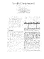
The autopsy incidence of FPE over time in Heidelberg, Germany showed a clear rela-
tionship b etween pulmonary embolism and wartime conditions. The lowest incidence
of FPE, expressed as a percentage of all hospitalized patients, was registered during the
post-Second World War years with a relative and absolute minimum between 1945
and 1949. The 1947 value (0.04%) was lower than 1932 (0.45%) or 1955 (0.38%) [36]
(Fig. 1).
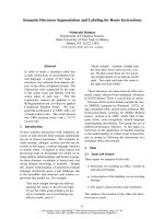
In Vienna after the First World War, FPE accounted for less than 0.5% of deaths ver-
sus 2.5% in the early 1930s. Again, in the late 1940s, inc idence of FPE at autopsy was
<1% versus almost 8% by the early 1970s [30] (Fig. 2).
These historical studies have limitat ions but suggest that the high-complex-carbohy-
drate, low-fat diet associated with war-time food rationing and perhaps increased exer-
cise may have markedly reduced the tendency to form thrombi and/or lessened the
consequences of those that do form. Judging from the autopsy data, the effects of
these lifestyle influences on VTE risk had a rapid onset and offset, and wartime condi-
tions afforded substantial protection against VTE, especially FPE.
Figure 1 Fatal PE from 1915 to 1964 in Hei delberg, Germany [36]. Absolute numbers of patients with
autopsy-proven FPE in black, and percentage of in hospital patient deaths related on autopsy to PE in
white. Reproduced from Linder et al. [88].
Cundiff et al. Theoretical Biology and Medical Modelling 2010, 7:31
/>Page 9 of 24
Prospective observational studies of diet and lifestyle factors associated with VTE
In the “Longitudinal Investigation of Thromboembolism Etiology (LITE) “ prospective
study, hazard ratios (95% CIs) of VTE incidence across quintiles of fruit and vegetable
intake were: 1.0 (reference: lowest quintile), 0.73 (0.48 to 1.11), 0.57 (0.37 to 0.90), 0.47
(0.29 to 0.77), and 0.59 (0.36 to 0.99) with Ptrend = 0.03 [32]. The fruit and vegetable
intake in the lowest quintile, 2.0 servings per day, was far less than rec ommended by
the United States Center for Dis ease Control (i.e. >5 servin gs per day for most people)
[37]. The highest quintile averaged 6.7 servings per day. Meat intake was a predictor of
VTE risk in LITE (HRs of VTE across quintiles of red and processed meat intake–1.0
(lowest quintile), 1.24 (0.78 to 1.98), 1.21 (0.74 to 1.98), 1.09 (0.64 to 1.87), and 2.01
(1.15 to 3.53) with the Ptrend = 0.02 [32]). Since fruit/vegetable intake in LITE corre-
lated negatively with meat intake (r = - 0.28), the two most influential dietary variables
may have acted synergistically on VTE risk.
In contrast, the Iowa Women ’s Health Study (IWHS) [38], t o date the only other
large prospective study of diet related to VTE risk, found no associations of VTE risk
with intake of fruit s/vegetables, meat, fish, or other foods. It also found no significant
associations with dietary patterns or individual nutrients. The IWHS found an associa-
tion of daily alcohol consu mption with lowered VTE risk, wherea s LITE only affirmed
that adjusting for alcohol consumption di d not diminish the strength of the correla-
tions between diet and VTE [32]. The following differences between the LIT E and
IWHS studies may account for the discrepancies:
• Only women were surveyed in the IWHS (99% white women); the LITE study
included relatively fewer women (55%) and more non-whites (27%).
Figure 2 Vienna, Austria p ercentages of autopsies with fatal PE (Quoted by Nielsen from Sigg
[30,36]).
Cundiff et al. Theoretical Biology and Medical Modelling 2010, 7:31
/>Page 10 of 24
• LITE’s dietary assessment had an interviewer-administered food frequency ques-
tionnaire (FFQ), considered more precise than the self-administered IWHS ques-
tionnaire [32].
• WhiletheIWHSFFQassessmentwasdoneonlyonceandrelatedtothesubse-
quent 19 year (mean 13 year) follow-up for VTE incidence, the LITE FFQ assess-
ment was done twice, six years apart (r ranged from 0.49 to 0.56 between the two
assessments). This allowed the LITE diet assessment to be based on a cumulative
average dietary intake. The designers of t he FFQs used in both studies found that
using the cumulat ive averages, in general, yielded stronger associations than utiliz-
ing either baseline diet or the most recent diet alone [39].
Therefore, the LITE study findings relating fruit and vegetable intake to reduced
VTE risk and meat intake to increased VTE risk have more scientific validity than the
IWHS finding of no dietary factor influences on VTE. The LITE findings are also
consistent with the historical observations (above).
VTE risk related to lipids, inflammation, and hemostatic parameters
A meta-analysis of VTE ca se-control studies found that VTE patients had significantly
lower HDL cholesterol levels and higher triglycerides but no differences in total choles-
terol or LDL-cholesterol [40]. Prospective studies of associations between lipid levels
and VTE risk are conflicting but possibly suggest that VTE risk is reduced when
serum levels of HDL cholesterol are higher and triglyceride lower [41].
In LITE, VTE was not correlated with C-reactive protein or white cell count [42].
However, LITE did not differentiate between idiopathic versus secondary VTE cases.
Luxembourg and colleagues reported a case-control study comparing idiopathic VTE
with risk-associated VTE patients (post-op, etc.) and controls. Idiopathic VTE patients
had significantly higher levels of high-sensitivity C-reactive protein (hs-CRP) than sec-
ondary VTE patients (mean 1.8 versus 1.5, P = 0.05), who had significantly higher
levels than controls (mean 1.5 versus 1.2, P = 0.02) [43].
Case-control and prospective studies show correlations of VTE with serum levels of
factor VIII [43-47]. The VTE case control study by Luxembourg and colleagues found
significantly higher fibrinogen levels in patients with idiopathic VTE than those with
risk-associated VTE (median: 33 1 versus 299 mg/dl, p = 0.004) [43]. Controls had
levels similar to risk-associate d VTE pa tients (median: 302 mg/dl). In LITE, factor VIII
levels and von Willebrand factor levels correlated significantly with VTE (P for trend
in quartiles <0.0001 for both) but fibrinogen levels did not. LITE did not assess platelet
aggregation or indices of fibrinolysis.
Alcohol and VTE
TheIowaWomen’s Health Study reported that daily alcohol imbibers suffered fewer
episodes of VTE. Thi s finding is questionable, since a single assessment of alcohol
intake was related to VTE incidence over the ensuing 19 years [38]. The LITE study
found that alcohol consumption neither increased nor decreased VTE risk [32].
There is no other prospective epide miological evide nce linking alcohol use with
either increased or decreased risk of VTE.
Cundiff et al. Theoretical Biology and Medical Modelling 2010, 7:31
/>Page 11 of 24
Fish intake and VTE
In LITE, fish intake was deemed protective against VTE on the b asis of a comparison
of VTE incidence between the first quintile of f ish consumption and the sum of the
four subsequent quintiles: 1.0 (first quintile) 0.58 (0.37-0.90) 0.60 (0.3 9-0.92) 0.55
(0.35-0.88) 0.70 (0.44-1.10), Ptrend= 0.30). This post hoc comparison is dubious
because fish consumption correlated positively with intake of fruits/vegetables (r =
0.27, P < 0.001) [32]. The c orrelation between fish and meat consumption was not
reported. In an analysis of t he Diabetes Control and Complications Trial database by
Cundiff and colleagues [4 8], long chain omega-3 fatty acid intake (a marker for fish
consumption) correlated inversely with percentage of calories from saturated fatty
acids (r = -0.21, P < 0.0001), and directly with dietary fiber intake (g/1,000 kcal) (r =
0.20, P < 0.0001), suggesting that fish eating is correlated with a more plant-based than
animal-based diet. In a Greek study of diet in peo ple with and without acute coronary
syndromes [49], fish intake was associated with consumption of:
• red meat - inversely related in patient and control groups (P<0.001 for both);
• vegetables - directly correlated (P<0.001 for both);
• fruit - directly correlated (P<0.001 for both); and
• legumes - directly correlated (P<0.001 for both).
Becausefishintakeisconfoundedwithother healthy dietary choices in the LITE
database and other studies, fish consumption does not appear to be an independent
protective factor for VTE. The possibility may nevertheless merit further study taking
account of the confounding variables.
Diets to consider for lowering VTE risk
Low VTE risk diets (i.e., h igh in fruits and vegetabl es and low in red and processed
meats) to consider as the experimental arm of non-inferiority randomized trials evalu-
ating standard anticoagulants for prophylaxis and treatment of VTE are as follows:
American Heart Association (AHA) step 1 and step 2 diets
AHA step 1 and step 2 diets recommend plenty of fruits and vegetables, lean meat and
two servings of fish per week [50]. A meta-analysis of randomized trials of these diets
versus regular diets (27 trials with more than 30,000 patient years of follow-up) shows
no significant reductio n of overall mortality (RR: 0.98, 95% CI 0.86 to 1.12), or cardio-
vascular disease mortality (RR: 0.91, 95% CI 0.77 to 1.07) [51,52].
There is no evidence that the AHA step 1 or step 2 diets would reduce VTE risk any
more than overall cardiovascular risk, so they would not be good low VTE risk diet
candidates.
Mediterranean diet (MD)
While the MD does not significantly benefit serum lipids, blood pressure, or body mass
index, it reduced overall cardiovascular disease risk by 70% in the “Lyon Diet Heart
Study “ [53].
Studies in Table 5 suggest that the MD benefits m arkers of coagulation , inflamma-
tion, and cardiovascular disease risk.
Cundiff et al. Theoretical Biology and Medical Modelling 2010, 7:31
/>Page 12 of 24
Vegetarian diets
Epide miological studies show that people consumi ng vegetarian diets (lacto-ovo, lacto,
ovo, or not otherwise specified (NOS)) and vegan diets (no meat, dairy, or eggs) have
lower overall vascular disease incidence than omnivores [54-58].
A lacto-ovo vegetarian diet contains plenty of fruits and vegetables and no meat, lar-
gely conforming to the LITE prospective data regarding a low VTE risk diet [32]. How-
ever, the lack of decreased vascular disease risk in Denmark during and after the
Second World War (see above) [35] might be considered a point in favor of a vegan
diet.
Tables 6, 7, and 8 show representative studies of vegetarian and vegan diets related
to VTE risk and inflammatory, lipid, and hematological markers, respectively, demon-
strating trends favorable to these diets.
A study from Rotterdam assessed the relationship of dietary fat and fiber with coagu-
lation factor VII in 3,007 elderly men and women subjects. Total fat and saturated fat
Table 5 MD studies of serum markers of inflammation and coagulation
Author Study
Design
Population Exposure
variable
Outcome variable Results
Esposito [76] RCT Metabolic
syndrome
patients
MD 1. Nutrient intake
2. endothelial function
3. lipid and glucose
parameters
4. insulin sensitivity
5. hs-CRP
6. IL-6
7. IL-7
8. IL-18
With MD
1. hs-CRP decreased
(P = 0.01)
2. IL6 decreased
3. (P = 0.01)
4. Endothelial function
improved (P < 0.001)
5. lipid and glucose
parameters improved
(P < 0.001)
6. decreased insulin
resistance (P < 0.001)
Mezzano [77] RCT Healthy
volunteers
MD versus high
fat diet
Fat content
Fibrinogen
factor VIIc
factor VIIIc
protein S
Fat content
- MD: 27.3%
- HFD: 39.9%
With MD
Fibrinogen reduced
(P = 0.03)
factor VIIc reduced
(P = 0.034)
factor VIIIc reduced
(P = 0.0057)
protein S increased
(P = 0.013)
Antonopoulou
[75]
Observa-
tional
Healthy
volunteers
and type 2
DM patients
MD platelet aggregation in
response to platelet
aggregating factor or
thrombin
Platelet activity
reduced in both
groups
Chrysohoou
[74]
Observa-
tional
People in
Greece
Adherence to MD
comparing the
highest and
lowest tertile
CRP
IL-6
Homocysteine
WBCs
highest tertile
participants averaged
- 20% lower CRP levels
(P = 0.015)
- 17% lower
interleukin-6 levels
(P = 0.025)
- 15% lower
homocysteine levels
(P = 0.031)
- 14% lower white
blood cell counts
(P = 0.001)
- 6% lower fibrinogen
levels (P = 0.025)
Cundiff et al. Theoretical Biology and Medical Modelling 2010, 7:31
/>Page 13 of 24
intake were significantly associated with factorVIIconlyinwomen.Fiberintakewas
inversely associated with factor VIIc in both men and women [59].
Mediterranean, vegetarian or vegan diets could all be reasonable choices for the
experimental arm of non-inferiority trials with anticoagulants for prophylaxis and
treatment of VTE. Given the challenges to patients in changing diets and the likelihood
that RCTs would show that anticoagulation prophylaxis and treatment for VTE are
themselves ineffective as sugge sted in the background section above, RCTs of VTE
patients should allow considerable flexibility in the diet interventio ns so that they are
not burdensome.
Vitamin K intake and VTE risk
Studies on vitamin K and VTE risk or coagulation profiles are very limited, because
there is no established role of vitamin K supplementation except in people who are
deficient in vitamin K. Based on the hypothesis that vitamin K supplementation may
protect against atherosclerosis, a placebo-controlled randomized trial evaluated the
Table 6 Studies of vegetarian diets and serum markers of inflammation
Author Study
Design
Population Exposure
variable
Outcome
variable
Results
Mezzano [70] Case-
control
52 Chilean subjects Lacto or lacto-ovo
Vegetarians v.
Omnivores
CRP NS
Chen [65] Case-
control
198 healthy Taiwanese
subjects
Vegetarians (NOS)
v. Omnivores
CRP NS
Harvinder
[121]
Case-
control
47 USA subjects with
CAD or CAD risk factors
Vegans v.
Omnivores
CRP Vegans had significantly
lower levels of CRP
Kjeldsen-
Kragh [64]
Case-
control
53 Rheumatoid arthritis
patients
Lacto vegetarians
v. Omnivores
WBC
count
Lacto vegetarians had
significantly lower WBCs
Kjeldsen-
Kragh [72]
Case-
control
Rheumatoid arthritis
patients
Vegetarians (NOS)
v. Omnivores
WBC
count
Vegetarians had
significantly lower WBCs
Nazarewicz
[62]
Case-
control
22 vegetarian and 19
omnivore Pols
Vegetarians (NOS)
v. Omnivores
WBC
count
Vegetarians had
significantly lower WBCs
Pongstaporn
[122]
Case-
control
178 vegetarian and 58
omnivore Thais
Vegetarians (NOS)
v. Omnivores
WBC
count
Vegetarians had
significantly lower WBCs
Arm-strong
[63]
Case-
control
431 vegetarian and 131
omnivore Seventh-day
Adventists
Vegetarians (NOS)
v. Omnivores
WBC
count
Vegetarian men but not
women had significantly
lower WBCs
Haddad [68] Case-
control
25 vegan and 20
omnivore Californians
Vegans v.
Omnivores
WBC
count
Vegans had significantly
lower WBCs
Tungtrong-
chitr [67]
Case-
control
132 vegetarians and 47
omnivores from Thailand
Vegetarians (NOS)
v. Omnivores
WBC
count
NS
Malter [66] Case-
control
German male
vegetarians and
omnivores
Vegetarians (NOS)
v. Omnivores
WBC
count
NS
Table 7 Studies of vegetarian diets and serum lipid markers
Author Study
Design
Population Exposure
variable
Outcome
variable
Results
Li [73] Case-
control
139 healthy male
subjects aged 20-55
Melbourne
Vegetarians
(NOS) v.
Omnivores
ratios of
triglycerides/HDL-
cholesterol
Vegetarians had lower ratios
of triglycerides/HDL-
cholesterol
Chen
[65]
Case-
control
198 healthy Taiwanese
subjects
Vegetarians
(NOS) v.
Omnivores
levels of total
cholesterol and
LDL-C
Vegetarians had lower levels
of total cholesterol and
LDL-C
Cundiff et al. Theoretical Biology and Medical Modelling 2010, 7:31
/>Page 14 of 24
effect of phylloquinone supplementation on blood lipids, inflammatory markers, and
fibrinolytic activity in postmenopausal women. No effect was seen on inflammatory or
fibrinolytic markers and lipid markers worsened (i.e., increased triacylglyc erols and
decreased HDL-C) [60]. A systematic review of RCTs of vitamin K supplementation
for preventing bone loss and fractures yielded 13 trials. None of the trials reported an
increase in VTE or other adver se reaction s [61]. Vitamin K status does not seem to be
a significant factor in VTE risk.
Effects of diet in relation to the VCHH
The simplest and most plausible mechanistic link between diet and DVT/VTE is
through other aspects of lifestyle: people who eat unhealthy diets are likely to exercise
less and remain sedentary for longer than those who eat healthy diets, exposing them
to a greater risk of prolonged non-pulsatile venous blood flow and consequent valve
pocket hypoxemia. However, subtler links between diet and venous thrombogenesis
can be inferred from the VCHH.
Compared with subjects taking omnivorous diets, a non-vegan vegetarian diet is
associated with a lower neutrophil count in some studies [62-64] though not all
[65-67]. Subjects on vegan diets consistently show lower neutrophil counts [68,69].
Studies have provided m ixed findings regarding platelet counts and function in vege-
tarians and vegans [62,68,70-73].
A reduced number of neutrophils would tend to attenuate the invasion of a hyp oxi-
cally injured ( necrotic) valve cusp endothelium by leukocytes, which according to the
VCHH would militate against the events initiating thrombogenesis. This suggests a
mechanism by which vegan diets could reduce the risk of VTE.
In that the MD has been found to decrease the white blood cell count [74], reduce
platele t activity [75], de crease markers of inflammation [76], improve endothelial func-
tion [76], and lower factor VII and VIII levels [77]; i t may be consistent with reduced
neutrophil margination and sequestration on a hypoxically damaged valve cusp, asso-
ciated with elevation of EDRF and non-elevation of p38 MAPK and thus with a
decreased risk for venous thrombogenesis (see chapter 12 in [2]) [78].
Table 8 Studies of vegetarian diets and serum markers of coagulation
Author Study
Design
Population Exposure
variable
Outcome variable Results
Li [73] Case-
control
139 healthy
male subjects
aged 20-55 in
Melbourne
Vegetarians
(NOS) v.
Omnivores
factor VII activity Lacto-ovo vegetarians had
significantly lower plasma
factor VII activity
Mezzano
[70]
Case-
control
52 Chilean
subjects
Lacto or
lacto-ovo
Vegetarians
v.
Omnivores
PT, fibrinogen, factor Vc,
factor VIIc, factor VIIIc,
antithrombin III, protein S,
plasminogen, protein C
Lacto-ovo vegetarians had
significantly lower levels of
fibrinogen, factor Vc, factor
VIIc, factor VIIIc, antithrombin
III, protein S, plasminogen,
prothrombin, protein C
Pan [123] Case-
control
203 healthy
Taiwanese age
<30
60
vegetarians
and 143
omnivores
PT, APTT, fibrinogen, factor
VIIc, factor VIIIc,
antithrombin III,
plasminogen,
Vegetarian men did not differ
from omnivore men.
Women: factor VIIIc higher and
APTT shorter in vegetarian
women versus omnivore
women
Cundiff et al. Theoretical Biology and Medical Modelling 2010, 7:31
/>Page 15 of 24
Randomized controlled non-inferiority trials of anticoagulants for VTE
proposed
Given the risks of anticoagul ation for VTE and new data concerning the current
reduced risk of hospital-acquired VTE, researchers should consider non-inferiority ran-
domized trials to test the efficacy and safety of anticoagulants for prophylaxis of VTE
for medical and surgical patients. Likewise, given the l ack of RCT evidence of efficacy
of anticoagulant treatment of VTE, a non-inferiority RCT would be in order to test the
efficacy and safety of anticoagulants for treatment of VTE.
To enlist researchers and patients in these proposed trials, the rationale must be
clearly explained and appropriate experimental arms determined. Experimental arm
options include using placebos or non-steroidal anti-inflammatory drugs (NSAIDs).
However, a Cochrane review of anticoagulation treatment for VTE c oncluded that:
“The use of anticoagulants is widely accepted in clinical p ractice, so a further RCT
comparing anticoagulants to placebo could not ethically be carried out “ [20]. A n
NSAID would have the advantage of being a weak antithro mbotic with less bleeding
risk than anticoagulants; however, epidemiological evidence suggests that NSAIDs may
increase the risk of VTE [79].
Given the relationship between diet and VTE noted above, low VTE risk diet inter-
ventions should be explored as possible experimental arms i n randomized non-infer-
iority trials testing the efficacy and safety of anticoagulants for prophylaxis and
treatment of VTE.
If such trial s yield positive results, they may be especially useful in developing coun-
tries, where there is a significant and increasing risk of DVT and VTE in medically ill
patients but only a minority receives anticoagulan t prophylaxis [80,81]. Pra ctitioners in
these countries recognize the economic and other difficulties of providing anticoagulants
for all patients for whom they are not contraindicated and recommend careful risk stra-
tification and education [80,82]. Education could include dietary advice. Interestingly,
there is strong evidence that oral contraceptives currently used in developing countries
entail a statistically significant risk of VTE, but only among women with high body mass
indices [83,84]; again, this could indicate that dietary prophylaxis would be beneficial.
Statistical considerations in designing non-inferiority trials
To provide convincing evidence for a change in standard-of-care treatment/prophylaxis
for VTE, from anticoagulants to a lower-risk alternative like low-VTE-risk diet, a defi-
nitive clinical trial would have to accomplish two goals simultaneously:
1. demonstrate that the diet is significantly safer than anticoagulants with respect
to hemorrhage, and
2. demonstrate that the diet is non-inferior to anticoagulants with respec t to the
prevention or amelioration of major complications of VTE.
This requires a safety/efficacy trial with two co-primary endpoints
1. a primary safety endpoint – an undesired hemorrhagic event, and
2. a primary efficacy endpoint –an undesired thromboembolic event, such as fatal
or non-fatal PE, recurrent symptomatic PE, or symptomatic DVT.
Cundiff et al. Theoretical Biology and Medical Modelling 2010, 7:31
/>Page 16 of 24
The choice of t he two co-primary endpoints is of utmost importance because t hese
endpo ints determine the sample size needed for the study – there must be a sufficient
number of each endpoint observed, so that the study will have sufficient statistical
power to yield simultaneous significant outcomes in testing each of the two co-primary
study hypotheses – better safety and non-inferior efficacy for the low VTE risk diet,
relative to the anticoagulants.
So the co-primary safety and efficacy endpoints must be consequential enough to
provide convincing motivation to c hange medical practice, and frequent enough to
yield a sufficiently powered study with a reasonable sample size. Of course, a set of
secondary safety and efficacy endpoints would also be monitored and analyzed. For
any given choice of endpoints, the study design would take the form of a prospective,
parallel, randomized, active-comparator, unblinded interventional clinical trial.
The occurrence or non-occurrence of each safety and efficacy endpoint within the
stated follow-up interval w ill be recorded. Potential confounders (subject age, gender,
medical history, type and severity illness, type and duration of surgery, etc.) will also
be recorded. These will be tested for balance between the two treatment groups by
Student’s t or chi-square tests. Variables found to be significantly unbal anced between
groups (p ≤ 0.1) will be adjusted for in the analyses, as described below.
Statistical analysis will consist of a comparison, between treatment groups, of the
fraction of subjects experiencing each of the two co-primary endpoints.
If no adjustment for potential confounders is necessary, the rates will be compared
by cross-tabulating, for each endpoint, the treatment group by the occurrence/non-
occurrence of the endpoint in a 2-by-2 table, and determining the “raw “ (unadjusted)
odds ratio for the occurrence of the event, for the diet, r elative to the anticoagulant,
along with its upper 1-sided 95% confidence limit. If adjustment for potential confoun-
ders is necessar y, the odds ratio and its upper 1-sided 95% confidence limit will be
obtained by multivariate logistic regression, where the occurrence of the endpoint is
the dependent variable, the treatment group is the main independent variable, and the
confounders are covariates. Alternativel y, a propensity score can be calculated for each
subject, incorporating all significant confounders. This score can be used in place of
the individual confounders in the logistic regression.
Regardless of whic h way the odds ratios are calculated, significantly better safety for
the diet will be inferred if the confid ence interval around the odds ratio (diet/antic oa-
gulant) for the primary safety endpoint lies entirely below the value of 1. Non-inferior
efficacy for the diet will be inferred if the confidence interval around the odds ratio
(diet/an ticoagulant) for the primary efficacy endpoint lies entirely below the value 1.5,
corresponding to an “allowable non-inferiority tolerance “ of 50%.
The sample size required for 80% power to infer improved safety and non-inferior
efficacy simultaneously will depend on the expected incidence rates of the prima ry
safety and effic acy endpoints, the expected amount of improvement in safety for diet
relative to anticoagulants, and the expected difference in efficacy (if any) between the
two treatments.
While the FPE rate ma y appear to be the logical primary efficacy endpoint for RCTs
comparing standard anticoagulant prophylaxis and treatment with a low VTE risk diet,
FPE data are unreliable for several reasons:
Cundiff et al. Theoretical Biology and Medical Modelling 2010, 7:31
/>Page 17 of 24
• Autopsy rates have been low in recent years, so trial data and conclusions would
rely on clinical diagnoses of FPE.
• Clinician FPE diagnoses corr elate very poorly with autopsy diagnoses of FPE in
both directions (i.e., more than 50% false positives and more than 50% false nega-
tives [85]).
• At autopsy, the pathologist has to d ecide if the PE was the fatal event or just a
contributing condition. This is quite subjective.
◦ Studies in the literature report postmortem exams inconsistently (i.e., classif i-
cations such as “incidental,”“contributing to death” or “fatal” PE vary).
◦ In a meta-analysis of cohort studies wit h a low proportion of deaths investi-
gated with autopsies, the overall mortality of patients undergoing elective hip
and knee replacement was a bout 2 1/2 times the rate of FPE (total mortality:
0.93% versus FPE rate: 0.36%) with most of the non FPE deaths from heart fail-
ure or myocardial infarction [86]. FPE very frequently occurs in those with
underlying severe cardiovascular disease, so FPE could not be ru led out without
autopsies on all cardiovascular deaths.
• Using FPE at the sole primary endpoint takes no account of deaths from anticoa-
gulant-induced bleeding.
Cons equently, ‘total mortality’ should be the primary effica cy endpoint, because it is
more reliable, consequential, and frequent than FPE. Screening for asymptomatic PE or
DVT as efficacy endpoints would not be necessary, because they correlate poorly with
FPE and total mortality.
The most consequential and frequent primary safety endpoint for RCTs would be
‘total bleeding’. This includes minor bleeding, wound hematomas, and major bleeding.
Secondary endpoints should be FPE, symptomatic PE, symptomatic DVT, fatal bleed-
ing, and major bleeding.
Example–RCT for VTE prophylaxis of hospitalized medical inpatients: Low VTE risk diet
versus standard anticoagulants
For a hypothetical example anticoagulant prophylaxis RCTs, consider acutely ill
patients admitted to hospital medical wards. America n Association of C hest Physician
(ACCP) Guidelines suggest that such patients receive LMWH, lo w dose heparin, or
fondaparinux as VTE prophylaxis
11
. We may use total bleeding (minor, major, and
fatal bleeding) as the primary safety endpoint. A recent Cochrane Review of prophylac-
tic anticoagulation for m edical patients found that bleeding occurred in 117/2,405
(4.9%) of anticoagulated patients and 62/2,404 (2.6%) of untreated patients [87].
Subjects would be recruited from patients admitted t o acute medical services who
have VTE risk factors qualifying them for anticoagulant prophylaxis according to
AACP Guidelines [11].
Subjects meeting the inclusion/exclusion criteria would be randomized to receive
either a standard anticoagulant per AACP Guidelines (i.e., LMWH, low dose heparin,
or fondaparinux) or a low-VTE-risk diet regimen. The diet (Mediterranean, vegetarian
or vegan per the patient’s choice) would be recommended for the entire duration of
the trial, but compliance would be left to the discretion of the patient and treating
physician. This flexibility in the diet arm a ssures that the diet intervention would not
Cundiff et al. Theoretical Biology and Medical Modelling 2010, 7:31
/>Page 18 of 24
be a burden to patients. Flexibi lity in a dministering the low VTE diet is reasonable,
since the RCTs are in large part testin g the safety and efficac y of standard anticoagula-
tion prophylaxis a nd treatment, and the low VTE diet would not be expected t o do
harm.
All outcomes will be recorded into the patients’ medical records. In addition, follow-
up RCT-related visits or calls as appropriate will be conducted approximately every
10 days up to 60 days after anticoagulation prophylaxis is c ompleted, to determine if
any of the primary or secondary safety or efficacy outcomes had occurred. Compliance
with the low VTE risk diet will also be assessed, asking each patient to estimate his/
her compliance on a scale of 0 - 100, with ‘0’ representing completely non-compliant
and ‘100’ representing completely compliant. If follow-up takes place before discharge
from the hospital, the information will be entered into the patient’s medical record and
separate data collection f orms; if the follow-up takes place after discharge, the infor-
mation will be recorded on to separate data collection forms.
The data collection forms will be assessed by study monitors at each participating
medical center and reviewed by the responsible investigator at each medical center
before anonymized data are sent to the study PI.
As noted above, we would use the overall death rate as the primary efficacy end-
point. Since the aforementioned Cochrane Review showed that prophylactic anticoagu-
lation does not significantly reduce FPE rates or overall deaths in acute medical
patients (for anticoagulation versus placebo, FPE: 0.94 [0.82, 1.07] and overall deaths:
RR 0.95 [0.83, 1.07] [87]), a 50% non-inferiority tolerance i s reasonable. The death rate
in this Cochrane review was about 5% with or without anticoagulant prophylaxis. So
with anticoagulant prophylaxis, the primary safety endpoint (overall bleeding) and the
primary efficacy endpo int (overall mortality) would each be expected to occur in about
5% of patients. Expecting that the absence of anticoagulation and a low VTE diet can
reduce the incidence of the total bleeding safety endpoint down to 2.5% with no
change in ef ficacy, about 1,400 subjects would be required in each treatment group in
order to have 80% power to meet both co-primary hypotheses (superior safety and
non-inferior efficacy).
Conclusion
Because of the bleeding and ‘rebound hypercoagulability’ risks of anticoagulation for
VTE prophylaxis and the possibility that a low VTE risk diet might reduce the inci-
dence of hospital-acquired VTE; randomized controlled non-inferiority clinical trials
should be undertaken to compare standard anticoagulant treatment with a potentially
low VTE risk diet (vegetarian, vegan, or Mediterranean) for VTE prophylaxis of medi-
cal and surgical patients. Likewise for VTE treatment, a randomized non-inferiority
clinical trial comparing standard anticoagulation treatment with a low VTE risk diet
should also be considered. We call upon the U. S. National Institutes of Health and
the British National Institute for Health and Clinical Excellence to design and fund
those trials.
Author details
1
333 Orizaba Avenue, Long Beach, CA 90814, USA.
2
Theoretical Medicine and Biology Group, 26 Castle Hill, Glossop,
Derbyshire, SK13 7RR, UK.
3
129 Viceroy Close, Birmingham, B5 7UY, UK.
4
Department of Medicine, Georgetown
University, Washington, DC, USA.
Cundiff et al. Theoretical Biology and Medical Modelling 2010, 7:31
/>Page 19 of 24
Authors’ contributions
DKC proposed the concept of dietary prophylaxis for VTE and a non-inferiority trial for comparison with
anticoagulants, and reviewed the relevant literature. PCM was primarily responsible for formulating the VCHH and
evaluating its relevance to VTE prophylaxis and treatment. PSA investigated the molecular aspects of the VCHH and
their consistency with the dietary hypothesis of prophylaxis. JCP was responsible for the statistical considerations and
the design of the proposed non-inferiority trial. All authors read and agreed the final manuscript.
Competing interests
DKC withdrew warfarin from a patient with lower-limb deep venous thrombosis, disseminated tuberculosis,
alcoholism, liver failure, and anemia because the risk for bleeding in this patient seemed greater than the benefit of
anticoagulant treatment. The patient later died of pulmonary embolism. DKC lost his medical license because of this
case, having had no other medical board discipline during 25 years of clinical practice. Otherwise, the authors declare
that they have no competing interests.
Received: 28 April 2010 Accepted: 11 August 2010 Published: 11 August 2010
References
1. Malone PC, Agutter PS: The aetiology of deep venous thrombosis. Quart J Med 2006, 99:581-593.
2. Malone PC, Agutter PS: The aetiology of deep venous thrombosis: a critical, historical and epistemological survey Springer:
Dordrecht 2008.
3. Bagot CN, Arya R: Virchow and his triad: a question of attribution. Br J Haematol 2008, 143:180-190.
4. Barritt DW, Jordan SC: Anticoagulant drugs in the treatment of pulmonary embolism – A controlled trial. Lancet
1960, 1:1309-1312.
5. Karimova A, Pinsky DJ: The endothelial response to oxygen deprivation: biology and clinical implications. Intensive
Care Med 2001, 27:19-31.
6. Carrier M, Le Gal G, Wells PS, Rodger MA: Systematic review: case-fatality rates of recurrent venous
thromboembolism and major bleeding events among patients treated for venous thromboembolism. Ann Intern
Med 2010, 152:578.
7. Fihn SD, McDonell M, Martin D, Henikoff J, Vermes D, Kent D, White RH: Risk factors for complications of chronic
anticoagulation. A multicenter study. Warfarin optimized outpatient follow-up study group. Ann Intern Med 1993,
118(7):511-520.
8. van der Meer FJ, Rosendaal FR, Vandenbroucke JP, Briet E: Bleeding complications in oral anticoagulant therapy. An
analysis of risk factors. Arch Int Med 1993, 153(13):1557-1562.
9. Levine MN, Hirsh J, Landefeld S, Raskob G: Hemorrhagic complications of anticoagulant treatment. Chest 1992, 102(4
Suppl):352S-363S.
10. Cundiff DK: Anticoagulation Therapy for Venous Thromboembolism. MedGenMed 2004, 6(3) [scape.
com/viewarticle/487577].
11. Geerts WH, Bergqvist D, Pineo GF, Heit JA, Samama CM, Lassen MR, Colwell CW: Prevention of venous
thromboembolism. Chest 2008, 133(6 suppl):381S-453S [ />full].
12. Dahl OE, Quinlan DJ, Bergqvist D, Eikelboom JW: A critical appraisal of bleeding events reported in venous
thromboembolism prevention trials of patients undergoing hip and knee arthroplasty. J Thromb Haemost 2010.
13. Eikelboom JW, Quinlan DJ, O’Donnell M: Major bleeding, mortality, and efficacy of fondaparinux in venous
thromboembolism prevention trials. Circulation 2009, 120(20):2006-2011 [ />abstract/120/20/2006].
14. Goldhaber SZ: Venous thromboembolism risk among hospitalized patients: Magnitude of the risk is staggering. Am
J Hematol 2007, 82(9):775-776.
15. Antithrombotics - current market and future outlook. IMS Health 2005 [ />IMSinclude/i_article_20050630.asp], />Accessed August 25, 2005.
16. Egermayer P: Value of anticoagulants in the treatment of pulmonary embolism: a discussion paper. Journal of the
Royal Society of Medicine 1981, 74(9):675-681.
17. Nielsen HK, Husted SE, Krusell LR, Fasting H, Charles P, Hansen HH, Nielsen BO, Petersen JB, Bechgaard P: Anticoagulant
therapy in deep venous thrombosis. A randomized controlled study. Thrombosis Research 1994, 73(3-4):215-226.
18. Ott P, Eldrup E, Oxholm P: The value of anticoagulant therapy in deep venous thrombosis in the lower limbs in
elderly, mobilized patients. A double-blind, placebo-controlled investigation with open therapeutic guidance.
Ugeskr Laeger 1988, 150:218-221.
19. Kakkar VV, Flanc C, O’Shea M, Flute P, Howe CT, Clarke MB: Treatment of deep-vein thrombosis–a random trial. Br J
Surg 1968, 55(11):858.
20. Cundiff D, Manyemba J, Pezzullo J: Anticoagulants versus non-steroidal anti-inflammatories or placebo for
treatment of venous thromboembolism. The Cochrane Database of Systematic Reviews 2006, , 1: CD003746 [http://
www.mrw.interscience.wiley.com/cochrane/clsysrev/articles/CD003746/frame.html], Feedback letter: http://medgenmed.
medscape.com/viewarticle/557263.
21. Goldhaber S, Dunn K, MacDougall R: New onset of venous thromboembolism among hospitalized patients at
Brigham and Women’s Hospital is caused more often by prophylaxis failure than by withholding treatment. Chest
2000, 118:1680-1684 [ />22. Cundiff DK: Reply to letters re: Anticoagulation therapy for venous thromboembolism. Med Gen Med 2005, 6(4),
/>23. Lindblad B, Eriksson A, Bergqvist D: Autopsy-verified pulmonary embolism in a surgical department: analysis of the
period from 1951 to 1988. Br J Surg 1991, 78(7):849-852.
Cundiff et al. Theoretical Biology and Medical Modelling 2010, 7:31
/>Page 20 of 24
24. Dahl OE, Aspelin T, Arnesen H, Seljeflot I, Kierulf P, Ruyter R, Lyberg T: Increased activation of coagulation and
formation of late deep venous thrombosis following discontinuation of thromboprophylaxis after hip replacement
surgery. Thrombosis Research 1995, 80(4):299-306.
25. Cundiff DK: Clinical evidence for ‘rebound hypercoagulability’ after discontinuing oral anticoagulants for venous
thromboembolism. Medscape J Med 2008, 10(11):258 [ />26. Cundiff DK: A systematic review of Cochrane anticoagulation reviews. Medscape J Med 2009, 11(1):5 [http://www.
medscape.com/viewarticle/584084].
27. Clagett GP, Reisch JS: Prevention of venous thromboembolism in general surgical patients. Results of meta-analysis.
Ann Surg 1988, 202(2):227-240.
28. Nurmohamed MT, van Riel AM, Henkens CM, Koopman MM, Que GT, d’Azemar P, Buller HR, ten Cate JW, Hoek JA, van
der Meer J, van der Heul C, Turpie AG, Haley S, Sicurella A, Gent M: Low molecular weight heparin and compression
stockings in the prevention of venous thromboembolism in neurosurgery. Thromb Haemost 1996, 75(2):233-238.
29. Collins R, Scrimgeour A, Yusuf S, Peto R: Reduction in fatal pulmonary embolism and venous thrombosis by
perioperative administration of subcutaneous heparin. Overview of results of randomized trials in general,
orthopedic, and urologic surgery. New Eng J Med 1988, 318(18):1162-1173.
30. Nielsen H, Bechgaard P, Nielsen P, Husted S, Geday E: 178 fatal cases of pulmonary embolism in a medical
department. Acta Med Scand 1981, 209(5):351-355.
31. Karwinski B, Svendsen E: Comparison of clinical and post-mortem diagnosis of pulmonary embolism. J Clin Pathol
1989, 42:135-139.
32. Steffen L, Folsom A, Cushman M, Jacobs D, Rosamond W: Greater fish, fruit, and vegetable intakes are related to
lower incidence of venous thromboembolism: The Longitudinal Investigation of Thromboembolism Etiology.
Circulation 2007, 115(2):188-195[ />33. Gaston S: The Surgeon General’s call to action to prevent deep vein thrombosis and pulmonary embolism. US
Department of Health and Human Services [ />34. Strom A: Examination into the diet of Norwegian families during the war-years 1942-45. Acta Med Scand Suppl
1948, 214:1-47.
35. Malmros H: Acta Med Scand 1950, 246(Suppl):137.
36. Sigg K: Varicen, Ulcus cruris und Thrombosen Berlin, Heidelberg, and New York: Springer-Verlag 1976.
37. How Many fruits and vegetables do you need?. Center for Disease Control 2009 [itsandveggiesmatter.
gov/], Accessed March 2, 2010.
38. Lutsey P, Steffen L, Virnig B, Folsom A: Diet and incident venous thromboembolism: The Iowa Women’s Health
Study. Am Heart J 2009, 157(6)
:1081-1087.
39. Hu FB, Stampfer MJ, Rimm E, Ascherio A, Rosner BA, Spiegelman D, Willett WC: Dietary fat and coronary heart
disease: A comparison of approaches for adjusting for total energy intake and modeling repeated dietary
measurements. Am J Epidemiol 1999, 149(6):531-540[ />40. Ageno W, Becattini C, Brighton T, Selby R, Kamphuisen PW: Cardiovascular risk factors and venous
thromboembolism. A meta-analysis. Circulation 2008, 117(1):93-102[ />CIRCULATIONAHA.107.709204v1].
41. Doggen CJM, Smith NL, Lemaitre RN, Heckbert SR, Rosendaal FR, Psaty BM: Serum lipid levels and the risk of venous
thrombosis. Arterioscler Thromb Vasc Biol 2004, 24:1970-1975.
42. Tsai A, Cushman M, Rosamond WD, Heckbert SR, Tracy RP, Aleksic N, Folsom AR: Coagulation factors, inflammation
markers, and venous thromboembolism: the Longitudinal Investigation of Thromboembolism Etiology (LITE). Am J
Med 2002, 113:636-642.
43. Luxembourg B, J S, M H, M G, D D, E S, E L-L: Cardiovascular risk factors in idiopathic compared to risk-associated
venous thromboembolism: A focus on fibrinogen, factor VIII, and high-sensitivity C-reactive protein (hs-CRP).
Thromb Haemost 2009, 102(4):668-675.
44. Luxembourg B, Schmitt J, Humpich M, Glowatzki M, Seifried E, Lindhoff-Last E: Intrinsic clotting factors in dependency
of age, sex, body mass index, and oral contraceptives: definition and risk of elevated clotting factor levels. Blood
Coagul Fibrinolysis 2009, 20(7):524-534.
45. Martinelli I: von Willebrand factor and factor VIII as risk factors for arterial and venous thrombosis. Sem Hematol
2005, 42:49-55.
46. Kraaijenhagen R, in’t Anker PS, Koopman MM, Reitsma PH, Prins MH, van den Ende A, Buller HR: High plasma
concentration of factor VIIIc is a major risk for venous thromboembolism. Thromb Haemost 2000, 83:5-9.
47. Goldenberg NA, Knapp-Clevenger R, Manco-Johnson MJ, Group tMSRT: Elevated plasma factor VIII and d-dimer levels
as predictors of poor outcomes of thrombosis in children. N Engl J Med 2004, 351(11):1081-1088[m.
org/cgi/content/abstract/351/11/1081].
48. Cundiff DK, Lanou AJ, Nigg CR: Relation of omega-3 fatty acid intake to other dietary factors known to reduce
coronary heart disease risk. Am J Cardiol 2007, 99(9):1230-1233.
49. Panagiotakosa DB, Pitsavosb C, Zampelasa A, Chrysohooub C, Griffinc BA, Stefanadisb C, Toutouzasd P: Fish
consumption and the risk of developing acute coronary syndromes: the CARDIO2000 study. Int J Cardiol 2005,
102(3):403-409.
50. Lichtenstein AH, Appel LJ, Brands M, Carnethon M, Daniels S, Franch HA, Franklin B, Kris-Etherton P, Harris WS,
Howard B, Karanja N, Lefevre M, Rudel L, Sacks F, Van Horn L, Winston M, Wylie-Rosett J: Diet and lifestyle
recommendations revision 2006: A scientific statement from the American Heart Association Nutrition Committee.
Circulation 2006, 114(1):82-96[ />51. Hooper L, Summerbell CD, Higgins JPT, Thompson RL, Clements G, Capps N, Davey Smith G, Riemersma RA, Ebrahim S:
Reduced or modified dietary fat for preventing cardiovascular disease (Cochrane Review). Cochrane Database of
Systematic Reviews 2000, , 2: CD002137.
52. Hooper L, Summerbell CD, Higgins JPT, Thompson RL, Capps NE, Smith GD, Riemersma RA, Ebrahim S: Dietary fat
intake and prevention of cardiovascular disease: systematic review. BMJ
2001, 322(7289):757-763[http://bmj.
bmjjournals.com/cgi/content/full/322/7289/757].
Cundiff et al. Theoretical Biology and Medical Modelling 2010, 7:31
/>Page 21 of 24
53. de Lorgeril M, Renaud S, Mamelle N, Salen P, Martin JL, Monjaud I, Guidollet J, Touboul P, Delaye J: Mediterranean
alpha-linolenic acid-rich diet in secondary prevention of coronary heart disease. Lancet 1994, 343(8911):1454-1459.
54. Craig WJ: Health effects of vegan diets. Am J Clin Nutr 2009, 89(5):1627S-1633[ />abstract/89/5/1627S].
55. Leitzmann C: Vegetarian diets: what are the advantages? Forum Nutr 2005, 57:147-156.
56. Willett W: Lessons from dietary studies in Adventists and questions for the future. Am J Clin Nutr 2003, 78(3
Suppl):539S-543S.
57. Singh PN, Sabate J, Fraser GE: Does low meat consumption increase life expectancy in humans? Am J Clin Nutr 2003,
78(3):526S-532[ />58. Ornish D, Scherwitz LW, Billings JH, Brown SE, Gould KL, Merritt TA, Sparler S, Armstrong WT, Ports TA, Kirkeeide RL,
Hogeboom C, Brand RJ: Intensive lifestyle changes for reversal of coronary heart disease. JAMA 1998,
280(23):2001-2007.
59. Mennen L, Witteman J, den Breeijen J, Schouten E, de Jong P, Hofman A, Grobbee D: The association of dietary fat
and fiber with coagulation factor VII in the elderly: the Rotterdam Study. Am J Clin Nutr 1997, 65(3):732-736[http://
www.ajcn.org/cgi/reprint/65/3/732?ijkey=fc4b980f6cecf9f04571ea31d25d0f61a2f6a983].
60. Kristensen M, Kudsk J, Bügel S: Six weeks phylloquinone supplementation produces undesirable effects on blood
lipids with no changes in inflammatory and fibrinolytic markers in postmenopausal women. Eur J Nutr 2008,
47(7):375-379.
61. Cockayne S, Adamson J, Lanham-New S, Shearer MJ, Gilbody S, Torgerson DJ: Vitamin k and the prevention of
fractures: Systematic review and meta-analysis of randomized controlled trials. Arch Intern Med 2006,
166(12):1256-1261[ />62. Nazarewicz R: The effect of vegetarian diet on selected biochemical and blood morphology parameters. Rocz
Panstw Zakl Hig 2007, 58(1):23-27.
63. Armstrong BK, Davis RE, Nicol DJ, van Merwyk AJ, Larwood CJ: Hematological, vitamin B12, and folate studies on
Seventh-day Adventist vegetarians. Am J Clin Nutr 1974, 27(7):712-718[ />712].
64. Kjeldsen-Kragh J: Rheumatoid arthritis treated with vegetarian diets. Am J Clin Nutr 1999, 70(3):594S-600[http://www.
ajcn.org/cgi/content/abstract/70/3/594S].
65. Chen CW, Lin YL, Lin TK, Lin CT, Chen BC, Lin CL: Total cardiovascular risk profile of Taiwanese vegetarians. Eur J Clin
Nutr 2008, 62(1):138-144.
66. Malter M, Schriever G, Eilber U: Natural killer cells, vitamins, and other blood components of vegetarian and
omnivorous men. Nutr Cancer 1989, 12(3):271-278.
67. Tungtrongchitr R, Pongpaew P, Prayurahong B, Changbumrung S, Vudhivai N, Migasena P, Schelp FP: Vitamin B12,
folic acid and haematological status of 132 Thai vegetarians.
Int J Vitam Nutr Res 1993, 63(3):201-207.
68. Haddad EH, Berk LS, Kettering JD, Hubbard RW, Peters WR: Dietary intake and biochemical, hematologic, and
immune status of vegans compared with nonvegetarians. Am J Clin Nutr 1999, 70(3 Suppl):586S-593S[http://www.
ajcn.org/cgi/reprint/70/3/586S].
69. McCarty MF: A taurine-supplemented vegan diet may blunt the contribution of neutrophil activation to acute
coronary events. Med Hypotheses 2004, 63(3):419-425.
70. Mezzano D, Muñoz X, Martínez C, Cuevas A, Panes O, Aranda E, Guasch V, Strobel P, Muñoz B, Rodríguez S, Pereira J,
Leighton F: Vegetarians and cardiovascular risk factors: hemostasis, inflammatory markers and plasma
homocysteine. Thromb Haemost 1999, 81(6):913-917.
71. Famodu AA, Osilesi O, Makinde YO, Osonuga OA, Fakoya TA, Ogunyemi EO, Egbenehkhuere IE: The influence of a
vegetarian diet on haemostatic risk factors for cardiovascular disease in Africans. Thromb Res 1999, 95(1):31-36.
72. Kjeldsen-Kragh J, Mellbye OJ, Haugen M, Mollnes TE, Hammer HB, Sioud M, Førre O: Changes in laboratory variables
in rheumatoid arthritis patients during a trial of fasting and one-year vegetarian diet. Scand J Rheumatol 1995,
24(2):85-93.
73. Li D, Sinclair A, Mann N, Turner A, Ball M, Kelly F, Abedin L, Wilson A: The association of diet and thrombotic risk
factors in healthy male vegetarians and meat-eaters. Eur J Clin Nutr 1999, 53(8):612-619[ />pubmed/10477247?dopt=Abstract].
74. Chrysohoou C, Panagiotakos DB, Pitsavos C, Das UN, Stefanadis C: Adherence to the Mediterranean diet attenuates
inflammation and coagulation process in healthy adults: The ATTICA Study. J Am Coll Cardiol 2004, 44(1):152-158.
75. Antonopoulou S, Fragopoulou E, Karantonis HC, Mitsou E, Sitara M, Rementzis J, Mourelatos A, Ginis A, Phenekos C:
Effect of traditional Greek Mediterranean meals on platelet aggregation in normal subjects and in patients with
type 2 diabetes mellitus. J Med Food 2006, 9(3):356-362.
76. Esposito K, Marfella R, Ciotola M, Di Palo C, Giugliano F, Giugliano G, D’Armiento M, D’Andrea F, Giugliano D: Effect of
a Mediterranean-style diet on endothelial dysfunction and markers of vascular inflammation in the metabolic
syndrome: a randomized trial. JAMA 2004, 292(12):1440-1446[ />1440].
77. Mezzano D, Leighton F, Martínez C, Marshall G, Cuevas A, Castillo O, Panes O, Muñoz B, D Pérez DD, Mizón C,
Rozowski J, San Martín A, Pereira J: Complementary effects of Mediterranean diet and moderate red wine intake on
haemostatic cardiovascular risk factors. Eur J Clin Nutr 2001, 55:444-451[ />pdf/1601202a.pdf].
78. Ju H, Behm DJ, Nerurkar S, Eybye ME, Haimbach RE, Olzinski AR, Douglas SA, Willette RN: p38 MAPK inhibitors
ameliorate target organ damage in hypertension: Part 1. p38 MAPK-dependent endothelial dysfunction and
hypertension. J Pharmacol Exp Ther 2003, 307(3):932-938.
79. Huerta C, Johansson S, Wallander M-A, Garcia Rodriguez LA: Risk factors and short-term mortality of venous
thromboembolism diagnosed in the primary care setting in the United Kingdom. Arch Intern Med 2007,
167(9):935-943[ />80. Pandey A, Patney N, Singh M, Guleria R: Assessment of risk and prophylaxis for deep vein thrombosis and
pulmonary embolism in medically ill patients during their early days of hospital stay at a tertiary care center in a
developing country. Vasc Health Risk Manag 2009, 5:643-648.
Cundiff et al. Theoretical Biology and Medical Modelling 2010, 7:31
/>Page 22 of 24
81. De Lorenzo F, Dotsenko O, Kakkar VV: Low molecular weight heparins in cardiovascular medicine. Minerva
Cardioangiol 2005, 53:585-603.
82. Awidi A, Obeidat N, Magablah A, Bsoul N: Risk stratification for venous thromboembolism in hospitalized patients in
a developing country: a prospective study. J Thromb Thrombolysis 2009, 28:309-313.
83. No authors listed: Venous thromboembolic disease and combined oral contraceptives: results of international
multicentre case-control study. World Health Organization Collaborative Study of Cardiovascular Disease and
Steroid Hormone Contraception. Lancet 1995, 346:1575-1582.
84. Drife J: Thromboembolism. Br Med Bull 2003, 67:177-190.
85. Dismuke SE, VanderZwaag R: Accuracy and epidemiological implications of the death certificate diagnosis of
pulmonary embolism. J Chronic Dis 1984, 37(1):67-73.
86. Dahl OE, Caprini JA, Colwell CW Jr, Frostick SP, Haas S, Hull RD, Laporte S, Stein PD: Fatal vascular outcomes following
major orthopedic surgery. Thromb Haemost 2005, 93(5):860-866.
87. Alikhan R, Cohen AT: Heparin for prevention of venous thromboembolism in general medical patients (excluding
stroke and MI). Cochrane Database of Systematic Reviews 2009, , 4: CD003747[ />cochrane/clsysrev/articles/rel0002/CD003747/pdf_fs.html].
88. Linder F, Schmitz W, Encke A, Trede M, Storch H: A study of 605 fatal pulmonary embolisms and two successful
embolectomies. Surg Gynecol Obstet 1967, 125:82-86.
89. Mahé I, Bergmann JF, d’Azémar P, Vaissie JJ, Caulin C: Lack of effect of a low-molecular-weight heparin (nadroparin)
on mortality in bedridden medical in-patients: a prospective randomised double-blind study. Eur J Clin Pharmacol
2005, 61(5-6):347-351.
90. Cohen AT, Davidson BL, Gallus AS, Lassen MR, Prins MH, Tomkowski W, Turpie AGG, Egberts JFM, Lensing AWA,
Investigators A: Efficacy and safety of fondaparinux for the prevention of venous thromboembolism in older acute
medical patients: randomised placebo controlled trial. BMJ 2006, 332(7537):325-329[ />content/full/332/7537/325].
91. Testroote M, Stigter W, de Visser DC, Janzing H: Low molecular weight heparin for prevention of venous
thromboembolism in patients with lower leg immobilization. Cochrane Database of Systematic Reviews 2008, , 1:
CD006681[ Feedback letter:
/>92. Bergman JF, Caulin C: Heparin prophylaxis in bedridden patients. Lancet 1996, 348(9021):205-206.
93. Bergman JF, Neuhart E: A multicenter randomised double-blind study of enoxaparin compared with unfractionated
heparin in the prevention of venous thromboembolic disease in elderly in-patients bedridden for an acute
medical illness. Thromb Haemostasis 1996, 76(4):529-534.
94. Fraisse F, Holzapfel L, Coulard JM, Simonneau G, Bedock B, Feissel M, Herbecq P, Pordes R, Poussel JF, Roux L:
Nadroparin in the prevention of deep vein thrombosis in acute decompensated chronic obstructive pulmonary
disease. Am J Respir Crit Care Med 2000, 161(4 Pt1)
:1109-1114.
95. Turpie AG, Bauer KA, Caprini JA, Comp PC, Gent M, Muntz JE: Fondaparinux combined with intermittent pneumatic
compression versus intermittent pneumatic compression alone for prevention of venous thromboembolism after
abdominal surgery: a randomized, double-blind comparison. J Thromb Haemost 2007, 5:1854-1861[http://www1853.
interscience.wiley.com/cgi-bin/fulltext/117993947/HTMLSTART].
96. Kosir MA, Kozol RA, Perales A: Is deep vein thrombosis prophylaxis overemphasized? A randomized prospective
trial. JSurgRes1996, 60:289-292.
97. Kosir MA, Schmittinger L, Barno-Winarski L, Duddella P, Pone M, Perales A, Lange P, Brish LK, McGee K, Beleski K,
Pawlak J, Mammen E, Sajahan NP, Kozol RA: Prospective double-arm study of fibrinolysis in surgical patients. J Surg
Res 1998, 74(1):96-101.
98. Rasmussen MS, Jørgensen LN, Wille-Jørgensen P: Prolonged thromboprophylaxis with low molecular weight heparin
for abdominal or pelvic surgery. Cochrane Database of Systematic Reviews 2009, , 1: CD004318[erscience.
wiley.com/cochrane/clsysrev/articles/CD004318/frame.html].
99. Sasaki S, Miyakoshi N, Matsuura H, Saitoh H, Kudoh D, Shimada Y: Prospective randomized controlled trial on the
effect of fondaparinux sodium for prevention of venous thromboembolism after hip fracture surgery. J Orthop Sci
2009, 14(5):491-496.
100. Bi C, Wen J, Jiang K, Chen S, Wang C, Sun W, Sun Y, Liang Z, Cheng Y, Cheng T, Hu H: Chinese drugs for
supplementing Qi and activating blood circulation in preventing deep venous thrombosis after big operations in
orthopaedics and traumatology. Zhongguo Zhong Yao Za Zhi 2009, 34(5):625-627.
101. Goel DP, Buckley R, deVries G, Abelseth G, Ni A, Gray R: Prophylaxis of deep-vein thrombosis in fractures below the
knee: a prospective randomised controlled trial. J Bone Joint Surg Br 2009, 91(3):388-394.
102. Agarwal NK, Mathur N: Deep vein thrombosis in acute spinal cord injury. Spinal Cord 2009, 47(10):769-772.
103. Eriksson BI, Wille-Jorgensen P, Kalebo P, Mouret P, Rosencher N, Bosch P, et al: A comparison of recombinant hirudin
with a lowmolecular-weight heparin to prevent thromboembolic complications after total hip replacement. NEng
JMed1997, 337(19):1329-1335[ />104. Eriksson BI, Dahl OE, Büller HR, Hettiarachchi R, Rosencher N, Bravo ML, Ahnfelt L, Piovella F, Stangier J, Kälebo P,
Reilly P, Group BIS: BISTRO II Study Group. A new oral direct thrombin inhibitor, dabigatran etexilate, compared
with enoxaparin for prevention of thromboembolic events following total hip or knee replacement: the BISTRO II
randomized trial. J Thromb Haemost 2005, 3(1):103-111.
105. Heit JA, Colwell CW, Francis CW, Ginsberg JS, Berkowitz SD, Whipple J, Astrazeneca Arthroplasty Study Group:
Comparison of the oral direct thrombin inhibitor ximelagatran with enoxaparin as prophylaxis against venous
thromboembolism after total knee replacement: A phase 2 dose-finding study. Arch Int Med 2001,
161(18):2215-2221.
106. Eriksson BI, Arfwidsson AC, Frison L, Eriksson UG, Bylock A, Kalebo P: A dose-ranging study of the oral direct thrombin
inhibitor, ximelagatran, and its subcutaneous form, melagatran, compared with dalteparin in the prophylaxis of
thromboembolism after hip or knee replacement: METHRO I. MElagatran for THRombin inhibition in Orthopaedic
surgery. Thromb Haemostasis 2002, 87(2):231-237.
Cundiff et al. Theoretical Biology and Medical Modelling 2010, 7:31
/>Page 23 of 24
107. Francis CW, Berkowitz SD, Comp PC, Lieberman JR, Ginsberg JS, Paiement G, EXULT A Study Group: Comparison of
ximelagatran with warfarin for the prevention of venous thromboembolism after total knee replacement. NEngJ
Med 2003, 349(18):1703-1712.
108. Eriksson BI, Dahl OE, Rosencher N, Kurth AA, van Dijk, Frostick SP, RE-MODEL Study Group, et al: Oral dabigatran
etexilate vs. subcutaneous enoxaparin for the prevention of venous thromboembolism after total knee
replacement: the RE-MODEL randomized trial. J Thromb Haemostasis 2007, 5(11):2178-2185.
109. Turpie AGG, Bauer KA, Eriksson BI, Lassen MR: Fondaparinux vs enoxaparin for the prevention of venous
thromboembolism in major orthopedic surgery: A meta-analysis of 4 randomized double-blind studies. Arch Intern
Med 2002, 162(16):1833-1840[ />ijkey=1c22b7f98d6e9661f700253520c77cf9181873be].
110. Ramos J, Perrotta C, Badariotti G, Berenstein G: Interventions for preventing venous thromboembolism in adults
undergoing knee arthroscopy. Cochrane Database of Systematic Reviews 2007, , 2: CD005259[.
interscience.wiley.com/cochrane/clsysrev/articles/CD005259/frame.html], Feedback letter: hranefeedback.
com/cf/cda/citation.do?id=9681#968.
111. Ginsberg JS, Davidson BL, Comp PC, Francis CW, Friedman RJ, Huo MH, Lieberman JR, Muntz JE, Raskob GE,
Clements ML, Hantel S, Schnee JM, Caprini JA, RE-MOBILIZE Writing Committee: Oral thrombin inhibitor dabigatran
etexilate vs North American enoxaparin regimen for prevention of venous thromboembolism after knee
arthroplasty surgery. J Arthroplasty 2009, 24(1):1-9.
112. Agnelli G, Haas S, Ginsberg JS, Krueger KA, Dmitrienko A, Brandt JT: A phase II study of the oral factor Xa inhibitor
LY517717 for the prevention of venous thromboembolism after hip or knee replacement. J Thromb Haemost 2007,
5(4):746-753.
113. Turpie AG, Fisher WD, Bauer KA, Kwong LM, Irwin MW, Kälebo P, Misselwitz F, Gent M, OdiXa-Knee Study Group: BAY
59-7939: an oral, direct factor Xa inhibitor for the prevention of venous thromboembolism in patients after total
knee replacement. A phase II dose-ranging study. J Thromb Haemost 2005, 3(11):2479-2486.
114. Colwell CW Jr, Berkowitz SD, Davidson BL, Lotke PA, Ginsberg JS, Lieberman JR, Neubauer J, McElhattan JL, Peters GR,
Francis CW: Comparison of ximelagatran, an oral direct thrombin inhibitor, with enoxaparin for the prevention of
venous thromboembolism following total hip replacement. A randomized, double-blind study. J Thromb
Haemostasis 2003, 1(10):2119-2130[ />115. Eriksson BI, Bergqvist D, Kalebo P, Dahl OE, Lindbratt S, Bylock A, Frison L, Eriksson UG, Welin L, Gustafsson D,
Melagatran for Thrombin inhibition in Orthopaedic surgery: Melagatran for thrombin, inhibition in orthopaedic
surgery. Ximelagatran and melagatran compared with dalteparin for prevention of venous thromboembolism
after total hip or knee replacement: the METHRO II randomised trial. Lancet 2002, 360(9344):1441-1447.
116. Eriksson BI, Agnelli G, Cohen AT, Dahl OE, Lassen MR, Mouret P, Rosencher N, Kälebo P, Panfilov S, Eskilson C,
Andersson M, Freij A, EXPRESS Study Group: The direct thrombin inhibitor melagatran followed by oral ximelagatran
compared with enoxaparin for the prevention of venous thromboembolism after total hip or knee replacement:
the EXPRESS study. J Thromb Haemostasis 2003, 1(12):2490-2496.
117. Eriksson BI, Agnelli G, Cohen AT, Dahl OE, Mouret P, Rosencher N, Eskilson C, Nylander I, Frison L, Ogren M, METHRO III
Study Group: Direct thrombin inhibitor melagatran followed by oral ximelagatran in comparison with enoxaparin
for prevention of venous thromboembolism after total hip or knee replacement. Methro III. Thromb Haemostasis
2003, 89(2):288-296.
118. Colwell CW Jr, Berkowitz SD, Lieberman JR, Comp PC, Ginsberg JS, Paiement G, McElhattan J, Roth AW, Francis CW,
EXULT B Study Group: Oral direct thrombin inhibitor ximelagatran compared with warfarin for the prevention of
venous thromboembolism after total knee arthroplasty. J Bone Joint Surg Am 2005, 87(10):2169-2177.
119. Rosenzweig T:
Post-operative deep vein thrombosis is infrequent in Alaska Natives. Int J Circumpolar Health 2003,
62(4):388-396[ />120. Nurmohamed MT, Rosendaal FR, Buller HR, Dekker E, Hommes DW, Vandenbroucke JP, Briet E: Low-molecular-weight
heparin versus standard heparin in general and orthopaedic surgery: a meta-analysis. Lancet 1992,
340(8812):152-156.
121. Dod HS, Bhardwaj R, Weidner G, Hobbs GR, Konat GW, Manivannan S, Gharib W, Warden BE, Nanda NC, Beto RJ,
Ornish D, Jain AC: Effect of intensive lifestyle changes on endothelial function and on inflammatory markers of
atherosclerosis. Am J of Cardiology 2010, 105(3):362-367.
122. Pongstaporn W, Bunyaratavej A: Hematological parameters, ferritin and vitamin B12 in vegetarians. J Med Assoc Thai
1999, 82(3):304-311.
123. Pan W, Chin C, Sheu C, Lee M: Hemostatic factors and blood lipids in young Buddhist vegetarians and omnivores.
Am J Clin Nutr 1993, 58(3):354-359[ />ijkey=12c4ddee7425c7953b26f5f656cb172638fad2fc].
doi:10.1186/1742-4682-7-31
Cite this article as: Cundiff et al.: Diet as prophylaxis and treatment for venous thromboembolism?. Theoretical
Biology and Medical Modelling 2010 7:31.
Cundiff et al. Theoretical Biology and Medical Modelling 2010, 7:31
/>Page 24 of 24