Báo cáo y học: "Time course of angiopoietin-2 release during experimental human endotoxemia and sepsis" doc
Bạn đang xem bản rút gọn của tài liệu. Xem và tải ngay bản đầy đủ của tài liệu tại đây (343.89 KB, 9 trang )
Open Access
Available online />Page 1 of 9
(page number not for citation purposes)
Vol 13 No 3
Research
Time course of angiopoietin-2 release during experimental human
endotoxemia and sepsis
Philipp Kümpers
1
*, Matijs van Meurs
2,3
*, Sascha David
1
, Grietje Molema
3
, Johan Bijzet
3
,
Alexander Lukasz
1
, Frank Biertz
4
, Hermann Haller
1
and Jan G Zijlstra
2
1
Department of Nephrology & Hypertension, Hannover Medical School, Carl-Neuberg-Straße 1, 30625 Hannover, Germany
2
Department of Critical Care, University Medical Center Groningen, Hanzeplein 1, 9713 GZ, Groningen, The Netherlands
3
Department of Pathology and Medical Biology, University Medical Center Groningen, Hanzeplein 19713 GZ Groningen, The Netherlands
4
Department of Biometrics, Hannover Medical School, Carl-Neuberg-Straße 1, 30625 Hannover, Germany
* Contributed equally
Corresponding author: Philipp Kümpers,
Received: 12 Feb 2009 Revisions requested: 3 Apr 2009 Revisions received: 21 Apr 2009 Accepted: 5 May 2009 Published: 5 May 2009
Critical Care 2009, 13:R64 (doi:10.1186/cc7866)
This article is online at: />© 2009 Kümpers et al.; licensee BioMed Central Ltd.
This is an open access article distributed under the terms of the Creative Commons Attribution License ( />),
which permits unrestricted use, distribution, and reproduction in any medium, provided the original work is properly cited.
Abstract
Introduction Endothelial activation leading to vascular barrier
breakdown denotes a devastating event in sepsis. Angiopoietin
(Ang)-2, a circulating antagonistic ligand of the endothelial
specific Tie2 receptor, is rapidly released from Weibel-Palade
and has been identified as a non-redundant gatekeeper of
endothelial activation. We aimed to study: the time course of
Ang-2 release during human experimental endotoxemia; the
association of Ang-2 with soluble adhesion molecules and
inflammatory cytokines; and the early time course of Ang-2
release during sepsis in critically ill patients.
Methods In 22 healthy volunteers during a 24-hour period after
a single intravenous injection of lipopolysaccharide (LPS; 4 ng/
kg) the following measurement were taken by immuno
luminometric assay (ILMA), ELISA, and bead-based multiplex
technology: circulating Ang-1, Ang-2, soluble Tie2 receptor, the
inflammatory molecules TNF-alpha, IL-6, IL-8 and C-reactive
protein, and the soluble endothelial adhesion molecules inter-
cellular adhesion molecule-1 (ICAM-1), E-selectin, and P-
selectin. A single oral dose of placebo or the p38 mitogen
activated protein (MAP) kinase inhibitor drug, RWJ-67657, was
administered 30 minutes before the endotoxin infusion. In
addition, the course of circulating Ang-2 was analyzed in 21
septic patients at intensive care unit (ICU) admission and after
24 and 72 hours, respectively.
Results During endotoxemia, circulating Ang-2 levels were
significantly elevated, reaching peak levels 4.5 hours after LPS
infusion. Ang-2 exhibited a kinetic profile similar to early pro-
inflammatory cytokines TNF-alpha, IL-6, and IL-8. Ang-2 levels
peaked prior to soluble endothelial-specific adhesion molecules.
Finally, Ang-2 correlated with TNF-alpha levels (r = 0.61, P =
0.003), soluble E-selectin levels (r = 0.64, P < 0.002), and the
heart rate/mean arterial pressure index (r = 0.75, P < 0.0001).
In septic patients, Ang-2 increased in non-survivors only, and
was significantly higher compared with survivors at baseline, 24
hours, and 72 hours.
Conclusions LPS is a triggering factor for Ang-2 release in men.
Circulating Ang-2 appears in the systemic circulation during
experimental human endotoxemia in a distinctive temporal
sequence and correlates with TNF-alpha and E-selectin levels.
In addition, not only higher baseline Ang-2 concentrations, but
also a persistent increase in Ang-2 during the early course
identifies septic patients with unfavorable outcome.
Introduction
Microvascular capillary leakage resulting in tissue edema,
vasodilation refractory to vasopressors, and increased recruit-
ment of leukocytes denote key features of sepsis-related
endothelial-cell activation. During the course of severe sepsis
and septic shock, widespread endothelial cell activation con-
ALI: acute lung injury; Ang: angiopoietin; ANOVA: analysis of variance; ARDS: acute respiratory distress syndrome; AUC: area under the curve; CI:
confidence interval; CRP: C-reactive protein; ELISA: enzyme linked immuno sorbent assay; ICAM-1: inter-cellular adhesion molecule-1; ICU: intensive
care unit; IL: interleukin; ILMA: immunoluminometric sandwich assay; LPS: lipopolysaccharide; MAP: mitogen activated protein; OR: odds ratio; ROC:
receiver operator characteristics; TNF-alpha: tumor necrosis factor-alpha; VCAM-1: vascular cell adhesion molecule-1; WPB: Weibel-Palade body.
Critical Care Vol 13 No 3 Kümpers et al.
Page 2 of 9
(page number not for citation purposes)
tributes to the initiation and progression of multi-organ failure
[1]. Recently, Angiopoietin (Ang)-2 has emerged as a key reg-
ulator of endothelial cell activation [2]. In critically ill patients,
Ang-2 increases endothelial permeability and is considered a
key molecule in the pathogenesis of acute lung injury (ALI) and
acute respiratory distress syndrome (ARDS) [3,4].
Ang-1 and Ang-2 are antagonistic ligands, which bind to the
extracellular domain of the Tie2 receptor, which is almost
exclusively expressed by endothelial cells [5,6]. Binding of the
agonist Ang-1 to the endothelial Tie2 receptor maintains ves-
sel integrity, inhibits vascular leakage, suppresses inflamma-
tory gene expression, and prevents recruitment and
transmigration of leukocytes [7,8]. In contrast, binding of Ang-
2 to the Tie2 receptor disrupts protective Ang-1/Tie2 signaling
and facilitates endothelial inflammation in a dose-dependent
fashion [9].
In vitro, Ang-2 simultaneously mediates disassembly of cell–
cell and cell–matrix contacts, and causes active endothelial
cell contraction in a Rho kinase-dependent fashion, followed
by massive plasma leakage and loss of vasomotor tone [3,10].
Furthermore, Ang-2 facilitates up-regulation of inter-cellular
adhesion molecule-1 (ICAM-1), vascular cell adhesion mole-
cule -1 (VCAM-1), and E-selectin [3,7,10,11].
In vivo, Ang-2-deficient mice do not exhibit any vascular inflam-
matory responses in experimental sepsis, and vessels in Ang-
1-overexpressing mice are resistant to leakage to inflammatory
stimuli [12,13]. As a Weibel-Palade body-stored molecule
(WPB), Ang-2 is rapidly released upon endothelial stimulation
and is regarded the dynamic regulator within the Ang/Tie sys-
tem [7,12]. Consistently, exceptionally high levels of circulat-
ing Ang-2 have been detected in critically ill patients with
sepsis and sepsis-related organ dysfunction [14-16].
Beyond its role as a mediator, Ang-2 has been identified as a
promising strong marker of endothelial activation in various
diseases [17-19]. In critically ill septic patients, we recently
showed that admission levels of circulating Ang-2 correlates
with surrogate markers of tissue hypoxia, disease severity, and
is a strong and independent predictor of mortality [20]. How-
ever, the exact time course of Ang-2 release during sepsis and
the role of inflammatory cytokines thereof remain elusive. Fur-
thermore, the tempting sequential concept [7] of Ang-2 as a
primer for excess endothelial adhesion molecule (e.g. ICAM-1,
VCAM-1, and E-selectin) expression in sepsis has not been
investigated in human sepsis.
To address these issues, we wanted to study the time course
of Ang-2 release, and the association of Ang-2 with soluble
adhesion molecules and inflammatory cytokines in a graded
and well-defined human endotoxemia model. Therefore, we re-
measured circulating Ang-2, cytokines, and adhesion mole-
cules in blood samples from a placebo-controlled interven-
tional trial on pharmacologic p38 mitogen-activated protein
(MAP) kinase inhibition during experimental human endotox-
emia [21]. Furthermore, we analyzed circulating Ang-2 in sep-
tic patients during a 72 hour time course after admission to the
intensive care unit (ICU).
Materials and methods
Subjects
Twenty-one healthy male subjects, mean age 29 (range 19 to
44) years, were admitted to the research unit of our ICU (Med-
ical Department) at University Medical Center of Groningen,
Groningen, The Netherlands. The local Medical Ethics Com-
mittee approved the study and written informed consent was
obtained from the subjects. A radial arterial catheter was
placed for blood sampling. Thirty minutes before the infusion
of lipopolysaccharide (LPS), the volunteers received a single
oral dose of RWJ-67657 (4-(4-(Fluorophenyl)-1-(3-phenylpro-
pyl)-5-(4-pyrindinyl)-1H-imidazol-2-yl)-3-butyn-1-ol), supplied
in an oral pharmaceutical formulation (R.W. Johnson Pharma-
ceutical Research Institute, Bassersdorf, Switzerland). Three
dose levels were tested, placebo-controlled: placebo (n = 6),
350 mg (n = 5), 700 mg (n = 6), and 1400 mg (n = 4). At time
point t = 0, LPS (Escherichia coli, batch EC-6, US Pharmaco-
peia, Twinbrook Parkway, Rockville, MD, USA) was adminis-
tered as a one minute infusion at a dose of 4 ng/kg body
weight (10.000 LPS units/mg). Blood samples were drawn at
several time points between pre-medication (t = 0) and 24
hours after administration of LPS. Samples were placed on
ice, centrifuged, stored at -80°C, and analyzed in a blinded
fashion [21].
Patients
The time course of Ang-2 release during the early course of
human sepsis was studied in 21 ICU patients (Internal Medi-
cine Department) recruited at Hannover Medical School (terti-
ary care university hospital), Hannover, Germany. Patient
characteristics are shown in Table 1. Enrollment was per-
formed after obtaining written informed consent from the
patient or his/her legal representatives. If the patient was
recovering and able to communicate, he/she was informed of
the study purpose and consent was required to further main-
tain status as a study participant. Twenty-eight day survival
was the primary outcome studied and was calculated from the
day of ICU admission to day of death from any cause. Patients
who did not die within the follow-up were censored at the date
of last contact. The study was carried out in accordance with
the declaration of Helsinki and was approved by the institu-
tional review board. Serum samples were obtained at baseline
(admission), 24 hours, and 72 hours, placed on ice, centri-
fuged, stored at -80°C, and analyzed in a blinded fashion.
Quantification of circulating angiopoietin-1 and 2, and
soluble Tie2
Ang-1 and Ang-2 were measured by in-house immuno lumino-
metric assay (ILMA), and ELISA as previously described
Available online />Page 3 of 9
(page number not for citation purposes)
[17,18,20]. Soluble Tie2 was measured by commercially avail-
able ELISA kit (R&D Systems, Oxford, UK) according to the
manufacturer's instructions.
Quantification of soluble endothelial-adhesion
molecules and cytokines
Soluble ICAM-1, E-selectin, and P-selectin were measured
using Fluorokine
®
MultiAnalyte Profiling kits and a Luminex
®
Bioanalyzer (R&D Systems, Oxford, UK) according to the man-
ufacturers' instructions. TNF-alpha, IL-6, IL-8, and c-reactive
protein (CRP) were determined using Medigenix Easia kits
from BioSource (BioSource, Nivelles, Belgium) and reported
previously [22].
Statistical analysis
The modified Kolmogorov-Smirnov test was used to test for a
normal distribution of continuous variables. In the human endo-
toxemia model, a non-parametric analysis of variance
(ANOVA) (Friedman's test) with Dunn's test for multiple com-
parison (two-sided) was used to demonstrate statistical
changes in Ang-2, cytokines, and adhesion molecules. Corre-
lations of Ang-2 with TNF-alpha, E-selectin, and the heart rate/
mean arterial pressure index were calculated with Pearsons's
correlation and linear regression analysis after log-transforma-
tion. Data are presented as mean ± standard error of the mean
unless otherwise stated.
In patients, differences between survivors and non-survivors at
baseline and during follow-up were compared by non-para-
metric two-sided Mann Whitney U test. Receiver operator
characteristic (ROC) procedures identified optimal cut-off val-
ues for Ang-2 to differentiate between survivors and non-sur-
vivors. Contingency table-derived data and likelihood ratios
were calculated using the StatPages website [23]. Two-sided
P < 0.05 were considered statistically significant for all statis-
tical procedures used. All statistical analyses were performed
using the SPSS package (SPSS Inc., Chicago, IL, USA) and
the GraphPad Prism software (GraphPad Prism Software Inc.
San Diego, CA, USA).
Results
Angiopoietin-2 is released in a distinctive pattern after
endotoxin challenge in healthy volunteers
Normal Ang-2 concentrations (0.57 ± 0.20 ng/mL) were
present at baseline in healthy volunteers (Table 2). Ang-2 lev-
els started to increase at two hours, were significantly elevated
from 2.5 hours until 6.5 hours (P < 0.01), reaching peak levels
(2.42 ± 0.54 ng/mL) 4.5 hours after LPS infusion (P < 0.0001;
Figure 1; n = 6, placebo group).
In our cohort of healthy volunteers, neither endogenous sTie2,
nor circulating Ang-1 concentrations changed during 24
hours after endotoxin challenge (Table 2).
Angiopoietin-2 release runs in parallel with early pro-
inflammatory cytokines and precedes endothelial
inflammation after endotoxin challenge
Plasma levels of TNF-alpha were already significantly elevated
at 1.5 hours (P < 0.01) compared with baseline, and 30 min-
utes earlier compared with Ang-2 and IL-6 (Figure 1a). IL-8
appeared in the circulation about 30 minutes later than Ang-2
and IL-6. Elevated Ang-2 levels declined more slowly than that
of TNF-alpha, IL-6, and IL-8.
Soluble E-selectin appeared in the circulation later than Ang-
2 and E-selectin levels were elevated from 4.5 hours until 24
hours (all P < 0.0001). Similarly, ICAM-1 levels were elevated
from 6.5 hours until 24 hours after LPS infusion (all P <
0.0001; Figure 1b). However, P-selectin did not increase after
endotoxin challenge in the present study (P = 0.151).
Angiopoietin-2 release after endotoxin challenge is
attenuated by p38 MAP kinase inhibition
Our previous studies have shown that inhibition of the intrac-
ellular p38 MAP kinase attenuated inflammatory responses
during human endotoxemia [21]. Thus, we hypothesized that
p38 MAP kinase inhibition would also have an impact on Ang-
2 release. In addition to LPS-treated subjects that received
Table 1
Characteristics of septic ICU patients on admission
Characteristics Value
Patients, number 21
Male 8 (38.1%)
Female 13 (61.9%)
Age, years, median (min to max) 57 (36 to 72)
Reason for medical ICU admission
Pneumonia 12 (57.1%)
Peritonitis 4 (19.0%)
Urinary tract infection 2 (9.5%)
Systemic mycosis 2 (9.5%)
Endocarditis 1 (4.8%)
Mediastinitis 1 (4.8%)
Mean arterial pressure (mmHg) 78 (58 to 108)
Heart rate (beats/minute) 95 (53 to 125)
Vasopressor support, number 12 (57.1%)
Mechanically ventilated, number 19 (90.5%)
Fraction of inspired oxygen (%) 40 (25 to 95)
APACHE II score 22 (12 to 48)
SOFA score 10 (3 to 19)
Mortality, number 11 (52.4%)
APACHE = Acute Physiology and Chronic Health Evaluation; ICU =
intensive care unit; SOFA = Sequential Organ Failure Assessment.
Critical Care Vol 13 No 3 Kümpers et al.
Page 4 of 9
(page number not for citation purposes)
placebo (n = 6), circulating Ang-2 was determined in LPS-
treated subjects that were randomized to different doses of an
oral p38 MAP kinase inhibitor [21]. In contrast to the placebo
group (LPS without p38 MAP kinase inhibitor), no statistically
significant Ang-2 release occurred in any of the three interven-
tional groups (i.e. 350 mg, 700 mg, or 1400 mg of RWJ-
67657). However, when the areas under the curves (AUC)
during the time course were calculated, a dose dependent
effect of RWJ-67657 on Ang-2 release was present.
The AUC of absolute Ang-2 values (ng/ml) were 39.8, 31.0,
32.1, and 17.8 in the placebo and the three interventional
groups, respectively. Correspondingly, the AUC of percent-
age increase in Ang-2 from baseline were 9850, 4765, 3435,
and 2567 in the placebo and the three interventional groups,
respectively.
Circulating angiopoietin-2 correlates with TNF-alpha
levels, soluble E-selectin levels, and the heart rate/mean
arterial pressure index
TNF-alpha levels correlated well with Ang-2 at 3.5 (r = 0.44, P
= 0.04), 4.5 hours (r = 0.54, P = 0.012), 6.5 hours (r = 0.61,
P = 0.003), and 8 hours (r = 0.49, P = 0.024; Figure 2a). Like-
wise, levels of soluble E-selectin were closely associated with
Ang-2 at 4.5 hours (r = 0.5, P = 0.005), 6.5 hours (r = 0.64,
P = 0.0013), and 24 hours (r = 0.69, P < 0.0004; Figure 2b),
when all subjects in the endotoxin model were analyzed (n =
21). Finally, we analyzed the increase in heart rate/mean arte-
rial pressure index as a dynamic surrogate marker of hemody-
namic compromise. Indeed, a close correlation was found
between the increase in circulating Ang-2 and the increase in
heart rate/mean arterial pressure index at 4.5 hours (r = 0.6, P
= 0.003), 6.5 hours (r = 0.58, P = 0.006), and 8 hours (r =
0.75, P < 0.0001; Figure 2c), when all subjects were analyzed
(n = 21).
Excess Ang-2 on admission and increasing Ang-2 level
during the early course indicate unfavorable 28-day
survival in septic patients
First, circulating Ang-2 on admission was 9.8 ± 3.2 ng/ml in
septic patients (n = 21). Regarding the kinetics of Ang-2 dur-
ing follow-up, mean Ang-2 levels remained unchanged at 24
hours (14.3 ± 4.0 ng/ml) and 72 hours (18.2 ± 6.0 ng/ml)
when all patients were analyzed (non-parametric repeated
measures ANOVA (Friedman's test); P = 0.146; Figure 3).
Second, when analyzed separately, non-survivors (n = 11) had
higher Ang-2 levels compared with survivors (n = 10) on
admission (9.7 ± 1.6 ng/ml vs. 4.7 ± 1.3 ng/ml; P = 0.032),
after 24 hours (13.3 ± 3.2 ng/ml vs. 5.0 ± 1.3 ng/ml; P =
0.027) and 72 hours (21.5 ± 6.0 ng/ml vs. 4.3 ± 1.6 ng/ml; P
= 0.008). In non-survivors, Ang-2 levels were significantly
increased after 72 h (9.7 ± 1.6 ng/ml vs. 21.5 ± 6.0 ng/ml; P
= 0.019). In contrast, no increase in Ang-2 level was detected
in survivors during follow-up (4.7 ± 1.3 ng/ml vs. 4.3 ± 1.6 ng/
ml; P = 0.83; Figure 3).
Finally, we calculated sensitivity, specificity, and predictive val-
ues by 2 × 2 tables including all patients (n = 21) to compare
the predictive value between absolute Ang-2 at baseline,
absolute Ang-2 at 72 hours, and the decrease/increase of
Ang-2 between baseline and during 72 hours. At baseline
(admission), a ROC-optimized Ang-2 cut-off value more than
5.9 ng/ml best identified non-survivors with 90% specificity
and 81% sensitivity. The positive predictive value was 90%
and the negative predictive value 81%. In patients with Ang-2
values more than 5.9 ng/ml, the odds ratio (OR) was 40.5
(95% confidence interval (CI) = 3.7 to 398.1) for death during
28-day follow-up (Fisher exact test P = 0.002). Essentially the
same results were obtained at 72 hours when a ROC-opti-
mized Ang-2 cut-off value of more 5.0 ng/ml was used (Fisher
exact test P = 0.002). In a similar fashion, albeit with a lower
statistical significance, the Ang-2 time course (as a categorical
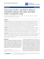
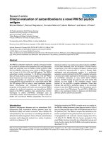
Figure 1
Time course of Ang-2, cytokines, and adhesion molecules after LPS challenge in healthy subjectsTime course of Ang-2, cytokines, and adhesion molecules after LPS
challenge in healthy subjects. (a) Concentrations of circulating angi-
opoietin (Ang)-2 compared with plasma levels of TNF-alpha. IL-6, IL-8,
and C-reactive protein (CRP) after lipopolysaccharide (LPS) challenge
in six healthy volunteers. (b) Concentrations of circulating Ang-2 com-
pared with plasma levels of endothelial adhesion molecules E-selectin,
P-selectin, and inter-cellular adhesion molecule-1 (ICAM-1) after LPS
challenge in six healthy volunteers. Non-parametric analysis of variance
(Friedman's test) with Dunn's test for multiple comparison (two-sided)
was used to demonstrate statistical changes in Ang-2, cytokines, and
adhesion molecules (y-axes denote percentage increase; baseline =
100%).
Available online />Page 5 of 9
(page number not for citation purposes)
Table 2
Time course after LPS challenge in healthy subjects
Time course after LPS challenge
Variables Pre-dose 1 hour 1.5 hours 2 hours 2.5 hours 3.5 hours 4.5 hours 6.5 hours 8 hours 24 hours P value
Systolic BP
(mmHg)
140 ± 14 139 ± 11 152 ± 11 158 ± 18 151 ± 18 142 ± 22 121 ± 18 107 ± 11 104 ± 11 131 ± 11 < 0.0001
Diastolic BP
(mmHg)
74 ± 10 74 ± 7 80 ± 7 77 ± 12 65 ± 12 61 ± 14 54 ± 11 55 ± 8 55 ± 7 66 ± 7 < 0.0001
MAP (mmHg) 96 ± 11 96 ± 9 104 ± 7 104 ± 13 94 ± 14 88 ± 16 77 ± 13 72 ± 8 71 ± 7 88 ± 7 < 0.0001
Heart rate
(beats/minute)
61 ± 16 59 ± 11 78 ± 19 78 ± 18 92 ± 12 98 ± 8 101 ± 8 97 ± 11 96 ± 13 81 ± 16 < 0.0001
HR/MAP index 0.64 ± 0.11 0.62 ± 0.11 0.75 ± 0.17 0.76 ± 0.22 1.01 ± 0.22 1.15 ± 0.26 1.36 ± 0.32 1.36 ± 0.27 1.4 ± 0.22 0.92 ± 0.16 < 0.0001
Body
temperature
(°C)
35.4 ± 0.38 36.2 ± 0.54 36.4 ± 0.87 37.0 ± 1.04 37.6 ± 1.11 38.5 ± 0.66 38.9 ± 0.53 38.14 ± 0.28 37.9 ± 0.19 36.2 ± 0.29 < 0.0001
White blood
count (10
3
/μl)
5.5 ± 0.7 10.9 ± 1.60.03
C-reactive
protein (mg/l)
1.1 ± 2.4 0.9 ± 1.8 - - 1.5 ± 2.4 2.2 ± 2.5 1.1 ± 2.3 1.8 ± 2.8 4.8 ± 2.0 60.0 ± 21.5 < 0.0001
Ang-1 (ng/ml) 67.0 ± 20.7 58.2 ± 24.4 - - 61.2 ± 25.0 54.3 ± 19.5 - 64.9 ± 29.1 60.3 ± 31.4 52.3 ± 21.6 0.053
Ang-2 (ng/ml) 0.57 ± 0.50 0.63 ± 0.20 1.04 ± 0.65 1.63 ± 0.89 2.33 ± 0.69 2.35 ± 1.06 2.42 ± 1.32 2.23 ± 1.18 1.61 ± 1.07 1.51 ± 1.03 < 0.0001
Tie2 (ng/ml) 1.34 ± 0.31 1.23 ± 0.29 1.33 ± 0.32 1.53 ± 0.52 1.23 ± 0.20 1.3 ± 0.16 1.31 ± 0.35 1.4 ± 0.3 1.25 ± 0.32 1.43 ± 0.42 0.085
A non-parametric repeated-measures analysis of variance (Friedman's test) was used to test for significant changes of variables during the time course after LPS challenge (placebo group; n = 6). ANG =
angiopoietin; BP = blood pressure; HR = heart rate; LPS = lipopolysaccharide; MAP = mean arterial pressure.
Critical Care Vol 13 No 3 Kümpers et al.
Page 6 of 9
(page number not for citation purposes)
variable: increase vs. non-increase in Ang-2 during 72 hours)
identified non-survivors with 81% specificity and 80% sensi-
tivity. The positive predictive value was 81% and the negative
predictive value 80%. In patients with increasing Ang-2 values
(during 72 hours), the OR was 18.0 (95% CI = 2.2 to 144.6)
for death during 28-day follow-up (Fisher exact test P =
0.009).
Discussion
The present study dissects the time course of Ang-2 release
after experimental LPS administration in healthy subjects. The
decisive results are: LPS (4 ng/ml) is a triggering factor for
Ang-2 release in vivo; circulating Ang-2 reached peak levels
4.5 hours after LPS infusion; Ang-2 exhibited a kinetic profile
similar to that of TNF-alpha, IL-6, and IL-8, and peaked explic-
itly prior to soluble endothelial-specific adhesion molecules,
and correlated with TNF-alpha levels, soluble E-selectin levels,
and the heart rate/mean arterial pressure index; in septic
patients, not only higher baseline Ang-2 but also a persistent
increase in Ang-2 predicts unfavorable 28-day survival.
Clinical studies of pathophysiological changes during sepsis
are potentially confounded by the absence of a well-defined
onset time of inflammation, by significant co-morbid condi-
tions, as well as by considerable delays from the presumed ini-
tiation of inflammation until study inclusion. Animal studies,
although indispensable for investigating early and late events
during systemic inflammation, are potentially confounded by
major inter-species differences in the sensitivity and immune
response to various types of inflammatory stimuli [24,25]. As
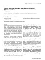
Figure 2
Correlation of Ang-2 with TNF-α, E-selectin and heart rate/mean arterial pressure index after LPS challenge in healthy subjectsCorrelation of Ang-2 with TNF-α, E-selectin and heart rate/mean arterial
pressure index after LPS challenge in healthy subjects. Dot blots show-
ing the correlation between circulating angiopoietin (Ang)-2 and (a)
plasma levels of TNF-alpha, (b) plasma levels of the soluble endothelial
specific adhesion molecule E-selectin, and (c) the heart rate/mean arte-
rial pressure index (HR/MAP index) at 6.5 hours after lipopolysaccha-
ride (LPS) challenge in 21 subjects (placebo (n = 6), and medication
groups: 350 mg (n = 5). 700 mg (n = 6), and 1400 mg (n = 4), respec-
tively). Pearsons correlation coefficient was used after logarithmic
transformation of variables (axes denote percentage increase after log-
arithmic transformation; baseline = 100%).
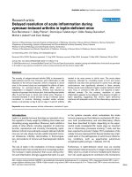
Figure 3
Time course of Ang-2 in critically ill patients with sepsisTime course of Ang-2 in critically ill patients with sepsis. Dot blots
showing the concentration of angiopoietin (Ang)-2 (ng/ml) in 21 septic
on intensive care unit (ICU) admission, 24 hours and 72 hours after
admission, respectively. Of note, median Ang-2 levels increased in non-
survivors (P = 0.019) (continuous line), but remained unchanged in sur-
vivors (P = 0.83) (dotted line) during the time course. Mean Ang-2 level
was higher in non-survivors (filled circles, n = 11) compared with survi-
vors (open circles, n = 10) on admission (P = 0.032), after 24 hours (P
= 0.027), and 72 hours (P = 0.008) (two-sided Mann-Whitney test).
Available online />Page 7 of 9
(page number not for citation purposes)
already indicated, the present study is a re-analysis of blood
samples from a placebo-controlled interventional trial on phar-
macologic p38 MAP kinase inhibition in endotoxemia. The
design of this trial enabled us to investigate the time course of
Ang-2 release in humans in a highly standardized experimental
model with a graded inflammatory response [21,22].
After LPS infusion, peak Ang-2 levels (2.4 ± 0.5 ng/ml) are
four-fold lower than Ang-2 levels in critically ill patients at the
ICU (9.8 ± 3.2 ng/ml). Emerging data from our group, as well
as a recent study by Siner and colleagues [16] suggest the
notion that survival is good in critically ill patients with low Ang-
2 (< 7 to 8 ng/ml), whereas outcome is explicitly worse above
this threshold. Indeed, septic patients with circulating Ang-2
levels below 5.9 and 5.0 ng/ml (admission and 72 hours,
respectively) identified patients with good 28-day survival in
the present study. Compared with the experimental endotox-
emia model (single dose of LPS), the inflammatory stimuli in
critical illness are probably more intense, often persistent, and
multiple in nature. Thus, a rather low but significant Ang-2 peak
level of 2.4 ng/ml (four-fold vs. baseline) during experimental
endotoxemia is probably adequate and well in line with the
aforementioned data. Although we cannot rule out loss of Ang-
2 immunoreactivity due to deep-freeze storage for several
years, in our experience this phenomenon is negligible [26].
Ang-2 exhibited a kinetic profile that is similar to the early pro-
inflammatory cytokines TNF-alpha, IL-6, and IL-8. In the
present study, TNF-alpha level increased somewhat earlier
than Ang-2 levels did. As previously shown, the release of
TNF-alpha and several other cytokines during human endotox-
emia is blocked by p38 MAP kinase inhibition in a dose-
dependent manner [22]. In the present study, p38 MAP kinase
inhibition blocked Ang-2 release in a similar fashion. In addi-
tion, Ang-2 correlated well with TNF-alpha throughout the time
course after LPS infusion. This implies that either the Ang-2
release is mediated by TNF-alpha, or that a p38-MAP kinase-
dependent upstream signaling pathway controls both TNF-
alpha and Ang-2 release. Well in line with this data, Orfanos
and colleagues reported a strong relationship of Ang-2 with
TNF-alpha in critically ill patients, suggesting that the latter
may participate in the regulation of Ang-2 production in sepsis
[27]. In contrast, Fiedler and colleagues showed that even
high concentrations of TNF-alpha are not sufficient to induce
Ang-2 release from human umbilical vein cells in vitro [28].
However, we cannot exclude that there is an independent
route with a slower signaling pathway, and the fact that TNF-
alpha preceded Ang-2 release does not prove causality.
Expression of endothelial adhesion molecules such as E-
selectin, VCAM-1, and ICAM-1, are a consistent feature of
sepsis [29,30]. As a functional antagonist of Ang-1/Tie2 sign-
aling, Ang-2 promotes up-regulation of endothelial adhesion
molecules (i.e. endothelial activation), by sensitizing endothe-
lial cells toward cytokine-induced adhesion molecule expres-
sion. Consistently, firm leukocyte adhesion and subsequent
transmigration is almost absent during experimental sepsis in
Ang-2 deficient animals [3,7,12]. In the present study, Ang-2
levels increased and peaked explicitly prior to soluble endothe-
lial-specific adhesion molecules E-selectin and ICAM-1. Fur-
thermore, soluble E-selectin correlated well with circulating
Ang-2 throughout the time course. This temporal sequence is
in line with the concept proposed by Fiedler and colleagues
[7] that endothelial-cell activation might indeed represent a
predominantly Ang-2 driven process in vivo.
Although (circulating) Ang-2 has a significant adverse effect
on pulmonary vascular barrier properties in sepsis [3,4] its role
in extra-pulmonary endothelial activation and systemic loss of
barrier function is less well defined. However, Ang-1 increases
arteriolar vasoconstriction to phenylephrine in the presence of
LPS in vitro [31] and preserves blood pressure and cardiac
output in septic rats in vivo [10]. Indeed, we found a close cor-
relation between circulating Ang-2 and the heart rate/mean
arterial pressure as a surrogate marker for hemodynamic com-
promise in the present study. Although this observation does
not prove causality, it is in line with a significant association of
circulating Ang-2 levels with MAP, and vasopressor require-
ment in a large cohort of critically ill patients with acute kidney
injury (Kümpers P, Hafer C, David S, Kielstein JT, Hecker H,
Lukasz A, Fliser D, Haller H, Faulhaber-Walter R, unpublished
data).
Over the past few years, it has been appreciated that multiple
components of WPB, such as Ang-2, P-selectin, and IL-8, are
co-stored with von Willebrand Factor (vWF), the major constit-
uent of WPBs [7]. It has been shown that storage of Ang-2
and P-selectin in WPB is mutually exclusive. Interestingly, we
detected a selective release of the aforementioned WPB
stored mediators: Ang-2 is released first, then IL-8, and P-
selectin is not released at all. The phenomenon that some
components are selectively released from WPB has already
been show in vitro [32] and deserves further attention in in
vitro studies of differential regulation of WPB exocytosis.
As a prepackaged constituent of WPB, it is not surprising that
Ang-2 levels on admission are increased in response to early
endothelial activation in critically ill patients [15,16,20]. In line
with this finding, high Ang-2 levels on admission are associ-
ated with unfavorable 28-day survival. However, it still
remained unanswered whether Ang-2 levels either decline or
increase during the course of sepsis, as recently summarized
by Giuliano and Wheeler [33].
In vitro intracellular Ang-2 pools are rapidly replenished after
stimulated depletion with a protein kinase C activator (phorbol
12-myristate 13-acetate) [28]. Furthermore, LPS administra-
tion has been shown to increase Ang-2 in a murine sepsis
model [34]. Based on available data we hypothesized that lev-
els of circulating Ang-2 might even increase in patients with
Critical Care Vol 13 No 3 Kümpers et al.
Page 8 of 9
(page number not for citation purposes)
persistent inflammation and/or clinical deterioration. Indeed, in
our cohort of septic patients, non-survivors, not only presented
with higher admission Ang-2 levels but also showed a signifi-
cant increase in Ang-2 during a period of 72 hours. It is tempt-
ing to speculate that LPS regulates both, the release of Ang-2
from WPB and the transcriptional induction at the same time.
However, additional pre-clinical studies and also clinical stud-
ies are urgently needed to clarify this issue.
Our study has several limitations. The human endotoxin model
carries a risk of inappropriate extrapolation from experimental
findings to the clinical setting. However, this is the only model
that renders an opportunity to study the early mechanisms of
endothelial activation during a time course in human subjects.
Because vWF (as the major constituent of WPBs) was not
determined in this study and citrated plasma samples from the
original trial were not available for re-evaluation, we cannot
exclude that Ang-2 might have derived from endothelial cells
exclusively. At least murine macrophages seem to express
smaller quantities of Ang-2, but this has not been tested in
experimental sepsis [35]. However, the time course of Ang-2
release in the present study was well in line with that of vWF
release during endotoxemia [36]. Further, the sample size of
the septic cohort was small. Thus, ROC procedures and 2 ×
2 tables should be interpreted with caution. However, Fisher's
exact test (still appropriate even with small sample size) con-
firmed the significance of our findings. Finally, elevated circu-
lating Ang-2 is not an exclusive feature of endotoxemia and
sepsis, but rather reflects endothelial activation and vascular
damage in diseases that share a significant inflammatory
endothelial phenotype [17,18,37].
Conclusions
We could show for the first time that LPS administration is a
triggering factor for Ang-2 release in men. Circulating Ang-2
appears in the systemic circulation during experimental human
endotoxemia in a distinctive temporal sequence and correlates
with TNF-alpha and E-selectin levels. In addition, not only
higher baseline Ang-2 concentrations, but also a persistent
increase in Ang-2 during the early course identifies septic
patients with unfavorable outcomes.
Competing interests
The authors declare that they have no competing interests.
Authors' contributions
PK had the initial idea, supervised the project, analyzed the
data, prepared the figures and wrote the manuscript. MM
supervised the project, analyzed the data, made figures and
contributed to the manuscript. SD contributed to the idea,
identified patients, participated in analysis of the results and
contributed to the manuscript. GM participated in analysis of
the results and contributed to the manuscript. JB performed
the multiplex assays and reviewed the manuscript. AL identi-
fied patients, established and performed the immunoassays
and reviewed the manuscript. FB collected and analyzed
patient data and reviewed the manuscript. HH supervised the
project and reviewed the manuscript. JZ designed and super-
vised the entotoxemia model, enrolled patients, analyzed the
data and contributed to the manuscript. PK and MM contrib-
uted equally to the work and are both considered first authors.
Acknowledgements
We would like to thank Dr Stefan Stuth for extensive monitoring of the
patients and Dr Ulrike Kümpers for critical discussion and proofreading
of the manuscript. We also would like to thank Rianne M. Jongman
(UMCG, Groningen) for excellent technical assistance.
References
1. Vincent JL, De Backer D: Microvascular dysfunction as a cause
of organ dysfunction in severe sepsis. Crit Care 2005, 9(Suppl
4):S9-12.
2. van Meurs M, Kümpers P, Ligtenberg JJ, Mertens JH, Molema G,
Zijlstra JG: Bench-to-bedside review: Angiopoietin signalling in
critical illness: a future target? Crit Care 2009, 13:207.
3. Parikh SM, Mammoto T, Schultz A, Yuan HT, Christiani D,
Karumanchi SA, Sukhatme VP: Excess circulating angiopoietin-
2 may contribute to pulmonary vascular leak in sepsis in
humans. PLoS Med 2006, 3:e46.
4. Heijden M van der, Nieuw Amerongen GP, Koolwijk P, van Hins-
bergh VW, Groeneveld AB: Angiopoietin-2, permeability
oedema, occurrence and severity of ALI/ARDS in septic and
non-septic critically ill patients. Thorax 2008, 63:903-909.
5. Fiedler U, Krissl T, Koidl S, Weiss C, Koblizek T, Deutsch U, Mar-
tiny-Baron G, Marme D, Augustin HG: Angiopoietin-1 and angi-
opoietin-2 share the same binding domains in the Tie-2
receptor involving the first Ig-like loop and the epidermal
growth factor-like repeats. J Biol Chem 2003, 278:1721-1727.
6. Wong AL, Haroon ZA, Werner S, Dewhirst MW, Greenberg CS,
Peters KG: Tie2 expression and phosphorylation in angiogenic
and quiescent adult tissues. Circ Res 1997, 81:567-574.
7. Fiedler U, Augustin HG: Angiopoietins: a link between angio-
genesis and inflammation. Trends Immunol 2006, 27:552-558.
8. Thurston G, Rudge JS, Ioffe E, Zhou H, Ross L, Croll SD, Glazer
N, Holash J, McDonald DM, Yancopoulos GD: Angiopoietin-1
protects the adult vasculature against plasma leakage. Nat
Med 2000, 6:460-463.
9. Scharpfenecker M, Fiedler U, Reiss Y, Augustin HG: The Tie-2 lig-
and angiopoietin-2 destabilizes quiescent endothelium
through an internal autocrine loop mechanism. J Cell Sci
2005, 118:771-780.
10. Witzenbichler B, Westermann D, Knueppel S, Schultheiss HP,
Tschope C: Protective role of angiopoietin-1 in endotoxic
shock. Circulation 2005, 111:97-105.
11. Roviezzo F, Tsigkos S, Kotanidou A, Bucci M, Brancaleone V, Cir-
ino G, Papapetropoulos A: Angiopoietin-2 causes inflammation
in vivo by promoting vascular leakage. J Pharmacol Exp Ther
2005, 314:738-744.
Key messages
• LPS administration is a triggering factor for Ang-2
release in human endotoxemia.
• Circulating Ang-2 appears in the systemic circulation
during experimental human endotoxemia in a distinctive
temporal sequence and correlates with TNF-alpha and
E-selectin levels.
• High Ang-2 concentrations at baseline, as well as a per-
sistent increase in Ang-2 during the early course identi-
fies septic patients with unfavorable outcome.
Available online />Page 9 of 9
(page number not for citation purposes)
12. Fiedler U, Reiss Y, Scharpfenecker M, Grunow V, Koidl S,
Thurston G, Gale NW, Witzenrath M, Rosseau S, Suttorp N,
Sobke A, Herrmann M, Preissner KT, Vajkoczy P, Augustin HG:
Angiopoietin-2 sensitizes endothelial cells to TNF-alpha and
has a crucial role in the induction of inflammation. Nat Med
2006, 12:235-239.
13. Thurston G, Suri C, Smith K, McClain J, Sato TN, Yancopoulos
GD, McDonald DM: Leakage-resistant blood vessels in mice
transgenically overexpressing angiopoietin-1. Science 1999,
286:2511-2514.
14. Gallagher DC, Parikh SM, Balonov K, Miller A, Gautam S, Talmor
D, Sukhatme VP: Circulating angiopoietin 2 correlates with
mortality in a surgical population with acute lung injury/adult
respiratory distress syndrome. Shock 2008, 29:656-661.
15. Giuliano JS Jr, Lahni PM, Harmon K, Wong HR, Doughty LA, Car-
cillo JA, Zingarelli B, Sukhatme VP, Parikh SM, Wheeler DS:
Admission angiopoietin levels in children with septic shock.
Shock 2007, 28:650-654.
16. Siner JM, Bhandari V, Engle KM, Elias JA, Siegel MD: Elevated
serum angiopoietin 2 levels are associated with increased
mortality in sepsis. Shock 2009, 31:348-353.
17. Kumpers P, Koenecke C, Hecker H, Hellpap J, Horn R, Verhagen
W, Buchholz S, Hertenstein B, Krauter J, Eder M, David S, Gohring
G, Haller H, Ganser A: Angiopoietin-2 predicts disease-free
survival after allogeneic stem cell transplantation in patients
with high-risk myeloid malignancies. Blood 2008,
112:2139-2148.
18. Kumpers P, David S, Haubitz M, Hellpap J, Horn R, Brocker V,
Schiffer M, Haller H, Witte T: The Tie2 receptor antagonist Angi-
opoietin-2 facilitates vascular inflammation in Systemic Lupus
Erythematosus. Ann Rheum Dis 2008 in press. doi:10.1136/
ard.2008.094664
19. Lukasz A, Hellpap J, Horn R, Kielstein JT, David S, Haller H, Kump-
ers P: Circulating angiopoietin-1 and angiopoietin-2 in critically
ill patients: development and clinical application of two new
immunoassays. Crit Care 2008, 12:R94.
20. Kumpers P, Lukasz A, David S, Horn R, Hafer C, Faulhaber-Walter
R, Fliser D, Haller H, Kielstein JT: Excess circulating angiopoie-
tin-2 is a strong predictor of mortality in critically ill medical
patients. Crit Care 2008, 12:R147.
21. Fijen JW, Tulleken JE, Kobold AC, de Boer P, Werf TS van der,
Ligtenberg JJ, Spanjersberg R, Zijlstra JG: Inhibition of p38
mitogen-activated protein kinase: dose-dependent suppres-
sion of leukocyte and endothelial response after endotoxin
challenge in humans. Crit Care Med 2002,
30:841-845.
22. Fijen JW, Zijlstra JG, De Boer P, Spanjersberg R, Tervaert JW, Van
Der Werf TS, Ligtenberg JJ, Tulleken JE: Suppression of the clin-
ical and cytokine response to endotoxin by RWJ-6 a p38
mitogen-activated protein-kinase inhibitor, in healthy human
volunteers. Clin Exp Immunol 2001, 124:16-20.
23. StatPages [ />]
24. Dyson A, Singer M: Animal models of sepsis: why does preclin-
ical efficacy fail to translate to the clinical setting? Crit Care
Med 2009, 37:S30-S37.
25. Rittirsch D, Hoesel LM, Ward PA: The disconnect between ani-
mal models of sepsis and human sepsis. J Leukoc Biol 2007,
81:137-143.
26. David S, Kümpers P, Lukasz A, Kielstein JT, Haller H, Fliser D: Cir-
culating angiopoietin-2 in essential hypertension: relation to
artherosclerosis, vascular information, and treatment with
olmesartan/pravastatin. J Hypertens 2009 in press.
27. Orfanos SE, Kotanidou A, Glynos C, Athanasiou C, Tsigkos S,
Dimopoulou I, Sotiropoulou C, Zakynthinos S, Armaganidis A,
Papapetropoulos A, Roussos C: Angiopoietin-2 is increased in
severe sepsis: correlation with inflammatory mediators. Crit
Care Med 2007, 35:199-206.
28. Fiedler U, Scharpfenecker M, Koidl S, Hegen A, Grunow V,
Schmidt JM, Kriz W, Thurston G, Augustin HG: The Tie-2 ligand
angiopoietin-2 is stored in and rapidly released upon stimula-
tion from endothelial cell Weibel-Palade bodies. Blood 2004,
103:4150-4156.
29. Aird WC: The role of the endothelium in severe sepsis and
multiple organ dysfunction syndrome. Blood 2003,
101:3765-3777.
30. Hein OV, Misterek K, Tessmann JP, van DV, Krimphove M, Spies
C: Time course of endothelial damage in septic shock: predic-
tion of outcome. Crit Care 2005, 9:R323-R330.
31. Hall E, Brookes ZL: Angiopoietin-1 increases arteriolar vaso-
constriction to phenylephrine during sepsis. Regul Pept 2005,
131:34-37.
32. Babich V, Meli A, Knipe L, Dempster JE, Skehel P, Hannah MJ,
Carter T: Selective release of molecules from Weibel-Palade
bodies during a lingering kiss. Blood 2008, 111:5282-5290.
33. Giuliano JS Jr, Wheeler DS: Excess circulating angiopoietin-2
levels in sepsis: harbinger of death in the intensive care unit?
Crit Care 2009, 13:114.
34. Mofarrahi M, Nouh T, Qureshi S, Guillot L, Mayaki D, Hussain SN:
Regulation of angiopoietin expression by bacterial lipopoly-
saccharide. Am J Physiol Lung Cell Mol Physiol 2008,
294:L955-L963.
35. Hubbard NE, Lim D, Mukutmoni M, Cai A, Erickson KL: Expres-
sion and regulation of murine macrophage angiopoietin-2.
Cell Immunol 2005, 234:102-109.
36. Jilma B, Blann A, Pernerstorfer T, Stohlawetz P, Eichler HG, Von-
drovec B, Amiral J, Richter V, Wagner OF: Regulation of adhe-
sion molecules during human endotoxemia. No acute effects
of aspirin. Am J Respir Crit Care Med 1999, 159:857-863.
37. Kumpers P, Hellpap J, David S, Horn R, Leitolf H, Haller H, Haubitz
M: Circulating angiopoietin-2 is a marker and potential media-
tor of endothelial cell detachment in ANCA-associated vascu-
litis with renal involvement. Nephrol Dial Transplant 2009 in
press.