Báo cáo y học: "Infra-red thermometry: the reliability of tympanic and temporal artery readings for predicting brain temperature after severe traumatic brain injur" pot
Bạn đang xem bản rút gọn của tài liệu. Xem và tải ngay bản đầy đủ của tài liệu tại đây (241.16 KB, 8 trang )
Open Access
Available online />Page 1 of 8
(page number not for citation purposes)
Vol 13 No 3
Research
Infra-red thermometry: the reliability of tympanic and temporal
artery readings for predicting brain temperature after severe
traumatic brain injury
Danielle Kirk
1
, Timothy Rainey
1
, Andy Vail
2
and Charmaine Childs
1
1
Brain Injury Research Group, School of Translational Medicine, University of Manchester, Salford Royal Foundation Trust, Stott Lane, Salford, M6
8HD UK
2
Biostatistics Group, University of Manchester, Salford Royal Foundation Trust, Stott Lane, Salford, M6 8HD UK
Corresponding author: Charmaine Childs,
Received: 12 Mar 2009 Revisions requested: 17 Apr 2009 Revisions received: 8 May 2009 Accepted: 27 May 2009 Published: 27 May 2009
Critical Care 2009, 13:R81 (doi:10.1186/cc7898)
This article is online at: />© 2009 Kirk et al.; licensee BioMed Central Ltd.
This is an open access article distributed under the terms of the Creative Commons Attribution License ( />),
which permits unrestricted use, distribution, and reproduction in any medium, provided the original work is properly cited.
Abstract
Introduction Temperature measurement is important during
routine neurocritical care especially as differences between
brain and systemic temperatures have been observed. The
purpose of the study was to determine if infra-red temporal
artery thermometry provides a better estimate of brain
temperature than tympanic membrane temperature for patients
with severe traumatic brain injury.
Methods Brain parenchyma, tympanic membrane and temporal
artery temperatures were recorded every 15–30 min for five
hours during the first seven days after admission.
Results Twenty patients aged 17–76 years were recruited.
Brain and tympanic membrane temperature differences ranged
from -0.8 °C to 2.5 °C (mean 0.9 °C). Brain and temporal artery
temperature differences ranged from -0.7 °C to 1.5 °C (mean
0.3 °C). Tympanic membrane temperature differed from brain
temperature by an average of 0.58 °C more than temporal artery
temperature measurements (95% CI 0.31 °C to 0.85 °C, P <
0.0001).
Conclusions At temperatures within the normal to febrile range,
temporal artery temperature is closer to brain temperature than
is tympanic membrane temperature.
Introduction
Temperature measurement is important during routine neuro-
critical care. There is retrospective evidence that moderate to
high body temperature is an independent predictor of inten-
sive care unit (ICU) and hospital length of stay and leads to a
higher mortality and worse outcome in a mixed population of
neurosurgical ICU patients [1]. Recent prospective data of
brain temperature and outcome in a relatively homogenous
population of patients with severe traumatic brain injury (TBI)
show that outcome is worse at temperature extremes (high
and low) [2]. Current opinion favours treatment of pyrexia in
patients with neurological injury. However, there are no pub-
lished guidelines or recommendations for the management of
raised temperature [3]. The focus of the most recent (2007)
Brain Trauma Foundation (BTF) guidelines for the manage-
ment of temperature after human TBI was on the management
of hypothermia (a treatment which is limited to a level III recom-
mendation only [4]). Popular opinion has, therefore, consid-
ered controlled normothermia as a clinical therapeutic option,
but whether normothermia has the potential for therapeutic
benefit for the TBI patient remains untested.
As body core temperature frequently dissociates from brain
temperature [5,6] there remains some doubt about the reliabil-
ity of traditional body temperature methods for brain tempera-
ture estimation. While measurement of deep body core
temperature using traditional monitoring sites such as rectum,
oesophagus, urinary bladder or pulmonary artery might be
expected to provide a reasonable surrogate for brain temper-
ature during neurocritical care, we have shown that tympanic
membrane temperature is currently the most popular, non-sur-
gical method of brain temperature estimation in UK neurosur-
AIS: abbreviated injury scale; BTF: Brain Trauma Foundation; CBF: cerebral blood flow; CT: computed tomography; ED: emergency department;
GCS: Glasgow coma scale; ICP: intracranial pressure; ICU: intensive care unit; TBI: traumatic brain injury.
Critical Care Vol 13 No 3 Kirk et al.
Page 2 of 8
(page number not for citation purposes)
gical practice [7]. Thus, most (70%) neurosurgical centres do
not measure 'true' body core temperature, rather they employ
a measurement method about which serious doubts regarding
accuracy are documented [8-10].
A new, non-invasive method for core temperature estimation is
now available which captures infra-red heat energy from the
skin overlying the course of the temporal artery [11]. The tech-
nique is quick; the instrument is easy to clean and is relatively
inexpensive. The aim of the study was to determine if infra-red
temporal artery thermometry provides a better performance as
a 'surrogate' for brain temperature than the most common
method (tympanic temperature) used in UK neurocritical care.
Materials and methods
The study was approved by the local research ethics commit-
tee (reference number 06/Q1406/115). Approval to under-
take the study was obtained from the patient's spouse, relative
or partner before measurements were made.
Patients
All patients aged 16 years or above, who were admitted to our
16 bed, level 3, university teaching hospital ICU within 24
hours of severe TBI were eligible for recruitment to the study.
Patients were admitted either as direct referrals from the emer-
gency department (ED) or as tertiary referrals from EDs of
other hospitals within the greater Manchester region. All the
patients were sedated, intubated and mechanically ventilated;
all had an intra or extra-axial lesion on computed tomography
(CT), with or without systemic trauma and a Glasgow Coma
Scale (GCS) of eight or less on admission to the ICU. The
patients were treated in accordance with local neurointensive
care guidelines to maintain cerebral perfusion pressure at 60
mmHg or higher and intracranial pressure (ICP) below 20
mmHg. To manage raised ICP, patients were positioned at a
30° angle head up and received sedation, analgesia, neu-
romuscular blockade and osmotherapy with mannitol (0.5 g/
kg) as required.
In the event of a rise in ICP in excess of 20 mmHg, refractory
to the standard treatment (including surgical removal of hae-
matoma), a barbiturate coma was induced.
Guidelines for the management of body temperature have
been developed to form a clinical protocol. At our centre, tem-
perature management is directed towards maintenance of nor-
mothermia; therapeutic hypothermia is not included as a part
of routine neurocritical care. Briefly, the temperature manage-
ment protocol involves a four level, step-up method for control
of body temperature beginning with antipyretic drugs (level 1)
with the addition of surface cooling (level 2) neuromuscular
blockade (level 3) and intragastric cooling with ice cold water
lavage (level 4) with an intention to achieve a target brain tem-
perature of 37°C (normothermia). In step 2 of our body surface
cooling protocol we applied wet (hand hot) cotton sheets to
the patient's body (from chest to mid thigh) and renewed the
sheets on an hourly basis when starting to dry. In step 4 of the
protocol, patients received 500 ml of iced water into the stom-
ach via a nasogastric tube and the residual volume was aspi-
rated after 10 minutes. This procedure was repeated every 15
minutes for a maximum of five hours with regular clinical
assessment of blood sugar and electrolytes during gastric lav-
age. Enteral feeding was resumed at the end of level 4 cooling.
Assessment of injury severity was made from the information
obtained in the patient's case notes. Details of all the injuries
sustained at the time of the accident were noted. The abbrevi-
ated injury scale (AIS) [12,13] was used to 'grade' the severity
of trauma to the head. The AIS for the head region (Table 1)
includes trauma to the brain and cranium. Patients were eligi-
ble for recruitment if brain temperature was being recorded
during routine neurocritical care.
Temperature measurement
Intraparenchymal temperature
Brain temperature was measured continuously using a com-
bined ICP/temperature probe (Neurovent-PTemp™, Raumedic
AG, Münchberg, Germany). Although no published data are
available to show the precision of the Neurovent-PTemp,
recent unpublished data by the author indicates that sensor
performance exceeds the manufacturer's stated accuracy.
The sensor was inserted into parenchyma, under aseptic con-
ditions at the bedside or during emergency neurosurgery.
Using aseptic techniques, the sensor tip was positioned 3 to
4 cm into deep white matter of the right frontal lobe, via a
standard burr hole. Temperature measurements (alongside
other routine vital signs and clinical parameters) were dis-
played in real-time via a patient data acquisition system (Mar-
quette Electronics, Milwaukee, WI, USA), updated and stored
to a bedside computer at 10-minute intervals.
Infra-red Thermometry
Infra-red techniques were used to obtain body temperature
using either an established site and method (tympanic mem-
brane thermometry) or a novel infra-red method (temporal
artery thermometry). Both methods use a non-contact temper-
ature measurement device to detect the infra-red energy emit-
ted from a specific body site at temperatures above absolute
zero (-273°C).
Measurement of tympanic membrane temperature was made
using a Core-Check thermometer (Model 2090 IVAC Corpo-
ration, San Diego, California, USA). To ensure that the 'lens'
was directed at the tympanum, the pinna was gently held and
the thermometer inserted into the external auditory meatus,
turned upwards and directed towards the eye. The probe
remained briefly in this position until the machine 'bleeped' to
signal a temperature reading.
Available online />Page 3 of 8
(page number not for citation purposes)
To obtain a temporal artery temperature reading, a small, hand-
held infra-red scanner (Model TAT 5000, Exergen, Watertown,
MA, USA) was used. The temporal artery thermometer [10]
incorporates an infra-red sensor which is placed on the skin at
the centre of the patient's forehead. The thermometer housing
includes a measurement button which, when pressed, allows
measurement to begin. The infra-red sensor (at the head of the
hand-held thermometer) is then swept horizontally along the
forehead to the hairline (crossing part of the course of the tem-
poral artery). Keeping the button pressed down, the sensor is
then removed briefly from the skin and placed behind the ear-
lobe to touch the skin overlying the mastoid process and in the
manner recommended in the product guidelines. This skin tap
is used to control for evaporative cooling of the forehead if the
patient is sweating. If the temperature at the mastoid is greater
than the measurement over the temporal artery then the tem-
perature at the mastoid process will be recorded [11]. On
release of the button, a digital temperature measurement is
displayed. The infra-red thermometer provides an estimate of
body core temperature using a proprietary algorithm which
incorporates a factor compensating for measured ambient
temperature [11]. In each patient a measurement of brain tem-
perature, tympanic membrane temperature and temporal
artery temperature was recorded as a temperature 'triplet'.
Each measurement triplet was made every 15 to 30 minutes
for a total duration of five hours.
Statistics
To undertake this descriptive study, a target of 20 patients
with severe TBI was set as a pragmatic sample size to test
whether temporal artery temperature performs better to pre-
dict brain temperature after TBI than tympanic membrane tem-
perature does. The Bland and Altman method [14] was used
to display the spread of data points. Using standard meta-anal-
ysis calculations, the absolute difference between each brain
and tympanic temperature pair and the corresponding brain
and temporal artery pair was calculated.
Table 1
Patient demographics: injury aetiology, brain pathology diagnosis and injury severity scores
Patient Aetiology Brain Pathology Measurements made on
(day after TBI)
Number of hours studied on ICU
†
n AIS ISS
AFall ICH 3 5 13417
B Fall Bilateral frontal haematoma 5, 6 5 17 5 30*
CFall SDH 3, 5 5 14416
DFall DAI 4 5 11526
E RTA Temporal contusions 6, 7 5 20 4 16
F RTA DAI 2, 3 5 26 5 45*
GFall ICH 2 5 12525
HAssault SDH 5, 6 5 23417
I Assault Temporal contusions 2 5 9 3 18*
JFall SDH 2 5 10416
KRTA SDH 3 5 11416
L Fall SDH, SAH, cerebral contusions 4 5 21 5 42*
M RTA SDH, cerebral oedema 4 5 21 4 24*
N RTA Cerebral oedema, SAH, contusions 5 5 21 4 21*
O Fall Cerebral contusions 4 5 21 4 16
PRTA DAI 2 5 19566*
Q RTA Cerebral oedema 4 5 21 3 17*
R Fall EDH, cerebral oedema, cerebral contusions 3 5 21 4 16
S RTA SDH, cerebral contusions 2 5 21 4 41*
T RTA SAH, cerebral contusions 2, 3 5 21 4 18*
* significant systemic trauma,
†
number of measurements made. AIS 1 = minor injury; AIS 5 = the most severe of survivable injuries.
AIS = abbreviated injury scale; DAI = diffuse axonal injury; EDH = extradural haematoma; ICH = intracerebral haemorrhage; ICU = intensive care
unit; ISS = injury severity score; RTA = road traffic accident; SAH = subarachnoid haemorrhage; SDH = subdural haematoma; TBI = traumatic
brain injury.
Critical Care Vol 13 No 3 Kirk et al.
Page 4 of 8
(page number not for citation purposes)
Results
Twenty patients (16 male, 4 female) aged 17 to 76 years
(median 33 years) with severe TBI (median AIS 4) due to road
traffic accidents (n = 9), falls (n = 9) or assault (n = 2) were
each studied over the course of five hours during the first two
to seven days (median three days) after injury. The ambient
temperature of the ICU during the study ranged from 22.5 to
23.6°C (median 23.1°C). Between 9 to 26 (median 20) tem-
perature measurement 'triplets' were obtained predominately
during the period 10.00 to 15.00 hours on the day of the
study. Three (15%) patients had diffuse axonal injury, 15
(75%) had haemorrhage and contusions, one patient (5%)
had a bilateral frontal haematoma and one patient (5%) had
cerebral oedema. Ten (50%) patients had significant systemic
trauma in addition to severe brain damage (Injury Severity
Score 17 to 66, median 27). Two sets of readings were pro-
vided by 353 temperature 'triplets': brain and tympanic mem-
brane readings, and brain and temporal artery readings. The
temperature measurements were made by one member of the
research team (DK) only. Two patients (B and H) received sur-
face cooling (level 2). Differences between brain and tympanic
membrane temperature readings ranged from -0.8°C to 2.5°C
and for brain and temporal artery temperature readings, -0.7°C
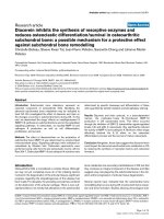
to 1.5°C (Figure 1). The mean difference between brain tem-
perature and tympanic membrane temperature was 0.91°C
and the mean difference between brain temperature and tem-
poral artery temperature was 0.26°C.
The absolute temperature difference between brain tempera-
ture and tympanic membrane temperature pairs and brain tem-
perature and temporal artery temperature pairs for each
patient studied is given in Figure 2. This graph shows which of
the two body temperatures agrees most closely with brain
temperature. If both of the measurement techniques (temporal
artery and tympanic membrane readings) were in agreement
with brain temperature it would be reasonable to expect that
Figure 1
Bland and Altman plot graphs of the difference between brain temperature and its respective temperature pair (comparator) versus the average of the temperature pairBland and Altman plot graphs of the difference between brain temperature and its respective temperature pair (comparator) versus the average of
the temperature pair. (a) Differences between brain and tympanic temperature readings. (b) Differences between brain and temporal artery readings
(n = 353 data sets). The data points for each of 20 patients are distinguished by letters of the alphabet (A-T; Table 1).
Available online />Page 5 of 8
(page number not for citation purposes)
there should be no difference between the temporal artery
thermometer reading and the tympanic membrane reading to
determine brain temperature; therefore, the mean difference
between the two pairs of temperature differences should be
zero. Using a standard meta-analysis method to describe
these data, the mean weighted difference between the two
infra-red measurement techniques (using absolute tempera-
ture values and ignoring the sign) is 0.58°C (Figure 2). This
implies that overall, tympanic membrane temperature differs, in
either direction, from brain temperature by an average of 0.6°C
(95% confidence interval (CI) = 0.31 to 0.85, P < 0.0001)
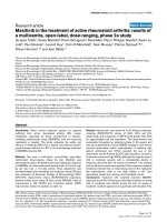
more than temporal artery temperature does. Figure 3 shows
a typical example of the temporal pattern of brain, tympanic
membrane and temporal artery temperature for one patient
(patient R; Figure 2).
In this study, two patients were noted to be sweating on the
forehead during the measurement period. In patient B, onset
of sweating led to a 1°C increase in temporal artery tempera-
ture without a corresponding change in tympanic or brain tem-
perature (or rectal temperature, data not shown). A similar,
approximately 1°C rise in temporal artery temperature was
noted once again (patient H) over a similar time period and
again without corresponding effects on brain or tympanic (or
rectal) temperature.
Figure 2
Weighted mean differences between brain and tympanic membrane temperatures and brain and temporal artery temperatures for each patient stud-ied, with 95% confidence intervalsWeighted mean differences between brain and tympanic membrane temperatures and brain and temporal artery temperatures for each patient stud-
ied, with 95% confidence intervals. A positive value indicates that on average, temporal artery temperature is closer to brain temperature than tym-
panic membrane readings. |T
br
- T
tymp
| - |T
br
- T
t.a
| denotes the difference between absolute temperature differences of the respective brain-body
temperature pairs. Data points to the right of the vertical line indicate that differences between brain and tympanic temperature readings are greater
than for brain and temporal artery readings, i.e. the arrow to the right of the vertical line indicates that readings favour temporal artery temperature.
The summary symbol (ᮀ) denotes the overall average by meta-analysis. T
br
= brain temperature; T
t.a
= temporal artery temperature; T
tymp
= tympanic
membrane temperature.
Critical Care Vol 13 No 3 Kirk et al.
Page 6 of 8
(page number not for citation purposes)
Discussion
In this study we found that on average, for normothermic and
febrile TBI patients, temporal artery temperature was closer to
brain temperature than tympanic temperature was, by approx-
imately 0.6°C. This suggests that within physiological and
fever-range temperatures there is greater accuracy and less
variability in the estimation of brain temperature using the tem-
poral artery thermometer. As the patients in this study did not
undergo therapeutic hypothermia, these results cannot be
extrapolated to below-normal temperature readings.
Although studies in rodents clearly show that raised tempera-
ture leads to an increase in infarct volume after experimental
cerebral ischaemia [15], there are no comparable data to con-
firm that raised temperature causes a worse outcome in
patients with stroke or severe head injury. Even so, the current
weight of opinion for brain injured patients is that a rise in body
temperature (and by assumption, a rise in neuronal tempera-
ture) is damaging and should be treated [16]. There are, how-
ever, important gaps in our knowledge about the impact of
raised (or below-normal) temperature on outcome in patients
with brain damage. For example, it has not yet been estab-
lished: if and how fever-range temperatures worsen outcome;
whether control of fever improves outcome; or if hypothermia
is appropriate for neurological patients. With regard to the role
of fever-range temperatures on outcome after TBI, two recent
studies from our centre [2] (and R.H. Sacho, unpublished MD
thesis) suggest that a modest early fever of 39°C or below is
not deleterious to outcome at either three or six months. If tem-
perature does play a key role in influencing patient outcome in
brain damaged patients, accurate measurement of 'at-risk' tis-
sue (i.e. brain) must be a priority. However, it is not always pos-
sible to measure brain temperature directly. Furthermore, we
must recognise a limitation to any clinical investigation involv-
ing brain temperature measurement because it is difficult to
measure brain temperature directly at more than one site. We
can not therefore be certain that measurements made in one
focal area (e.g. uninjured tissue) represent the temperature in
other brain regions (e.g. in areas of contusion, haemorrhage
and ischaemia).
The search for a 'surrogate' non-invasive body site, which best
reflects brain temperature remains of interest to clinicians.
Tympanic membrane temperature is currently the most com-
monly used, non-invasive method of brain temperature estima-
tion in the UK [7]; however, recent published data has raised
concerns about its accuracy, the cause of which may be due
to measurement error, user technique or 'true' temperature dif-
ferences between the ears. Measurement of the temperature
of the tympanum as a substitute for brain temperature has
been justified because it is the closest anatomical structure to
the brain that can be accessed without the need for surgery
[11]. Most studies assessing the accuracy of tympanic mem-
brane thermometry have been conducted in children. Craig
and colleagues [10] in their systematic review found that the
mean differences between body (rectal) and tympanic mem-
brane measurements were small but the wide limits of agree-
ment observed suggested that tympanic membrane
temperature is not a good approximation of deep body core
temperature. There is little information available, however,
about the accuracy of the tympanic membrane temperature
technique with regards to estimation of brain temperature.
Figure 3
Temporal pattern of T
br
(ᮀ), T
t.a
. (O) and tympanic membrane temperature (᭝) for patient R during 270 minutes of studyTemporal pattern of T
br
(boxes), T
t.a
. (O) and tympanic membrane temperature (᭝) for patient R during 270 minutes of study. T
br
= brain temperature;
T
t.a
= temporal artery temperature.
Available online />Page 7 of 8
(page number not for citation purposes)
Many studies have shown that blood flow in the head is altered
in patients with severe TBI, most studies showing a tri-phasic
blood flow pattern [17]. During the acute phase correspond-
ing to the initial hours after injury, cerebral blood flow (CBF) is
low, falling on average to approximately 50% of normal
[17,18]. The second phase beginning around 12 hours after
injury is marked by a rise in CBF that approaches or exceeds
normal values in some patients, typically persisting for the next
four to five days. A third phase of low CBF follows, lasting for
up to two weeks [19-21]. As measurement of tympanic mem-
brane temperature detects heat emitted (via the tympanum)
from blood flowing through branches of the maxillary and mid-
dle meningeal arteries [11], temperature measurements in
haemodynamically unstable patients may be different from that
in healthy people or in patients who have a stable cardiovas-
cular function.
One might propose that alterations in blood flow may also
influence the temperature obtained using the temporal artery
scanner but as the frontal branch of the superficial temporal
artery lacks arteriovenous anastomoses, it is not subject to the
same thermoregulatory vasomotor stimuli [22] as occurs in
other skin regions. Thus the skin overlying the temporal artery
may be an ideal site for temperature measurement, even under
conditions of haemodynamic instability.
However, a note of caution should be considered in the esti-
mation of brain temperature when sweating over the forehead
is observed. We noted a 1°C rise in infra-red temporal artery
readings during local (forehead) sweating. This finding might
be explained by the fact that during the 'sweep' across the
forehead (followed by the 'behind the ear' tap over the mas-
toid) the temporal artery thermometer records the 'peak' tem-
perature value of the completed measurement. As the skin
over the mastoid would be warmer than the skin over the
cooler (sweating) forehead, this might offer an explanation for
the apparently higher readings observed during forehead
sweating. Wiping away visible sweat might improve the accu-
racy of the reading under such circumstances.
In a recent publication, tympanic membrane temperature was
shown to drift from brain temperature by as much as 3°C [23].
In the present study we have shown that the average differ-
ence between brain and tympanic temperature readings was
0.9°C but individual readings could differ by up to 2.5°C. Such
differences might be attributable to inaccuracies in measure-
ment at either site, although as the brain temperature sensor
was inserted directly into the brain parenchyma it is more likely
to be inaccuracies using the tympanic membrane method.
When using the infra-red tympanic membrane thermometer,
the probe, once inserted into the ear, must 'see' the tympanic
membrane [24]. If it does not, the infra-red radiation energy
detected will be that of the ear canal rather than of the tympa-
num per se; the reading may therefore be inaccurate. Further
inaccuracies can be avoided by ensuring the ear canal is free
from cerumen [25]. This is clearly of clinical importance as
tympanic temperature is currently one of the 'first-line' meth-
ods (along with skin folds, axilla and groin) for temperature
measurement in UK neurocritical care patients [7]. How can
we improve our ability to find a surrogate measurement when
brain temperature monitoring stops or, as in many cases, is not
performed at all? A possible solution is the infra-red temporal
artery technique. A previous study [26] comparing temporal
artery to pulmonary artery measurements in adults, however,
showed poor performance. As far as we are able to tell, none
have assessed the agreement between temporal artery and
brain temperature.
Conclusions
The infra-red temporal artery thermometer is a new option for
clinicians to estimate brain temperature but there are a number
of possible limitations to its use. For example, when sweat is
observed over the forehead, the possibility for erroneous read-
ings should be considered. In this study, differences between
brain and systemic temperature methods were investigated in
normothermic and pyrexial patients only. Whether comparable
differences between brain and body sites occur in hypother-
mic patients (spontaneous and deliberate therapeutic hypo-
thermia) require further investigation.
While this study is limited to a small sample size, on the basis
of results presented, further work is needed to validate our
findings in a larger population of brain injured patients where
improvements in the conventional monitoring methods are
desirable.
Competing interests
The authors declare that they have no competing interests.
Authors' contributions
CC conceived and designed the study and wrote the paper.
AV performed the statistical analysis. DK performed all tem-
perature measurements and contributed to the manuscript
preparation during an undergraduate medical student
research project option. TR provided technical assistance and
contributed to the preparation of the manuscript. All authors
have given final approval of the version to be published.
Acknowledgements
The authors would like to thank all the staff of the intensive care unit of
the Salford Royal Foundation Trust for their support during the study.
Key messages
• In this pilot study temporal artery temperature per-
formed better as a surrogate for brain temperature than
tympanic temperature did.
• During visible sweating the performance of the temporal
artery thermometer to reflect brain temperature may limit
its usefulness as a brain temperature surrogate.
Critical Care Vol 13 No 3 Kirk et al.
Page 8 of 8
(page number not for citation purposes)
References
1. Diringer MN, Reaven NL, Funk SE, Uman GC: Elevated body
temperature independently contributes to increased length of
stay in neurologic intensive care patients. Crit Care Med 2004,
32:1489-1495.
2. Childs C, Vail A, Leach P, Rainey T, Protheroe R, King A: Brain
temperature and outcome after severe traumatic brain injury.
Neurocrit Care 2006, 5:10-14.
3. Aiyagari V, Diringer MN: Fever control and its impact on out-
comes: What is the evidence? J Neurol Sci 2007, 261:39-46.
4. The Brain Trauma Foundation, American Association of Neurolog-
ical Surgeons, Congress of Neurological Surgeons, Joint Section
on Neurotrauma and Critical Care, AANS/CNS, Bratton SL,
Chestnut RM, Ghajar J, McConnell Hammond FF, Harris OA, Hartl
R, Manley GT, Nemecek A, Newell DW, Rosenthal G, Schouten J,
Shutter L, Timmons SD, Ullman JS, Videtta W, Wilberger JE,
Wright DW: Guidelines for the management of severe trau-
matic brain injury. J Neurotrauma 2007, 24(Suppl 1):S21-S25.
5. Rumana CS, Gopinath SP, Uzura M, Valadka AB, Robertson CS:
Brain temperature exceeds systemic temperature in head-
injured patients. Crit Care Med 1998, 26:562-567.
6. Childs C, Vail A, Protheroe R, King AT, Dark PM: Differences
between brain and rectal temperature during routine neuro-
critical care of patients with severe traumatic brain injury.
Anaesthesia 2005, 60:759-765.
7. Johnston NJ, King AT, Protheroe R, Childs C: Body temperature
management after severe traumatic brain injury: Methods and
protocols used in the United Kingdom and Ireland. Resuscita-
tion 2006, 70:254-262.
8. Hooker EA: Use of tympanic thermometers to screen for fever
in patients in a pediatric emergency department. South Med J
1993, 86:855-858.
9. Brogan P, Childs C, Philips BM, Moulton C: Evaluation of a tym-
panic thermometer in children. Lancet 1993, 342:1364-1365.
10. Craig JV, Lancaster GA, Taylor S, Williamson PR, Smyth RL: Infra-
red ear thermometry compared with rectal thermometry in
children: a systematic review. Lancet 2002, 360:603-609.
11. Pompei F, Pompei M: Noninvasive temporal artery thermome-
try: physics, physiology, and clinical accuracy. Proc SPIE
2004,
5404:61-67.
12. Greenspan L, McLellan BA, Greig H: Abbreviated Injury Scale
and Injury Severity Score: a scoring chart. J Trauma 1985,
25:60-64.
13. Baker SP, O'Neill B, Haddon W Jr, Long WB: The injury severity
score: a method for describing patient with multiple injuries
and evaluating emergency care. J Trauma 1974, 14:187-196.
14. Bland JM, Altman DG: Statistical methods for assessing agree-
ment between two methods of clinical measurement. Lancet
1986, 1:307-310.
15. Minamisawa H, Smith ML, Siesjo BK: The effect of mild hyper-
thermia and hypothermia on brain damage following 5, 10, 15
minutes of forebrain ischemia. Ann Neurol 1990, 28:26-33.
16. Kilpatrick MM, Lowry DW, Firlik AD, Yonas H, Marion DW: Hyper-
thermia in the neurosurgical intensive care unit. Neurosurgery
2000, 47:850-856.
17. Kelly DF, Martin NA, Kordestani R, Counelis G, Hovda DA, Bergs-
neider M, Shalmon E, McBride DQ, Herman D, Becker DP: Cere-
bral blood flow as a predictor of outcome following traumatic
brain injury. J Neurosurg 1997, 86:633-641.
18. Bouma GJ, Muizelaar JP, Stringer WA: Ultra-early evaluation of
regional cerebral blood flow in severely head-injured patients
using xenon-enhanced computerized tomography. J Neuro-
surg 1992, 77:360-368.
19. Marion DW, Darby J, Yonas H: Acute regional cerebral blood
flow changes caused by severe head injuries. J Neurosurg
1991, 74:407-414.
20. Bouma GJ, Muizelaar JP, Choi : Cerebral circulation and metab-
olism after severe traumatic brain injury: the elusive role of
ischemia. J Neurosurg 1991, 75:685-693.
21. Martin NA, Benalcazar HE, Alexander M: Characterization of cer-
ebral hemodynamic phases that follow severe head trauma
(hypoperfusion, hyperemia, vasospasm, resolution). J Neuro-
trauma 1995, 12:432.
22. Bergersen TK: A search for arteriovenous anastomoses in
human skin using ultrasound Doppler.
Acta Physiol Scand
1993, 147:195-201.
23. Childs C: Human brain temperature: regulation, measurement
and relationship with cerebral trauma: Part 1. Br J Neurosurg
2008, 22:486-496.
24. Childs C, Harrison R, Hodkinson C: Tympanic membrane tem-
perature as a measure of core temperature. Arch Dis Child
1999, 80:262-266.
25. Ducharme M, Friml F, Bourdon L: Infrared tympanic thermome-
try: Methodological considerations. Sixth Int Conf on Envir
Ergon Ergan 1994, 2530:146-147.
26. Seuleman MI, Doufas AG, Akca O, Ducharme M, Sessler DI:
Insufficiency in a new temporal-artery thermometer for adult
and pediatric patients. Anesth Analg 2002, 95:67-71.