Báo cáo y học: "Rapid development of intestinal cell damage following severe trauma: a prospective observational cohort study" doc
Bạn đang xem bản rút gọn của tài liệu. Xem và tải ngay bản đầy đủ của tài liệu tại đây (418.66 KB, 7 trang )
Open Access
Available online />Page 1 of 7
(page number not for citation purposes)
Vol 13 No 3
Research
Rapid development of intestinal cell damage following severe
trauma: a prospective observational cohort study
Jacco J de Haan
1
, Tim Lubbers
1
, Joep P Derikx
1,2
, Borna Relja
3
, Dirk Henrich
3
, Jan-
Willem Greve
4,1
, Ingo Marzi
3
and Wim A Buurman
1
1
Department of Surgery, NUTRIM School for Nutrition, Toxicology and Metabolism, Maastricht University Medical Center, Universiteitssingel 50, 6229
ER, Maastricht, The Netherlands
2
Department of Surgery, Orbis Medisch Centrum, Dr. H. van der Hoffplein 1, 6162 BG, Sittard-Geleen, The Netherlands
3
Department of Trauma Surgery, J.W. Goethe University, Theodor-Stern-Kai 7, 60590, Frankfurt am Main, Germany
4
Department of Surgery, Atrium Medisch Centrum, Henri Dunantstraat 5, 6419 PC, Heerlen, The Netherlands
Corresponding author: Wim A Buurman,
Received: 24 May 2009 Revisions requested: 8 Jun 2009 Revisions received: 8 Jun 2009 Accepted: 8 Jun 2009 Published: 8 Jun 2009
Critical Care 2009, 13:R86 (doi:10.1186/cc7910)
This article is online at: />© 2009 de haan et al.; licensee BioMed Central Ltd.
This is an open access article distributed under the terms of the Creative Commons Attribution License ( />),
which permits unrestricted use, distribution, and reproduction in any medium, provided the original work is properly cited.
Abstract
Introduction Loss of intestinal integrity has been implicated as
an important contributor to the development of excessive
inflammation following severe trauma. Thus far, clinical data
concerning the occurrence and significance of intestinal
damage after trauma remain scarce. This study investigates
whether early intestinal epithelial cell damage occurs in trauma
patients and, if present, whether such cell injury is related to
shock, injury severity and the subsequent inflammatory
response.
Methods Prospective observational cohort study in 96 adult
trauma patients. Upon arrival at the emergency room (ER)
plasma levels of intestinal fatty acid binding protein (i-FABP), a
specific marker for damage of differentiated enterocytes, were
measured. Factors that potentially influence the development of
intestinal cell damage after trauma were determined, including
the presence of shock and the extent of abdominal trauma and
general injury severity. Furthermore, early plasma levels of i-
FABP were related to inflammatory markers interleukin-6 (IL-6),
procalcitonin (PCT) and C-reactive protein (CRP).
Results Upon arrival at the ER, plasma i-FABP levels were
increased compared with healthy volunteers, especially in the
presence of shock (P < 0.01). The elevation of i-FABP was
related to the extent of abdominal trauma as well as general
injury severity (P < 0.05). Circulatory i-FABP concentrations at
ER correlated positively with IL-6 and PCT levels at the first day
(r
2
= 0.19; P < 0.01 and r
2
= 0.36; P < 0.001 respectively) and
CRP concentrations at the second day after trauma (r
2
= 0.25;
P < 0.01).
Conclusions This study reveals early presence of intestinal
epithelial cell damage in trauma patients. The extent of intestinal
damage is associated with the presence of shock and injury
severity. Early intestinal damage precedes and is related to the
subsequent developing inflammatory response.
Introduction
Severe trauma and major surgery frequently result in the devel-
opment of inflammatory complications, including systemic
inflammatory response syndrome, sepsis, and organ failure.
These conditions are associated with a poor clinical prognosis
[1,2]. For many years, the gut has been an organ of interest in
the initiation and perpetuation of the inflammatory response
following trauma or surgery [3-6]. In a rodent model of hemor-
rhagic shock that resembles the clinical situation of severe
blood loss-induced splanchnic hypoperfusion, intestinal cell
damage developed within one hour after shock induction [7].
Enterocyte damage following shock was paralleled by disrup-
tion of tight junction complexes and subsequent failure of the
gut barrier. This resulted in translocation of luminal bacteria
and toxins into the gut wall, which has been associated with
the development of the inflammatory response [8-12]. Moreo-
ver, intracellular proteins that are released by damaged cells
may contribute to the unfolding systemic inflammatory
response by acting as damage-associated molecular patterns
[13-15].
AIS: abbreviated injury scale; CRP: C-reactive protein; ER: emergency room; i-FABP: intestinal fatty acid binding protein; IL: interleukin; ISS: injury
severity score; PCT: procalcitonin; SI: shock index.
Critical Care Vol 13 No 3 de Haan et al.
Page 2 of 7
(page number not for citation purposes)
Although various animal studies indicate a role for gut integrity
loss in the development of excessive inflammation following
trauma, it remains to be clarified whether intestinal damage is
present early after trauma in humans [16]. Some reports indi-
cate that gut permeability as measured by sugar absorption
tests is increased within 48 hours after trauma, which sug-
gests that the intestine is compromised [17,18]. However, it is
not resolved whether this is the cause or the consequence of
systemic inflammation. Data on the state of the gut early after
trauma are absent because the value of standard permeability
tests is limited in the first hours [19].
This study aimed to clarify the early presence of enterocyte
damage following trauma. To this end, on arrival at the emer-
gency room (ER) circulating intestinal fatty acid binding pro-
tein (i-FABP), a specific biomarker for damage of differentiated
enterocytes, was measured [20-24]. A second aim of this
study was to gain insight into the factors that influence the
development of intestinal cell damage following multiple trau-
mas, such as presence of shock and injury severity. In addition,
the relation between intestinal cell damage and the inflamma-
tory response to trauma was explored.
Materials and methods
Patient selection
This prospective observational cohort study was approved by
the Ethics Committee of J.W. Goethe University, performed in
accordance with the Declaration of Helsinki and reported fol-
lowing the STrengthening the Reporting of OBservational
studies in Epidemiology (STROBE) guidelines [25]. Informed
consent was obtained by all patients or their relatives.
Between April 2006 and December 2007, all trauma patients
between 18 and 65 years were included at admittance to the
ER. Exclusion criteria were burns, acute myocardial infarction,
chronic inflammatory diseases, and lethal injury, resulting in a
cohort of 96 patients.
Assessment of shock and injury severity
Upon arrival at the ER, vital parameters of all patients were
recorded. The shock index (SI) was calculated as a ratio
between the first heart rate and systolic blood pressure regis-
tered. A normal SI was defined as a ratio of 0.7 or less [26].
Next, each injury was assigned an abbreviated injury scale
(AIS) score ranging from 0 to 5. Each AIS score was allocated
to one of six body regions (head/neck, face, chest, abdomen,
extremities, and external) [27]. Of each body region, the high-
est AIS score was used. The injury severity score (ISS) was
determined by squaring and adding together the scores of the
three most severely injured body regions [28].
Blood processing and analysis
Blood was withdrawn on arrival at the ER and daily during the
patient's stay in the J.W. Goethe University Hospital until the
second day after trauma. Samples were collected in pre-
chilled EDTA vacuum tubes (BD vacutainer, Becton Dickinson
Diagnostics, Aalst, Belgium) and kept on ice. Blood was cen-
trifuged at 2000 g for 15 minutes at 4°C. The supernatant was
stored at -80°C until batch sample analysis. Blinded speci-
mens (n = 7) from trauma patients were used for duplicate
measurement of i-FABP levels. i-FABP was determined using
ELISA (kindly provided by Hycult Biotechnology, Uden, the
Netherlands). i-FABP levels were also determined in 57
healthy volunteers between 18 and 65 years. For statistical
analyses, the detection limit for i-FABP of 41 pg/mL was
adjusted to samples in which i-FABP was not detectable (ER:
2 samples, day 1: 7 samples, day 2: 17 samples; and control:
20 samples). In the first 68 trauma patients, sufficient plasma
was stored to study inflammatory parameters. Plasma concen-
trations of IL-6 were measured by ELISA (Diaclone, Hoelzel
Diagnostica, Cologne, Germany) and C-reactive protein
(CRP) using the Tina-quant CRP assay (Roche, Mannheim,
Germany). Procalcitonin (PCT) levels were detected using a
Kryptor-Assay (Brahms, Henningsdorf, Germany).
Statistical analysis
First, the plasma i-FABP levels of all trauma patients on admit-
tance and at days 1 and 2 were compared with healthy control
values. Next, the relation between i-FABP values and the pres-
ence of shock and extent of injury severity were studied (gen-
eral injury: ISS classified in five categories and abdominal
injury: AIS). IL-6, PCT, and CRP levels in plasma were meas-
ured to analyze the inflammatory response in relation to early
intestinal cell damage. A Kolmogorov-Smirnov test showed
that plasma concentrations of i-FABP and inflammatory mark-
ers were not Gaussian distributed. Kruskal-Wallis test was
used to analyze differences between groups with regard to the
presence of shock, injury severity, and inflammatory markers.
Mann-Whitney U test was used to compare separate groups.
Data are expressed as median, 25th and 75th percentiles, and
range in the figures and as median (range) in the text. A P value
below 0.05 was considered to indicate statistical significance.
After transformation of the data into natural logarithms, Spear-
man's correlation was used to assess the association between
i-FABP and peak inflammatory parameters. Prism 4.0 for Win-
dows (GraphPad Software Inc., San Diego, CA, USA) was
used for computations.
Results
Intestinal cell damage is increased in trauma patients
arriving at the emergency room
The mean age of trauma patients (n = 96) was 40 years; 83%
was male. Blood samples at admission to the ER were col-
lected at a mean period of 85 minutes following trauma induc-
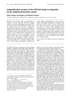
tion. Concentrations of i-FABP in trauma patients were
significantly increased in comparison with healthy controls
(303 (41 to 84,846) pg/mL vs. 87 (41 to 413) pg/mL; P <
0.001; Figure 1). i-FABP levels at ER were also elevated com-
pared with levels at day 1 (174 (41 to 1805) pg/mL; P <
0.001) and day 2 (103 (41 to 1049) pg/mL; P < 0.001). At
day 1, i-FABP concentrations were still increased compared
Available online />Page 3 of 7
(page number not for citation purposes)
with day 2 in control samples (both P < 0.001). i-FABP at day
2 was not significantly increased compared with control values
(P = 0.21). Of all trauma patients at the ER, i-FABP levels of
89 patients (93% of all trauma patients) exceeded 87 pg/mL,
which is the median of i-FABP plasma concentration in healthy
controls.
The extent of intestinal cell damage is related to
presence of shock and injury severity
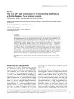
To investigate the relation between hemodynamic stability and
intestinal cell damage, i-FABP concentrations in trauma
patients in shock were compared with patients without shock
(SI > 0.7 vs. ≤ 0.7, respectively). On admittance to the ER, the
SI was increased in 42% of the patients. Plasma i-FABP con-
centrations were significantly higher in patients with an ele-
vated SI in comparison with patients with a normal SI (455 (41
to 84,242) pg/mL vs. 259 (41 to 1957) pg/mL; P < 0.01) or
healthy controls (P < 0.01, Figure 2a). Also in trauma patients
with a SI in the normal range, i-FABP levels were elevated in
comparison to healthy controls (P < 0.01).
On admittance, the ISS of all patients was calculated and cat-
egorized. All ISS categories comprised 12 patients or more. i-
FABP levels in patients with high ISS scores (ISS 31 to 40
and 41 to 50) were significantly increased compared with ISS
0 to 10, 11 to 20, and 21 to 30 categories (428 (142 to
84,846) pg/mL and 682 (52 to 8206) pg/mL vs. 189 (41 to
735) pg/mL, 210 (58 to 1860) pg/mL and 235 (54 to 1957)
pg/mL, each P < 0.05; Figure 2b). In all ISS categories, intes-
tinal cell damage was increased compared with healthy con-
trols (P < 0.01).
Next, the severity of local abdominal trauma was assessed
using the AIS scores of the abdomen. Scores of 0 (no abdom-
Figure 1
Intestinal cell damage increased rapidly following severe traumaIntestinal cell damage increased rapidly following severe trauma.
Plasma intestinal fatty acid binding protein (i-FABP) in trauma patients
at the emergency room (ER) was significantly higher compared with
samples collected at day 1 († P < 0.001), day 2 and healthy controls
(both * P < 0.001). i-FABP concentrations at day 1 were elevated in
comparison with day 2 and controls (both * P < 0.001).
Figure 2
Early intestinal cell damage is related to presence of shock and the extent of injury severityEarly intestinal cell damage is related to presence of shock and the
extent of injury severity. (a) Plasma intestinal fatty acid binding protein
(i-FABP) concentrations were significantly increased in patients with an
elevated shock index (SI > 0.7) compared with patients with a normal
SI (= 0.7) († P < 0.01) or healthy controls (* P < 0.01). Also in trauma
patients with a normal SI, i-FABP levels were higher in comparison to
healthy controls (* P < 0.01). (b) i-FABP concentrations in patients
with an injury severity score (ISS) of more than 30 were significantly
elevated compared with ISS of 30 categories or less († P < 0.05).
Intestinal cell damage in all ISS categories was increased compared
with healthy controls (* P < 0.01). (c) i-FABP levels in patients with
severe abdominal trauma (abbreviated injury scale (AIS) = 3) were sig-
nificantly increased compared with patients without abdominal injury
(AIS = 0; * P < 0.01) and healthy controls († P < 0.001). i-FABP levels
in patients without abdominal trauma were significantly higher com-
pared with healthy controls († P < 0.001).
Critical Care Vol 13 No 3 de Haan et al.
Page 4 of 7
(page number not for citation purposes)
inal injury), 3, and 4 (serious and severe abdominal injury)
occurred most frequently (n = 48, 21, and 14 patients, respec-
tively), whereas scores of 1, 2, and 5 were assigned less often
(n = 8, 1, and 4 patients, respectively). As the abdominal AIS
score of 2 was assigned only once, the i-FABP concentration
detected in this patient (783 pg/mL) was not used for statisti-
cal evaluation. Taken together, at the ER abdominal trauma
was diagnosed in 50% of the patients. i-FABP levels were sig-
nificantly increased in patients with serious, severe, and critical
abdominal injury (AIS 3: 364 (122 to 1194) pg/mL, AIS 4:
1185 (52 to 2753) pg/mL and AIS 5: 1806 (287 to 8206) pg/
mL) compared with patients without abdominal injury (AIS 0:
231 (41 to 84,846) pg/mL; all P < 0.01) and healthy controls
(all P < 0.001; Figure 2c). Interestingly, also i-FABP concen-
trations in patients without abdominal trauma were signifi-
cantly elevated compared with healthy controls (P < 0.001).
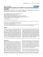
Remarkably elevated i-FABP values at admittance
indicate abdominal emergencies
In a few patients extremely elevated i-FABP levels were meas-
ured, far exceeding the values of other patients (Figure 3).
Examination of the medical records revealed that the highest
10% of i-FABP values at ER belonged to patients with severe
abdominal trauma that required acute surgical intervention,
such as ruptures of the diaphragm, liver, and spleen. The high-
est i-FABP concentration (84,846 pg/mL) was measured in a
patient assigned an AIS score of 0 at ER admission who was
diagnosed at day 2 with intestinal perforation. In this patient, i-
FABP concentrations at day 1 and 2 were 1181 pg/mL and
175 pg/mL, respectively.
Intestinal mucosal cell damage correlates with the
subsequent inflammatory response
Circulating levels of IL-6, PCT, and CRP were measured on
arrival at the ER and at the following days to explore the inflam-
matory response following trauma. Plasma IL-6 strongly
increased at the first day (0.11 (0.01 to 18.35) ng/mL vs. ER:
0.04 (0.00 to 5.16) ng/mL, P < 0.05) and remained elevated
at the second day (0.12 (0.00 to 11.37) ng/mL). Levels of PCT
were barely detectable on presentation (0.06 (0.02 to 1.06)
ng/mL), whereas elevated levels were measured at day 1 and
2 (0.22 (0.04 to 18.23) ng/mL and 0.22 (0.03 to 18.55) ng/
mL, each P < 0.001 to ER). Consecutive measurements of
acute phase protein CRP showed highest plasma values on
the second day post-trauma (1.21 (0.06 to 2.72) mg/mL) com-
pared with the first day (0.42 (0.05 to 1.57) mg/mL, P <
0.001) and to CRP concentrations on admittance (0.01 (0.00
to 0.28) mg/mL, P < 0.001; Figure 4a). Next we analyzed the
relation between intestinal cell damage and the development
of inflammation. Concentrations of i-FABP at admittance cor-
related positively with values of IL-6 (r
2
= 0.19, P < 0.01; Fig-
ure 4b) and PCT (r
2
= 0.36, P < 0.001, Figure 4c) on the first
day after trauma. Furthermore, early i-FABP levels correlated
with CRP in plasma at the second day (r
2
= 0.25, P < 0.01;
Figure 4d).
Discussion
Compromised intestinal integrity is considered to contribute to
the inflammatory response to trauma [12]. This study sought
to clarify the occurrence of intestinal damage and thus com-
promised integrity in the direct phase following severe trauma.
Here, we showed presence of intestinal epithelial cell damage
in a cohort of 96 trauma patients on arrival at the ER.
In the current study, evidence for intestinal cell damage after
trauma was provided by increased plasma i-FABP levels. i-
FABP is a small intracellular protein (14 kD) solely expressed
in differentiated enterocytes of the small intestine and to a
lower extent in the colon [20,22,24]. Following cell damage, i-
FABP is released and readily detectable in circulation [22].
The fast clearance of FABP (T1/2 = 11 minutes) implies that
the enhanced plasma i-FABP levels reflect ongoing intestinal
damage in our study [29].
A strong increase of i-FABP was observed in trauma patients
in shock. In the setting of shock, blood flow to the splanchnic
region is hampered in favor of perfusion of vital organs such as
the brain [30,31]. Therefore, the finding that i-FABP release is
increased in shock is in line with studies that established
splanchnic hypoperfusion as a major cause of i-FABP release.
In a human model of gut ischemia and reperfusion, short-term
ischemia induced a strong increase of plasma i-FABP, paral-
leled by histological damage of the epithelial layer and break-
down of intestinal barrier [22]. Moreover, elevated i-FABP
levels were detected in settings of splanchnic hypoperfusion
during non-abdominal surgery and critically illness [23,32]. In
the present study shock was determined using the SI, which
is considered more sensitive for shock than standard vital
signs alone [26,33]. The observed mucosal epithelial damage
in this patient cohort stresses the importance of rapid and ade-
quate fluid administration after severe trauma [34].
Figure 3
Strongly elevated i-FABP levels at ER indicate major abdominal trauma requiring immediate surgeryStrongly elevated i-FABP levels at ER indicate major abdominal trauma
requiring immediate surgery. The highest 10% of intestinal fatty acid
binding protein (i-FABP) values at emergency room (ER) were found in
patients with severe abdominal trauma requiring acute intervention,
such as rupture of the intestine, diaphragm, kidney, liver, or spleen.
Available online />Page 5 of 7
(page number not for citation purposes)
An increase of intestinal cell damage was also present in
patients with a normal SI, so the relation between intestinal
damage and other trauma characteristics was explored. The
ISS is a frequently used anatomical scoring system that corre-
lates linearly with mortality, morbidity, hospital stay, and other
measures of trauma severity [28]. In the current study, the
extent of intestinal cell damage was found to be related to the
ISS. It should be noted that the ISS is a composite of the
scores of six body regions, including the abdomen. As abdom-
inal trauma is a likely cause of intestinal cell damage, the rela-
tion between abdominal trauma and intestinal cell damage
was then investigated. Half of the patients included in this
study had trauma in the abdominal region, as determined using
the AIS score. The highest i-FABP levels after trauma were
detected in patients with severe abdominal trauma that
required acute surgical intervention, such as rupture of the
intestine, diaphragm, liver, and spleen. Further studies are
needed to explore the sensitivity and specificity of i-FABP as
an early marker for small intestinal organ damage following
trauma. In addition to accepted diagnostic tools such as com-
puted tomography, i-FABP assessment may help to detect
abdominal emergencies in the early phase after trauma and
support the decision to perform surgical intervention
[24,34,35] (Relja and colleagues, unpublished data). In con-
clusion, the extent of intestinal cell damage is related to shock,
ISS, and abdominal trauma.
In search for a potential role of the compromised gut in the
development of inflammation following trauma, the relation
between intestinal cell damage and the early inflammatory
response was investigated. i-FABP levels on arrival at the ER
correlated with concentrations at day 1 of IL-6, a potent
cytokine in the early post-injury immune response that was
identified as a useful predictor of complications as well as mor-
tality [36,37]. Furthermore, i-FABP levels strongly correlated
with day 1 plasma levels of PCT, an inflammation marker that
is used to distinguish septic from non-septic patients [38]. In
line, CRP concentrations at day 2 also correlated positively to
Figure 4
Intestinal mucosal cell damage after trauma correlates positively with the inflammatory responseIntestinal mucosal cell damage after trauma correlates positively with the inflammatory response. (a) Peak concentrations of circulating IL-6 and pro-
calcitonin (PCT) were reached at the first day after trauma, whereas highest levels of C-reactive protein (CRP) were measured at the second day (all
parameters: * P < 0.05 vs. emergency room (ER); † P < 0.001 vs. day 1). (b to d) i-FABP concentrations at ER correlated positively with peak con-
centrations of IL-6 (r
2
= 0.19, P < 0.01), PCT (r
2
= 0.36, P < 0.001), and CRP (r
2
= 0.25, P < 0.01). All data are shown in natural logarithmic scale.
Critical Care Vol 13 No 3 de Haan et al.
Page 6 of 7
(page number not for citation purposes)
early i-FABP values. Taken together, early intestinal cell dam-
age clearly precedes and is related to the subsequent inflam-
matory response to severe trauma. Further studies are
required to determine the causative involvement and predic-
tive value of early enterocyte damage and gut barrier loss in
the development of inflammatory complications.
The gut has long since been considered to play a role in the
pathophysiology of complications following trauma [3-6]. Clar-
ification of the role of the intestine in the development of
excessive inflammation after trauma is not only interesting from
an etiologic viewpoint, but may also contribute to the selection
of patients for novel therapeutic strategies directed at preser-
vation of intestinal integrity and attenuation of the inflammatory
response [39].
Conclusions
To the best of the authors' knowledge, this paper is the first to
show that a significant proportion of trauma patients rapidly
develops intestinal mucosal cell damage. The extent of intesti-
nal damage is readily detectable in blood withdrawn on pres-
entation at the ER. Circulatory concentrations of enterocyte
damage marker i-FABP are related to the presence of shock
and the extent of general injury as well as abdominal trauma,
indicating that the level of intestinal cell damage is determined
by both systemic and local factors. Moreover, early i-FABP val-
ues correlate positively with the inflammatory response that
develops in the days following trauma. Further studies are
needed to clarify the importance of early intestinal damage in
the pathophysiologic response to trauma and its diagnostic
and therapeutic implications.
Competing interests
WB is a shareholder of Hycult Biotechnology that provided the
FABP assays.
Authors' contributions
JdH coordinated the overall design of the study, analyzed the
data, and drafted the manuscript. TL contributed to the design
of the study, helped to interpret the data, and was involved in
drafting the manuscript. JD helped to interpret the data and to
draft the manuscript. BR collected the data and helped to ana-
lyze them. DH collected the data and helped to interpret the
data. JG aided in defining the clinical context and revised the
manuscript. IM conceived of the study, supervised the overall
design, and revised the manuscript. WB conceived of the
study, supervised the overall design, and helped to draft the
manuscript. All authors read and approved the final manu-
script.
Acknowledgements
The authors thank Dr K.P. Hunfeld (Institut für Medizinische Mikrobiol-
ogie, J.W. Goethe University, Frankfurt am Main, Germany) and Mrs A.A.
van Bijnen (Department of Surgery, MUMC, Maastricht, the Nether-
lands) for excellent technical assistance. Dr M.D. Luyer (Department of
Surgery, Orbis Medisch Centrum, Sittard, the Netherlands) and Dr M.
Poeze (Department of Surgery, MUMC, Maastricht, the Netherlands) are
gratefully acknowledged for critical review of the manuscript. This study
was supported by Danone Research Centre for Specialised Nutrition,
Wageningen, the Netherlands and by AGIKO-stipendia 920-03-522 to
TL and 920-03-438 to JD from the Netherlands Organization for Health
Research and Development. The funding sources had no involvement in
study design.
References
1. Osborn TM, Tracy JK, Dunne JR, Pasquale M, Napolitano LM: Epi-
demiology of sepsis in patients with traumatic injury. Crit Care
Med 2004, 32:2234-2240.
2. Angus DC, Linde-Zwirble WT, Lidicker J, Clermont G, Carcillo J,
Pinsky MR: Epidemiology of severe sepsis in the United
States: analysis of incidence, outcome, and associated costs
of care. Crit Care Med 2001, 29:1303-1310.
3. Rotstein OD: Pathogenesis of multiple organ dysfunction syn-
drome: gut origin, protection, and decontamination. Surg
Infect (Larchmt) 2000, 1:217-223.
4. Carrico CJ, Meakins JL, Marshall JC, Fry D, Maier RV: Multiple-
organ-failure syndrome. Arch Surg 1986, 121:196-208.
5. Rowlands BJ, Soong CV, Gardiner KR: The gastrointestinal tract
as a barrier in sepsis. Br Med Bull 1999, 55:196-211.
6. Moore FA: The role of the gastrointestinal tract in postinjury
multiple organ failure. Am J Surg 1999, 178:449-453.
7. Thuijls G, de Haan JJ, Derikx JP, Daissormont I, Hadfoune M, Hein-
eman E, Buurman WA: Intestinal cytoskeleton degradation pre-
cedes tight junction loss following hemorrhagic shock. Shock
2009, 31:164-169.
8. Deitch EA, Xu D, Kaise VL: Role of the gut in the development
of injury- and shock induced SIRS and MODS: the gut-lymph
hypothesis, a review. Front Biosci 2006, 11:520-528.
9. Fink MP, Delude RL: Epithelial barrier dysfunction: a unifying
theme to explain the pathogenesis of multiple organ dysfunc-
tion at the cellular level. Crit Care Clin 2005, 21:177-196.
10. Luyer MD, Buurman WA, Hadfoune M, Jacobs JA, Konstantinov
SR, Dejong CH, Greve JW: Pretreatment with high-fat enteral
nutrition reduces endotoxin and tumor necrosis factor-alpha
and preserves gut barrier function early after hemorrhagic
shock. Shock 2004, 21:65-71.
11. Van Leeuwen PA, Boermeester MA, Houdijk AP, Ferwerda CC,
Cuesta MA, Meyer S, Wesdorp RI: Clinical significance of trans-
location. Gut
1994, 35:S28-34.
12. Clark JA, Coopersmith CM: Intestinal crosstalk: a new paradigm
for understanding the gut as the "motor" of critical illness.
Shock 2007, 28:384-393.
13. Coopersmith CM, Stromberg PE, Dunne WM, Davis CG, Amiot
DM, Buchman TG, Karl IE, Hotchkiss RS: Inhibition of intestinal
epithelial apoptosis and survival in a murine model of pneu-
monia-induced sepsis. JAMA 2002, 287:1716-1721.
14. Matzinger P: The danger model: a renewed sense of self. Sci-
ence 2002, 296:301-305.
Key messages
• Intestinal mucosal cell damage develops early following
trauma.
• The extent of intestinal damage is detectable in blood
drawn on presentation at the ER.
• The presence of shock and the severity of local and
overall injury are related to the extent of early intestinal
cell damage.
• Early plasma values of intestinal epithelial cell damage
marker i-FABP correlate positively with the subse-
quently developing inflammatory response.
Available online />Page 7 of 7
(page number not for citation purposes)
15. Rubartelli A, Lotze MT: Inside, outside, upside down: damage-
associated molecular-pattern molecules (DAMPs) and redox.
Trends Immunol 2007, 28:429-436.
16. Soeters PB, Luyer MD, Greve JW, Buurman WA: The signifi-
cance of bowel permeability. Curr Opin Clin Nutr Metab Care
2007, 10:632-638.
17. Langkamp-Henken B, Donovan TB, Pate LM, Maull CD, Kudsk KA:
Increased intestinal permeability following blunt and penetrat-
ing trauma. Crit Care Med 1995, 23:660-664.
18. Kompan L, Kompan D: Importance of increased intestinal per-
meability after multiple injuries. Eur J Surg 2001, 167:570-574.
19. Bjarnason I, MacPherson A, Hollander D: Intestinal permeability:
an overview. Gastroenterology 1995, 108:1566-1581.
20. Lieberman JM, Sacchettini J, Marks C, Marks WH: Human intes-
tinal fatty acid binding protein: report of an assay with studies
in normal volunteers and intestinal ischemia. Surgery 1997,
121:335-342.
21. Pelsers MM, Namiot Z, Kisielewski W, Namiot A, Januszkiewicz M,
Hermens WT, Glatz JF: Intestinal-type and liver-type fatty acid-
binding protein in the intestine. Tissue distribution and clinical
utility. Clin Biochem 2003, 36:529-535.
22. Derikx JP, Matthijsen RA, de Bruine AP, van Bijnen AA, Heineman
E, van Dam RM, Dejong CH, Buurman WA: Rapid reversal of
human intestinal ischemia-reperfusion induced damage by
shedding of injured enterocytes and reepithelialisation. PLoS
ONE 2008, 3:e3428.
23. Derikx JP, van Waardenburg DA, Thuijls G, Willigers HM, Koen-
raads M, van Bijnen AA, Heineman E, Poeze M, Ambergen T, van
Ooij A, van Rhijn LW, Buurman WA: New insight in loss of gut
barrier during major non-abdominal surgery. PLoS ONE 2008,
3:e3954.
24. Derikx JP, Vreugdenhil AC, Van den Neucker AM, Grootjans J, van
Bijnen AA, Damoiseaux JG, van Heurn LW, Heineman E, Buurman
WA: A pilot study on the noninvasive evaluation of intestinal
damage in celiac disease using I-FABP and L-FABP. J Clin
Gastroenterol 2009 in press.
25. von Elm E, Altman DG, Egger M, Pocock SJ, Gotzsche PC,
Vandenbroucke JP: The Strengthening the Reporting of Obser-
vational Studies in Epidemiology (STROBE) statement: guide-
lines for reporting observational studies.
Bull World Health
Organ 2007, 85:867-872.
26. Rady MY, Smithline HA, Blake H, Nowak R, Rivers E: A compari-
son of the shock index and conventional vital signs to identify
acute, critical illness in the emergency department. Ann Emerg
Med 1994, 24:685-690.
27. The Abbreviated Injury Scale, 1990 Revision, Update 98. Asso-
ciation for the Advancement of Automotive Medicine; Barrington,
IL 1998.
28. Baker SP, O'Neill B, Haddon W Jr, Long WB: The injury severity
score: a method for describing patients with multiple injuries
and evaluating emergency care. J Trauma 1974, 14:187-196.
29. Poll MC van de, Derikx JP, Buurman WA, Peters WH, Roelofs HM,
Wigmore SJ, Dejong CH: Liver manipulation causes hepatocyte
injury and precedes systemic inflammation in patients under-
going liver resection. World J Surg 2007, 31:2033-2038.
30. Ceppa EP, Fuh KC, Bulkley GB: Mesenteric hemodynamic
response to circulatory shock. Curr Opin Crit Care 2003,
9:127-132.
31. Tamion F, Richard V, Sauger F, Menard JF, Girault C, Richard JC,
Thuillez C, Leroy J, Bonmarchand G: Gastric mucosal acidosis
and cytokine release in patients with septic shock. Crit Care
Med 2003, 31:2137-2143.
32. Derikx JP, Poeze M, van Bijnen AA, Buurman WA, Heineman E:
Evidence for intestinal and liver epithelial cell injury in the early
phase of sepsis. Shock 2007, 28:544-548.
33. King RW, Plewa MC, Buderer NM, Knotts FB: Shock index as a
marker for significant injury in trauma patients. Acad Emerg
Med 1996, 3:1041-1045.
34. Blow O, Magliore L, Claridge JA, Butler K, Young JS: The golden
hour and the silver day: detection and correction of occult
hypoperfusion within 24 hours improves outcome from major
trauma. J Trauma 1999, 47:964-969.
35. Evennett NJ, Petrov MS, Mittal A, Windsor JA: Systematic review
and pooled estimates for the diagnostic accuracy of serologi-
cal markers for intestinal ischemia. World J Surg 2009,
33:1374-1383.
36. Stensballe J, Christiansen M, Tonnesen E, Espersen K, Lippert FK,
Rasmussen LS: The early IL-6 and IL-10 response in trauma is
correlated with injury severity and mortality. Acta Anaesthesiol
Scand 2009, 53:515-521.
37. Biffl WL, Moore EE, Moore FA, Peterson VM: Interleukin-6 in the
injured patient. Marker of injury or mediator of inflammation?
Ann Surg 1996, 224:647-664.
38. Tang BM, Eslick GD, Craig JC, McLean AS: Accuracy of procal-
citonin for sepsis diagnosis in critically ill patients: systematic
review and meta-analysis. Lancet Infect Dis 2007, 7:210-217.
39. de Haan JJ, Lubbers T, Hadfoune M, Luyer MD, Dejong CH, Buur-
man WA, Greve JW: Postshock intervention with high-lipid
enteral nutrition reduces inflammation and tissue damage.
Ann Surg 2008, 248:842-848.