Báo cáo y học: " Theoretical study of the Usutu virus helicase 3D structure, by means of computer-aided homology modelling" ppsx
Bạn đang xem bản rút gọn của tài liệu. Xem và tải ngay bản đầy đủ của tài liệu tại đây (3.37 MB, 9 trang )
BioMed Central
Page 1 of 9
(page number not for citation purposes)
Theoretical Biology and Medical
Modelling
Open Access
Research
Theoretical study of the Usutu virus helicase 3D structure, by
means of computer-aided homology modelling
Dimitrios Vlachakis
Address: Institute of Biology, National Centre for Scientific Research Demokritos, 15310 Ag. Paraskevi Attikis, Greece
Email: Dimitrios Vlachakis -
Abstract
Background: Usutu virus belongs to the Flaviviridae viral family and constitutes an important
pathogen. The viral helicase is an ideal target for inhibitor design, since this enzyme is essential for
the survival, proliferation and transmission of the virus.
Results: Towards a drug-design approach, the 3D model of the Usutu virus helicase structure has
been designed, using conventional homology modelling techniques and the known 3D-structure of
the Murray Valley Encephalitis virus helicase, of the same viral family, as template. The model was
then subjected to extended molecular dynamics simulations in a periodic box, filled with explicit
water molecules for 10 nanoseconds. The reliability of the model was confirmed by obtaining
acceptable scores from a variety of in silico scoring tools, including Procheck and Verify3D.
Conlcusion: The 3D model of the Usutu virus helicase exhibits in silico all known structural
characteristics of the Flaviviridae viral family helicase enzymes and could provide the platform for
further de novo structure-based design of novel anti-Usutu agents.
Background
The viral family Flaviviridae comprises the genera Flavivi-
rus, Pestivirus and Hepacivirus and includes numerous
important human and animal pathogens [1]. The small,
enveloped virions of the different members of the Flaviv-
iridae family contain a single-stranded, positive-sense
RNA genome of about 9.5–12.5 kb. The genome consists
of a single, long open reading frame (ORF), which is
flanked by untranslated regions (UTRs) at the 5' and 3'
ends.
Recent studies on sub-genomic Pestivirus and Flavivirus
RNA replicons have revealed that the non-structural (NS)
proteins, which are encoded by the C-terminal part of the
polyprotein, play a crucial role in viral RNA replication
[2,3]. Accordingly, these proteins are assumed to form
replication complexes in conjunction with genomic RNA
and possibly with other cellular factors.
Sequence alignments of the Usutu viral helicase identified
several conserved sequence motifs that are important for
biological functions. So far, the crystal structures of heli-
cases from various RNA viruses have been determined,
including the helicases from hepatitis C virus, Dengue
virus, Yellow Fever Virus and Kunjin virus [4].
Usutu virus is the cause of one of the most important
arthropod-borne viral (arbovirus) diseases, transmitted
mainly by the Culex pipiens mosquito [5]. Usutu virus was
first detected in Africa, and four decades later it emerged
in Austria. Usutu virus is closely related to the more com-
mon West Nile virus, Japanese Encephalitis virus and Yel-
Published: 25 June 2009
Theoretical Biology and Medical Modelling 2009, 6:9 doi:10.1186/1742-4682-6-9
Received: 21 October 2008
Accepted: 25 June 2009
This article is available from: />© 2009 Vlachakis; licensee BioMed Central Ltd.
This is an Open Access article distributed under the terms of the Creative Commons Attribution License ( />),
which permits unrestricted use, distribution, and reproduction in any medium, provided the original work is properly cited.
Theoretical Biology and Medical Modelling 2009, 6:9 />Page 2 of 9
(page number not for citation purposes)
low Fever virus. Usutu virus caused mass mortalities of
birds, especially blackbirds (Turdus merula) and great grey
owls (Strix nebulosa) initially in Austria, but then spread to
other central European countries such as Hungary, Swit-
zerland, and Northern Italy (in chronological order) [6].
In the present work, the three-dimensional structure of
the helicase enzyme of Usutu virus was modelled by
applying homology modelling techniques and using the
crystal structure of the Murray Valley Encephalitis virus
helicase of the Flaviviridae family as template.
Methods
Sequence alignment
The amino acid sequence of Usutu viral helicase was
obtained from the GenBank database (accession no.:
NC_006551
, entry name: Usutu virus, complete genome).
Using the Gapped-BLAST [7] through NCBI [8]. the
homologous structure of Murray Valley Encephalitis virus
helicase was identified, which was used as template for the
homology modelling of the Usutu viral helicase. The
sequence alignment was done using the online version of
ClustalW [9].
Secondary structure prediction
Secondary structure predictions were performed using the
NPS (Network Protein Sequence Analysis) web-server
and the GeneSilico MetaServer
/> in order to confirm that the chosen
Murray Valley Encephalitis virus helicase template is the
most appropriate one [10].
Homology modelling
The homology modelling of the Usutu viral helicase was
carried out using the MOE (Molecular Operating Environ-
ment, version: 2004.03) package and its built-in homol-
ogy modelling application [11]. The RCSB entry 2V80,
which corresponds to the crystal structure of the Murray
Valley Encephalitis virus helicase was used as template.
The sequence alignment between the raw sequence of the
Usutu and the sequence of the Murray Valley Encephalitis
virus template revealed almost 90% identity, which
allows for a conventional homology modelling approach
to be considered. The homology model method of MOE
comprises the following steps: First an initial partial
geometry specification, where an initial partial geometry
for each target sequence is copied from regions of one or
more template chains. Secondly, the insertions and dele-
tions task, where residues that still have no assigned back-
bone coordinates are modelled. Those residues may be in
loops (insertions in the model with respect to the tem-
plate), they may be outgaps (residues in a model sequence
which are aligned before the C-terminus or after the N-ter-
minus of its template) or may be deletions (regions where
the template has an insertion with respect to the model).
For this study though outgaps have not been included in
the homology modelling process. Third step is the loop
selection and sidechain packing, where a collection of
independent models is created. Last step is the final model
selection and refinement one, where the final models are
scored and ranked, after they have been stereochemically
tested and evaluated with the built-in module "Protein
Geometry" for errors.
Molecular electrostatic potential (MEP)
Electrostatic potential surfaces were calculated by solving
the nonlinear Poisson-Boltzmann equation using finite
difference method as implemented in the PyMOL Soft-
ware [12]. The potential was calculated on grid points per
side (65, 65, 65) and the grid fill by solute parameter was
set to 80%. The dielectric constants of the solvent and the
solute were set to 80.0 and 2.0, respectively. An ionic
exclusion radius of 2.0Å, a solvent radius of 1.4Å and a
solvent ionic strength of 0.145 M were applied. Amber99
[13] charges and atomic radii were used for this calcula-
tion.
Model optimization
Energy minimisation was done in MOE initially using the
Amber99 [13] forcefield implemented into the same
package, up to a RMSd gradient of 0.0001 to remove the
geometrical strain. The model was subsequently solvated
with SPC water using the truncated octahedron box
extending to 7Å from the model and molecular dynamics
were performed after that at 300 K, 1 atm with 2 fsecond
step size and for a total of ten nanoseconds, using the NVT
ensemble in a canonical environment. NVT stands for
Number of atoms, Volume and Temperature that remain
constant throughout the calculation. The results of the
molecular dynamics simulation were collected into a
database by MOE and can be further analysed.
Model evaluation
The produced models were initially evaluated within the
MOE package by a residue packing quality function,
which depends on the number of buried non-polar side
chain groups and on hydrogen bonding. Moreover, the
suite PROCHECK [14] was employed to further evaluate
the quality of the produced Usutu virus helicase model.
Finally, Verify3D [15,16] was used to evaluate whether the
model of Usutu virus helicase is similar to known protein
structures.
Results and discussion
The NS3 domain of Flaviviridae contains both the protease
and the helicase coding regions. For this study, the Usutu
virus helicase sequence was aligned with the Murray Val-
ley Encephalitis virus helicase template sequence and all
the major helicase motifs, characteristic of the Flaviviridae
family were found to be conserved (Figure 1).
Theoretical Biology and Medical Modelling 2009, 6:9 />Page 3 of 9
(page number not for citation purposes)
The Murray Valley Encephalitis virus helicase structure has
been established by X-ray crystallography at 1.9 Å resolu-
tion [17]. The Murray Valley Encephalitis virus helicase is
an appropriate template because the virus belongs to the
same viral family (Flaviviridae). Among the available heli-
case structureswithin this family, the Murray Valley
Encephalitis virus enzyme presented the highest identity
percentage to the Usutu virus helicase. That is also sup-
ported by the analysis of the helicase structures available
for the whole Flaviviridae family that revealed that the
overall 3D structure for the Murray Valley Encephalitis
virus helicase ishighly conserved [17,18]. Moreover using
a fold recognition approach (FR), the Murray Valley
Encephalitis virus helicase was confirmed as the most
appropriate model. The secondary structure prediction for
the Usutu virus helicase turned out to be very similar to
the actual structure of the Murray Valley Encephalitis virus
helicase. Methods of protein fold recognition attempt to
detect similarities between protein 3D structure that are
not accompanied by any significant sequence similarity.
The sequence alignment between the Usutu virus helicase sequence and the corresponding sequence of the Murray Valley Encephalitis virus helicase template (RCSB entry: 2V80)Figure 1
The sequence alignment between the Usutu virus helicase sequence and the corresponding sequence of the
Murray Valley Encephalitis virus helicase template (RCSB entry: 2V80). All seven major conserved motifs of Flaviviri-
dae helicases have been highlighted.
Theoretical Biology and Medical Modelling 2009, 6:9 />Page 4 of 9
(page number not for citation purposes)
There are many approaches, but the unifying theme is to
try and find folds that are compatible with a particular
sequence. Unlike sequence-only comparison, these meth-
ods take advantage of the extra information made availa-
ble by 3D structure information. In effect, the turn the
protein folding problem on it's head: rather than predict-
ing how a sequence will fold, they predict how well a fold
will fit a sequence [19-22].
The model was firstly structurally superimposed and sub-
sequently compared to its template, where exhibited an
alpha-carbon RMSD less than 0,65 angstroms. Further-
more it was evaluated with MOE and PROCHECK for its
geometry [see additional file 1] and then subjected to the
Verify3D algorithm for a more thorough evaluation.
Verify3D assessed the compatibility of the 3D model of
Usutu virus helicase with its own amino acid sequence.
Based on location and environment, a structural class is
assigned for each residue. A collection of reference struc-
tures is used as a control in order to calculate a score for
each residue. The Usutu virus helicase model ranged from
+0.32 to +0.84. That confirmed that the model is of high
quality, since it is Verify3D scores below +0.1 that are
indicative of serious problems in the model [23]. A more
important criterion was the compliance of the model with
the known unique characteristics of the helicases that
belong to the Flaviviridae family of viruses. The Murray
Valley Encephalitis virus, Hepatitis C virus, Dengue virus,
West Nile virus and Usutu virus helicases all belong to the
superfamily II of helicases and share seven common
motifs within their domains, all of which have been struc-
turally conserved on the Usutu virus helicase model [17].
Description of the Usutu virus helicase model
As expected from the sequence alignment (Figure 1) and
the secondary structure prediction (data not shown), the
Usutu virus helicase model exhibited the structural fea-
tures of known Flaviviridae helicases. Namely, the three
distinct domains of helicases as well as the various motifs
(as reported by Kim [24] – Figure 1) were structurally con-
served in the Usutu virus helicase model (Figures 2 and 3).
One of the most crucial motifs in Flaviviridae helicases is
the GxGKT/S motif in domain 1, which is conserved to the
same loop in kinases. It is a Walker A motif and its role
involves binding of the β-phosphate of ATP [25]. Site
directed mutagenesis studies of that motif have reported
that the mutant protein is inactive. Furthermore another
crucial motif for the helicase is the DExH motif. The DExH
motif is responsible for the binding of the Mg2+-ATP sub-
strate. Studies in adenylate and thymidine kinases
revealed that an aspartate (Asp170) binds the Mg2+ and
helps to establish the optimum orientation of ATP for
nucleophilic attack [26]. Finally, another crucial motif is
the QRxGRxGR motif. The role of this motif is exception-
ally crucial to the Flaviviridae helicase function as it is
involved in nucleic acid binding [27-29].
The model shared a very similar topology (shape and size)
to its template (Figures 2 and 3). The Flaviviridae helicases,
in general, have three domains in total, which are sepa-
rated by two channels. The first and third domains are
interacting much more together than they do with
domain two. Domain two is supposed to undergo signifi-
cant movements compared to the other two domains,
during the unwinding of double-stranded nucleic acids. It
is the channel between domain 3 and 1–2 that accommo-
dates the ssRNA during the viral unwinding process. RNA
binds to the helicase at the arginine-rich site of the 2
nd
domain. The ATP and ssRNA sites were found to have
been conserved on the Usutu virus helicase model (Fig-
ures 4 and 5) [30].
ssRNA – ATP substrates and MD simulations
In order to evaluate the substrate binding site, the model
was subjected to energy minimization and molecular
dynamics (MD) simulations in the presence of the heli-
case substrates. The coordinates of the ssRNA and ADP/
Mn
++
were transferred to the model from the Hepatitis C
(1A1V) and the Dengue virus helicase structure (2JLS)
respectively, for this purpose. Invariant residues of various
motifs in the vicinity of either substrate in the Murray Val-
ley Encephalitis virus template structure were conserved
structurally in the Usutu virus helicase model (Figures 2
and 4). Most interactions between the ssRNA fragment
and the Usutu helicase enzyme are established with the
backbone of the ssRNA fragment, which is typical for non-
specific protein – nucleic acid interactions. The bases in
the middle of the ssRNA do not seem to interact much
with the receptor (Figure 2D). The receptor's contacts
emerge mostly from domains one and two of the Usutu
helicase and most specifically, by loops between second-
ary structure elements of the latter domains. More
detailed (i.e. per residue) comparison of the ssRNA inter-
action pattern between the Usutu virus helicase model
and the Murray Valley Encephalitis virus helicase structure
has been drawn using LigPlot, which is a built-in module
of MOE, and is shown in Figure 2D.
The 10 ns MD simulations revealed that soon the Usutu
virus helicase model reached a conformational equilib-
rium similar to that of Murray Valley Encephalitis virus
helicase structure (Figure 5B and 5C). Taken together
these observations illustrated the viability of the homol-
ogy modelling of the Usutu virus helicase model. To eval-
uate further the Usutu virus helicase model, the latter was
compared with its template structure by calculating the
root mean square deviations (RMSd) between equivalent
atoms for the full MD course. Large RMSd values are indic-
ative of systems of poor quality. The C
α
RMSd of the Usutu
Theoretical Biology and Medical Modelling 2009, 6:9 />Page 5 of 9
(page number not for citation purposes)
virus helicase model from the equivalent domains of the
template structures was less than 0.65. The low RMSd
value indicated that this model remains conformationally
close to the template structure upon the minimization
and the molecular dynamics simulation course that fol-
lowed, reflecting its high quality.
Molecular surface analysis
In order to analyze the molecular surface of the produced
Usutu virus helicase model, the electrostatic potential sur-
face was calculated. For direct comparison with the tem-
plate structures used in this study, electrostatic potential
surfaces were also calculated for the Murray Valley
Encephalitis virus helicase (Figure 6). The two helicases
exhibited almost identical electrostatic surfaces, sharing
common features such as a negatively charged ssRNA
entrance to the helicase tunnel. This observation verified
the validity of the model, which was found to share simi-
lar electrostatic surface to its X-ray crystal structure tem-
plate.
Conclusion
The 3D model of the Usutu virus helicase was designed
using the homologous X-ray crystal structure of the Mur-
ray Valley Encephalitis viral helicase as template. The
model was successfully evaluated both in terms of its
geometry, fold recognition and compliance to the criteria
required as a member to the Flaviviridae viral family. It is
A: Ribbon representation of the produced Usutu virus helicase modelFigure 2
A: Ribbon representation of the produced Usutu virus helicase model. B: the Usutu virus helicase model superim-
posed with its Murray Valley Encephalitis virus helicase template (RCSB entry: 2V80). The Usutu virus helicase model is in red,
whereas the Murray Valley Encephalitis virus helicase template is in green and the ssRNA substrate (coordinates obtained from
1A1V [24]) is in magenta. C: All residues surrounding the ssRNA fragment, color-coded as B. The ssRNA interacting regions
are almost identical between the Usutu virus helicase model and the MVE helicase template. D: The interaction map of the per-
residue ssRNA interaction pattern from Ligplot for Usutu virus helicase model (left) and the Murray Valley Encephalitis virus
helicase template (right). Conserved residues that are involved in the ssRNA binding and are common between the Usutu virus
helicase model and the MVE helicase template have been connected with a line.
Theoretical Biology and Medical Modelling 2009, 6:9 />Page 6 of 9
(page number not for citation purposes)
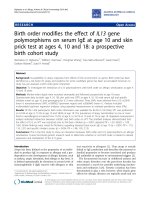
The conserved motifs of the Murray Valley Encephalitis virus helicase (in green color) next to the corresponding motifs on the Usutu virus helicase model (colored in Red)Figure 3
The conserved motifs of the Murray Valley Encephalitis virus helicase (in green color) next to the correspond-
ing motifs on the Usutu virus helicase model (colored in Red). The major motifs have been color-coded according to
the conventions of Fig. 1, and are showing in CPK format (Usual space filling) along with the rest of the helicase motifs.
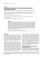
The ATP site of the three helicase structuresFigure 4
The ATP site of the three helicase structures. Left: The Murray Valley Encephalitis helicase is in green color, the Dengue
helicase is in orange color, the Usutu helicase model is colored red and the ADP molecule, is showing in CPK format (ADP
conformation adopted from the X-ray structure of the Dengue Virus helicase: 2JLS [30]). Right: Zoom-in of the ADP binding
site and the conformation of the ADP molecule after the MD simulation color-coded for each receptor. The binding mode of
the ADP molecule is exactly the same in the Usutu virus helicase model, as it is in the Murray Valley Encephalitis virus template
structure.
Theoretical Biology and Medical Modelling 2009, 6:9 />Page 7 of 9
(page number not for citation purposes)
Figure 5 (see legend on next page)
Theoretical Biology and Medical Modelling 2009, 6:9 />Page 8 of 9
(page number not for citation purposes)
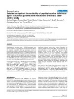
A: The ssRNA channel site of the four helicase structures: the Murray Valley Encephalitis helicase is in green color, the Yellow fever helicase is in pink color, the Dengue helicase is in orange color and the Usutu helicase model is colored redFigure 5 (see previous page)
A: The ssRNA channel site of the four helicase structures: the Murray Valley Encephalitis helicase is in green
color, the Yellow fever helicase is in pink color, the Dengue helicase is in orange color and the Usutu helicase
model is colored red. The ssRNA substrate, indicating the nucleic acid binding channel is showing in magenta spacefill repre-
sentation and the NTP binding site is in the proximity of the ADP molecule, showing in CPK format. B: The energy vs time plot
of the full MD simulation of the Usutu helicase model. The 10 ns MD simulation quickly reached equilibrium and remained sta-
ble. C: Energy vs time plot of 10 ns MD simulation for each of the two X-ray structures (Murray Valley Encephalitis and Yellow
Fever helicases) and the Usutu helicase model in the presence of the ssRNA fragment, borrowed from the Murray Valley
Encephalitis structure (id: 2V80). It was demonstrated that the Usutu helicase model + ssRNA complex (yellow) behaved in a
way very similar to that of the Murray Valley Encephalitis X-ray structure + ssRNA (blue) and Yellow Fever X-ray structure +
ssRNA (magenta) complexes.
Electrostatic surfaces potential for the Usutu virus helicase model and the Murray Valley Encephalitis virus helicase template (X-ray structure: 2V80)Figure 6
Electrostatic surfaces potential for the Usutu virus helicase model and the Murray Valley Encephalitis virus
helicase template (X-ray structure: 2V80). White transparent arrow is showing the nucleic acid channel (ssRNA).
Theoretical Biology and Medical Modelling 2009, 6:9 />Page 9 of 9
(page number not for citation purposes)
therefore proposed that the Usutu virus helicase model
will be suitable for further in silico structure-based de novo
drug design experiments. These computer-based method-
ologies are now becoming integral part of the drug discov-
ery process that may eventually lead to the development
of potential inhibitor structures of the Usutu virus heli-
case enzyme in the future.
Competing interests
The author declares that they have no competing interests.
Authors' contributions
DV is the sole author of this paper and is responsible for
developing the concepts and for writing and revising the
manuscript.
Additional material
References
1. Calisher CH, Gould EA: Taxonomy of the virus family Flaviviri-
dae. Adv Virus Res 2003, 59:1-19.
2. Behrens S-E, Grassmann CW, Thiel H-J, Meyers G, Tautz N: Char-
acterization of an autonomous subgenomic pestivirus RNA
replicon. Journal of Virology 1998, 72:2364-2372.
3. Khromykh AA, Westaway EG: Subgenomic replicons of the fla-
vivirus Kunjin: construction and applications. Journal of Virology
1997, 71:1497-1505.
4. Wu J, Bera AK, Kuhn RJ, Smith JL: Structure of the flavivirus hel-
icase: implications for catalytic activity, protein interactions,
and proteolytic processing. J Virol 2005, 79:10268-10277.
5. Bakonyi T, Erdélyi K, Ursu K, Ferenczi E, Csörgo T, Lussy H, Chvala
S, Bukovsky C, Meister T, Weissenböck H, Nowotny N: Emergence
of Usutu virus in Hungary. J Clin Microbiol 2007, 45:3870-3874.
6. Bakonyi T, Gould EA, Kolodziejek J, Weissenböck H, Nowotny N:
Complete genome analysis and molecular characterization
of Usutu virus that emerged in Austria in 2001: comparison
with the South African strain SAAR-1776 and other flavivi-
ruses. Virology. 2004, 328(2):301-310.
7. Altschul SF, Madden TL, Schäffer AA, Zhang J, Zhang Z, Miller W:
Gapped BLAST and PSI-BLAST: a new generation of protein
database search programs. Nucleic Acids Res 1997, 25:3389-3402.
8. Benson DA, Karsch-Mizrachi I, Lipman DJ, Ostell J, Wheeler DL:
GenBank. Nucleic Acids Res 2007, 35:21-25.
9. Thompson JD, Higgins DG, Gibson TJ: CLUSTAL W: improving
the sensitivity of progressive multiple sequence alignment
through sequence weighting, position specific gap penalties
and weight matrix choice. Nucleic Acids Res 1994, 22:4673-4680.
10. Kurowski MA, Bujnicki JM: GeneSilico protein structure predic-
tion meta-server. Nucleic Acids Res 2003, 31:3305-3307.
11. MOE, Chemical Computing Group, 1010 Sherbrooke St.
West, Suite 910, Montreal, Canada, H3A 2R.
.
12. Warren L: DeLano "The PyMOL Molecular Graphics Sys-
tem.". [
]. DeLano Scientific LLC, San Carlos,
CA, USA
13. Wang J, Cieplak P, Kollman PA: How well does a restrained elec-
trostatic potential (resp) model perform in calculating con-
formational energies of organic and biological molecules. J
Comp Chem 2000, 21:1049-1074.
14. Laskowski RA, Rullmannn JA, MacArthur MW, Kaptein R, Thornton
JM: AQUA and PROCHECK-NMR: programs for checking
the quality of protein structures solved by NMR. J Biomol NMR
1996, 8:477-486.
15. Eisenberg D, Lüthy R, Bowie JU: VERIFY3D: assessment of pro-
tein models with three-dimensional profiles. Methods Enzymol
1997, 277:396-404.
16. Lüthy R, Bowie JU, Eisenberg D: Assessment of protein models
with three-dimensional profiles. Nature 1992, 356:83-85.
17. Mancini EJ, Assenberg R, Verma A, Walter TS, Tuma R, Grimes JM,
Owens RJ, Stuart DI: Structure of the Murray Valley encephali-
tis virus RNA helicase at 1.9 Angstrom resolution. Protein Sci
2007, 16(10):2294-300.
18. Sampath A, Padmanabhan R: Molecular targets for flavivirus drug
discovery. Antiviral Res 2009, 81(1):6-15.
19. Wodak SJ, Rooman MJ: Generating and testing protein folds.
Current Opinion in Structural Biology 1993, 3:247-259.
20. Jones D, Thornton J: Protein fold recognition. Journal of Computer
Aided Molecular Design 1993, 7:439-456.
21. Bowie JU, Eisenberg D: Inverted protein structure prediction.
Current Opinion in Structural Biology 1993, 3:437-444.
22. Lemer C, Rooman MJ, Wodak SJ: Protein Structure Prediction
By Threading Methods: Evaluation Of Current Techniques.
Proteins. 1996, 23(3):337-355.
23. Bujnicki Janusz, Rychlewski Leszek, Fischer Daniel: Fold-recogni-
tion detects an error in the Protein Data Bank. Bioinformatics
2002, 18(10):1391-1395.
24. Kim JL, Morgenstern KA, Griffith JP, Dwyer MD, Thomson JA,
Murcko MA, Lin C, Caron PR:
Hepatitis C virus NS3 RNA heli-
case domain with a bound oligonucleotide: the crystal struc-
ture provides insights into the mode of unwinding. Structure
1998, 6:89-100.
25. Saraste M, Sibbald PR, Wittinghofer A: The P-loop – a common
motif in ATP- and GTP-binding proteins. Trends Biochem Sci
1990, 15:430-434.
26. Ruff M, Krishnaswamy S, Boeglin M, Poterszman A, Mitschler A, Pod-
jarny A, Rees B, Thierry JC, Moras D: Class II aminoacyl transfer
RNA synthetases: crystal structure of yeast aspartyl-tRNA
synthetase complexed with tRNA(Asp). Science 1991,
252:1682-1689.
27. Lohman TM, Bjornson KP: Mechanisms of helicase-catalyzed
DNA unwinding. Annu Rev Biochem 1996, 65:169-214.
28. Pause A, Sonenberg N: Mutational analysis of a DEAD box RNA
helicase: the mammalian translation initiation factor elF-4A.
EMBO J 1992, 11:2643-2654.
29. Gross CH, Shuman S: The QRxGRxGRxxxG motif of the vac-
cinia virus DExH box RNA helicase NPH-II is required for
ATP hydrolysis and RNA unwinding but not for RNA bind-
ing. J Virol 1996, 70:1706-1713.
30. Luo D, Xu T, Watson RP, Scherer-Becker D, Sampath A, Jahnke W,
Yeong SS, Wang CH, Lim SP, Strongin A, Vasudevan SG, Lescar J:
Insights Into RNA Unwinding and ATP Hydrolysis by the Fla-
vivirus Ns3 Protein. EMBO J 2008, 27:3209-3219.
Additional file 1
Extended Procheck Results for both the Usutu virus helicase model
and the Murray Valley Encephalitis virus helicase template. Extended
Procheck Results for both the Usutu virus helicase model (LEFT COL-
UMN) and the Murray Valley Encephalitis virus helicase template
(RIGHT COLUMN, X-ray structure: 2V80). A: Ramachandran plot, B:
Bond Length Plot, C: Bond Angles plot, D: Dihedrals plot, E: Rotamers
plot and F: Contact Energies Plot. It is therefore concluded that the Usutu
virus helicase model has inherited all structural characteristics of its Mur-
ray Valley Encephalitis virus helicase template.
Click here for file
[ />4682-6-9-S1.jpeg]