Heart Disease in Pregnancy - part 7 doc
Bạn đang xem bản rút gọn của tài liệu. Xem và tải ngay bản đầy đủ của tài liệu tại đây (1023.95 KB, 37 trang )
should be encouraged and measures set in place to maintain a lifelong healthy
lifestyle.
Congenital coronary anomalies
Congenital coronary anomalies are occasionally encountered in pregnancy and
patients with repaired tetralogies and other defects have now lived long enough
to be seen with acquired atheromatous coronary disease. Occasional patients
with previously unrecognized corrected transposition are referred with angina
and ‘mitral’ regurgitation thought to be ischemic in origin. They often have
poor function of the systemic right ventricle and atrial fibrillation or atrioven-
tricular conduction defects.
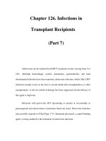
A continuous murmur caused by a coronary cameral fistula may first be de-
tected during antenatal examination. It is usually distinguishable from a patent
duct by an unusual location. Echocardiography will usually display the anom-
aly but small ones may be hard to spot (Figure 15.2). Even large fistulae may be
symptom free and cause no trouble in pregnancy but should be closed after the
pregnancy. Small fistulae should be left alone. Connections can be multiple and
are best tackled percutaneously.
Anomalous origin of a coronary artery (usually the left) from the pulmonary
artery, with poor left ventricular function as a result of neonatal infarction or
progressive ischaemia caused by increasing fistulous flow from right to left
coronary artery, may present with angina,
45
mitral regurgitation or left ventric-
ular failure. The patient illustrated in Figure 15.3 had undergone two unevent-
ful pregnancies before she was referred with angina, mitral regurgitation and
failure. She did well after ligation of the left coronary artery at its ostium and
212 Chapter 15
Figure 15.2 One frame from a left
coronary angiogram of a young girl
who was found to have a murmur at a
routine examination. This was
continuous and placed maximally at
the third left interspace too low for a
patent ductus. Echocardiography
showed flow into the main
pulmonary artery just distal to the
valve. Coronary angiography showed
a coronary artery fistula with
abnormal branches from the anterior
descending coronary artery draining
into the main pulmonary artery
which is opacified from the left
coronary injection. This rare
abnormality carries no adverse
prognostic significance.
internal mammary artery bypass into the left anterior descending artery
and mitral valve replacement.
Pregnancy after myocardial infarction
The occurrence of a heart attack in pregnancy is deeply distressing and likely to
be followed by considerable depression and insecurity even if recovery is good.
Little is known about the risk of further pregnancies, which depends on the
mechanism and the residual left ventricular function. Subsequent successful
pregnancies have been reported but both patients and their doctors will usually
be fearful because the risk of repetition of dissection is unknown.
Conclusion
Myocardial infarction is a rare complication of pregnancy with a high mortality.
Rapid intervention with coronary stenting or bypass is usually indicated. The
most common cause is probably spontaneous dissection but the prevalence of
atheroma has increased in association with the older age of many pregnant
women.
References
1 Wenger NK. Coronary heart disease: the female heart is vulnerable. Prog Cardiovasc
Dis 2003;46:199–229.
2 Von der Lohe E. Coronary Heart Disease in Women. Berlin: Springer, 2003.
Coronary artery disease 213
Figure 15.3 Aortogram showing the
dilated right coronary artery in a patient
with anomalous origin of the left coronary
artery from the pulmonary artery
(described in the text). The left coronary
artery is faintly opacified by fistulous flow
from the right coronary artery but has no
connection with the aorta.
3 Brosius FC, Waller BF, Roberts WC. Radiation heart disease: analysis of 16 young
(aged 15–33 years) necropsy patients who received over 3500 rads to the heart. Am J
Med 1981;70:519–30.
4 Mallilos-Perez M, Orteger-Carnicer O, Gutierrez-Millet V, Pazmino-Narvaez L. Post
partum acute myocardial infarction associated with polyarteritis nodosa. Med Clin
1982;78:32–4.
5 Rallings P, Exner T, Abraham R. Coronary artery vasculitis and myocardial infarction
associated with antiphospholipid antibodies in a pregnant woman. Aust NZ J Med
1989;19:347–50.
6 Parry G, Goudevenos J, Williams DO. Coronary thrombosis postpartum in a young
woman with Still’s disease. Clin Cardiol 1992;15:305–7.
7 Nolan TE, Savage RW. Peripartum myocardial infarction from presumed Kawasaki’s
disease. Southern Med J 1990;83:1360–1.
8 Hankins GDV, Wendel GD, Leveno KL, Stoeham J. Myocardial infarction during
pregnancy: a review. Obstet Gynecol 1985;65:139–46.
9 Jaffe BD, Broderick TM, Leier CV. Cocaine induced coronary artery dissection. N Eng
J Med 1994;330:510–11.
10 Liu SS, Forrester RM, Murphy GS, Chen K, Glassenberg R. Anaesthetic management
of a parturient with myocardial infarction related to cocaine use. Can J Anaesth
1992;39:858–61.
11 Livingston JC, Mabie BC, Ramanathan J. Crack cocaine, myocardial infarction and
troponin I levels at the time of caesarean delivery. Anesth Analg 2000;91:913–15.
12 Bonnet J, Aumailley M, Thomnas D, Grosgogeat Y, Broustet JP, Bricaud H. Sponta-
neous coronary artery dissection; case report and evidence for a defect in collagen
metabolism. Eur Heart J 1986;7:904–9.
13 Anderson RA, Fineron FW: Aortic dissection in pregnancy: importance of pregnancy
induced changes in the vessel wall and bicuspid aortic valve in pathogenesis. Br J
Obstet Gynaecol 1994;101:1085–18.
14 Basso C, Morgagni GL, Thiene G. Spontaneous coronary artery dissection: a
neglected cause of acute myocardial ischaemia and sudden death. Heart 1996;75:
451–4.
15 Dhawan R, Singh G, Fesniack H. Spontaneous coronary artery dissection: the clinical
spectrum. Angiology 2002;53:5383–93.
16 Maeder M, Ammann P, Angehrn W, Rickli H. Idiopathic spontaneous coronary artery
dissection: incidence, diagnosis and treatment. Int J Cardiol 2005;101:363–9.
17 Sheikh AU, Harper MA. Myocardial infarction during pregnancy: management and
outcome of two pregnancies. Am J Obstet Gynecol 1993;163:279–83.
18 Antoniucci D, Magdidilgenti I. Spontaneous dissection of the three major coronary
arteries. Eur Heart J 1990;11:1130–4.
19 Black MD, Catzavelos C, Boyd D, Walley VM. Simultaneous spontaneous dissections
in three coronary arteries. Can J Cardiol 1991;7:34–6.
20 Emori T, Goto, Y, Maeda T, Chiba Y, Haze K. Multiple coronary artery dissections
diagnosed in vivo in a pregnant woman. Chest 1993;104:289–90.
21 Togni M, Ammann FW, Follath F. Spontaneous multivessel coronary artery dissec-
tion in a pregnant woman treated successfully with stent implantation. Am J Med
1999;107:407–8.
22 Greenblatt JM, Kochar GS, Albornoz MA. Multivessel spontaneous coronary artery
dissection in a patient with severe systolic hypertension: a possible association. A case
report. Angiology 1999;50:509–13.
214 Chapter 15
23 Choi JW, Davidson CJ. Spontaneous multivessel coronary artery dissection in a long
distance runner successfully treated with oral antiplatelet therapy. J Invasive Cardiol
2002;14:675–8.
24 De Maio JJ Jr, Kinsella SH, Silverman ME. Clinical course and long term prognosis of
spontaneous coronary artery dissection. Am J Cardiol 1989;64:471–4.
25 Jorgensen MB, Aharonian V, Mansukhani V, Mahrer PR. Spontaneous coronary dis-
section; a cluster of cases with this rare finding. Am Heart J 1994;127:1382–7.
26 Dowling GP, Buja LM. Spontaneous coronary artery dissection occurs with and with-
out periadventitial inflammation. Arch Pathol Lab Med 1987;111:470–2.
27 Chanler Smith J. Dissecting aneurysms of coronary arteries. Arch Pathol 1975;99:
1127–31.
28 Robinowitz M, Virmani R, McAllister H. Spontaneous coronary dissection and
eosinophilic inflammation: a cause and effect relationship? Am J Med 1982;72:
923–8.
29 Curiel P, Petrella A et al. Postpartum coronary artery dissection followed by heart
transplantation. Am J Obstet Gynecol 1990;163:538–9.
30 Movsesiam MA, Wray RB. Postpartum myocardial infarction. Br Heart J 1989;62:
154–6.
31 Thayer JO, Healy RW, Maggs PR. Spontaneous coronary artery dissection. Ann Thorac
Surg 1987;44:97–102.
32 Engelman DT, Thayer J, Derossi J, Scheinerman J, Brown N. Pregnancy related
coronary artery dissection: a case report and collective review. Conn Med 1993;57:
135–9.
33 Koller PT, Cliffe CM, Ridley DJ. Immunosuppressive therapy for peripartum-type
spontaneous coronary artery dissection: case report and review. Clin Cardiol
1998;21:40–6.
34 Ferrari E, Tozzi P, von Segesser LK. Spontaneous coronary artery dissection in a
young woman: from emergency coronary artery bypass grafting to heart transplanta-
tion. Eur J Cardiothorac Surg 2005;28:349–51.
35 Liao JK, Cockrill BA, Yurchak PM. Acute myocardial infarction after ergonovine
administration. Am J Cardiol 1991;68:623–4.
36 Fujiwara Y, Yamanaka O, Nakamura T, Yokoi H, Yamaguchi H. Acute myocardial
infarction induced by ergonovine administration for artificially induced abortion.
Jpn Heart J 1993;34:803–8.
37 Hayashi Y, Ibe T, Kawato H, Futamura N et al. Post partum acute myocardial infarc-
tion induced by ergonovine administration. Intern Med 2003;42:983–6.
38 Ruch A, Duhring JL. Postpartum myocardial infarction in a patient receiving
bromocriptine. Obstet Gynecol 1989;74:448–9.
39 Ottman EH, Gall SA. Myocardial infarction in the third trimester of pregnancy sec-
ondary to an aortic valve thrombus. Obstet Gynecol 1993;
81:804–5.
40 Janion M, Kurzawski J, Konstantinowicz H et al. Myocardial infarction in pregnancy.
Kardiologia Polska 1993;38:351–3.
41 Box LC, Hanak V, Arciniegas JG. Dual coronary emboli in peripartum cardiomyopa-
thy. Tex Heart Inst J 2004;31:442–4.
42 Butters L, Kennedy S, Rubin PC. Atenolol in essential hypertension during preg-
nancy. BMJ 1990;301:587–9.
43 Hameed AB, Tummala PP, Goodwin TM et al. Unstable angina during pregnancy in
two patients with premature atherosclerosis and aortic stenosis in association with
familial hypercholesterolaemia. Am J Obstet Gynecol 2000;182:1152–5.
Coronary artery disease 215
44 Leiserowitz GS, Evans AT, Samuels SJ, Omand K, Kost GJ. J Reprod Med
1992;37:910–16.
45 Zavalloni D, Belli G, Caratti A, Presbitero P. Anomalous origin of the left coronary ar-
tery from the pulmonary artery in an adult pregnant patient: surgical and percuta-
neous myocardial revascularisation. Ital Heart J 2005;6:348–52.
216 Chapter 15
CHAPTER 16
Heart rhythm disorders
David Lefroy, Dawn Adamson
Cardiac arrhythmias occur commonly during pregnancy and are a frequent
cause for concern for the well-being of both the mother and the fetus. For some
mothers the arrhythmias may simply be a recurrence of a previously diagnosed
arrhythmia or a manifestation of known heart disease. However, in most cases,
there is no previous history of heart disease, and the new occurrence of a cardiac
problem generates considerable alarm. Fortunately, most arrhythmias that
occur during pregnancy are benign, and simply troublesome, rather than inca-
pacitating or life threatening. Advice about appropriate actions during sympto-
matic episodes, together with reassurance, is usually all that is needed. In the
remaining minority of cases, judicious use of anti-arrhythmic drugs will lead to
a safe and successful outcome for both mother and baby. Maternal death from
arrhythmia is extremely rare.
The aims of investigating a suspected cardiac arrhythmia apply irrespective of
whether or not the patient is pregnant. The first aim is accurate diagnosis of the
arrhythmia by clinical assessment and appropriate ECG investigation. This en-
ables the clinician to give a reliable opinion about the prognosis and appropriate
treatment. The temptation to treat symptoms empirically should be resisted be-
cause it will frequently lead to the use of ineffective, inappropriate and possibly
harmful therapy.
1,2
The second aim is to determine whether or not there is additional heart disease
associated with the arrhythmia. For this the echocardiogram is an invaluable ad-
junct to the clinical examination, e.g. a patient with atrial fibrillation may be
found to have previously undiagnosed mitral stenosis, and this in turn will have
an important implication for the use of anticoagulation during pregnancy.
The third aim is that systemic disorders may present with arrhythmias and
should be actively sought and excluded by appropriate clinical investigation,
e.g. abnormalities of thyroid function should always be excluded, and hemor-
rhage, pulmonary embolism, infections and inflammatory states must be con-
sidered in cases of unexplained sinus tachycardia.
217
Practice points
Essential investigations for suspected arrhythmia during pregnancy include:
• Resting 12-lead ECG
• ECG recorded during tachycardia, 12-lead if at all possible
• Echocardiogram
• Thyroid function tests.
Heart Disease in Pregnancy, Second Edition
Edited by Celia Oakley, Carole A Warnes
Copyright © 2007 by Blackwell Publishing
It is in the realm of treatment that the management of arrhythmias during preg-
nancy varies significantly from the approach used in the non-pregnant patient.
There are a number of reasons for this. First, the potential for harm to the fetus
mandates against the use of procedures that require X-ray fluoroscopy, includ-
ing radiofrequency catheter ablation or pacemaker implantation, which are
standard treatments for arrhythmias in non-pregnant patients. Second, con-
cern about adverse effects on the fetus may preclude the use of several anti-
arrhythmic drugs. Third, the altered physiological state of pregnancy may have
profound effects on the pharmacokinetics of anti-arrhythmic drugs, leading to
unpredictable plasma levels that may limit the safety and efficacy of drug treat-
ment. Finally, compared with a non-pregnant patient, a pregnant woman may
better accept arrhythmia symptoms without recourse to drug treatment simply
because her symptoms are likely to improve spontaneously after delivery.
3–5
This chapter is intended to serve as a guide to understanding and managing
arrhythmias in pregnant women by covering the underlying principles and dis-
cussing individual arrhythmias that may be encountered.
Incidence and prevalence of arrhythmia
during pregnancy
The sinus rate increases by about 10 beats/minute during pregnancy, and sinus
tachycardia greater than 100 beats/min is common.
1,2
Ectopic beats, intermit-
tent sinus tachycardia and non-sustained arrhythmia are very commonly en-
countered in more than 50% of pregnant women who are investigated for
symptoms of arrhythmia.
6.7
Sustained tachycardias are less common, and the
prevalence in women of child-bearing age has been estimated at around
2–3/1000.
1,5
Some arrhythmias that occur during pregnancy represent a recur-
rence of a pre-existing problem, but a substantial number of cases present for
the first time in pregnancy.
8,9
Bradyarrhythmias presenting during pregnancy
are rare with a prevalence of about 1–20 000, and are usually caused by sinoa-
trial disease or congenital complete heart block. Death as a result of maternal
tachyarrhythmia is extremely rare, with none recorded in the UK during
a 12-year period in women with no evidence of underlying structural heart
disease.
1
Tachycardia mechanisms and arrhythmogenic effects
of pregnancy
The cardiovascular adaptations to pregnancy include increased resting heart
rate, raised intravascular volume, increased cardiac output, reduced systemic
vascular resistance, dilatation of the cardiac chambers, augmented stroke vol-
ume and enhanced catecholaminergic tone. Atrial and ventricular myocardial
wall stress is probably increased, and stretch-dependent ionic currents in car-
diac myocytes may be activated. In addition to these changes, a state of height-
ened visceral awareness in pregnancy may lead a patient to pay attention to
218 Chapter 16
symptoms of sinus tachycardia or occasional ectopic activity which are within
normal limits and which otherwise would have been ignored.
3,4
Tachycardias are initiated and perpetuated by one or more of three mecha-
nisms
—
focal, re-entrant or ion channelopathy
—
all of which may be initiated or
modified by the physiological changes of pregnancy.
Focal tachycardia
A focal tachycardia can arise from a small cluster of abnormal cells called an ‘ec-
topic focus’. An ectopic focus may occur anywhere within atrial or ventricular
myocardium, but some locations are more common, such as right ventricular
outflow tract and the regions adjacent to the atrial connections of the pul-
monary and caval veins. An ectopic focus is able to generate depolarizations
that pre-empt the next sinus beat, thus generating atrial or ventricular
ectopic beats. These may occur singly or in runs of tachycardia. An individual
ectopic focus has a unique ECG signature in the form of an abnormal P wave
(in the case of an atrial focus) or abnormal QRS complex (in the case of a
ventricular focus).
The cardiovascular adaptations to pregnancy promote the activity of ectopic
foci, and ectopic beats are particularly common during pregnancy. Sustained
focal atrial or ventricular tachycardia may present for the first time during preg-
nancy. A focal mechanism for tachycardia is suspected clinically when there are
frequent ectopic beats and recurrent self-terminating episodes of tachycardia.
Focal tachycardias may be triggered by physical exertion and terminate spon-
taneously when exercise ceases. They often respond to anti-arrhythmic drugs
that act on nodal tissue such as beta blockers, verapamil or digoxin.
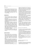
Re-entrant tachycardia
An abnormal electrical circuit (‘re-entry circuit’) may be present within the
heart and consists of one or more of the following components: atrial myocar-
dium, ventricular myocardium, atrioventicular (AV) node, accessory AV
pathway (Figure 16.1). The common feature of re-entrant arrhythmias is that a
depolarizing impulse can travel repeatedly around the re-entry circuit, generat-
ing one heart beat for each cycle. The greater the distance that the impulse has to
travel around the re-entry circuit, the more likely it is that each part of the circuit
will have recovered electrical excitability by the time the impulse returns for the
next cycle. This condition for sustained re-entry can be expressed as follows:
Length of re-entry circuit (mm) > Impulse propagation speed (mm/ms) ×
Refractory period (ms)
The physiological changes of pregnancy make it more likely that this condition
will be fulfilled. Dilatation of the cardiac chambers increases the length of a
re-entrant circuit, and the increased catecholaminergic tone reduces the
refractory period.
Re-entrant tachycardias are more common than focal tachycardias and tend
to have a more stable heart rate. Class I and III anti-arrhythmic drugs, which act
Heart rhythm disorders 219
on atrial and/or ventricular myocardium, tend to be more effective. They work
by prolonging the myocardial refractory period and thus preventing the condi-
tion for sustained re-entry described above.
Ion channelopathy
Mutations of the genes coding for cardiac K
+
and Na
+
channel proteins result
in impaired and delayed cardiac repolarization and cause various forms of
congenital long QT syndrome. Genetic polymorphisms may also underlie
the susceptibility of individuals to drug-induced and other forms of the
acquired long QT syndrome. Other ion channel mutations have been implicat-
ed as the cause of some cases of familial atrial fibrillation and Brugada
syndrome, in which affected individuals have a characteristic ECG with
partial right bundle-branch block and ST segment elevation in leads V1–3,
and are prone to syncope and sudden death as a result of ventricular
tachycardia.
220 Chapter 16
A
FED
CB
r atrium
ivc
svc
tricuspid valve
sa node
r ventricle
aorta
l ventricle
cs ostium
av node
l atrium
pa
mitral valve
pv ostia
accessory
pathway
infarct
scar
surgical
atriotomy
scar
Figure 16.1 Diagram of re-entrant circuits. Panel A: anatomy of diagram. av,
atrioventricular; cs, coronary sinus; ivc, inferior vena cava; l, left; pa, pulmonary artery;
pv, pulmonary vein; r, right; svc, superior vena cava. Panel B: AVNRT re-entrant circuit
in the region of the AV node. Panel C: Reentrant circuit for the common form of atrial
flutter which is anticlockwise around the tricuspid annulus in the right atrium. Panel D:
Atrioventricular re-entrant circuit using an accessory pathway to complete the
retrograde limb of the circuit in a patient with Wolff–Parkinson–White syndrome. Panel
E: Reentrant ventricular tachycardia around a region of infarct scar in the left ventricle.
Panel F: Reentrant tachycardia around an old healed surgical incision in the right
atrium.
The effect of pregnancy on cardiac ion channels, long QT syndrome and poly-
morphic ventricular tachycardia has not been studied in detail. The preponder-
ance of long QT syndrome in women, despite most cases being autosomal
dominant, strongly suggests that expression of the condition is dependent on
sex hormones, which in turn suggests that expression of the relevant cardiac ion
channels may be affected by the changing hormonal milieu of pregnancy.
Heart rhythm disorders 221
Tachycardia mechanisms: practice points
Focal tachycardias
• Frequent ectopic beats of same QRS morphology as tachycardia
• Tendency for frequent ‘stop–starts’
• May be exacerbated by exercise and increased catecholaminergic tone
• Usually the heart is structurally normal
• Typically respond to beta blockers and verapamil
• Cardioversion is often unhelpful; early re-initiations are common.
Re-entrant tachycardias
• Re-entry is the most common tachycardia mechanism
• The echocardiogram and resting ECG may be normal or show evidence of
underlying disease
• Ectopic beats occur occasionally, may initiate and terminate tachycardias,
and are usually different from the morphology of tachycardia
• Class I and III anti-arrhythmic drugs are useful, particularly when the AV
node is not part of the re-entrant circuit
• Cardioversion is an option.
Ion channelopathies and long QT syndrome
• There are abnormalities of the ST segment and/or T wave on the resting
ECG
• Syncope and cardiac arrest may occur
• Polymorphic ventricular tachycardia and pause-dependent initiation
• Acquired forms are related to certain drug classes or electrolyte depletion
• Familial tendency
• Beta blockers are effective, but other anti-arrhythmic drugs should be
avoided because they make the problem worse.
Clinical presentation and investigation
History
Palpitations are the most common presenting symptom, are usually intermit-
tent and only rarely indicate a serious problem. From the history, the inter-
mittent thumping and missed beats caused by ectopic beats can be readily
distinguished from the rapid palpitation of tachycardia. Ectopic beats that are
most noticeable at rest but disappear during physical exertion are benign.
The irregularity of atrial fibrillation distinguishes it from regular tachycardia.
An abrupt onset of symptoms at the start of an episode is common to many
tachycardias, but an abrupt cessation of symptoms, either spontaneously or
with a self-administered vagotonic maneuver such as breath-holding, straining
or taking a cold drink, is fairly typical for supraventricular tachycardia (SVT),
and may help distinguish this from a sinus tachycardia, which typically slows
down over a few minutes.
Presyncope or syncope at the start of a first episode of SVT, with rapid recov-
ery of consciousness, is quite common, but occurs rarely with recurrent SVT be-
cause the patient learns to recognize the warning symptoms and take action to
avoid syncope by sitting or lying down. Presyncope or syncope at the end of an
episode of palpitations may suggest an ‘offset’ pause resulting from delay in re-
sumption of sinus rhythm; this is a marker of intrinsic sinus node disease which
may be exacerbated by beta blockers.
Recurrent syncope with or without palpitation is a worrying symptom be-
cause the fetus may be endangered by the reduction in placental blood flow,
and, in some cases, it may be a harbinger of sudden maternal death. Syncope
caused by cardiac arrhythmia involves complete and abrupt loss of conscious-
ness, and frequently results in injury. Syncope that occurs during or immedi-
ately after exertion is worrying and indicates a catecholaminergic-dependent
arrhythmia mechanism. In contrast, syncope caused by vasovagal mechanisms
is typically more gradual in onset, and the patient is able to avoid injury. When
the loss of cerebral blood flow is prolonged, the patient may suffer a secondary
convulsion, leading to a misdiagnosis of epilepsy. If there is no head injury or
convulsion, recovery of consciousness and full orientation are rapid and occur
within a few minutes. The presence of residual focal neurological deficit
raises the possibility of a neurological cause of syncope and warrants urgent
investigation.
Patients with arrhythmia may also present with fatigue, breathlessness,
peripheral edema and chest discomfort resulting from cardiac insufficiency.
Symptoms of thromboembolism may be the presenting feature of atrial fibrilla-
tion or atrial flutter.
A history of previous heart disease increases the likelihood that an arrhyth-
mia is threatening. Inquiry should be made about the family history,
particularly with reference to cases of premature sudden death. Patients with
congenital heart disease that has been surgically treated in childhood are now
frequently surviving to adulthood. They are particularly vulnerable to arrhyth-
mias, which may be hemodynamically compromising and warrant special
consideration. Patients who have received atrial surgery, such as a Mustard pro-
cedure for transposition, or a Fontan procedure, are particularly vulnerable to
atrial flutter, as are those with any cause of right ventricular impairment.
Patients who have undergone correction of Fallot’s tetralogy may experience
atrial flutter or ventricular tachycardia arising from the right ventricular
outflow tract, particularly if the correction was incomplete and there is a
residual hemodynamic abnormality.
222 Chapter 16
Examination
The pulse may be abnormal during symptoms, variation in the intensity of
the first heart sound, and intermittent cannon waves in the jugular venous
pulse during symptoms, suggest AV dissociation, and are features of third-
degree AV block or ventricular tachycardia. The clinician should focus on look-
ing for signs of heart disease that may be associated with arrhythmia, including
scars from previous surgery, murmurs of structural heart disease and signs of
cardiac failure. It is also important to look for systemic problems such as thyro-
toxicosis that may manifest as an arrhythmia.
Resting 12-lead ECG
A patient without previous heart disease will usually have a normal ECG be-
tween episodes of arrhythmia. Infrequently, a patient may have 12-lead ECG
abnormalities indicative of primary ‘electrical’ disease, such as frequent ectopic
beats or Wolff–Parkinson–White syndrome (Figure 16.2).
In patients with suspected bradycardia, abnormalities that should be sought
include evidence of sinus node disease such as resting sinus bradycardia or in-
termittent pauses, and conduction system disease causing prolongation of the
P–R interval, QRS-axis deviation or bundle-branch block.
A patient with previous heart disease may have an abnormal resting ECG that
reflects their condition and any surgical intervention that they may have re-
ceived in the past. There may be Q waves from previous myocardial infarction,
increased QRS voltage and QRS axis shift, with repolarization changes caused by
ventricular hypertrophy, P wave abnormalities associated with atrial enlarge-
ment and right bundle-branch block in patients with repaired Fallot’s tetralogy.
The 12-lead ECG during arrhythmia
The 12-lead ECG recorded during symptoms is most helpful for arrhythmia di-
agnosis, but is not always available. A tachycardia with chaotically irregular QRS
complexes is usually the result of atrial fibrillation. Less frequently, it is caused by
atrial tachycardia or flutter with variable AV conduction. A regular narrow QRS
complex (<120 ms) tachycardia (rate > 100/min) is either the result of sinus
tachycardia if normal P waves precede each QRS complex, or of SVT if there are
no visible P waves or the P waves are abnormal (Figure 16.3). Vagotonic maneu-
vers such as carotid sinus massage or adenosine injection may help differentiate
sinus tachycardia from SVT. A regular broad QRS complex (>12 ms) tachycardia
is usually the result of SVT with bundle–branch aberrant conduction or ventric-
ular tachycardia, or SVT with pre-excitation caused by an accessory pathway
(Table 16.1). In a patient with a pacemaker, broad QRS tachycardia may be the
result of a pacemaker-mediated tachycardia. A careful search should be made for
pacing spikes on the ECG, which may be very low amplitude if the pacing system
is bipolar and the ECG is highly filtered.
Bradycardia is the result of either reduced sinus node automaticity (P–P
intervals >1s on ECG), or second- or third-degree AV block (fewer QRS com-
plexes than P waves on surface ECG with P wave rate < 100/min).
Heart rhythm disorders 223
224 Chapter 16
(a)
(b)
Figure 16.2 (a) 12-lead ECG from a 34-year-old pregnant woman with syncope and
a structurally normal heart. There are frequent ventricular ectopic beats with a left
bundle-branch block (LBBB) morphology and a QRS axis of around +100°, indicating
a right ventricular outflow tract origin. The patient was successfully treated with beta
blockers during pregnancy and had a successful ablation after delivery. (b) 12-lead ECG
from a 31-year-old pregnant women with frequent tachycardias and Wolff–Parkinson–
White syndrome. Delta waves (slurring of the upstroke to the QRS complex) together
with a short P–R interval are seen in this ECG during sinus rhythm. The tachycardias
were controlled with flecainide plus atenolol, and the pathway was successfully ablated
after delivery.
Prolonged ECG recording
Arrhythmia symptoms are typically intermittent and a 12-lead ECG recording
during symptoms may not be available. Prolonged ECG monitoring with either
inpatient bedside monitoring or a portable Holter monitor may pick up an
episode in a patient with frequent symptoms (Figure 16.4). Where symptoms
are less frequent, it is appropriate for the patient to wear a cardiac rhythm event
monitor for 7 days or longer, with a patient-activation function for use during
episodes of symptoms. It is helpful for the patient to keep a diary of symptoms,
which can then be related to the recorded rhythm. Most types of recorder will
also record asymptomatic episodes when the heart rate falls outside certain pre-
programmed limits. The patient should be encouraged to pursue all her normal
activities during the recording, in particular those activities that have pre-
viously triggered her symptoms.
Implantable loop recorders are increasingly being used to diagnose unex-
plained syncope. There is no published experience of the use of these devices in
pregnancy but there is no contraindication to their use. The battery life is typi-
cally around 18 months and the diagnostic yield is more than 50% in most pub-
lished series in non-pregnant patients.
Echocardiography
Echocardiography is of undisputed value in the diagnosis and follow-up of
structural and functional heart disease, and should be considered an integral
part of the investigation of any pregnant patient with arrhythmia. It is non-
invasive and poses no risk to the fetus. It is the best way to exclude puerperal
cardiomyopathy.
Exercise ECG
Exercise ECG testing can reasonably be used during pregnancy except in cases
where bedrest is indicated for obstetric reasons, and is particularly useful in
Heart rhythm disorders 225
Figure 16.3 Supraventricular tachycardia in a 36-year-old pregnant woman. There
is a narrow QRS tachycardia, at 230 beats/min with no clearly discernible P waves. The
acute episode was treated with adenosine, and her subsequent ECGs were normal. She
had no further episodes and did not require additional anti-arrhythmic drug treatment.
She was offered ablation after delivery, but declined.
cases where symptoms are repeatedly provoked by exercise, but the arrhy-
thmia has not been documented by other means. Care should be taken
during the test not to exceed greatly the patient’s accustomed maximal
level of physical exertion during her daily routine. Hypotension during
exercise poses a risk to the fetus and should result in immediate cessation of
the test.
226 Chapter 16
Table 16.1 Criteria that distinguish ventricular tachycardia from SVT with bundle-
branch block aberrant conduction in cases of broad QRS complex tachycardia
Ventricular tachycardia likely
Past history of:
Myocardial infarction
Cardiomyopathy
Ventricular surgery (e.g. Fallot repair)
Other ventricular damage or scarring
Examination shows:
Variable intensity of the first heart sound
Intermittent cannon waves in the JVP
Tachycardia ECG shows:
Dissociated P waves
Fusion and capture beats
QRS concordance in the pre-cordial leads
QRS morphology atypical for bundle-branch block
Sinus rhythm ECG shows:
Broad QRS complex with a substantially different pattern to tachycardia QRS complexes
No response to carotid sinus massage of high doses of adenosine
SVT will bundle-branch aberrant conduction likely
Past history of:
Recurrent similar episodes over many years with no evidence of disease progression
Previous known SVT
Rapid throbbing in neck in time with pulse
Tachycardia ECG shows:
Typical RBBB or LBBB pattern
Sharp upstroke/downstroke to QRS complexes
Sinus rhythm ECG shows:
No abnormality
Wolff–Parkinson–White syndrome
Tachycardia slows transiently or terminates with carotid sinus massage or adenosine
JVP, jugular venous pulse; LBBB, left bundle-branch block; RBBB, right bundle-branch block; SVT,
supraventricular tachycardia.
Heart rhythm disorders 227
(a)
Figure 16.4 (a) Recording from a bedside monitor in a 33-year-old pregnant woman
who had recurrent syncopal episodes at 32 weeks of pregnancy. A dual chamber
pacemaker was implanted while the abdomen was shielded, and X-ray exposure kept to
a minimum.
Tilt-table testing
Tilt-table testing is useful to confirm the presence of vasovagal mechanisms as
the cause for recurrent syncope when this is suspected from the history. As the
test may induce quite profound and long-lasting hypotension, there are some
concerns that it may endanger the fetus. However, there are reports that it can
be safely carried out.
10
Vasovagal syncope rarely presents for the first time
during pregnancy because women are relatively protected by the increased
circulating blood volume and catecholaminergic tone. It is more commonly
seen in the postpartum period, when rapid fluid losses, relative intravascular
volume depletion and readjustment to non-pregnant physiology may increase
vulnerability.
Invasive cardiac electrophysiological testing and catheter ablation
Electrophysiological testing is nowadays usually undertaken as a prelude to
catheter ablation of tachycardia, but both procedures confer risk to the fetus as
a result of exposure to ionizing radiation. In nearly all cases, the arrhythmia can
successfully be managed till delivery by drugs, and ablation can be undertaken
subsequently. In patients with uncontrollable and life-threatening arrhy-
thmias, ablation can be undertaken. Exposure of the fetus to radiation can
be minimized by lead shielding of the mother’s abdomen, and the use of
228 Chapter 16
(b)
Figure 16.4 Continued (b) Ambulatory Holter monitor recording from a patient with
paroxysmal atrial fibrillation, probably caused by a pulmonary vein focus and a
structurally normal heart. The top ECG strip shows sinus rhythm with frequent atrial
ectopic beats. The P waves of the ectopic beat fall on the T waves of the preceding beats,
and so are not clearly seen. The middle strip shows atrial fibrillation. The bottom strip
shows a rapid atrial flutter. The patient’s symptoms were controlled with beta blockers
and she received thromboprophylaxis with aspirin. The arrhythmias subsided
spontaneously after delivery.
echocardiography or non-fluoroscopic catheter locator systems (such as
CARTO or EnSite NavX) to guide catheter placement.
Pharmacological challenge
In certain circumstances, a pharmacological challenge may provide important
diagnostic information. The use of adenosine during narrow QRS-complex
tachycardia will either terminate the tachycardia (AV nodal re-entrant tachy-
cardia or AVNRT, AV re-entrant tachycardia or AVRT, some cases of atrial tachy-
cardia) or transiently slow the tachycardia before it resumes at its former rate
(atrial flutter, atrial tachycardia, sinus tachycardia) (Figure 16.5). In cases
where the SVT does not terminate, slowing of tachycardia is usually sufficient to
reveal the characteristics of the underlying P waves or flutter waves to allow the
correct diagnosis. In broad QRS-complex tachycardia, adenosine can usefully
be used to differentiate between SVT (slowing or termination) and VT (no ef-
fect). Adenosine selectively slows AV nodal conduction, while not affecting ac-
cessory pathway conduction. It may therefore reveal latent pre-excitation in
patients with Wolff–Parkinson–White syndrome, where pre-excitation is not
apparent on the resting 12-lead ECG.
In some patients, Brugada syndrome may be considered, because of either a
family history of sudden death or typical or suggestive ECG changes. A fle-
cainide provocation test may be helpful in elucidating the characteristic ECG
changes where the diagnosis is in doubt (Figure 16.6).
Genetic testing
Several cardiac conditions increase the vulnerability to arrhythmias and have a
defined genetic basis. The list is growing and includes long QT syndrome, Bru-
gada syndrome, hypertrophic cardiomyopathy, familial dilated cardiomyopa-
thy and arrhythmogenic right ventricular dysplasia. Although routine genetic
testing for these conditions is not currently available for the evaluation of ar-
rhythmia risk in pregnancy, a detailed family history should always be taken,
Heart rhythm disorders 229
Adenosine 12 mg
Figure 16.5 Rhythm strip from a pregnant women with a previous mitral valve
replacement who had an abrupt onset of a narrow QRS complex tachycardia. The
diagnosis of atrial flutter became clear when the ventricular rate was transiently slowed
by intravenous adenosine.
and include specific questioning about premature sudden death. Counseling
about the risk of transmission of these conditions to offspring is essential, and it
is likely that there will be an increasing role for pre-implantation diagnosis of
these conditions in affected families.
Anti-arrhythmic drugs
The physiological changes of pregnancy affect drug absorption, action and met-
abolism in several ways. It may therefore be difficult to achieve adequate thera-
peutic drug levels while avoiding toxicity. This may explain why some women
experience recurrence of symptoms of arrhythmia during pregnancy despite
continuing therapy that had previously been effective.
4
The greatest risk of drug-induced congenital malformation occurs during
fetal organogenesis, which occurs during weeks 3–11, and is therefore complete
by the end of the first trimester. Thereafter, the risk is mainly of impaired growth
and functional development, or direct toxicity to fetal tissues. Drugs given
shortly before term or during labour may have adverse effects on labour or the
neonate after delivery. Most antiarrhythmic drugs are categorized by the US
Food and Drug Administration (FDA) as category C during pregnancy which
signifies that:
‘. . . risk (to the foetus) cannot be ruled out. Adequate well-controlled
human studies are lacking, and animal studies have shown a risk to the
foetus or are lacking as well. There is a chance of foetal harm if the drug is
administered during pregnancy; but the potential benefits may outweigh
the risks.’
4,5
230 Chapter 16
Figure 16.6 A patient with recurrent syncope and a normal echocardiogram. Subtle
non-specific change on the baseline ECG became markedly abnormal after the
administration of flecainide 80 mg intravenously. The ST-segment elevation in leads
V1–3 is diagnostic of Brugada syndrome. This is associated with the risk of sudden death
and this patient received an implantable cardioverter defibrillator.
Drug treatment should be used if there is either hemodynamic compromise,
or a risk of tachycardia cardiomyopathy, or there are other disabling symptoms.
Drug treatment is not required for a benign tachycardia that is well tolerated. A
low dose should be used initially with titration according to response, and this
must be accompanied by regular monitoring. The selection of an appropriate
drug depends on knowledge of the arrhythmia mechanisms, and only those
drugs that have a safe track record in pregnancy should be used.
Adenosine
Adenosine is useful for the emergency management of SVT and broad QRS-
complex tachycardias, and has been used safely in pregnant women.
4,5,11
It is
administered as a bolus dose and it has a very short duration of action of no more
than 5–10 s. It depresses sinus and AV nodal function causing transient brady-
cardia and AV block in the mother, but it has no detectable effect on the fetal car-
diac rhythm. Adenosine is contraindicated in patients with brittle asthma, in
whom it may cause bronchospasm, and in those taking dipyridamole because of
the risk of prolonged asystole.
Digoxin
There is a long history of digoxin use during pregnancy and it is considered
to be safe.
5
It crosses the placenta, but is not teratogenic. It is renally cleared,
although renal excretion is inhibited by concomitant use of amiodarone.
It is mainly used for the control of ventricular rate in patients with persis-
tent atrial fibrillation, but may also be effective in some cases of focal atrial
tachycardia.
Beta blockers
Propranolol is the beta blocker with the longest track record and is considered
safe in pregnancy.
5
Beta blockers are not teratogenic, but beta
1
-selective block-
ers such as atenolol or metoprolol may be preferred because they may interfere
less with beta
2
-receptor-mediated uterine relaxation. However, beta
1
-selective
agents achieve less complete cardiac beta blockade because there are functional
beta
2
-receptors on cardiac myocytes, and these may therefore be less effective
anti-arrhythmic agents. There have been reports linking beta blockers to fetal
bradycardia, hypotonia, hypoglycemia and intrauterine growth retardation.
Sotalol
Sotalol is a combined beta blocker and class III anti-arrhythmic drug. It does
cross the placenta and is renally excreted. It has achieved class B classification
with the FDA and its use in pregnant patients has been reported with no adverse
outcome.
4,5
Flecainide
Flecainide has been used frequently during pregnancy and is a reasonable
choice for patients with a structurally normal heart.
4,5
It should probably be
Heart rhythm disorders 231
avoided in those with myocardial disease, and in particular patients with ven-
tricular tachycardia or vulnerability to myocardial ischaemia.
Amiodarone
The conflict between the interests of the mother and the well-being of the
fetus is thrown into sharp relief with amiodarone. Amiodarone is a very effec-
tive anti-arrhythmic drug to treat and prevent life-threatening ventricular ar-
rhythmias in patients with ventricular disease. However, there is a real concern
about fetal hypothyroidism and brain damage.
12,13
Amiodarone crosses the pla-
centa and achieves fetal concentrations of around 10% of maternal serum val-
ues.
14
Maternal amiodarone use may cause a goiter, which in turn compromises
the upper airway in the neonate. For these reasons, amiodarone should be used
only when the mother’s life is significantly threatened and no other agent
will do.
4,5
Verapamil
There are no reports of teratogenicity, but verapamil does cross the placenta and
may have cardiovascular effects in the fetus.
15
Intravenous verapamil is a useful
alternative to adenosine for emergency termination of SVT, and oral verapamil
may be used in patients to prevent SVT recurrence when beta blockers are con-
traindicated or not tolerated.
4
DC cardioversion
DC cardioversion may be used to terminate sustained tachycardias. It should be
carried out with general anesthesia, or deep sedation with midazolam or di-
azepam. Traditionally, the cardioversion electrodes are placed at the right ster-
nal edge and cardiac apex, but for atrial fibrillation, it may be more effective to
use an anterior–posterior configuration. Firm downward pressure on the ster-
nal paddle reduces the electrode separation and increases the intensity of the
electrical field, thus maximizing the chance of success. A waveform that re-
verses polarity during delivery (biphasic waveform) achieves cardioversion at
energy thresholds that are half those required when a monophasic waveform is
used. For all tachyarrhythmias except ventricular fibrillation, the shock should
be synchronized to the R wave to minimize the risk of inducing ventricular
fibrillation.
Patients with atrial flutter or fibrillation are particularly vulnerable to sys-
temic thromboembolism after restoration of sinus rhythm. DC cardioversion
should not be carried out on patients who have been in atrial fibrillation for
longer than 24 hours unless the arrhythmia results in serious cardiovascular
compromise, or the patient has been fully anticoagulated since the onset of ar-
rhythmia or the absence of thrombus in the left atrium has been verified by
transesophageal echocardiography. Anticoagulation should be continued for a
minimum of 4 weeks after DC cardioversion.
DC cardioversion seems to be quite safe in all stages of pregnancy because the
intensity of the electrical field to which the fetus is exposed is low. Nevertheless,
232 Chapter 16
the fetus should be carefully monitored throughout the procedure. In the latter
stages of pregnancy, some anesthetists prefer to carry out the procedure using
full general anesthesia and intubation in view of the more difficult airway and
increased risk of gastric aspiration.
Heart rhythm disorders 233
Practice points
Tips for DC cardioversion during pregnancy
• Anticoagulation is needed in patients with atrial flutter or fibrillation
• DC cardioversion when should be performed under a general anesthetic
• Fetal monitoring is necessary
• Anterior and posterior paddle positions work best for atrial arrhythmia
• Apply firm pressure on the anterior paddle
• Use a defibrillator with a biphasic waveform if available
• Start with 50 J for regular tachycardias
• Start with maximal output (e.g. 360 J) for atrial fibrillation.
Implantable cardioverter defibrillators
Women with an implanted cardioverter defibrillator (ICD) may undergo preg-
nancy successfully with a good reported outcome. Potentially threatening ar-
rhythmias are promptly detected and automatically terminated by an ICD by
either a series of rapid pacing impulses delivered via an endocardial right ven-
tricular pacing lead or delivery of a synchronized shock between a coil electrode
in the right ventricular cavity and a second electrode formed by the ICD box,
which is located in the pre-pectoral position on the left. Prompt detection and
termination of an arrhythmia by an ICD minimize the hemodynamic distur-
bance and thereby limit the risk of harm to the fetus. ICDs are configured to con-
centrate the maximal electrical field strength to the mother’s ventricular
myocardium, and the electrical energy to which the fetus is exposed is minimal.
Delivered energies (2–40 J) are about a tenth of those used for external DC car-
dioversion. One study of 44 women with ICDs who became pregnant showed
an 82% rate of successful completion of pregnancy without complication.
There were no maternal deaths, and only one stillborn child, despite eight
women receiving one or more ICD shocks.
16
Management of specific arrhythmias
Bradycardia
Bradycardia results from either dysfunction of the sinus node characterized by
a P-wave rate of <60/min on the ECG, or high-grade (second- or third-degree)
AV block where some or all P waves are not conducted.
The presence of new-onset sinus bradycardia with a resting heart rate of
<60 beats/min may indicate hypothyroidism or hypothermia. Heart rate-
slowing drugs such as beta blockers, calcium channel blockers or digoxin may
cause moderate sinus bradycardia of 45–60 beats/min at therapeutic concentra-
tions. Extreme bradycardia of <45 beats/min is often associated with symptoms
of excessive fatigue, effort intolerance, pre-syncope or syncope, and may result
from high doses of rate-slowing drugs, or even from moderate doses in patients
with underlying sinus node disease. Intermittent sinus bradycardia may be seen
in cases of sinoatrial disease, or occur with heightened vagal tone in patients
susceptible to vasovagal syncope. Sinus bradycardia and sinus pauses may
occur in patients with obstructive sleep apnea.
Congenital complete heart block is usually detected in childhood, and
asymptomatic patients with a mean heart rate of >50 beats/min probably do not
require a pacemaker. Acquired second- or third-degree AV nodal block is us-
ually the result of idiopathic conduction system disease but may occasionally
have an identifiable cause. Patients with symptomatic bradycardia that cannot
be alleviated by addressing an underlying cause require implantation of a per-
manent pacemaker (Table 16.2).
Ectopic beats
An increase in the frequency of atrial and ventricular ectopic beats occurs very
commonly during pregnancy. When these are symptomatic, a 12-lead ECG
during symptoms is helpful to define the origin. A Holter monitor may show
runs of ectopic beats and tachycardia. An echocardiogram is helpful for reassur-
ance when symptoms are persistent and troublesome, and is usually normal.
Reassurance is usually sufficient, but propranolol or another beta blocker can
be used if necessary.
Sinus tachycardia
Sinus tachycardia >100 beats/min is not uncommon during pregnancy and a
rate that is persistently in excess of 110/min should prompt consideration of un-
derlying causes of secondary tachycardia, including infection, inflammatory
disease, thyrotoxicosis and cardiomyopathy. A Holter monitor may be helpful
to differentiate the normal circadian variation in heart rate of a sinus tachycar-
dia from the virtually fixed heart rate of incessant atrial tachycardia. This dis-
tinction is important because a ventricular rate that remains high throughout
234 Chapter 16
Table 16.2 Causes of AV nodal block
Idiopathic
Congenital complete heart block, maternal lupus with anti-Ro anti-La antibodies
Vagal overactivity
Myocardial ischemia or infarction
Electrolyte imbalance
Drug toxicity
Iatrogenic post-cardiac surgery or post-catheter ablation
Infective endocarditis
Lyme disease
Sarcoidosis
Amyloidosis
the night may result in tachycardia cardiomyopathy, and therefore requires
treatment. Debilitating sinus tachycardia associated with pregnancy may be
treated with propranolol and typically resolves within days of delivery.
Supraventricular tachycardia
Supraventricular tachycardia is readily diagnosed when the QRS complexes are
narrow, regular and rapid, and the P waves abnormal or absent (see Figure
16.3). Broad QRS-complex tachycardia may be caused by SVT with aberrant
conduction in a left or right bundle-branch block pattern, or by VT. Discrimina-
tion between SVT and VT in a case of broad QRS-complex tachycardia is guided
by the characteristics indicated in Table 16.1.
Atrial flutter is usually distinguishable from other forms of SVT by the typical
saw-tooth pattern of the baseline, best seen in ECG leads II, III and aVF (Figure
16.7). AVNRT, AVRT and atrial tachycardia typically present with regular nar-
row QRS-complex tachycardia in a patient with an otherwise normal heart.
Analysis of the onset and termination of the arrhythmia, the P-wave morphol-
ogy and relationship of the P wave to the QRS complex, and the response to
adenosine, will often allow differentiation between different SVT mechanisms,
but this is not always the case. Whatever the precise mechanism, vagotonic ma-
neuvers such as carotid sinus massage may terminate the episode, and can be
self-administered by the patient to deal with recurrences. When vagotonic ma-
neuvers fail, intravenous bolus adenosine can be used, with escalating boluses
up to a maximum of 18–24 mg until the desired response is achieved.
Heart rhythm disorders 235
Figure 16.7 Atrial flutter with 2: 1 atrioventricular conduction. The typical sawtooth
pattern flutter waves are seen in leads II, III and aVF. The flutter waves occur at
300/min, and in this case every alternate wave is conducted to the ventricle, giving a
ventricular rate of 150/min.
Wolff–Parkinson–White syndrome
Wolff–Parkinson–White (WPW) syndrome is diagnosed on the basis of the
12–lead ECG in sinus rhythm where delta waves are seen (see Figure 16.2).
These are inscribed at the beginning of the QRS complex and cause a slurring of
the initial up- or downstroke of the QRS complex, together with an increase in
the width of the QRS complex and shortening of the P–R interval. The delta
wave represents ‘pre-excitation’ of a portion of the ventricular myocardium via
an accessory AV pathway. Accessory pathways are located around the annuli of
the mitral or tricuspid valves. In up to 10% of cases, there is more than one ac-
cessory pathway.
In most cases, the heart is otherwise normal, but WPW syndrome may be as-
sociated with hypertrophic cardiomyopathy or Ebstein’s anomaly. Patients
with WPW syndrome are prone to AVRT (see Figure 16.1), which may become
more frequent during pregnancy. WPW syndrome patients are also vulnerable
to atrial fibrillation, which may be conducted with high rates to the ventricles
via the accessory pathway, resulting in ventricular rates that may exceed
300 beats/min in some cases. This is a life-threatening situation with a signifi-
cant risk of degeneration to ventricular fibrillation and cardiac arrest. Drugs that
modulate AV nodal function, such as beta blockers, verapamil and digoxin, are
useless in this situation, and may even enhance conduction via the accessory
pathway. A class I drug such as flecainide will suppress or block conduction via
the accessory pathway and also have an anti-fibrillatory action on the atria, and
this is therefore the drug of choice for the emergency treatment of this condition
and prevention of recurrence. Patients should be referred for catheter ablation
of the pathway after delivery.
A patient with a WPW syndrome pattern on the ECG who has never had
symptoms of arrhythmia does not require treatment. If the patient has had
self-limiting palpitations but no documented tachycardia, assessment with
Holter monitoring is appropriate. Sometimes, delta waves are present only
intermittently, and this is a reassuring finding that indicates that the risk is
very low.
Atrial flutter and fibrillation
Atrial flutter and fibrillation are uncommon in pregnant women who do not
have structural heart disease. Heart conditions that increase hemodynamic
stress to the left atrium (e.g. mitral stenosis) tend to cause fibrillation, whereas
those affecting the right atrium (e.g. a Fontan circulation) tend to cause atrial
flutter. When the atria fibrillate or flutter, blood within the left atrial appendage
stagnates and may thrombose. The thrombus thus formed is often only loosely
adherent to the atrial endocardium, and may fragment and embolize to the sys-
temic arterial tree. The risk of thromboembolism and stroke is compounded by
the presence of mitral stenosis, left atrial dilatation or impaired ventricular
function, or a previous history of thromboembolism. The risk of embolization is
particularly high in the first few days after cardioversion to sinus rhythm, as
coordinated atrial contractile function gradually returns to normal, and may
236 Chapter 16