Báo cáo y học: "Association between regulatory T cell activity and sepsis and outcome of severely burned patients: a prospective, observational study" doc
Bạn đang xem bản rút gọn của tài liệu. Xem và tải ngay bản đầy đủ của tài liệu tại đây (725.89 KB, 10 trang )
Huang et al. Critical Care 2010, 14:R3
/>Open Access
RESEARCH
© 2010 Huang et al.; licensee BioMed Central Ltd. This is an open access article distributed under the terms of the Creative Commons
Attribution License ( which permits unrestricted use, distribution, and reproduction in
any medium, provided the original work is properly cited.
Research
Association between regulatory T cell activity and
sepsis and outcome of severely burned patients: a
prospective, observational study
Li-feng Huang, Yong-ming Yao*, Ning Dong, Yan Yu, Li-xin He and Zhi-yong Sheng
Abstract
Introduction: To investigate the significance of changes in regulatory T cells (Tregs) activity and its relationship with
sepsis, as well as outcome of patients with major burns.
Methods: The periphery blood samples of 106 patients were collected on post-burn days 1, 3, 7, 14, and 21. Tregs were
isolated and their phenotypes (cytotoxic T-lymphocyte-associated antigen 4 and forkhead/winged helix transcription
factor p3) were analyzed by flow cytometry, and the contents of cytokines (interleukin-10 and transforming growth
factor-β1) released into supernatants by Tregs were also determined by enzyme-linked immunosorbent assay kits.
Gene expressions of cytokines were assessed by real-time quantitative polymerase chain reaction.
Results: Expressions of Tregs phenotypes and gene/protein expression of cytokines were all elevated after burn, and
there were obvious differences among patients with various burn sizes. They were also higher in septic patients than
those without sepsis. Among septic patients, the expressions of Tregs phenotypes and the levels of cytokines were
markedly lower in the survival group than those in patients with fatal outcome.
Conclusions: Severe burn injury per se could lead to the changes in Tregs activities. Elevated levels of cytokines
produced by Tregs and activation markers on Tregs surface might play an important role in the pathogenesis of sepsis
and mortality in burned patients.
Introduction
There is accumulating evidence indicating that regulatory T
cells (Tregs) play important roles in the maintenance of
immunologic self-tolerance and in down-regulation of vari-
ous immune responses [1]. Tregs have been shown to be
important in regulating the immune responses in transplant
rejection, tumor immunity, infectious diseases and allergy.
Thus, there has recently been an increasing interest in
investigating the biology of Tregs as well as its potential
application in the treatment of immunity relevant illnesses.
Many types of Treg subsets have been reported in a vari-
ety of morbid conditions. It is now clear that immune regu-
latory cells consist of many distinct T cell subsets [2].
Among them, CD4
+
Tregs have been demonstrated in a
wide range of animal models and in humans [3,4], and the
forkhead/winged helix transcription factor p3 (FOXP3) has
been suggested to represent a reliable intracellular marker
for naturally occurring Tregs [5]. Most studies on CD4
+
Tregs use a combination of CD25, cytotoxic T-lymphocyte-
associated antigen 4 (CTLA-4), FOXP3, IL-10 and/or
transforming growth factor-β1 (TGF-β1) to define Treg
populations [6].
The stress response to burn injury is similar to that of
severe trauma or critical illness but differs in its severity
and duration. The inflammatory response is triggered
immediately after thermal injury and persists for almost
five weeks postburn [7]. Superimposed severe infections
can result in the suppression of one or more functions of the
host immune system after major burns. Multiple mecha-
nisms have been proposed to explain infection-induced
immunosuppression, including an imbalance in the cellular
T helper cell (Th1/Th2) or cytokine profile, induction of
anergy, depletion of effector cells and most recently the
activation of CD4
+
CD25
+
Tregs [8]. The role of both natu-
* Correspondence:
1
Department of Microbiology and Immunology, Burns Institute, First Hospital
Affiliated to the Chinese PLA General Hospital, 51 Fu-cheng Road, Haidian
District, Beijing 100048, PR China
Huang et al. Critical Care 2010, 14:R3
/>Page 2 of 10
rally occurring CD4
+
CD25
+
Tregs and IL-10-secreting
Tregs in infection has been the subject of several recent
excellent reviews [9,10]. However, it seems that its
response to trauma, burns, hemorrhagic shock, and micro-
bial infection is associated with only a transient proinflam-
matory period followed by a more prolonged period of
immune suppression [11]. Thus, it is speculated that there
are some other factors involved in this process.
Numerous studies show that an increased burn size leads
to higher mortality of burned patients [12,13]. It was also
implicated that the extent of burn size might be associated
with the development of sepsis. It is now believed that
along with the body's hyperinflammatory response desig-
nated to eliminate the invading pathogen, mechanisms pri-
marily aimed at controlling this initial response are
initiated, but may turn out to be deleterious with immune
dysfunctions and even death. A similar state of immune
suppression has been described after numerous forms of
severe trauma [14-16].
Although more and more evidence for immune dysfunc-
tion after sepsis has been accumulated the mechanisms
underlying its development and how it acts to worsen the
morbid state of the critically ill patient are yet to be eluci-
dated. In this context, although the majority of clinical and
basic researches conducted so far have focused on the roles
of myeloid cell populations [17], the contribution of T lym-
phocytes [18,19] and, in particular, of Tregs has been some-
what ignored. Whether CD4
+
CD25
+
Tregs participate
directly in sepsis-induced immunoparalysis resulting in
poor outcomes remains to be investigated. The purpose of
the present study was to investigate the significance of
changes in activity of Tregs in severely burned patients, and
its relation with pathogenesis of sepsis as well as the out-
come of the patients following major burns.
Materials and methods
Participants and demography
One hundred and six patients who were admitted to our
burns unit with total burn surface area (TBSA) larger than
30% were included in the present study over a time period
of 10 months. Patients were resuscitated according to the
Parkland formula using colloid and Ringer's lactate. Within
48 hours of admission all patients had undergone most burn
wound excision for full-thickness burns, and the excision
wounds were covered with available autologous skin, and
allograft was used to cover any remaining open wounds.
Five to ten days after healing of the donor area, the remain-
ing wounds were totally covered with autograft skin.
The thermally injured patients were stratified into three
groups according to burn size: 30 to 49% TBSA burns
(group I, n = 41), 50 to 69% TBSA burns (group II, n = 34),
and more than 70% TBSA burns (group III, n = 31).
According to whether there was development of sepsis or
not, patients were divided into sepsis group (n = 59) and
non-sepsis group based on the criteria for definition of
severe sepsis [20] (n = 47); then the patients with sepsis
were further divided into non-survival group (n = 17) and
survival group (n = 42). Twenty-five healthy volunteers
served as normal controls (17 men and 8 women, mean age
28.6 ± 6.2 years, range 21 to 45 years). The periphery blood
samples were collected on postburn days (PBD) 1, 3, 7, 14,
and 21. The study was reviewed and approved by the Insti-
tutional Review Board of the Burns Institute, First Hospital
Affiliated to the Chinese PLA General Hospital, Beijing,
China. Prior to the study, each patient or the patient's legal
guardian signed a written informed consent form.
Reagents and kits
RPMI 1640, FCS, glutamine, penicillin, streptomycin, and
HEPES were purchased from TianRunShanda Biotech Co.
Ltd (Beijing, China). Human CD4
+
CD25
+
Tregs isolation
kit and human fluorescein isothiocyanate (FITC)-conju-
gated anti-human CD4 (IgG1, Clone M-T466) were pur-
chased from Miltenyi Biotec GmbH (Bergisch Gladbach,
Germany). Antibodies used for flow cytometry analysis,
including FITC-conjugated anti-human CD152 (CTLA-4,
IgG2a, Clone 14D3), FITC-conjugated anti-human FOXP3
(IgG2a, κ. Clone PCH101) were purchased from eBiosci-
ence (San Diego, CA, USA). Total RNA isolation system
and RT-PCR system were purchased from Promega (Madi-
son, WI, USA). SYBR Green PCR Master MIX was pur-
chased from Applied Biosystems (Foster City, CA, USA).
ELISA kits of human IL-10 and TGF-β1 were purchased
from Biosource (Worcester, MA, USA).
Isolation of circulating Tregs
In an EDTA test tube, 10 ml of venous blood was collected.
It was then diluted in Hanks' balanced salt solution, and
Ficoll-Hypaque (Sigma Chemical Co., St. Louis, MO,
USA) was used for isolation and preparation of peripheral
blood lymphocytes. After centrifugation, the sedimentary
cells were collected. The cells were isolated using Micro-
Beads and a MiniMACS™ separator according to the man-
ufacturer's instructions. CD4
+
T cells were enriched by
depletion of cells expressing CD8, CD14, CD16, CD19,
CD36, CD56, CD123, TCRγ/δ and CD235a from lympho-
cytes with a CD4
+
CD25
+
Regulatory T Cell Isolation Kit.
CD4
+
CD25
+
Tregs and CD4
+
CD25
-
T cells were further
selected according to the expression of CD25. The purity of
isolated Tregs and CD4
+
CD25
-
T cells were verified by
flow cytometric analysis with anti-CD4 and anti-CD25
staining. Tregs were then cultured in RPMI 1640 FCS
(10%) overnight for recovery. The supernatants were col-
lected for the determination of IL-10 and TGF-β1 levels.
Flow cytometric analysis
To observe the CTLA-4 expression on the surface of Tregs,
cells were stained with anti-human CTLA-4-FITC antibody
Huang et al. Critical Care 2010, 14:R3
/>Page 3 of 10
for 30 minutes at 4°C in the dark. Concomitantly, for detec-
tion of intranuclear FOXP3, cells were reacted with 1 ml
freshly prepared fixation/permeabilization working solution
for two hours at 4°C. After washing cells with one times
permeabilization buffer twice, cells were stained with anti-
human FOXP3-FITC antibody for 30 minutes at 4°C in the
dark. After washing twice, cells were analyzed by flow
cytometry using a FACScan (BD Biosciences, Mountain
View, CA, USA). The fluorescence intensity was repre-
sented as a mean value.
Cytokine measurements by ELISA
IL-10 and TGF-β1 levels were determined by ELISA,
strictly following the protocols provided by the manufac-
turer. The color reaction was terminated by adding 100 μl of
ortho-phosphoric acid. Plates were read in a microplate
reader (Spectra MR, Dynex, VA, USA). The standard con-
centration curves for both IL-10 and TGF-β1 were from 0
to 2000 pg/ml.
SYBR green real-time RT-PCR
Total RNA was extracted from Tregs using the single-step
technique of acid guanidinium thiocyanate-chloroform
extraction, according to the manufacturer's instructions.
The concentration of purified total RNA was determined
spectrophotometrically at 260 nm. mRNA for IL-10 and
TGF-β1 in Tregs and GAPDH were quantified in duplicate
by SYBR Green two-step, real-time RT-PCR. After the
removal of potentially contaminating DNA with DNase I, 1
μg of total RNA from each sample was used for reverse
transcription with an oligo dT and a Superscript II to gener-
ate first-strand cDNA. PCR reaction mixture was prepared
using SYBR Green PCR Master Mix. Thermal cycling con-
ditions were 10 minutes at 95°C followed by 40 cycles of
95°C for 15 seconds and 60°C for one minute on a
Sequence Detection System (Applied Biosystems, Foster
City, CA, USA). Each gene expression was normalized
with GAPDH mRNA content. Sequences of human primer
for SYBR Green PCR were shown follows: IL-10 (79 bp) -
AAGGCGCATGTGAACTCCC (sense), ACGGCCTT-
GCTCTTG TTTTC (antisense) [21]; TGF-β1 (85 bp) -
TGAACCGGCCTTTCCTGCTTCTCATG (sense), GCG-
GAAGTCAATGTACAGCTGCCGC (antisense) [22];
GAPDH (147 bp) -ACTTCAACAGCGACACCCACT
(sense), GCCAAATTCGTTGTCATACCAG (antisense)
[23].
Statistical analysis
Data were expressed as mean ± standard deviation (SD) and
analyzed with analysis of variance (ANOVA; a mixed-
model, factorial ANOVA). Turkey Test was used to evalu-
ate significant differences between groups. A P value of
0.05 or less was considered to indicate statistical signifi-
cance.
Results
Demographics
One hundred and six patients with burn injury were
included in the present study. The patients' demographics
are illustrated in Table 1. The test for homogeneity of vari-
ance was considered and the ANOVA assumption was met.
The omnibus ANOVA was also found to be significant.
There was no significant difference in age among the
patients with different burn size. However, there were sig-
Table 1: Patient demographics
Variable n Age (years) Burn area
(TBSA%)
Average area
(TBSA%)
III° area
(TBSA%)
Total 106 33.6 ± 4.3 30-99 57.4 ± 5.6 33.3 ± 6.5
Group I 41 33.1 ± 6.9 30-49 39.4 ± 7.4 17.4 ± 3.9
Group II 34 29.3 ± 5.1 50-69 57.7 ± 10.7** 32.9 ± 6.3**
Group III 31 32.4 ± 6.3 70-99 82.6 ± 15.1
&&
63.7 ± 14.5
&&
Sepsis 59 34.3 ± 5.5 30-99 67.2 ± 11.3
##
45.3 ± 8.6
##
Non-sepsis 47 32.8 ± 5.7 30-72 45.0 ± 8.1 18.2 ± 3.8
Survivors 42 35.7 ± 6.8 30-88 56.2 ± 10.2
††
29.2 ± 5.8
††
Non-survivors 17 31.2 ± 10.5 30-99 77.1 ± 23.4 55.2 ± 16.2
Data presented as the mean ± standard deviation or percentage. TBSA = total body surface area (range); III° area = third-degree burn area.
Significant difference between Group II and Group I (**P < 0.01); significant difference between Group II and Group III (
&&
P < 0.01); significant
difference between sepsis group and non-sepsis group (
##
P < 0.01); Significant difference between survivors group and non-survivors (
††
P <
0.01).
Huang et al. Critical Care 2010, 14:R3
/>Page 4 of 10
nificant differences in burn area between Group II and
Group I (P < 0.01). The sepsis group had markedly large
burn areas compared with the non-sepsis group (P < 0.01).
Similarly, burn area in the non-survivors was much larger
than that in the survivors (P < 0.01).
Isolation of CD4
+
CD25
+
Tregs
CD4
+
CD25
+
Tregs were isolated from the peripheral blood
lymphocytes in two steps by a magnetic cell sorting
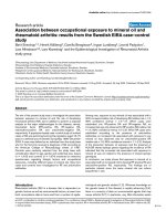
(MACS) system. As shown in Figure 1, the purity of posi-
tively sorted CD4
+
CD25
+
Tregs was 93.5 ± 1.7% with the
survival rate of 96.2 ± 2.9%. The purity of negatively sorted
CD4
+
CD25
-
T cells was 89.6 ± 2.5%.
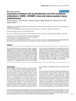
The phenotypic changes in Tregs after burns
To investigate the changes in Treg phenotypes, these cells
were analyzed at different time points and in different
groups after burns. A three (Group) times five (Day)
mixed-model, factorial ANOVA was conducted. As shown
in Figure 2, increased expressions of CTLA-4 and FOXP3
were found to be enhanced on the surface of Tregs from
burned patients on PBD 1 to 21 compared with normal con-
trols, and there were obvious differences among patients
with various burn sizes (P < 0.05 or P < 0.01). The expres-
sions of CTLA-4 and FOXP3 were significantly higher in
patients with serious burns at all time points, and they were
even higher in septic patients than those without sepsis on
PBD 3 to 21 (P < 0.01). Among septic patients, the expres-
sions of CTLA-4 and FOXP3 in the survival group were
significantly lower than those with fatal outcome on PBD 3
to 21 (P < 0.05 or P < 0.01).
Changes in protein and gene levels of cytokines released
by Tregs
The capacity of Tregs to produce IL-10 and TGF-β1, which
are two of the markers of function of Tregs, was analyzed in
Isolation of CD4
+
CD25
+
Tregs from the peripheral blood lymphocytes
Figure 1 Isolation of CD4
+
CD25
+
Tregs from the peripheral blood lymphocytes. CD4
+
CD25
+
regulatory T cells (Tregs) were isolated from the pe-
ripheral blood lymphocytes in two steps by magnetic cell sorting (MACS) system according to manufacturer's instructions. (a) T lymphocytes before
isolation and (b) CD4
+
T cells are shown. (c) The purity of positively sorted CD4
+
CD25
+
Tregs was 93.5 ± 1.7% with the survival rate of 96.2 ± 2.9%. (d)
The purity of negatively sorted CD4
+
CD25-T cells was 89.6 ± 2.5%.
Huang et al. Critical Care 2010, 14:R3
/>Page 5 of 10
the present experiment. As shown in Figures 3 and 4, ele-
vated protein and gene expressions of IL-10 and TGF-β1 in
Tregs from burned patients were detected on PBD 1 to 21 in
comparison with normal controls, and there were marked
differences among patients with different extents of burn
injury (P < 0.05 or P < 0.01). The protein and gene expres-
sions of IL-10 and TGF-β1 in Tregs were significantly
higher in septic patients than those without sepsis on PBD 3
to 21 (P < 0.05 or P < 0.01). Among septic patients, the
expression levels of IL-10 and TGF-β1 in the survival
group were obviously lower than those with non-survival
group on PBD 3 to 21 (P < 0.05 or P < 0.01).
Discussion
Severe burn injury induces detrimental changes in immune
function, often leaving the host highly susceptible to devel-
oping life-threatening opportunistic infections. Advances in
our understanding of how burn influences host immune
response suggest that thermal injury causes a phenotypic
imbalance in the regulation of Th1- and Th2-type immune
responses [24]. The immune response to infection repre-
Flow cytometric analysis of phenotypes of Tregs
Figure 2 Flow cytometric analysis of phenotypes of Tregs. Increased expressions of CTLA-4 and FOXP3 on the surface of regulatory T cells (Tregs)
from burned patients were found on postburn days (PBD) 1 to 21 compared with normal controls, and there were obvious differences among patients
with various burn sizes ((a and b) a mean of all days). The expressions of CTLA-4 and FOXP3 were significantly higher in patients with serious burns
during the whole observational period, and (c and d) they were much higher in septic patients than those without sepsis on PBD 3 to 21. (e and f)
Among septic patients, the expressions of CTLA-4 and FOXP3 in the survival group were obviously lower than those in non-survival group on PBD 3
to 21. * P < 0.05, ** P < 0.01, Group I vs. normal group or sepsis group vs. non-sepsis group or non-survivors vs. survivors; #P < 0.05, ## P < 0.01, Group
II vs. Group I; &P < 0.05, Group III vs. Group II.
Huang et al. Critical Care 2010, 14:R3
/>Page 6 of 10
sents a complex balance between the successful induction
of proinflammatory antipathogen response and anti-inflam-
matory response required to limit damage to host tissues.
The vast majority of clinical and basic science research on
the immune consequences of burn injury and sepsis con-
ducted during the past three decades has focused mainly on
the roles of macrophages, neutrophils, and, to a lesser
extent, conventional T lymphocytes [25,26]. During recent
years, however, it has become increasingly clear that minor
subsets of innate immune cells, innate regulatory lympho-
cytes in particular, are central to processes involved in both
protective immunity and immunopathology [27]. Tregs
undoubtedly play an important role in controlling this bal-
ance during infection, and the results can range from highly
detrimental to the host to highly beneficial to both the host
and pathogen [28].
In our previous observations, significant proliferation of
splenic T cells and IL-2 as well as IL-2Rα expression on T
ELISA analysis of IL-10 and TGF-β1 levels in Tregs supernatants
Figure 3 ELISA analysis of IL-10 and TGF-β1 levels in Tregs supernatants. Elevated protein expressions of IL-10 and TGF-β1 in regulatory T cells
(Tregs) from burned patients were detected on postburn days (PBD) 1 to 21 in comparison to normal controls, and there were obvious differences
among patients with different extent of burn injury ((a and b) a mean of all days). Protein levels of (c) IL-10 and (d) TGF-β1 in Tregs were significantly
higher in septic patients than those without sepsis on PBD 3 to 21. Among septic patients, (e) IL-10 and (f) TGF-β1 levels in the survivors were obvi-
ously lower than those with non-survivors on PBD 3 to 21. **P < 0.01, Group I vs. normal group, or sepsis group vs. non-sepsis group or non-survivors
group vs. survivors group;
##
P < 0.01, Group II vs. Group I;
&
P < 0.05,
&&
P < 0.01, Group III vs. Group II.
Huang et al. Critical Care 2010, 14:R3
/>Page 7 of 10
cells were simultaneously suppressed to a certain extent on
PBD 1 to 7 in rats [29]. Nuclear factor of activated T cell
activity of splenic T cells was markedly down-regulated on
PBD 1 to 3. It was revealed that T cells were polarized to
Th2 cells after burn injury. These data indicate that there is
a marked suppression of T cell function following major
burns. To collaborate with other findings, it has been
reported that Tregs in mice can inhibit the proliferation of T
cell and release of cytokine for polarization to antigen-spe-
cific Th1 cells after acute insults [30]. Similarly, we
recently reported increased Treg activity after thermal
injury in rats [31]. Because severe burn injury triggers both
excessive inflammation and suppressed adaptive immunity,
we would expect that there might be an activation of
immune activity of Tregs isolated from burn-injury
patients. Therefore, a major objective of this study was to
define how thermal injury influenced the maturation of
Tregs in peripheral blood in severely burned patients. We
SYBR green real-time RT-PCR analysis for mRNA expression of IL-10 and TGF-β1 in Tregs
Figure 4 SYBR green real-time RT-PCR analysis for mRNA expression of IL-10 and TGF-β1 in Tregs. Enhanced gene expressions of (a) IL-10 and
(b) TGF-β1 in regulatory T cells (Tregs) from burned patients were detected on postburn days (PBD) 1 to 21 in comparison with normal controls, and
there were obvious differences among patients with different extent of burn injury. mRNA expressions of (c) IL-10 and (d) TGF-β1 in Tregs were sig-
nificantly higher in septic patients than those without sepsis on PBD 3 to 21. Among septic patients, (e) IL-10 and (f) TGF-β1 mRNA expressions in the
survival group were markedly lower than those with fatal outcome on PBD 3 to 21. *P < 0.01, **P < 0.01, Group I vs. normal group or sepsis group vs.
non-sepsis group, or non-survivors group vs. survivors group;
#
P < 0.01,
##
P < 0.01, Group II vs. Group I;
&
P < 0.05,
&&
P < 0.01, Group III vs. Group II.
Huang et al. Critical Care 2010, 14:R3
/>Page 8 of 10
considered that this is an important question to be addressed
as regulation of Th1- and Th2-type responses against infec-
tious pathogens by Tregs can markedly affect host survival
[30].
In the current study, increased expressions of CTLA-4
and FOXP3 on the surface of Tregs from burned patients
were observed on PBD 1 to 21 compared with normal con-
trols. This finding was consistent with a previous report that
was published prior to the identification of Tregs [32]. In
that study, the authors demonstrated that cell-surface CD25
expression on CD4
+
T cells increased by five days after
injury and remained elevated for at least two weeks. Mean-
time, it was presumed the increase in CD25 expression was
due to injury-induced CD4
+
T cell activation. However, we
demonstrate here that the increased percentage of circulat-
ing Tregs in our burned patients was attributable to both T
cell activation and a significant increase in the percentage
of CD4
+
CD25
high
cells. This increase in circulating
CD4
+
CD25
high
T cells may represent Tregs expansion or
possibly migration of these cells from immunologically
active sites instigated by the injury. These are important
mechanistic issues that can be studied in more detail using
animal models of injury.
Numerous studies have shown that an increased burn size
leads to increased mortality in burn patients [12,33]. In a
large prospective clinical trial, Jeschke has indicated that
different burn sizes are associated with differences in inten-
sity of inflammation, in body composition, in protein syn-
thesis, and in organ function. In the present study in vivo,
we found there were obvious differences in the expressions
of CTLA-4 and FOXP3 on the surface of Tregs among
patients with various burn sizes. Taken together, these data
suggest that acute insults can induce or amplify
CD4
+
CD25
+
Tregs function and that CD4
+
CD25
+
T cells
contribute to the development of postinjury immunosup-
pression.
Using a mouse burn injury model, Ni Choileain noticed
that injury per se significantly enhances Tregs function
[33]. Such increase in Tregs activity was apparent on day 7
after injury and was restricted to CD4
+
CD25
+
T cells in
lymph nodes draining the injury site. Moreover, our recent
report implicated that the injury-induced increase in Tregs
activity was cell-contact dependent and was mediated in
part by increased cell surface TGF-β1 expression [31].
Depending on the different settings, cytokines (including
TGF-β1 and IL-10) as well as direct cell killing of conven-
tional T cells and antigen presenting cells (APCs) by the
Tregs have been proposed as the one of the mechanism of
immunosuppression [34-36]. Similarly, our results in this
study showed that gene/protein expression of IL-10 and
TGF-β1 in Tregs from burn patients was augmented on
PBD 1-21 in comparison to normal controls, and there were
obvious differences among patients with different extent of
burn injury. It appears that serious burn injury induces
activitiy of Tregs resulting in high expressions of certain
phenotypes, and this enhanced Tregs activity might play a
key role in modulating cell-mediated immunity of T lym-
phocytes.
Sepsis and subsequent multiple organ dysfunction syn-
drome are frequent complications of major trauma or burns,
and remain to be the most common cause of morbidity as
well as mortality in critical illnesses. It is well known that
marked immune depression is critically involved in the
development of sepsis. Immunoparalysis has recently be
thought to be a possible cause of explaining the failure of
numerous clinical therapeutic trials in septic shock [37].
Severe burn injury induced a marked reduction in HLA-DR
expression at both protein and messenger RNA levels [38].
Its persistent decrease was associated with mortality and the
development of septic complications [39]. However,
whether sepsis and mortality after burns are due to inflam-
mation, immune suppression or other pathophysiologic
contributing factors is not entirely elucidated. Furthermore,
it is also not very clear whether Tregs induced by severe
burns can contribute to the development of sepsis and out-
come of the patients.
The studies presented here described that the expressions
of activation markers of Tregs and cytokines produced by
Tregs were significantly higher in patients with serious
burns at all time points, and they were much higher in sep-
tic patients than those without sepsis on PBD 3 to 21.
Among septic patients, the expressions of these parameters
in the survival group were markedly lower than those with
fatal outcome on PBD 3 to 21. These findings support the
concept that CD4
+
CD25
+
Tregs contribute to the control of
immune response after being affected by thermal injury and
sepsis. The persistence of a pronounced immunoparalysis
induced by Tregs after severe sepsis is associated with a
poor outcome after burns. Recently, similar findings have
been reported by others [40-42]. Some authors suggest that
although CD4
+
CD25
+
Tregs induced by IL-10 seem to con-
tribute to sepsis-induced suppression of lymphoid depen-
dent immunity, the removal of CD25
+
cells does not provide
a survival advantage or disadvantage.
We therefore speculated that Tregs might also play an
essential role in initiating effective immunosuppression
response to sepsis. This may be related to their ability to
interact with components of the innate and adaptive
immune response, and to their potentiality to be activated
nonspecifically by bacterial products and/or cytokines, and
to regulate through direct cell-cell and/or soluble mediators.
It is our hope that a better understanding of the mechanism
through which those rare lymphocyte subsets, which is
found to exert such a profound effect on the immune
response, may help in improving our clinical ability not
only in diagnosis but also in treatment for the critically sep-
tic individual.
Huang et al. Critical Care 2010, 14:R3
/>Page 9 of 10
Conclusions
In summary, severe burn injury per se could result in acti-
vation and maturation of Tregs, thus invoking its immu-
nodepressive activity to the full extent, finally leading to
immunosuppression. Elevated levels of cytokines produced
by Tregs and activation markers on Tregs surface might
play an important role in the pathogenesis of sepsis and
mortality in burned patients.
Key messages
• Severe burn played an important role in activation and
expansion of Treg cells. This feature might allow Treg to
function for a prolonged period of time to regulate immune
responses and induce suppression of T lymphocyte immune
function.
• The elevated levels of cytokines producted by Treg and
activation markers on Treg surface may also be involved in
increased burn sizes, sepsis and mortality of burned
patients.
• This suggested that Treg might have a potential for sup-
pressing the proliferation and cytokine production of T cells
in vivo. It also suggested that the regulation of Treg cells as
a cellular therapy might be important to the Th1/Th2
cytokine balance in burned patients and sepsis patients.
Abbreviations
ANOVA: analysis of variance; bp: base pair; CTLA-4: cytotoxic T-lymphocyte-
associated antigen 4; ELISA: enzyme-linked immunosorbent assay; FCS: fetal
calf serum; FITC: fluorescein isothiocyanate; FOXP3: the forkhead/winged helix
transcription factor p3; IL: interleukin; MACS: magnetic cell sorting; PBD: post-
burn days; RT-PCR: reverse transcription polymerase chain reaction; SD: stan-
dard deviation; TBSA: total body surface area; TGF-β1: transforming growth
factor-β1; Tregs: regulatory T cells.
Competing interests
The authors declare that they have no competing interests.
Authors' contributions
YMY supervised the entire project and wrote the manuscript with LFH and
with comments from all coauthors. LFH and ZYS participated in the study
design. LFH, YMY, YY, and LXH conducted the clinical study. ND processed the
data analysis. All authors read and approved the final manuscript.
Acknowledgements
This work was supported, in part, by grants from the National Basic Research
Program of China (No. 2005CB522602), the National Natural Science Founda-
tion (No. 30872683, 30901561), and the National Natural Science Outstanding
Youth Foundation of China (No. 30125020).
Author Details
Department of Microbiology and Immunology, Burns Institute, First Hospital
Affiliated to the Chinese PLA General Hospital, 51 Fu-cheng Road, Haidian
District, Beijing 100048, PR China
References
1. Sakaguchi S: Naturally arising CD4+ regulatory T cells for immunologic
self-tolerance and negative control of immune responses. Annu Rev
Immunol 2004, 22:531-562.
2. Roncarolo MG, Bacchetta R, Bordignon C, Narula S, Levings MK: Type 1 T
regulatory cells. Immunol Rev 2001, 182:68-79.
3. Zhai Y, Kupiec-Weglinski JW: What is the role of regulatory T cells in
transplantation tolerance? Curr Opin Immunol 1999, 11:497-503.
4. Shevach EM: CD4+CD25+ suppressor T cells: more questions than
answers. Nat Rev Immunol 2002, 2:389-400.
5. Ramsdell F: Foxp3 and natural regulatory T cells: key to a cell lineage?
Immunity 2003, 19:165-168.
6. Morgan ME, van Bilsen JH, Bakker AM, Heemskerk B, Schilham MW,
Hartgers FC, Elferink BG, Zanden L van der, de Vries RR, Huizinga TW,
Ottenhoff TH, Toes RE: Expression of FOXP3 mRNA is not confined to
CD4+CD25+ T regulatory cells in humans. Hum Immunol 2005,
66:13-20.
7. Finnerty CC, Herndon DN, Przkora R, Pereira CT, Oliveira HM, Queiroz DM,
Rocha AM, Jeschke MG: Cytokine expression profile over time in
severely burned pediatric patients. Shock 2006, 26:13-19.
8. O'Garra A, Vieira PL, Vieira P, Goldfeld AE: IL-10-producing and naturally
occurring CD4+ Tregs: limiting collateral damage. J Clin Invest 2004,
114:1372-1378.
9. Venet F, Chung CS, Kherouf H, Geeraert A, Malcus C, Poitevin F, Bohé J,
Lepape A, Ayala A, Monneret G: Increased circulating regulatory T cells
(CD4(+)CD25 (+)CD127 (-)) contribute to lymphocyte anergy in septic
shock patients. Intensive Care Med 2009, 35:678-686.
10. Belkaid Y, Rouse BT: Natural regulatory T cells in infectious disease. Nat
Immunol 2005, 6:353-360.
11. Efron P, Moldawer LL: Sepsis and the dendritic cell. Shock 2003,
20:386-401.
12. Peden M, McGee K, Sharma G: The injury chart book: a graphical
overview of the global burden of injuries. Geneva: World Health
Organization; 2002:29.
13. Pereira CT, Barrow RE, Sterns AM, Hawkins HK, Kimbrough CW, Jeschke
MG, Lee JO, Sanford AP, Herndon DN: Age-dependent differences in
survival after severe burns: a unicentric review of 1,674 patients and
179 autopsies over 15 years. J Am Coll Surg 2006, 202:536-548.
14. Mannick JA, Rodrick ML, Lederer JA: The immunologic response to
injury. J Am Coll Surg 2001, 193:237-244.
15. Shelley OT, Murphy H, Paterson JA, Mannick JA: Interaction between the
innate and adaptive immune systems is required to survive sepsis and
control inflammation after injury. Shock 2003, 20:123.
16. Roncarolo MG, Gregori S, Levings M: Type 1 T regulatory cells and their
relationship with CD4+CD25+ T regulatory cells. Novartis Found Symp
2003, 252:115-127. discussion 127-131, 203-21017.Yende S, Kellum JA:
Understanding genetics of sepsis: will new technology help? Crit Care
2009, 13:141
18. Venet F, Bohé J, Debard AL, Bienvenu J, Lepape A, Monneret G: Both
percentage of gammadelta T lymphocytes and CD3 expression are
reduced during septic shock. Crit Care Med 2005, 33:2836-2840.
19. O'Brien JM Jr, Ali NA, Abraham E: Year in review 2007: Critical Care
multiple organ failure and sepsis. Crit Care 2008, 12:228
20. Greenhalgh DG, Saffle JR, Holmes JH, Gamelli RL, Palmieri TL, Horton JW,
Tompkins RG, Traber DL, Mozingo DW, Deitch EA, Goodwin CW, Herndon
DN, Gallagher JJ, Sanford AP, Jeng JC, Ahrenholz DH, Neely AN, O'Mara
MS, Wolf SE, Purdue GF, Garner WL, Yowler CJ, Latenser BA: American
Burn Association consensus conference to define sepsis and infection
in burns. J Burn Care Res 2007, 28:776-790.
21. Kurokawa CS, Araujo JP Jr, Soares AM, Sugizaki MF, Peraçoli MT: Pro- and
anti-inflammatory cytokines produced by human monocytes
challenged in vitro with Paracoccidioides brasiliensis. Microbiol
Immunol 2007, 51:421-428.
22. Rostkowska-Nadolska B, Kapral M, Mazurek U, Gawron W, Preś K: Co-
expression of the TGF-beta1 and TGF- beta2 isoforms in nasal polyps
and in healthy mucosa. Postepy Hig Med Dosw (Online) 2007, 61:702-707.
23. Lyng MB, Laenkholm AV, Pallisgaard N, Ditzel HJ: Identification of genes
for normali zation of real-time RT-PCR data in breast carcinomas. BMC
Cancer 2008, 8:20
24. Guo Z, Kavanagh E, Zang Y, Dolan SM, Kriynovich SJ, Mannick JA, Lederer
JA: Burn Injury promotes antigen-driven Th2-type responses in vivo. J
Immunol 2003, 171:3983-3990.
25. Zhang LT, Yao YM, Lu JQ, Yan XJ, Yu Y, Sheng ZY: Recombinant
bactericidal/permeability- increasing protein inhibits endotoxin-
induced high-mobility group box 1 protein gene expression in sepsis.
Shock 2008, 29:278-284.
Received: 3 September 2009 Revisions Requested: 5 November 2009
Revised: 2 December 2009 Accepted: 11 January 2010 Published: 11
January 2010
This article is available from: 2010 Huang et al.; licensee BioMed Central Ltd. This is an open access article distribute d under the terms of the Creative Commons Attribution License ( which permits unrestricted use, distribution, and reproduction in any medium, provided the original work isproperly cited.Critical Care 2010, 14:R3
Huang et al. Critical Care 2010, 14:R3
/>Page 10 of 10
26. Huang LF, Yao YM, Meng HD, Zhao XD, Dong N, Yu Y, Sheng ZY: The
effect of high mobility group box-1 protein on immune function of
human T lymphocytes in vitro. Zhongguo Wei Zhong Bing Ji Jiu Yi Xue
2008, 20:7-13.
27. Schneider DF, Glenn CH, Faunce DE: Innate lymphocyte subsets and
their immunoregulatory roles in burn injury and sepsis. J Burn Care Res
2007, 28:365-379.
28. MacConmara MP, Maung AA, Fujimi S, McKenna AM, Delisle A, Lapchak
PH, Rogers S, Lederer JA, Mannick JA: Increased CD4+ CD25+ T
regulatory cell activity in trauma patients depresses protective Th1
immunity. Ann Surg 2006, 244:514-523.
29. Zhang LT, Yao YM, Dong Ning Dong YQ, Yu Y, Sheng ZY: Relationship
between high mobility group box-1 protein release and T cell
suppression in rats after thermal injury. Shock 2008, 30:449-455.
30. Murphy TJ, Ni Choileain N, Zang Y, Mannick JA, Lederer JA: CD4+CD25+
regulatory T cells control innate immune reactivity after injury. J
Immunol 2005, 174:2957-2963.
31. Huang LF, Yao YM, Zhang LT, Dong N, Yu Y, Sheng ZY: The effect of high
mobility group box-1 protein on activity of regulatory T cells after
thermal injury in rats. Shock 2009, 31:322-329.
32. Walsh DS, Siritongtaworn P, Pattanapanyasat K, Thavichaigarn P,
Kongcharoen P, Jiarakul N, Tongtawe P, Yongvanitchit K, Komoltri C,
Dheeradhada C, Pearce FC, Wiesmann WP, Webster HK: Lymphocyte
activation after non-thermal trauma. Br J Surg 2000, 87:223-230.
33. Ni Choileain N, MacConmara M, Zang Y, Murphy TJ, Mannick JA, Lederer
JA: Enhanced regulatory T cell activity is an element of the host
response to injury. J Immunol 2006, 176:225-236.
34. Marie JC, Letterio JJ, Gavin M, Rudensky AY: TGF-β 1 maintains
suppressor function and Foxp3 expression in CD4+CD25+ regulatory T
cells. J Exp Med 2005, 201:1061-1067.
35. Pestka S, Krause CD, Sarkar D, Walter MR, Shi Y, Fisher PB: Interleukin-10
and related cytokines and receptors. Annu Rev Immunol 2004,
22:929-979.
36. Zhang Y, Yao YM, Yu Y, Wu Y, Sheng ZY: Influence of high mobility group
box-1 protein on the correlation between regulatory T cells and CD4+
CD25- T cells of spleen in mice. Zhonghua Wai Ke Za Zhi 2008,
46:217-220.
37. Monneret G, Debard AL, Venet F, Bohe J, Hequet O, Bienvenu J, Lepape A:
Marked elevation of human circulating CD4+CD25+ regulatory T cells
in sepsis-induced immunoparalysis. Crit Care Med 2003, 31:2068-2071.
38. Dong N, Yao YM, Cao YJ, He LX, Chai JK, Xu S, Sheng ZY: Clinical
significance of changes in quantitative expression of human leukocyte
antigen DR in severely burned patients. Zhonghua Wai Ke Za Zhi 2007,
45:766-769.
39. Venet F, Tissot S, Debard AL, Faudot C, Crampé C, Pachot A, Ayala A,
Monneret G: Decreased monocyte human leukocyte antigen-DR
expression after severe burn injury: Correlation with severity and
secondary septic shock. Crit Care Med 2007, 35:1910-1917.
40. Dong N, Yao YM, Cao YJ, He LX, Yu Y, Chai JK, Sheng ZY: The clinical
significance of changes in immunological function of T lymphocyte in
severe burn patients with sepsis. Zhonghua Shao Shang Za Zhi 2007,
23:84-87.
41. Venet F, Chung CS, Monneret G, Huang X, Horner B, Garber M, Ayala A:
Regulatory T cell populations in sepsis and trauma. J Leukoc Biol 2008,
83:523-535.
42. Wisnoski N, Chung CS, Chen Y, Huang X, Ayala A: The contribution of
CD4+ CD25+ T-regulatory-cells to immune suppression in sepsis.
Shock 2007, 27:251-257.
doi: 10.1186/cc8232
Cite this article as: Huang et al., Association between regulatory T cell activ-
ity and sepsis and outcome of severely burned patients: a prospective, obser-
vational study Critical Care 2010, 14:R3