Báo cáo y học: "Continuous glucose monitors prove highly accurate in critically ill children" pot
Bạn đang xem bản rút gọn của tài liệu. Xem và tải ngay bản đầy đủ của tài liệu tại đây (1.91 MB, 10 trang )
RESEARC H Open Access
Continuous glucose monitors prove highly
accurate in critically ill children
Brian C Bridges
1
, Catherine M Preissig
2,3,4
, Kevin O Maher
2,5
, Mark R Rigby
2,3*
Abstract
Introduction: Hyperglycemia is associated with increased morbidity and mortality in critically ill patients and strict
glycemic control has become standard care for adults. Recent studies have questioned the optimal targets for such
management and reported increased rates of iatrogenic hypoglycemia in both critically ill children and adults. The
ability to provide accurate, real-time continuous glucose monitoring would improve the efficacy and safety of this
practice in critically ill patients. The aim of our study is to determine if a continuous, interstitial glucose monitor
will correlate with blood glucose values in critically ill children.
Methods: We evaluated 50 critically ill children age 6 weeks to 16 years old with a commercially available
continuous glucose monitor (CGM; Medtronic Guardian®). CGM values and standard blood glucose (BG) values
were compared. During the study, no changes in patient management were made based on CGM readings alone.
Results: Forty-seven patients had analyzable CGM data. A total of 1,555 CGM and routine BG measurements were
compared using Clarke error grid and Bland-Altman analysis. For all readings, 97.9% were within clinically
acceptable agreement. The mean absolute relative difference between CGM and BG readings was 15.3 %. For the
1,555 paired CGM and BG measurements, there is a statistically significant linear relationship between CGM values
and BG (P <.0001). A high degree of clinical agreement existed in three subpopulation analyses based on age,
illness severity, and support measures. This included some of our smallest patients (that is, <12 months old), those
who required vasopressors, and those who were treated for critical illness hyperglycemia.
Conclusions: In one of the largest studies to date, in a highly vulnerable ICU population, CGM values have a
clinically acceptable correlation with the BG values now used diagnostically and therapeutically. Our data contest
the theoretical concerns posed by some regarding CGM use in the ICU. The existing medical evidence may now
support a role for CGM devices in the identification and management of hyperglycemia in diverse ICU settings.
Introduction
Hyperglycemia is a risk factor for morbidity and mortal-
ity in critical illness. Active glycemic control wi th i nsulin
can improve outcomes. This has been demonstrated in a
variety of adult settings and recently in a mixed medical/
surgical pediatric intensive care unit (ICU) [1-4]. The
most substantive drawback to aggressive glycemic control
in ICUs is iatrogenic hypoglycemia. Several recent, large
multi-center randomized controlled trials (RCTs), includ-
ing the Glucontrol, VISEP, and NICE-SUGAR trials, have
bee n p lagued with unacceptably high rates of hypoglyce-
mia in strict control arms [5-7]. This resulted in
prematurestudyclosureinsomeofthesetrials.Inthe
first pediatric ICU glycemic RCT, published in the Lancet
in February of 2009, the rate of hypoglycemia was 25% in
the strict control arm group [4]. Concerns regarding
hypoglycemia, substantiated by such trials, have caused
major medical oversight committees to recommend a
less strict approach to glycemic control [8-10]. Therapy-
induced hypoglycemia is the primary reason many pedia-
tric intensivists are reluctant to adopt standard glycemic
control approaches, likely due to the potential adverse
effects of low BG levels on the developing brain [11,12].
Both those who support or challenge glycemic control
efforts in critical care settings agree that glycemic man-
agement cou ld be made signifi cantly safer and more effi-
cient if there existed a mea ns to m ore frequently and
reliably track patients ’ glucose levels. Within the past
* Correspondence:
2
Department of Pediatrics, Emory University School of Medicine, 1405 Clifton
Road NE, Atlanta, GA 30322-1060, USA
Full list of author information is available at the end of the article
Bridges et al. Critical Care 2010, 14:R176
/>© 2010 Brid ges et al.; li censee BioMed Central Ltd. This i s an open access art icle distributed under the terms of the Creative Commons
Attribu tion License ( y/2.0), which permits u nrestricted us e, dis tribution, and re production in
any medium, provided t he original work is properly cited.
decade, continuous gluc ose measurement devices ha ve
been developed and approved to assist with outpatient
diabetes management. Due to concerns regarding al tered
perfusion in critical illness, many have questioned the
accuracy of such devices in ICUs.
Materials and methods
Study design, patient enrollment, CGM placement and
monitoring
We condu cted a single-cent er, prospective, non- blinded,
institutional review board-approved study. We enrolled 50
patients, ranging in age from 6 weeks to 16 years, admitted
to our mixed medical/surgical or cardiac ICU at Children’s
Healthcare of Atlanta at Egleston who required mechani-
cal ventilation and were at risk for developing critical ill-
ness hyperglycemia based on predefined risk factors.
Patients with known type I diabetes mellitus were not con-
sidered for enrollment. Following informed consent, an
area on the lower abdomen or thigh was cleaned with a
chlorhexidine gluconate/isopropyl alcohol solution and
the Medtronic Guardian®Real-Time Continuous Glucose
Monitoring System sensor (Medtronic, Northridge, CA,
USA) was placed via the manufacturer’s recommended
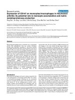
technique [13]. A wireless transmitter was attached to the
sensor and covered with a supplied transparent dressing
(Figure 1). Initial calibration of the CGM was performed
using arterial, venous, or capillary point-of-care (POC)
glucometry (iSTAT®, Abbo t Laborat ories, Princet on, NJ,
USA) at two hours and six hours after the sensor place-
ment. Subsequent calibrations occurred every 12 hours.
The CGM recorded a glucose reading every five minutes.
The sensor was replaced every five days durin g the study
unless contraindicated, and it was removed when partici-
pants no longer required mechanical ventilation and/or
vasoactive infusions. Although bedside nursing and physi-
cian teams were aware of enrollment, they did not assess
CGM readings a nd no changes in pa tient management
were based on values from the CGM. Audible alarms were
set for CGM values of ≤ 70 mg/dL (3.9 mmol/L) and ≥
200 mg/dL (11.1 mmol/L). With any alarm, the bedside
nurses were instructed to obtain a POC glucose measure-
ment, act accordingly to the POC value, and notify the
study staff.
Data acquisition and analysis
Demographic and clinical data were collected for all parti-
cipants. CGM values were compared to POC and labora-
tory BG measurements that occurred at or within five
minutes of CGM readings. A mixed model was performed
to assess the relationship between CGM and BG measure-
ments, as this accounts for the repeated measurements of
glucose levels. CGM versus BG agreement was assessed
using Clarke error grid analysis (Matlab® R2009A, Natick,
MA, USA) and Bland-Altman analysis. BG values used to
calibrate the CGM were not used for comparison, but all
other POC or laboratory BG measurements obtained dur-
ing the study period were compared to CGM values. This
included both BG measurements p erformed as part the
patient’s routine care and BG measurements obtained for
a high or low glucose alarm from the CGM.
Role of the funding source
This was an investigator-initiated study fo r which Med-
tronic® (Northridge, CA, USA) donated CGMs, but pro-
vided no other funding or support. Internal institution
funds were used to purchase the sensors for the GCM.
Results
Fifty patients were enrolled, and a total of 89 sensors
were used. There were 26 patients who had the sensor
removedinlessthanfivedays, because they no longer
met study criteria for critical illness hyperglycemia (that
is, they no longer required mechanical ventilation,
vasoactive medications, or continuous renal replacement
therapy). One patient did not have the device in place
long enough to re cord BG values, and two patients had
dysfunctional sensors with no data recorded. The two
patients with dysfunctional sensors were not signifi-
cantly different in degree of illness or condition than
the rest of the study patie nts. However, the sensors
used on t hese two pa tients came from the same box.
When a third patient s tarted the study with a sensor
from t his box, it also did not work. When a sensor
from a different box was used on this patient, it worked
very well. Therefore, we concluded that this box of sen-
sors was defective, and it was not used during the rest
of the study.
Of the 47 patients with accessible data, the mea n age
was 4.3 years (range 6 weeks to 16 years-old) and 31
(66%) were male. Twenty-nine (62%) were medical
pediatric ICU patients, 8 (17%) were surgical pediatric
ICU patients (including general surgery, trauma surgery,
neurosurgery, and otolaryngology), and 10 (21%) were
cardiac surgery patients. Six (13%) had traumatic brain
injury or intracranial hemorrhage. All patients required
mechanical ventilation, and 30 (63.8%) required vaso-
pressor or inotropic infusions. Twenty (42.6%) devel-
oped critical illness hyperglycemia, defined as persistent
BG levels >140 mg/dL (7.7 mmol/L), and received insu-
lin via our published pediatric-specific hyperglycemia
protocol [14,15]. Six (12.8%) required continuous renal
replacement therapy (CRRT), and three (6%) developed
the need for veno-venous extracorporeal membrane
oxygenation (ECMO). Participants had indicators consis-
tent with high level of illness s everity, including a mean
ICU length of stay (LOS) of 15 days. A total of 17 (36%)
patients had pediatric logistic organ dysfunction
(PELOD) scores ≥12 (Table 1).
Bridges et al. Critical Care 2010, 14:R176
/>Page 2 of 10
There were a total of 142 episodes of CGM readings
<40 mg/dL (2.2 mmol/L). Readings <40 mg/dL (2.2
mmol/L) from the CGM accounted for 0.2% of the total
64,315 CGM readings. All of the CGM readings of <40
mg/dL (2.2 mmol/L) took place in just five different
patients. When checked against BGs, these CGM read-
ings of <40 mg/dL (2.2 mmol/L) were shown to be fal-
sely low. The lowest BG drawn during an episode in
which the CGM reading was <40 mg/dL (2.2 mmol/L)
was 71 mg/dL (3.9 mmol/L). All of these episodes of fal-
sely low CGM readings corrected with sensor recalibra-
tion or replacement. There were n o episodes o f
hypoglycemia of <40 mg/dl (2.2 mmol/L) in any of the
POC or laboratory BGs during the entire study.
A total of 1,555 paired CGM and BG measurements
were analyzed using Clarke error grid and Bland-Altman
analysis (Figure 2). For the 1,555 paired CGM and BG
measurements, there is a statistically significant linear
relationship between CGM values and BG (P <.0001).
A one unit increase in CGM increases BG by an average
of .6537.
With Clarke analysis, readings in Zone A differ by
≤20%, whereas those in Zone B differ by >20% but do
not impact manag ement. Readings in Zone A and B are
considered to have clinically acceptable correlation.
Zone C values would lead to therapeutic overcorrection
and Zone D readings would not trigger intervention,
although warranted. Zone E values would result in treat-
ment aggravating a hypo- or hyperglycemic state. With
Clarke error grid analysis of all patients, 74.6% of read-
ings were in Zone A and 23.3% of readings were in
Zone B, equating to 97.9% of all readings with clinically
acceptable correlation (Zone A + B). A total of 2.1% of
rea ding s were in Zones C + D, and no readings were in
Zone E. Pearson’s correlation coefficient for all compari-
sons was 0.68. In Bland-Altman agreement analysis,
Figure 1 Application of the CGM sensor and transmitter to pediatric patients. The interstitial glucose sensor (solid arrow) was placed in the
subdermal, interstitial space on the abdomen (A) or upper thigh (B, C). The clam shell-appearing wireless transmitter (dotted line) was then
attached. In A, the sensor was placed on a 10 year-old female with H1N1 and respiratory failure who subsequently required veno-venous ECMO
(note the right femoral vein ECMO cannula and radial arterial catheter). Patient B depicts a three-year-old male trauma patient that had suffered
a gunshot wound. Patient C was a six-week-old male status-post traumatic brain injury. The supplied clear dressing facilitates site observation for
bleeding or reactions. Patients depicted in these photos were consented for photography.
Bridges et al. Critical Care 2010, 14:R176
/>Page 3 of 10
Table 1 Patient demographics
Category N Glucose
comparisons
Age (yr) Weight (kg) % Female
(N)
Ethnicity (N) ICU LOS (days) % CIH (N) CGMdays Pearson’s
correlation
coefficient
All 47 1555 4.3 (0.01 to 16) 20.6 (2.4 to 87) 34% (16) AA 1.1% (24) H 8.5% (4) C 36.2% (17) 15 (2 to 102) 79% (34) 6.1 (1 to 18) .68
A. Primary
diagnosis
Medical 29 895 4.6 (0.1 to 14) 22.5 (2.9 to 87) 41.4% (12) AA 51.7% (15) H 10.3% (3) C 31% (9) 13.4 (2 to 41) 66% (19) 6.2 (1 to 15) .71
Cardiac surgery 10 222 1.2 (0.01 to 7) 7.5 (2.4 to 20.2) 40% (4) AA 30% (3) H 10% (1) C 60% (6) 11 (2 to 44) 70% (7) 3.2 (1 to 7) .81
Surgery/other 8 438 7.1 (1.9 to 16) 30.2 (12.7 to 59) 0% (0) AA 75% (6) H 0% (0) C 25% [39] 25.6 (4 to 102) 100% (8) 9.4 (1 to 18) .50
TBI/ICH 6 113 5.3 (0.1 to 14) 21.4 (3.3 to 50) 16.7% (1) AA 33.3% [39] H 0% (0) C 66.7% (4) 10.2 (6 to 15) 14% [39] 5.5 (2 to 14) .59
B. Support
VPI/Inotropes 30 1184 4.4 (0.01 to 16) 19 (2.4 to 87) 43.3% (13) AA 50% (15) H 10% (3) C 33.3% (10) 17.6 (2 to 102) 80% (24) 6.5 (1 to 18) .73
IV Steroids 32 1321 4 (0.01 to 16) 20 (2.4 to 87) 31.3% (10) AA 65.6% (21) H 6.3% [39] C 25% (8) 17.4 (2 to 102) 81% (26) 6.6 (1 to 18) .69
CVVH 6 495 6.8 (1.3 to 16) 34.7 (8 to 87) 50% (3) AA 83.3% (5) H 0% (0) C 16.7% (1) 34.5 (12 to 102) 83% (5) 10.3 (4 to 18) .76
ECMO 3 216 5.8 (1.3 to 16) 39.7 (10 to 87) 33.3% (1) AA 66.7% [39] H 0% (0) C 33.3% (1) 25 (12 to 38) 100% (3) 12 (8 to 18) .57
C. CIH
No 13 96 2 (0.04 to 9) 12.8 (2.4 to 55) 30.8% (4) AA 53.8% (7) H 23.1% (3) C 23.1% (3) 10.1 (2 to 25) 0% (0) 3.3 (1 to 7) .68
Yes - without
insulin therapy
14 324 1.7 (0.01 to 14) 12.2 (2.9 to 74) 35.7% (5) AA 35.7% (5) H 0% (0) C 50% (7) 14.5 (2 to 41) 100% (14) 6.1 (1 to 13) .71
Yes - with
insulin therapy
20 1135 7.5 (0.01 to 16) 31.7 (3.5 to 87) 35% (7) AA 60% (12) H 5% (1) C 35% (7) 18.6 (2 to 102) 100% (20) 7.9 (1 to 18) .67
D. Age
Less than one
years
18 374 0.3 (0.01 to 0.8) 5.3 (2.4 to 9.2) 38.9% (7) AA 38.9% (7) H 11.1% [39] C 44.4% (8) 15.4 (2 to 44) 56% (10) 5.3 (1 to 12) .75
One to five
years
13 416 2.3 (1.2 to 4) 13.7 (10 to 20) 23.1% (3) AA 61.5% (8) H 7.7% (1) C 23.1% (3) 10.3 (2 to 33) 77% (10) 5.5 (1 to 13) .64
6 to 10 years 9 488 7.6 (6 to 10) 34.9 (20.2 to 87) 44.4% (4) AA 55.6% (5) H 11.1% (1) C 33.3% (3) 15.2 (2 to 38) 78% (7) 8 (1 to 18) .73
More than 10
years
7 277 14 (13 to 16) 54.6 (32.4 to 74) 28.6% [39] AA 57.1% (4) H 0% (0) C 42.9% (3) 22.4 (2 to 102) 100% (7) 6.7 (1 to 14) .55
E. PELOD
<12 30 604 4.1 (0.01 to 14) 20.3 (2.4 to 74) 30% (30) AA 46.7% (14) H 13.3% (4) C 33.3% (10) 11.8 (2 to 41) 63% (19) 4.9 (1 to 18) .69
≥12 17 951 4.6 (0.01 to 16) 21.3 (3.3 to 87) 41.2% (7) AA 58.8% (10) H 0% (0) C 41.2% (7) 20.6 (3 to 102) 88% (15) 8.2 (1 to 15) .68
F. ICU LOS
Less than three
days
5 45 5 (0.2 to 14) 25.2 (6.2 to 74) 41.2% (7) AA 60% (3) H 20% (1) C 20% (1) 2 (2 to 2) 60% (3) 1.4 (1 to 2) .74
Three to seven
days
12 139 3.8 (0.1 to 14) 17.9 (3.3 to 67.8) 8.3% (1) AA 33.3% (4) H 16.7% [39] C 41.7% (5) 5 (3 to 7) 58% (7) 2.2 (1 to 5) .85
More than
seven days
30 1371 4.3 (0.01 to 16) 21 (2.4 to 87) 43.3% (13) AA 56.7% (17) H 3.3% (1) C 36.7% (11) 21.2 (8 to 102) 80% (24) 8.4 (2 to 18) .66
Numbers indicate mean values unless otherwise indicated. Numbers in parentheses ( ) represent number or range. VPI, vasopressor infusions; CVVH, continuous veno-venous hemofiltration; AA, African American; C,
Caucasian; CIH, critical illness hyperglycemia; H, Hispanic; LOS, length of stay.
Bridges et al. Critical Care 2010, 14:R176
/>Page 4 of 10
approximately 95% of all CGM values were +/- 58 mg/
dL (3.2 mmol/L) from the mean difference of -1.5 mg/
dL (0.08 mmol/L) between the CGM and BG values.
The mean absolute relat ive difference (MARD) between
CGM and BG values was 15.3%. T he MARD gives the
average absolute difference between two methods of
measurement.
We compared BG and CGM glucose values in a variety
of subpopulations to investigate whether patient age,
diagnosis, support measures, or illness severity influenced
CGM function. We found a high correlation of glucose
values (that is, Clark Zone A + B >95%) in all age ranges,
including our youngest (<12 months old), and in all diag-
nostic categories. Sensor placement (thigh vs. abdomen)
did not impact reading accuracy. There was high correla-
tion in children who required support measures in addi-
tion to mechanical vent ilation, including vasopressors,
inotr opes, CRRT , and/or ECMO (Figures 2 and 3). Clini-
cally acceptable agreement was not obviously different in
those with low or high illness severity scores or according
to ICU LOS.
We also investigated whether CGM values correlated
with BG values in patients who developed critical illness
hyperglycemia and received insulin to maintain BG in the
80-140 mg/dL (4.4 to 7.7 mmol/L) range [5,12,14,15]. We
found a high correlation between BG and CG M readings
(98.2% in Z one A + B) i n p atients who required an insu-
lin infusion for critical illness hyperglycemia (Figure 2D).
No patient with CGM sensors in our study developed
a site infection, reaction, or bleeding. The insertion of
sensors was well tolerated with no o bvious discomfort,
albeit all patients were receiving sedation and/or analge-
sia, because they were intubated and mechanically venti-
lated. There was no obvious interference with the CGM
from ICU monitors or electronic support devices. The
sensors and transmitters are not MRI compatible, and
for study subjects who required such imaging, the sen-
sors and transmitters were removed and then replaced
after the exam was complete.
Using software provided with the CGM, we overlaid
CGM and BG readings. Shown in Figure 4 are t wo
examples; one of a patient who developed critical illness
hyperglycemia and was managed with insulin and
another of a patient who developed hyperglycemia but
did not receive insulin due to age restrictions.
Discussion
Our study demonstrates that commercially available,
interstitial CGMs correlate closely with BG measure-
ments in critically ill children. Glycemic control in criti-
cally ill adults im proves outcomes in a number of
studies from adult medical, surgical, and mixed ICUs,
and is recommended as standard practice by many med-
ical advisory committees [1,2,8,9,16]. A recent report by
Vlasselaers et al. also demonstrated clinical benefits of
strict glycemic control in a pediatric medical-surgical
ICU [4]. However, one issue that has plagued many gly-
cemic control trials in adults and children has been
unacceptably high rates of iatrogenic hypoglycemia in
strict control groups. Although the effect of hypoglyce-
mia on outcomes is unclear, these concerns have
resulted in a lack of adoption of routine glycemic man-
agement by some ICU practitioners (for example, those
in pediatric critical care).
Advocates and critics of glycemic control agree that a
reliable means to continuously sample and report glu-
cose levels will assist in understanding the effects of
ICU hyperglycemia and its management. Such technol-
ogywouldimproveboththeconsistencyandsafetyof
glycemic control approaches. Notably, in discussing the
high rates of hypoglycemia despite outcome benefit in
their recently published RCT in pediatric critical care,
Vlasselaers et al. state that “ for future studies, an accu-
rate continuous blood glucose sensor for use in the
PICU would be preferable tokeeptheriskofhypogly-
caemia to a minimum [4].”
CGMs, which regularly assess interstitial glucose levels,
have been developed, approved, and marketed for outpati-
ent diabetes management. Although ICU practitioners
support the concept of utilizing such devices, many have
dismissed the potential use of such technology in critical
care settings. Their argument is that there may be an inac-
curacy of interstitial glucose values compared to blood
levels in patients with edema, poor perfusion, or in those
requiring vasoactive infusions. Despite these theoretical
drawbacks of CGM and interstitial glucose a ssessments,
little evidence exists to substantiate these concerns. There
is now a small cohort of limited studies of ap proximately
160 adult and pediatric ICU patients suggesting that there
is a reasonable correlation between CGM and BG mea-
surements [17-27].
We report one of the largest studies to date evaluating
a Food and Drug Administration (FDA) approved, com-
mercially available CGM in a critical care setting. We
assessed patients that our previous studies have proved
to be at increased ri sk for critic al illness h yperglycemi a
[12,14,15,28,29]. Currently, t here is debate on which is
the gold standard blood supply (that is, arterial vs.
venous) to assess BG. In daily management, ICUs likely
use the most convenient and accessible vascular bed for
blood glucose dete rmi nation and glycemic management,
including capillary sampling. Our study may provide gen-
eralizable and relevant data for practitioners and staff
who may use different sources for BG measurements.
In all patients and in subpopulation analysis based on
age, diagnosis, and ICU support measures, we found a
high correlation between CGM readings and standard
BG measurements per Clarke error grid and Bland-
Bridges et al. Critical Care 2010, 14:R176
/>Page 5 of 10
Altman analysis. Although some vasoactive medications
can result in peripheral vasoconstriction, this did not
affect the accuracy of interstitial readings in our study.
Correlation was also strong in those requiring CRRT
and/or ECMO, despite significant edema and vascular
leak syndrome in these patients. A 2005 report demon-
strated that a CGM device attached to an ECMO circuit
providing supp ort for pediat ric patients, could supply
reasonable glucose readings [30]. Our study appears to
be the first to evaluate CGMs on patients who required
ECMO, with the important note that sensors were placed
on patients before they were placed on ECMO.
Patients who were more severely ill according to tradi-
tional illness severity indicators (PELOD scores, ICU
lengths of stay) had excellent correlation between CGM
and standard readings. In fact, those with an ICU stay
Figure 2 Clarke error grid and Bland-Altman agreement analysis of paired BG readings. A total of 1,555 CGM values from 47 pediatric ICU
patients were compared for agreement with BG measurements (A). In Clarke plots (upper panel), the routine POC readings (X-axis) were
compared to corresponding CGM values (Y-axis). Readings in Zone A differ by ≤20%, whereas those in Zone B differ by >20% but do not impact
management. The Zone A + B composite is considered “clinically acceptable” correlation. Zone C values would lead to therapeutic
overcorrection and Zone D readings would not trigger intervention, although warranted. Zone E values would result in treatment aggravating a
hypo- or hyperglycemic state. In Bland-Altman analysis (bottom panel), the mean of the paired readings is on the X-axis, and the difference is on
the Y-axis. The broken line represents the cutoff of two standard deviations from the mean difference. In addition to the R
2
, the solid line with
the indicated slope is shown and represents the correlation coefficient. Clarke and Bland-Altman plots are shown for patient subpopulations less
than one-year-old (B), those who received vasopressor or inotropic support (C), and those who developed critical illness hyperglycemia and
were managed with insulin using our protocol (D).
Bridges et al. Critical Care 2010, 14:R176
/>Page 6 of 10
of less than three days had the lowest Clark Zone A cor-
relation in our analysis. This may be due to the fact that
the precision of CGMs may improve with time after
insertion. It is interesting to note that one study that
found a poor correlation of CGMs in hospitalized
patients, sensors were in place for a maximum of three
days [27]. In addition, while some have questioned the
utility of such devices in very small pediatric patients,
we found e xcellent correlation in patients <12 months
of age. Also, we found excellent correlation in pediatric
ICU patients who were hyperglycemic and managed
with an insulin infusion.
A number of CGM devices have been approved for
outpatient use in children and adults [31-33]. FDA
approval applications have been supported by 90 to 99%
Clarke A + B correlation between CGM and standard
BG readings, and 1.6 to 10% of clinically inaccurate
readings(ZoneC+D+E).Such applications cite stu-
dies evaluating approximately 20 to 137 patients [34-36].
Findings from ou r study of 47 participants, with Zone A
+ B of 97.9% and Zone B + C + D of 2.1%, are consis-
tent with criteria used in other venues to determine
acceptable accuracy. In addition, we found no untoward
effects of CGM use. Although critically ill patients often
have coagulopathies, we found no evidence of bleeding
with device use. Of note, sensors were not in serted
when a known severe coagulopathy was present. In
addition, we also found no site infections, reactions, or
significant discomfort noted with sensor placement or
use.
Our data provide some of the most robust evidence to
date that these devices can be used safely and provide
clinically meaningful information, even in the smallest
and most critically ill pediatric patients. In conjunction
Figure 3 Percentage of clinically acceptable paired glucose readings in patient subpopulations.Patientsweregroupedbasedon
diagnosis (A), age or sensor site placement (B), support measures in addition to mechanical ventilation (C), development and/or treatment of
critical illness hyperglycemia (D), and severity of illness indicators (E). See Table 1 for corresponding subpopulation details. Clarke analysis was
conducted and shown as the percent of readings in Zone A and B. As is standard, we defined “clinically acceptable” correlations as the sum of
Zone A and B.
Bridges et al. Critical Care 2010, 14:R176
/>Page 7 of 10
with other CGM studies in ICU settings, it may be rea-
sonable to consider incorporating the use of such
devices into critical care practice, albeit with some
important limitations. We found that there is a signifi-
cant time and educational component needed to learn
to insert, calibrate and assess these devices. We also fre-
quently kept sensors in place for longer than the FDA-
recommended time. This sensor is currently approved
for 72 hours of use in the o utpatient setting, and there
are no guidelines for their use in the ICU setting.
However, there are reports of these sensors being accu-
rate and safe for longer than the 72-hour period [37].
Medtronic has recently completed a study testing the
use of this sensor for six days in an inpatient setting,
and these results are pending [38]. Although our find-
ings demonstrate a strong correlation even with
extended use, we did not make any clinical management
decisions based on CGM readings. Only after further
study in clinical trials should such devices be used to
assist in titrating insulin during hyperglycemia
Figure 4 Temporal plot comparing CGM readings and BG values. With software provided with the CGM, CGM values and BG values were
plotted over time. CGM values (gray dots that at times morph into a line) are acquired approximately every five minutes, and BG values (black
squares) sporadically per regular care. We define persistent BG >140 mg/dL (7.7 mmol/L) as critical illness hyperglycemia, and have traditionally
managed those more than six months old with insulin infusions to a target BG of 80 to 140 mg/dL (4.4 to 7.7 mmol/L) (black dashed lines). The
top plot (A) represents data from a 13 year-old male with traumatic brain injury requiring mechanical ventilation and norepinephrine to
maintain his cerebral perfusion pressure. The arrows indicate when insulin was started and stopped per our protocol to manage his
hyperglycemia. The data in B is from a five-month-old male with Trisomy 21, RSV, pulmonary hypertension, and refractory hypoxemia requiring
high frequency oscillatory ventilation, inhaled nitric oxide, milrinone, and an epinephrine infusion. At the time of this study, this patient was
below our typical age threshold for protocolized hyperglycemic management, and he was not treated with insulin.
Bridges et al. Critical Care 2010, 14:R176
/>Page 8 of 10
management in critical care settings. Active diagnosis
and/or manage ment of glucose disturbances in ICU set-
tings should only occur with time-tested BG
measurements.
The current state of evidence in this field may support
the use of such devices to trend patient BGs, and setting
high and low alarms to trigger standard BG assessment
may assist in improving the efficacy and safety of glyce-
mic control approaches. We experienced very few hyp o-
glycemic episodes in this study, and thus the accuracy
of CGM in low-norm al or hypoglycemic ranges is
unclear. Anecdotally, although our staff was essentially
blind to the readings of these monitors, there were
times when the “low” BG alarm was triggered, prompt-
ing our staff to perform a routine BG check. In one
notable case, a hyperglycemic patient treated with insu-
lin had her intravenous fluids switched from a high to
low dextrose-contain ing fluid without a change in insu-
lin dosing. The patient was not due to receive a routine
BG check, but after the CGM alarmed, a POC BG was
performed and confirmed a low reading. The IV fluid
discrepancy was noted, and the insulin infusion was
changed. As such, the most important utility of CGMs
at this time may be to trigger standard BG checks to
improve the safety of glycemic control.
Data from CGM devices will likely add to the under-
standing of glycemic control in the critically ill patient.
As shown in Figure 4, obtaining daily glucose trends via
CGM may help to identify the percent of time spent
outside of goal targe t ranges. Accurate, real-ti me assess-
ment of area-under-the-curve metrics can be obtained
from CGM devices. This may provide more meaningful
data compared to approaches currently used to define
and compare glycemic control.
Conclusions
Critically ill children encompass one of the widest spec-
trums of patients , in terms of size and condition, in any
ICU setting. The severity of illness and support mea-
sures of patients in this study were consid erable. It may
be reasonable to consider that if CGMs function well in
our study population, they will likely function well in
most other ICU populations, whether pediatric, adult,
medical, or surgical. Yet, there will likely be scenarios
where CGM performance will be sub-standard, and
careful assessment during its use is required. Despite
the proven benefits of glycemic control in many critical
care settings, there are recognized risks of this approach
and more studies are needed to better define optimal
BG target ranges in the spectrum of ICU patients.
Our study adds to the body of evidence that refutes
the theoretical concerns regarding the use of CGM
devices in the ICU setting. There is no doubt that future
technologies will help advance the field of glycemic con-
trol in critical illness. The evidence may now support
theuseofcurrentlyavailableCGMstoassistwiththe
safety and efficacy of glycemic control in critical illness.
Further study and application for clinical approval of
CGM use in ICU settings should be paramount in the
international effort to better understand critical illness
hyperglycemia.
Key messages
• Hyperglycemia, hypoglycemia and glucose variabil-
ity are associated with poor outcomes in critical ill-
ness. However, recent studies on the control of
criti cal illness hyperglycemia have shown unaccepta-
bly high rates of iatrogenic hypoglycemia.
• This study demonstrates that interstitial CGMs
have a high degree of correl ation with BG levels in a
wide variety of critically ill children.
• Our findings suggest that with further examination,
existing CGM technology may be a useful adjunct in
the detection and management of glucose disorders
in critically ill children.
Abbreviations
BG: blood glucose; CGM: continuous glucose monitor; CRRT: continuous
renal replacement therapy; ECMO: extracorporeal membrane oxygenation;
FDA: Food and Drug Administration; LOS: length of stay; MARD: mean
absolute relative differenc e; PELOD: pediatric logistic organ dysfunction; POC:
point-of-care; RCT: randomized controlled trial
Acknowledgements
We would like to thank the nurses and staff of the pediatric critical care unit
and cardiac critical care unit at Children’s Healthcare of Atlanta at Egleston.
Without your help, this study would not have been possible. We would also
like to thank the patients and families that participated in this study. Written
consent for publication was obtained from the guardians of the patients
that participated in this study. We would like to thank Traci Leong, PhD for
critical reading of this manuscript and providing statistical insight.
Author details
1
Department of Pediatrics, Division of Pediatric Critical Care, Vanderbilt
University School of Medicine, 2200 Children’s Way, Nashville, TN 37232-
9075, USA.
2
Department of Pediatrics, Emory University School of Medicine,
1405 Clifton Road NE, Atlanta, GA 30322-1060, USA.
3
Pediatric Critical Care,
Children’s Healthcare of Atlanta at Egleston, 1405 Clifton Road NE, Atlanta,
GA 30322-1060, USA.
4
Pediatric Critical Care, The Children’s Hospital at the
Medical Center of Central Georgia, 777 Hemlock Street, Macon, GA 31201-
2155, USA.
5
Pediatric Cardiac Critical Care, Sibley Heart Center Cardiology,
1405 Clifton Road NE, Atlanta, GA 30322-1060, USA.
Authors’ contributions
BCB, CMP, KOM and MRR contributed equally to the design, acquisition of
data, and analysis of data for this study. All of the authors were involved in
drafting the manuscript and revising it critically for important intellectual
content. They all gave final approval of the version to be published.
Competing interests
The authors declare that they have no competing interests.
Received: 5 May 2010 Revised: 22 July 2010 Accepted: 6 October 2010
Published: 6 October 2010
Bridges et al. Critical Care 2010, 14:R176
/>Page 9 of 10
References
1. Van den Berghe G, Wouters P, Weekers F, Verwaest C, Bruyninckx F,
Schetz M, Vlasselaers D, Ferdinande P, Lauwers P, Bouillon R: Intensive
insulin therapy in the critically ill patients. N Engl J Med 2001,
345:1359-1367.
2. Van den Berghe G, Wilmer A, Hermans G, Meersseman W, Wouters PJ,
Milants I, Van Wijngaerden E, Bobbaers H, Bouillon R: Intensive insulin
therapy in the medical ICU. N Engl J Med 2006, 354:449-461.
3. Krinsley JS: Effect of an intensive glucose management protocol on the
mortality of critically ill adult patients. Mayo Clin Proc 2004, 79:992-1000.
4. Vlasselaers D: Intensive insulin therapy for patients in paediatric intensive
care: a prospective, randomised controlled study. Lancet 2009,
373:547-556.
5. Preiser JC, Devos P, Ruiz-Santana S, Melot C, Annane D, Groeneveld J,
Iapichino G, Leverve X, Nitenberg G, Singer P, Wernerman J, Joannidis M,
Stecher A, Chiolero R: A prospective randomised multi-centre controlled
trial on tight glucose control by intensive insulin therapy in adult
intensive care units: the Glucontrol study. Intensive Care Med 2009,
35:1738-1748.
6. Brunkhorst FM, Engel C, Bloos F, Meier-Hellmann A, Ragaller M, Weiler N,
Moerer O, Gruendling M, Oppert M, Grond S, Olthoff D, Jaschinski U,
John S, Rossaint R, Welte T, Schaefer M, Kern P, Kuhnt E, Kiehntopf M,
Hartog C, Natanson C, Loeffler M, Reinhart K: Intensive insulin therapy and
pentastarch resuscitation in severe sepsis. N Engl J Med 2008,
358:125-139.
7. Finfer S, Chittock DR, Su SY, Blair D, Foster D, Dhingra V, Bellomo R, Cook D,
Dodek P, Henderson WR, Hebert PC, Heritier S, Heyland DK, McArthur C,
McDonald E, Mitchell I, Myburgh JA, Norton R, Potter J, Robinson BG,
Ronco JJ: Intensive versus conventional glucose control in critically ill
patients. N Engl J Med 2009, 360:1283-1297.
8. Moghissi ES, Korytkowski MT, DiNardo M, Einhorn D, Hellman R, Hirsch IB,
Inzucchi SE, Ismail-Beigi F, Kirkman MS, Umpierrez GE: American
Association of Clinical Endocrinologists and American Diabetes
Association consensus statement on inpatient glycemic control. Endocr
Pract 2009, 15:353-369.
9. IHI.org A Resource from the Institute for Healthcare Improvement.
[ />ImplementEffectiveGlucoseControl.htm].
10. Surviving Sepsis Campaign: Maintain Adequate Glycemic Control. [http://
www.survivingsepsis.org/Bundles/Individual_Changes/Pages/
maintain_glycemic.aspx].
11. Hirshberg E, Lacroix J, Sward K, Willson D, Morris AH: Blood glucose
control in critically ill adults and children: a survey on stated practice.
Chest 2008, 133:1328-1335.
12. Preissig CM, Rigby MR: A disparity between physician attitudes and
practice regarding hyperglycemia in pediatric intensive care units in the
United States: a survey on actual practice habits. Crit Care 14:R11.
13. Medtronic: User Guide Guardian Real-Time Continuous Glucose
Monitoring System. 2007.
14. Preissig CM, Hansen I, Roerig PL, Rigby MR: A protocolized approach to
identify and manage hyperglycemia in a pediatric critical care unit.
Pediatr Crit Care Med 2008, 9:581-588.
15. Preissig CM, Rigby MR, Maher KO: Glycemic control for postoperative
pediatric cardiac patients. Pediatr Cardiol 2009,
30:1098-1104.
16. Dellinger RP, Levy MM, Carlet JM, Bion J, Parker MM, Jaeschke R, Reinhart K,
Angus DC, Brun-Buisson C, Beale R, Calandra T, Dhainaut JF, Gerlach H,
Harvey M, Marini JJ, Marshall J, Ranieri M, Ramsay G, Sevransky J,
Thompson BT, Townsend S, Vender JS, Zimmerman JL, Vincent JL:
Surviving Sepsis Campaign: international guidelines for management of
severe sepsis and septic shock: 2008. Crit Care Med 2008, 36:296-327.
17. Corstjens AM, Ligtenberg JJ, van der Horst IC, Spanjersberg R, Lind JS,
Tulleken JE, Meertens JH, Zijlstra JG: Accuracy and feasibility of point-of-
care and continuous blood glucose analysis in critically ill ICU patients.
Crit Care 2006, 10:R135.
18. De Block C, Manuel YKB, Van Gaal L, Rogiers P: Intensive insulin therapy in
the intensive care unit: assessment by continuous glucose monitoring.
Diabetes Care 2006, 29:1750-1756.
19. Chee F, Fernando T, van Heerden PV: Closed-loop glucose control in
critically ill patients using continuous glucose monitoring system
(CGMS) in real time. IEEE Trans Inf Technol Biomed 2003, 7:43-53.
20. Goldberg PA, Siegel MD, Russell RR, Sherwin RS, Halickman JI, Cooper DA,
Dziura JD, Inzucchi SE: Experience with the continuous glucose
monitoring system in a medical intensive care unit. Diabetes Technol Ther
2004, 6:339-347.
21. De Block C, Manuel-y-Keenoy B, Rogiers P, Jorens P, Van Gaal L: Glucose
control and use of continuous glucose monitoring in the intensive care
unit: a critical review. Curr Diabetes Rev 2008, 4:234-244.
22. Allen HF, Rake A, Roy M, Brenner D, McKiernan CA: Prospective detection
of hyperglycemia in critically ill children using continuous glucose
monitoring. Pediatr Crit Care Med 2008, 9:153-158.
23. Piper HG, Alexander JL, Shukla A, Pigula F, Costello JM, Laussen PC, Jaksic T,
Agus MS: Real-time continuous glucose monitoring in pediatric patients
during and after cardiac surgery. Pediatrics 2006, 118:1176-1184.
24. Beardsall K, Ogilvy-Stuart AL, Ahluwalia J, Thompson M, Dunger DB: The
continuous glucose monitoring sensor in neonatal intensive care. Arch
Dis Child Fetal Neonatal Ed 2005, 90:F307-310.
25. Branco RG, Chavan A, Tasker RC: Pilot evaluation of continuous
subcutaneous glucose monitoring in children with multiple organ
dysfunction syndrome. Pediatr Crit Care Med 2010, 11:415-419.
26. Holzinger U, Warszawska J, Kitzberger R, Herkner H, Metnitz PG, Madl C:
Impact of shock requiring norepinephrine on the accuracy and reliability
of subcutaneous continuous glucose monitoring. Intensive Care Med 2009,
35:1383-1389.
27. Price GC, Stevenson K, Walsh TS: Evaluation of a continuous glucose
monitor in an unselected general intensive care population. Crit Care
Resusc 2008, 10:209-216.
28. Srinivasan V, Spinella PC, Drott HR, Roth CL, Helfaer MA, Nadkarni V:
Association of timing, duration, and intensity of hyperglycemia with
intensive care unit mortality in critically ill children. Pediatr Crit Care Med
2004, 5:329-336.
29. Verhoeven JJ, Brand JB, van de Polder MM, Joosten KF: Management of
hyperglycemia in the pediatric intensive care unit; implementation of a
glucose control protocol. Pediatr Crit Care Med 2009, 10:648-652.
30. Javid PJ, Halwick DR, Betit P, Thompson JE, Long K, Zhang Y, Jaksic T,
Agus MS: The first use of live continuous glucose monitoring in patients
on extracorporeal life support. Diabetes Technol Ther 2005, 7:431-439.
31. Garg S, Zisser H, Schwartz S, Bailey T, Kaplan R, Ellis S, Jovanovic L:
Improvement in glycemic excursions with a transcutaneous, real-time
continuous glucose sensor: a randomized controlled trial. Diabetes Care
2006, 29:44-50.
32. Kovatchev B, Anderson S, Heinemann L, Clarke W: Comparison of the
numerical and clinical accuracy of four continuous glucose monitors.
Diabetes Care 2008, 31:1160-1164.
33. Wilhelm B, Forst S, Weber MM, Larbig M, Pfutzner A, Forst T: Evaluation of
CGMS during rapid blood glucose changes in patients with type 1
diabetes. Diabetes Technol Ther 2006, 8:146-155.
34. Summary of safety and effectiveness data: Medtronic Guardian RT. 2006
[ />03-SSED-vol1.pdf].
35. Summary of safety and effectiveness data: DexCom STS. 2006 [http://
www.accessdata.fda.gov/cdrh_docs/pdf5/P050012b.pdf].
36. Summary of Safety and Effectiveness Data: FreeStyle Navigator
Continuous Glucose Monitoring System. [ />cdrh_docs/pdf5/P050020b.pdf].
37. Chamberlain J, Small D: Successful use of a single subcutaneous
continuous glucose monitor sensor for 28 days in a patient with type i
diabetes. Clinical Diabetes 2008, 26:138-139.
38. An Inpatient Evaluation of Six-Day Subcutaneous Glucose Sensor
Performance (6-Day FU). [ />39. Yates AR, Dyke PC, Taeed R, Hoffman TM, Hayes J, Feltes TF, Cua CL:
Hyperglycemia is a marker for poor outcome in the postoperative
pediatric cardiac patient. Pediatr Crit Care Med 2006, 7:351-355.
doi:10.1186/cc9280
Cite this article as: Bridges et al.: Continuous glucose monitors prove
highly accurate in critically ill children. Critical Care 2010 14:R176.
Bridges et al. Critical Care 2010, 14:R176
/>Page 10 of 10