Báo cáo y học: " Kinetic modeling of tricarboxylic acid cycle and glyoxylate bypass in Mycobacterium tuberculosis, and its application to assessment of drug targets" ppt
Bạn đang xem bản rút gọn của tài liệu. Xem và tải ngay bản đầy đủ của tài liệu tại đây (342.21 KB, 11 trang )
BioMed Central
Page 1 of 11
(page number not for citation purposes)
Theoretical Biology and Medical
Modelling
Open Access
Research
Kinetic modeling of tricarboxylic acid cycle and glyoxylate bypass in
Mycobacterium tuberculosis, and its application to assessment of
drug targets
Vivek Kumar Singh and Indira Ghosh*
Address: Bioinformatics Centre, University of Pune, Pune-411007, India
Email: Vivek Kumar Singh - ; Indira Ghosh* -
* Corresponding author
Abstract
Background: Targeting persistent tubercule bacilli has become an important challenge in the
development of anti-tuberculous drugs. As the glyoxylate bypass is essential for persistent bacilli,
interference with it holds the potential for designing new antibacterial drugs. We have developed
kinetic models of the tricarboxylic acid cycle and glyoxylate bypass in Escherichia coli and
Mycobacterium tuberculosis, and studied the effects of inhibition of various enzymes in the M.
tuberculosis model.
Results: We used E. coli to validate the pathway-modeling protocol and showed that changes in
metabolic flux can be estimated from gene expression data. The M. tuberculosis model reproduced
the observation that deletion of one of the two isocitrate lyase genes has little effect on bacterial
growth in macrophages, but deletion of both genes leads to the elimination of the bacilli from the
lungs. It also substantiated the inhibition of isocitrate lyases by 3-nitropropionate. On the basis of
our simulation studies, we propose that: (i) fractional inactivation of both isocitrate dehydrogenase
1 and isocitrate dehydrogenase 2 is required for a flux through the glyoxylate bypass in persistent
mycobacteria; and (ii) increasing the amount of active isocitrate dehydrogenases can stop the flux
through the glyoxylate bypass, so the kinase that inactivates isocitrate dehydrogenase 1 and/or the
proposed inactivator of isocitrate dehydrogenase 2 is a potential target for drugs against persistent
mycobacteria. In addition, competitive inhibition of isocitrate lyases along with a reduction in the
inactivation of isocitrate dehydrogenases appears to be a feasible strategy for targeting persistent
mycobacteria.
Conclusion: We used kinetic modeling of biochemical pathways to assess various potential anti-
tuberculous drug targets that interfere with the glyoxylate bypass flux, and indicated the type of
inhibition needed to eliminate the pathogen. The advantage of such an approach to the assessment
of drug targets is that it facilitates the study of systemic effect(s) of the modulation of the target
enzyme(s) in the cellular environment.
Background
Tuberculosis is an ancient disease that has plagued
humans for centuries, and presently there is an urgent
need for new drugs to combat drug-resistant tuberculosis
Published: 03 August 2006
Theoretical Biology and Medical Modelling 2006, 3:27 doi:10.1186/1742-4682-3-27
Received: 03 April 2006
Accepted: 03 August 2006
This article is available from: />© 2006 Singh and Ghosh; licensee BioMed Central Ltd.
This is an Open Access article distributed under the terms of the Creative Commons Attribution License ( />),
which permits unrestricted use, distribution, and reproduction in any medium, provided the original work is properly cited.
Theoretical Biology and Medical Modelling 2006, 3:27 />Page 2 of 11
(page number not for citation purposes)
and shorten the time of tuberculosis therapy. Tuberculosis
treatment is lengthy because of a population of persistent
bacilli that is not effectively eliminated by current drugs.
The persistent bacilli primarily use fatty acids as their car-
bon source [1]. This makes the glyoxylate bypass, consist-
ing of isocitrate lyase (ICL) and malate synthase (MS),
essential for the bacterium; in its absence there will be no
net formation of the intermediates required for synthesiz-
ing cellular materials. Inhibition of both ICL1 (prokaryo-
tic-like isoform) and ICL2 (eukaryotic-like isoform) has
been shown to block the growth of M. tuberculosis in mac-
rophages and in mice [2]. Hence, interference with the
glyoxylate bypass is a potential approach to the design of
new drugs against persistent mycobacteria. This is consist-
ent with the suggestion that the regulation of M. tubercu-
losis metabolism in response to the environment of the
bacterium makes large contributions to its virulence [3].
At the branch point of the tricarboxylic acid (TCA) cycle
and glyoxylate bypass, isocitrate dehydrogenase (ICD),
involved in the TCA cycle, and ICL, involved in the glyox-
ylate bypass, compete for the same substrate, namely isoc-
itrate (ICIT). In Escherichia coli, flux at this branch point is
predominantly controlled through the reversible inactiva-
tion of ICD by phosphorylation, catalyzed by ICD-kinase
[4]. We have already identified the kinase in M. tuberculo-
sis, equivalent to ICD-kinase in E. coli, that is responsible
for reversible inactivation of ICD1 (Rv3339c) by phos-
phorylation [5]. Moreover, a method has been described
for inhibiting a metabolic pathway that is essential for the
viability of a microorganism by diverting the substrate to
a different metabolic pathway, and it has been suggested
that inhibiting ICD1-kinase could inhibit the flux through
the glyoxylate bypass in M. tuberculosis [5]. Since inhibi-
tion of ICD1-kinase would increase the amount of
dephosphorylated (active) ICD1, the flux through the gly-
oxylate bypass would be diminished. However, enzymes
are not isolated entities in living organisms but act as
components of systems, so the effect of modulation of any
enzyme activity on a metabolic flux depends on the prop-
erties of the other enzymes in the pathway concerned [6].
Metabolic Control Analysis (MCA) is a theoretical frame-
work that relates the systemic properties of a metabolic
system to the properties of its components, in particular
the enzymes, in a quantitative manner [6]. Application of
MCA to the identification of potential drug targets is
exemplified by glycolysis in Trypanosoma brucei [7-9].
MCA also gives insight into the cellular effect(s) of inhibi-
tion of a particular enzyme. Eisenthal et al. [9] suggested
two basic metabolic methods for killing an organism:
decreasing the flux through an essential metabolic path-
way to a nonviable level, or increasing the concentration
of a metabolite to a toxic level. Therefore, if inhibition of
an enzyme kills an organism, MCA can elucidate the
mechanism involved.
Since modulation of target enzyme(s) activity is usually
aimed at altering the cell's metabolic profile, knowledge
of the metabolic profile is important for identifying the
target. Recent experiments have shown a positive correla-
tion between mRNA levels measured by DNA microarrays
and protein abundance in both E. coli [10] and yeast cells
[11,12], so the gene expression profile could be connected
to the metabolic profile via simulation of the pathway
under study. In E. coli, the in vivo kinetic parameters
required for estimating the metabolic profile of most
enzymes are available when the organism is grown using
glucose as the carbon source [13]. In contrast, when ace-
tate is used as the carbon source, the gene expression pro-
file of the TCA cycle and glyoxylate bypass enzymes
differed from that found with glucose [14]. The corre-
sponding metabolic flux distributions in central meta-
bolic pathways under both growth conditions are known
[15], so this seems an ideal system for testing the hypoth-
esis that the gene expression profile can be connected with
the metabolic profile via simulation of the pathway under
study.
In this communication, we describe the construction of a
kinetic model of the TCA cycle and glyoxylate bypass in
M. tuberculosis, and we study the likely metabolic conse-
quences of inhibiting ICLs and ICD1-kinase. To the best
of our knowledge, this is the first attempt to model any
specific metabolic pathway in M. tuberculosis, and no
kinetic model is available for the TCA cycle and glyoxylate
bypass in this bacterium. Initially, we constructed a
kinetic model for the TCA cycle and glyoxylate bypass in
E. coli to validate the pathway modeling protocol used
and to test how well the metabolic profile correlates with
the gene expression profile while trying to predict the met-
abolic flux distribution using the gene expression data.
The biochemical reactions considered for the models are
shown in figure 1 and the metabolites with known con-
centrations are listed in table 1. In M. tuberculosis H37Rv
strain there are two isoforms of ICD [17], ICD1 (Rv3339c)
and ICD2 (Rv0066c), and two isoforms of ICL [17,18],
ICL1 (Rv0467) and ICL2 (Rv1915 and Rv1916). In addi-
tion, the inability of Nathan and co-workers to detect α-
ketoglutarate dehydrogenase (KDH) activity in M. tubercu-
losis [13] was taken into account while constructing the
model. M. tuberculosis model-1 represents a standard TCA
cycle and glyoxylate bypass with KDH present, while
model-2 lacks KDH activity. Our aim was to check the
metabolic consequences of the presence and absence of
KDH in this organism.
Theoretical Biology and Medical Modelling 2006, 3:27 />Page 3 of 11
(page number not for citation purposes)
Results and discussion
Steady state solution for the models
Steady state fluxes in the E. coli model (table 2) were com-
pared to the experimental fluxes given by Zhao et al. [15];
the net fluxes were expressed in relative units. The unit
conversion is described in methods section. The steady
state fluxes calculated from the model accorded with the
experimental fluxes [15] (table 3), thus validating the pro-
tocol used.
Since the maximal reaction rates (Vmax) of the enzymes
during growth on acetate were estimated using gene
expression data, it is possible to estimate the changes in
metabolic flux distribution due to changes in gene expres-
sion via simulation of the biochemical pathway under
study. This was also noted in the study of branched chain
amino acid biosynthesis in E. coli [19].
The steady state fluxes in the M. tuberculosis model-1
(standard TCA cycle) and model-2 (absence of KDH activ-
ity) are shown in table 4. The fluxes in the two models of
the M. tuberculosis TCA cycle and glyoxylate bypass are
similar, with the following exceptions. (i) The entire flux
from α-ketoglutarate (αKG) towards the TCA cycle passes
through the α-ketoglutarate decarboxylase (KGD) and
succinic semialdehyde dehydrogenase (SSADH) steps in
model-2 (which has no other branch from αKG that con-
tinues in TCA cycle); in model-1, about 84% of the flux
from αKG passes through KDH and the remaining 16%
through KGD and SSADH, but the total flux from αKG
continuing in the TCA cycle is almost the same in both
models. (ii) Flux was observed through the succinyl-CoA
synthetase (ScAS) step in model-1 but was negligible in
model-2. This is expected because KDH converts αKG to
succinyl-CoA, and succinyl-CoA must be converted to suc-
cinate (SUC) for the continuation of the TCA cycle. This
conversion is brought about by ScAS. Model-2 does not
require ScAS because it converts αKG directly to SUC
using KGD and SSADH. The steady state fluxes computed
from the two models showed minor differences, but the
turnover of the TCA cycle and glyoxylate bypass was sim-
ilar in both models, indicating that M. tuberculosis can
manage without a functional KDH. Thus, this study illus-
trates that at the metabolic level, the absence of KDH
activity has no effect on the net flux through the TCA cycle
and glyoxylate bypass.
On the basis of the finding of Tian et al. [13], i.e. that KDH
activity is absent in M. tuberculosis, and of the observation
that there is little difference between the two models in
the turnover of the TCA cycle and glyoxylate bypass, M.
tuberculosis model-2 was taken as the reference model in
the remaining parts of this study.
Inactivation of ICDs in M. tuberculosis model
Inactivation of ICD1, which is brought about by ICD1-
kinase, leads to a change in the number of active ICD1
molecules. Since Vmax is a function of the amount of
enzyme, any change in the amount of enzyme will affect
the Vmax. Therefore, varying Vmax for ICD1 from 1% to
100% was used to monitor the effect of inactivation of
ICD1 by ICD1-kinase. Since there is no information about
any such kinase for ICD2, the activity value was kept at
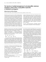
100%. Plots of the sum of flux through ICD1 and ICD2
(J
ICD1
+ J
ICD2
) and the sum of flux through ICL1 and ICL2
(J
ICL1
+ J
ICL2
) against Vmax for the forward ICD1 reaction
(Vf
ICD1
) (figure 2A) showed that even at 99% inactivation
there was no perceptible flux through the glyoxylate
bypass. We then studied the effect of inactivation of ICD2
by a hypothetical inactivator, along with the inactivation
Table 1: Metabolites of the models with known concentrations (with references indicated in square brackets)
Escherichia coli Mycobacterium tuberculosis
Metabolite Concentration in
glucose condition (in
mM)
Concentration in
acetate condition (in
mM)
Metabolite Concentration (in mM)
acetyl-CoA 0.5 [16] 0.5 [16] succinate 2.464 (derived from Tian
et. al [13])
citrate 3 [16] 9 [16] fumarate 0.08528 (derived from Tian
et. al [13])
isocitrate 0.018
a
[16] 0.15 [16] malate 0.408 (derived from Tian
et. al [13])
succinate 0.6 [16] 6 [16] oxaloacetate 0.0003 (assumed)
malate 1.8 [16] 5 [16] CoA 0.0001 (assumed)
oxaloacetate 0.004
b
0.0014 (assumed)
CoA 0.0001 (assumed) 0.0001 (assumed)
a
Isocitrate concentration was inferred from a graph shown by Walsh et. al [16]. The value in the graph was 0.025 mM at 30 minutes after addition
of glucose to the medium, but it had a negative slope, so, a value of 0.018 mM was taken.
b
Taken as 2.4 times the concentration of oxaloacetate under growth on acetate because flux leading to the synthesis of oxaloacetate under growth
on glucose is 2.4 times of that under growth on acetate [15].
Theoretical Biology and Medical Modelling 2006, 3:27 />Page 4 of 11
(page number not for citation purposes)
of ICD1. The plot of J
ICD1
+ J
ICD2
and J
ICL1
+ J
ICL2
against
Vmax for the ICD1 and ICD2 forward reactions (Vf
ICD1
and Vf
ICD2
respectively) (figure 2B) showed that the flux
through the glyoxylate bypass (J
ICL1
+ J
ICL2
) starts to
increase after Vf
ICD1
and Vf
ICD2
have fallen to approxi-
mately 30% of the original values, and becomes equal to
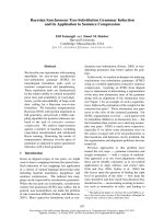
TCA cycle and glyoxylate bypass reactions considered in E. coli and M. tuberculosis modelsFigure 1
TCA cycle and glyoxylate bypass reactions considered in E. coli and M. tuberculosis models. Reactions 1, 2, 3, 5, 8,
9, 10, 11, 12 and 13 were present in all the models; reaction 4 was present only in the E. coli model and M. tuberculosis model-
1, but absent from M. tuberculosis model-2; and reactions 6 and 7 were present in the M. tuberculosis models, but absent from E.
coli model. 1, CS; 2, ACN; 3, ICD in E. coli model and ICD1 and ICD2 in M. tuberculosis models; 4, KDH; 5, ScAS; 6, KGD; 7,
SSADH; 8, SDH; 9, FUM; 10, MDH; 11, fraction of αKG utilized for precursor biosynthesis (SYN); 12, ICL in E. coli model and
ICL1 and ICL2 in M. tuberculosis models; 13, MS.
glyoxylate
citrate
isocitrate
alpha-
ketoglutarate
succinyl-CoA
succinate
fumarate
oxaloacetate
precursor
succinic
semialdehyde
1
2
3
4
5
6
7
8
9
10
11
12
13
malate
acetyl-CoA
acetyl-CoA
CoA
CoA
Theoretical Biology and Medical Modelling 2006, 3:27 />Page 5 of 11
(page number not for citation purposes)
J
ICD1
+ J
ICD2
when Vf
ICD1
and Vf
ICD2
have fallen to about
3% of the original values. Thus, flux through the glyoxy-
late bypass was observed only when both ICD1 and ICD2
were more than 70% inactivated. Inactivation of ICD1 has
already been demonstrated experimentally [5], but no
such phosphorylation-induced inactivation of ICD2 has
been reported. The possibility of inactivation of ICD2
along with ICD1 in persistent mycobacteria, leading to an
up-regulation of flux through the glyoxylate bypass, is
suggested by our study. A novel protein might bring about
this inactivation, or the kinase that acts on ICD1 might
also act on ICD2. Since no differential expression of ICD1
and ICD2 has been reported in the literature, both the
ICDs were kept active in our study. Interestingly, the
model also suggests that if 30% or more of ICD1 and
ICD2 are in the active state, there will be no flux through
the glyoxylate bypass. Since the glyoxylate bypass is essen-
tial for persistent bacilli, they would perish under such
conditions. Inhibition of ICD1-kinase and/or the pro-
posed inactivator of ICD2 would increase the amount of
active ICD1 and/or ICD2 respectively, suggesting that this
is a potential target for the development of drugs against
persistent mycobacteria.
Deletion of genes encoding ICLs in M. tuberculosis model
McKinney and co-workers showed that deletion of either
of the genes icl1 or icl2 had little effect on mycobacterial
growth in macrophages or in mice [2]. In our model, dele-
tion of icl1 could be simulated by deleting the ICL1 reac-
tion. Plots of J
ICD1
+ J
ICD2
and J
ICL2
as a function of Vf
ICD1
and Vf
ICD2
(figure 2C) showed that more than 90% inacti-
vation of both ICD1 and ICD2 is required to allow a per-
ceptible flux through the glyoxylate bypass in the absence
of ICL1. In contrast, when both ICLs were present, 70%
inactivation of both ICD1 and ICD2 sufficed to allow a
flux through the glyoxylate bypass (figure 2B). Simulating
icl2 gene deletion showed only a marginal difference in
the flux through the glyoxylate bypass or in J
ICD1
+ J
ICD2
when plotted against Vf
ICD1
and Vf
ICD2
(figure 2D), com-
pared to the fluxes observed in the presence of both ICLs
(figure 2B). Thus, the model correctly simulates the exper-
imental observation that deletion of either of the two ICL
genes has little effect on the growth of mycobacteria in
macrophages and in mice [2]. It also shows that a flux of
approximately 26% through the glyoxylate bypass
remains in the absence of icl1, compared to the flux when
both ICLs are present (with Vf
ICD1
and Vf
ICD2
kept at 5% of
Table 2: Steady state fluxes computed for E. coli model.
Reaction step Growth on glucose (mM/min) Growth on acetate (mM/min)
CS 4.187 8.006
ACN 4.187 8.006
ICD 4.179 6.125
KDH 3.394 5.916
ScAS 3.394 5.916
SDH 3.401 7.798
FUM 3.401 7.798
MDH 3.409 9.679
SYN 0.786 0.209
ICL 0.008 1.882
MS 0.008 1.882
Table 3: Comparison of the experimental fluxes to that computed from E. coli model. The reaction step SYN was not explicitly
mentioned by Zhao et al. [15], but was shown by a branch from αKG.
Reaction step Growth on glucose
(Experimental)
Growth on glucose
(Simulation)
Growth on acetate
(Experimental)
Growth on acetate
(Simulation)
CS 50 50 73.4 73.4
ACN 50 50 73.4 73.4
ICD 50 49.9 52.8 56.1
KDH 40.6 40.5 51.0 54.2
ScAS 40.6 40.5 51.0 54.2
SDH 40.6 40.6 71.6 71.5
FUM 40.6 40.6 71.6 71.5
MDH 40.6 40.7 86.3 88.7
SYN 9.4 9.4 1.8 1.9
ICL 0 0.1 20.6 17.2
MS 0 0.1 20.6 17.2
Theoretical Biology and Medical Modelling 2006, 3:27 />Page 6 of 11
(page number not for citation purposes)
the original values). In the absence of icl2, the flux
through the glyoxylate bypass decreases only by 7.6%
compared to the flux in presence of both ICLs (with Vf
ICD1
and Vf
ICD2
kept at 5% of the original values). Such a reduc-
tion in flux due to the deletion of either of the two ICL
genes would be too small to lead to elimination of the
bacilli.
Competitive inhibition of ICLs
The rate equations of the ICL1 and ICL2 reactions were
modified to account for competitive inhibition, i.e. com-
petition against isocitrate, as shown in equation (1). The
ratio of inhibitor concentration to inhibitor constant (I/
K
I
) was assumed to be the same for both ICL1 and ICL2.
Two simulations were performed, one with Vf
ICD1
and
Vf
ICD2
kept at 2.5%, the other at 5%, of the original values.
The plots of J
ICD1
+ J
ICD2
and J
ICL1
+ J
ICL2
against (I/K
I
)
showed that I/K
I
ratios of about 477 (figure 3A) and 105
(figure 3B) respectively were required to reduce J
ICL1
+ J
ICL2
by 90%.
An increase was observed in the efficiency of competitive
inhibition of ICL1 and ICL2 with an increase in Vf
ICD1
and
Vf
ICD2
from 2.5% to 5% of the original values, because at
lower Vf
ICD1
and Vf
ICD2
, inhibition of ICL1 and ICL2 leads
to an increase in isocitrate concentration, nullifying the
effect of competitive inhibition.
Uncompetitive inhibition of ICLs
The rate equations of the ICL1 (equation (2)) and ICL2
reactions were modified to account for uncompetitive
inhibition against isocitrate. The procedure used was sim-
ilar to that described for competitive inhibition. The plots
of J
ICD1
+ J
ICD2
and J
ICL1
+ J
ICL2
against (I/K
I
) showed that I/
K
I
ratios of about 35 (figure 4A) and 71 (figure 4B) respec-
tively were required to reduce J
ICL1
+ J
ICL2
by 90%. The cor-
responding reductions in J
ICL1
+ J
ICL2
by competitive
inhibition of ICL1 and ICL2 were 52.4% and 86.2%
respectively.
In contrast to competitive inhibition of ICL1 and ICL2,
the efficiency of uncompetitive inhibition decreased with
an increase in Vf
ICD1
and Vf
ICD2
from 2.5% to 5% of the
original values. This is because an increase in the Vmax of
the ICDs leads to a decrease in isocitrate concentration,
and hence to a decrease in the enzyme-substrate complex
concentration. Because an uncompetitive inhibitor binds
only to the enzyme-substrate complex, a decrease in
enzyme-substrate complex concentration leads to a
decrease in inhibitor binding, resulting in less inhibition.
The increase in efficiency of competitive inhibition with
an increase in the Vmax of the ICDs leads to an alternative
strategy for killing mycobacteria, i.e. by using a competi-
tive inhibitor of ICL1 and ICL2 along with inhibition of
ICD1-kinase and/or the proposed inactivator of ICD2.
Inhibition of ICD1-kinase and/or proposed inactivator of
ICD2 would increase the amount of active ICD1 and/or
v
Vf
ICIT
K
Vr
SUC
K
GLY
K
ICIT
K
ICL
MICIT
ICL
MSUC MGLY
MICIT
=
−
++
11
1
,,,
,
SSUC
K
GLY
K
ICIT
K
SUC
K
SUC
K
GLY
K
MSUC MGLY
M ICIT M SUC M SUC M G
,,
,, ,,
++
+
LLY I
I
K
+
⎛
⎝
⎜
⎜
⎜
⎜
⎜
⎞
⎠
⎟
⎟
⎟
⎟
⎟
()
equation
1
v
Vf
ICIT
K
Vr
SUC
K
GLY
K
ICIT
K
ICL
MICIT
ICL
MSUC MGLY
MICIT
=
−
++
11
1
,,,
,
IICIT
K
I
K
SUC
K
GLY
K
ICIT
K
SUC
K
S
MICIT I MSUC
M GLY M ICIT M SUC
,,
,,,
++
++
UUC
K
GLY
K
MSUC MGLY,,
⎛
⎝
⎜
⎜
⎜
⎜
⎜
⎞
⎠
⎟
⎟
⎟
⎟
⎟
()
equation
2
Table 4: Steady state fluxes computed for M. tuberculosis model-1 and model-2 (in persistent mycobacteria)
Reaction step Fluxes in model-1 (mM/min) Fluxes in model-2 (mM/min)
CS 0.988 0.988
ACN 0.988 0.988
ICD1 0.653 0.650
ICD2 0.331 0.333
KDH 0.797 -
ScAS 0.797 -5.65892 × 10
-11
KGD 0.154 0.950
SSADH 0.154 0.950
SDH 0.955 0.955
FUM 0.955 0.955
MDH 0.959 0.959
SYN 0.034 0.034
ICL1 0.004 0.004
ICL2 0.000 0.001
MS 0.004 0.005
Theoretical Biology and Medical Modelling 2006, 3:27 />Page 7 of 11
(page number not for citation purposes)
ICD2, i.e. would indirectly cause an increase in the Vmax
of ICD1 and/or ICD2, thus indirectly improving the effi-
ciency of competitive inhibition of the ICLs by the availa-
ble isocitrate and reducing the competition between the
substrate isocitrate and inhibitor. The points to note in this
strategy are: (i) a competitive inhibitor of ICLs can serve
the purpose; and (ii) the percentage inhibition of the ICD-
kinase and/or proposed inactivator of ICD2 required here
would be less than required to increase the amount of
active ICD1 and/or ICD2 sufficiently to stop the flux
through the glyoxylate bypass.
Mixed inhibition of ICLs
Here, an attempt has been made to simulate the inhibi-
tion of ICLs by 3-nitropropionate (3-NP), a dual-specific
ICL inhibitor that is known to block the growth of myco-
bacteria in macrophages at a concentration of 0.1 mM [2].
3-NP is competitive against succinate and uncompetitive
against either glyoxylate or isocitrate [20]. The ICL1 and
ICL2 rate equations were therefore modified to account
for mixed inhibition (rate equation for ICL1 is shown in
equation (3); 'I' denotes 3-NP concentration). A similar
equation was used for ICL2. The inhibitor constants (K
I
)
of 3-NP for ICL1 and ICL2 are 0.003 mM and 0.11 mM
respectively [18]. Using these K
I
values, simulations were
performed to study the effect of 3-NP concentration on
J
ICD1
+ J
ICD2
and J
ICL1
+ J
ICL2
in the model (figure 5). Vf
ICD1
and Vf
ICD2
were kept at 5% of the original values during
the simulation, driving the isocitrate towards the shunt
(glyoxylate bypass) pathway. The results showed that a
concentration of 0.38 mM 3-NP was required to reduce
the in vivo flux through glyoxylate bypass by 90%. An
almost 10-fold lower inhibitor concentration was
required for 50% inhibition of ICL1 in vitro compared to
the model (result not shown). A concentration of 0.1 mM,
which experimentally blocks the growth of mycobacteria
in macrophages [2], reduced the flux by 75.8%. It was also
observed that a concentration of 3 mM was required to
reduce the flux by 98.4%.
Effect on the flux through ICDs and ICLs with varying Vf
ICD1
and Vf
ICD2
Figure 2
Effect on the flux through ICDs and ICLs with varying
Vf
ICD1
and Vf
ICD2
. Effects of varying (A) Vf
ICD1
alone, (B)
both Vf
ICD1
and Vf
ICD2
simultaneously (abbreviated as Vf
ICDs
),
(C) Vf
ICD1
and Vf
ICD2
simultaneously (abbreviated as Vf
ICDs
)
with ICL1 reaction removed from the model to simulate
deletion of gene encoding ICL1, (D) Vf
ICD1
and Vf
ICD2
simulta-
neously (abbreviated as Vf
ICDs
), with ICL2 reaction removed
from the model to simulate deletion of gene encoding ICL2.
Broken line represents the sum of flux through ICD1 and
ICD2, and solid line represents the sum of flux through ICL1
and ICL2.
0 50 100
0
0.5
1
% Vf
ICD1
Fluxes
(mM / min)
0 50 10
0
0
0.5
1
% Vf
ICDs
Fluxes
(mM / min)
0 50 100
0
0.5
1
% Vf
ICDs
Fluxes
(mM / min)
0 50 10
0
0
0.5
1
% Vf
ICDs
Fluxes
(mM / min)
A
B
C
D
Competitive inhibition of ICLs by an inhibitor with concen-tration I and inhibitor constant K
I
Figure 3
Competitive inhibition of ICLs by an inhibitor with
concentration I and inhibitor constant K
I
. Inhibition of
ICL1 and ICL2, with Vf
ICD1
and Vf
ICD2
both kept at (A) 2.5%
of the original values, (B) 5% of the original values. Broken
line represents the sum of flux through ICD1 and ICD2, and
solid line represents the sum of flux through ICL1 and ICL2.
The effect of inhibitor is shown by varying the ratio of I/K
I
.
0 250 500
0
0.25
0.5
I / K
I
Fluxes (mM / min)
0 250 500
0
0.5
1
I / K
I
Fluxes (mM / min)
A
B
Theoretical Biology and Medical Modelling 2006, 3:27 />Page 8 of 11
(page number not for citation purposes)
Considering that we focused on the TCA cycle and glyox-
ylate bypass only, and that the model was built with a
number of permissible assumptions, the results obtained
agree satisfactorily with the experimental data. The obser-
vation that inhibition of ICLs results in no marked
changes in the concentrations of any other metabolites in
the model (result not shown), but to a decrease in the flux
through glyoxylate bypass, indicates that the clearing of
mycobacterial load from macrophages as observed by
McKinney and co-workers [2] can be correlated with a
decrease in the glyoxylate bypass flux, not with accumula-
tion of any toxic metabolite.
Conclusion
This study constitutes a proof of concept: one can use
kinetic modeling of biochemical pathways to investigate
potential drug targets and to infer the type of inhibition
appropriate for eliminating the pathogen. The study high-
lights the difference between the inhibitor concentrations
required in vitro and in vivo to inhibit the glyoxylate bypass
pathway enzymes. The advantage of this approach to
assessing drug targets is that it facilitates the study of sys-
temic effect(s) of modulating the target enzyme(s) on the
pathway. The applicability of the study is certainly limited
by the approximations and assumptions made while con-
structing the models, but these should be overcome soon
because the required data are accumulating rapidly in this
post-genomic era.
Methods
The steps in the construction of the kinetic model are
described below.
Biochemical reactions in the pathway
The biochemical reactions of the E. coli TCA cycle and gly-
oxylate bypass were obtained from EcoCyc [21], and those
of M. tuberculosis from MetaCyc [22]. These reactions for
the two organisms from the two different data sources
v
Vf
ICIT
K
Vr
SUC
K
GLY
K
ICIT
K
ICL
MICIT
ICL
MSUC MGLY
MICIT
=
−
++
11
1
,,,
,
IICIT
K
I
K
SUC
K
I
K
GLY
K
GLY
K
I
K
MICIT I
MSUC I
MGLY
MGLY I
,
,
,
,
+
+
⎛
⎝
⎜
⎞
⎠
⎟
++
+++
⎛
⎝
⎜
⎜
⎜
⎜
⎜
⎞
⎠
⎟
⎟
⎟
⎟
⎟
ICIT
K
SUC
K
SUC
K
GLY
K
M ICIT M SUC M SUC M GLY,, ,,
equaation
3
()
Simulation of the effect of inhibition of both ICL1 and ICL2 by 3-nitropropionate (3-NP)Figure 5
Simulation of the effect of inhibition of both ICL1 and
ICL2 by 3-nitropropionate (3-NP). Broken line repre-
sents the sum of flux through ICD1 and ICD2, and solid line
represents the sum of flux through ICL1 and ICL2. Vf
ICD1
and
Vf
ICD2
both kept at 5% of the original values during the simu-
lation.
0 1 2 3
0
0.5
1
I (mM)
Fluxes (mM / min)
Uncompetitive inhibition of ICLs by an inhibitor with concen-tration I and inhibitor constant K
I
Figure 4
Uncompetitive inhibition of ICLs by an inhibitor with
concentration I and inhibitor constant K
I
. Inhibition of
ICL1 and ICL2, with Vf
ICD1
and Vf
ICD2
both kept at (A) 2.5%
of the original values and (B) 5% of the original values. Bro-
ken line represents the sum of flux through ICD1 and ICD2,
and solid line represents the sum of flux through ICL1 and
ICL2. The effect of inhibitor is shown by varying the ratio of
I/K
I
.
0 250 500
0
0.25
0.5
I / K
I
Fluxes (mM / min)
0 250 500
0
0.5
1
I / K
I
Fluxes (mM / min)
A
B
Theoretical Biology and Medical Modelling 2006, 3:27 />Page 9 of 11
(page number not for citation purposes)
were identical. A reaction branching from α-ketoglutarate
(αKG = precursor; named SYN in the models) was added
to both the E. coli and M. tuberculosis models to account
for the fraction of αKG utilized for precursor biosynthesis
(as shown by Zhao et al. [15] in E. coli). A set of two reac-
tions catalyzed by α-ketoglutarate decarboxylase (KGD)
and succinic semialdehyde dehydrogenase (SSADH) that
together convert αKG to succinate (SUC) via succinic sem-
ialdehyde (SSA) was also included in the M. tuberculosis
model. The model also accounted for the presence of two
isoforms of ICD [17], ICD1 (Rv3339c) and ICD2
(Rv0066c), and two isoforms of ICL [17,18], ICL1
(Rv0467) and ICL2 (Rv1915 and Rv1916), in M. tubercu-
losis H37Rv strain. The requisite co-enzymes and co-fac-
tors were assumed to be present in large excess so their
effects on the reaction rates in the models were ignored.
The reactions considered in the construction of the mod-
els are shown in figure 1.
Recently, Nathan and co-workers failed to detect α-ketogl-
utarate dehydrogenase (KDH) activity in M. tuberculosis
[13]. They suggested that Rv1248c, annotated as encoding
SucA, the putative E1 component of KDH, encodes KGD
and produces SSA. SSA is then converted by SSADH to
SUC. This new finding was also incorporated into our
study by constructing another model for M. tuberculosis
(named M. tuberculosis model-2) in which the KDH reac-
tion was removed (see figure 1).
Reaction kinetics
Michaelis-Menten equations for one substrate and two-
substrate reactions were used to describe the reaction
kinetics in the models. The reversible Michaelis-Menten
equation for two non-competing product-substrate cou-
ples is shown in equation (4) [23]:
where v = net rate of the reaction; Vf, Vr = maximal rates
of the forward and reverse reaction, respectively; S
1
, S
2
=
concentrations of substrates S
1
and S
2
respectively; P
1
, P
2
=
concentrations of products P
1
and P
2
respectively; K
S1
, K
S2
,
K
P1
, K
P2
= Michaelis-Menten constants for S
1
, S
2
, P
1
and P
2
respectively.
The only reaction in which a different kinetic equation
was used was the reaction: ICIT = SUC + glyoxylate (GLY),
catalyzed by ICL. This is known to occur by an ordered
uni-bi mechanism [24] as described by Bakker et. al [7].
Parameters of the models
The kinetic parameters of the enzymes in the models (see
[additional file 1: Kinetic constants of the enzymes in E.
coli model'] and [additional file 2: Kinetic constants of the
enzymes in M. tuberculosis model-1 and model-2]) were
either obtained from publicly available databases, namely
CyberCell Database (CCDB) [25] and BRENDA [26], or
extracted from the literature. The maximal reaction rates
(Vmax) expressed in nmol/min/mg protein were con-
verted to mM/min by taking the intracellular volume of a
bacterial cell as 2 × 10
-12
ml [27] and the total protein con-
tent as 3.2 × 10
-10
mg [28]. We were interested in studying
the reactions of the pathway in the catabolic direction, i.e.
the direction in which it usually works in the cell; so in
cases where the value of Vr was not available it was taken
as a fraction of Vf (after some trial and error, Vr = Vf/100).
In cases where reverse reaction had been monitored and
Vr reported, Vf was taken as equal to Vr. Where a K
M
was
not available, usually for a reverse reaction, it was
assumed to be equal to 10 × K
M
of the substrate from
which that product was formed (by the same logic as used
for the Vr values). The metabolites acetyl-CoA, oxaloace-
tate and CoA were considered as boundary metabolites, so
their concentrations were fixed in the simulations. The
initial concentration of each variable metabolite was
taken as 2 × K
M
for the reaction for which that metabolite
is a substrate (except for those metabolites of which the
concentrations were known; see table 1).
In the E. coli model, the carbon flux through the pathway
was predicted under two growth conditions, viz. growth
on glucose and acetate as carbon sources. Most enzyme
kinetic parameters are available for E. coli grown on glu-
cose, but it is also necessary to estimate the enzyme kinetic
parameters for the acetate condition. The changes in E. coli
gene expression when growth shifts from glucose to ace-
tate were described by Oh et al. [14]. Assuming that the
change in mRNA level leads to a proportional change in
protein level (enzyme level in our study), there would be
a proportional change in the Vmax of that enzyme
(because Vmax is proportional to the amount of enzyme).
Thus, using the Vmax values of enzymes under the glucose
condition and the fold change in gene expression of the
corresponding enzymes, the Vmax values under the ace-
tate condition were calculated.
Calculation of Vmax from gene expression data
Let, the expression levels of a gene g1 under the acetate
and glucose conditions be g1
a
and g1
g
respectively. There-
fore, the fold change when growth shifts from glucose to
acetate is n = g1
a
/g1
g
. Taking account of the assumption
that a change in mRNA level leads to a proportional
change in protein level,
p1
a
/p1
g
= g1
a
/g1
g
= n equation (5)
v
Vf
S
K
S
K
Vr
P
K
P
K
S
K
P
K
S
K
P
K
SS PP
SP SP
=
−
++
⎛
⎝
⎜
⎞
⎠
⎟
++
12 12
11 22
12 12
11 2
11
22
4
⎛
⎝
⎜
⎞
⎠
⎟
()
equation
Theoretical Biology and Medical Modelling 2006, 3:27 />Page 10 of 11
(page number not for citation purposes)
where p1 is the amount of the protein encoded by g1 and
the subscripts 'a' and 'g' denote its level in acetate and glu-
cose respectively
Since Vmax = kcat × E (where kcat = turnover number, E =
amount of enzyme catalyzing the reaction) and kcat is a
constant, Vmax α E
Therefore, from equation (5), Vmax
a
/Vmax
g
= n
(where Vmax
a
, Vmax
g
= Vmax of the enzyme in acetate
and glucose respectively)
or Vmax
a
= n × Vmax
g
Thus, using the values of n and Vmax
g
, Vmax
a
values were
calculated and used as parameters for the model to simu-
late the condition of growth on acetate as the carbon
source.
The rate of the SYN reaction was maintained at 0.188
times (for glucose condition) and 0.0341 times (for ace-
tate condition) the rate of the ICD reaction in the E. coli
model, as shown experimentally [15]. Owing to the una-
vailability of data for M. tuberculosis, the rate of the SYN
reaction was maintained at that under acetate conditions
in E. coli. The kinetic parameters for M. tuberculosis KDH
were also assumed to be same as for E. coli. As ICL activity
in persistent mycobacteria is 4 times that in the normal
condition [28], the concentration of the ICLs were taken
as 4 times those in normal conditions.
Computation
Simulations were performed by writing scripts for Jarnac
2.14 [29]. First, steady states were calculated, then – start-
ing from the steady state solution for each model – a time-
dependent simulation was performed to test the stability
of the steady state. We checked that the program Gepasi
3.30 [30] generates the same results as Jarnac given the
same input, but we continued our work with Jarnac
because it offered us the flexibility of writing our own
scripts.
The fluxes computed from the models were expressed in
mM/min. To compare the steady state fluxes of the E. coli
model with experimental findings [15], they were con-
verted to the units in which experimental fluxes were
expressed. The experimental fluxes were expressed relative
to (a) molar glucose uptake or (b) molar acetate uptake
rate depending on the carbon source. The following steps
were used to convert the units: flux through citrate syn-
thase during growth on glucose = 50; flux through citrate
synthase during growth on glucose in the model = 4.187
mM/min; hence, conversion factor x = (50)/(4.187 mM/
min). Using this conversion factor (x), all the fluxes com-
puted from the model were converted to the units in
which experimental fluxes were expressed.
Example: flux through α-ketoglutarate dehydrogenase
(KDH) reaction step in the model = 3.394 mM/min =
(3.394 mM/min) × (x min/mM) = 40.5.
A similar conversion factor was calculated for growth on
acetate using flux through the citrate synthase step.
Abbreviations
ICL, isocitrate lyase; ACN, aconitase; αKG, α-ketoglutar-
ate; CS, citrate synthase; FUM, fumarase; GLY, glyoxylate;
I, inhibitor concentration; ICD, isocitrate dehydrogenase;
ICIT, isocitrate; J
ICD1
, flux through ICD1; J
ICD2
, flux
through ICD2; J
ICL1
, flux through ICL1; J
ICL2
, flux through
ICL2; KDH, α-ketoglutarate dehydrogenase; KGD, α-
ketoglutarate decarboxylase; K
I
, inhibitor constant of
inhibitor I; MCA, Metabolic Control Analysis; MDH,
malate dehydrogenase; MS, malate synthase; 3-NP, 3-
nitropropionate; ScAS, succinyl-CoA synthetase; SDH,
succinate dehydrogenase; SSA, succinic semialdehyde;
SSADH, succinic semialdehyde dehydrogenase; SUC, suc-
cinate; TCA, tricarboxylic acid; Vf, maximal rate of the for-
ward reaction; Vf
ICD1
, Vmax of the reaction catalyzed by
ICD1 in the forward direction; Vf
ICD2
, Vmax of the reac-
tion catalyzed by ICD2 in the forward direction; Vmax,
maximal rate of an enzymatic reaction; Vr, maximal rate
of the reverse reaction.
Competing interests
The author(s) declare that they have no competing inter-
ests.
Authors' contributions
VKS has contributed in developing the models, analysis
and interpretation of data, and writing the manuscript. IG
was involved in the overall design of this study, critical
analysis and interpretation of the data, and revision of the
draft of the manuscript.
Additional material
Additional File 1
Kinetic constants of the enzymes in E. coli model. Additional file 1 con-
tains a table that enlist the kinetic constants of the enzymes in E. coli
model.
Click here for file
[ />4682-3-27-S1.pdf]
Theoretical Biology and Medical Modelling 2006, 3:27 />Page 11 of 11
(page number not for citation purposes)
Acknowledgements
We thank Dr. S. Datta, AstraZeneca R&D, Bangalore, and Dr. S. Sinha,
Centre for Cellular and Molecular Biology, Hyderabad, for interesting dis-
cussions and help provided during this work. We are also grateful to Dr. V.
Shankar, Institute of Bioinformatics and Biotechnology, University of Pune,
Pune for his inputs on the presentation and flow of the manuscript. V. K.
Singh would like to thank Department of Biotechnology, Government of
India, for providing the Junior Research Fellowship. We are thankful to the
referees for painstakingly reading the manuscript and giving valuable sugges-
tions.
References
1. Bishai W: Lipid lunch for persistent pathogen. Nature 2000,
406:683-685.
2. Muñoz-Elías EJ, McKinney JD: Mycobacterium tuberculosis isoci-
trate lyases 1 and 2 are jointly required for in vivo growth and
virulence. Nat Med 2005, 11:638-644.
3. McAdam RA, Quan S, Smith DA, Bardarov S, Betts JC, Cook FC,
Hooker EU, Lewis AP, Woollard P, Everett MJ, Lukey PT, Bancroft GJ,
Jacobs WR Jr, Duncan K: Characterization of a Mycobacterium
tuberculosis H37Rv transposon library reveals insertions in
351 ORFs and mutants with altered virulence. Microbiology
2002, 148:2975-2986.
4. LaPorte DC, Walsh K, Koshland DE Jr: The branch point effect
ultrasensitivity and subsensitivity to metabolic control. J Biol
Chem 1984, 259:14068-14075.
5. Balganesh TS, Datta S, Ghosh I: WO 2004/087943 A1. 2004.
6. Fell DA: Metabolic Control Analysis: a survey of its theoreti-
cal and experimental development. Biochem J 1992,
286:313-330.
7. Bakker BM, Michels PAM, Opperdoes FR, Westerhoff HV: Glycoly-
sis in bloodstream form Trypanosoma brucei can be under-
stood in terms of the kinetics of the glycolytic enzymes. J Biol
Chem 1997, 272:3207-3215.
8. Bakker BM, Michels PAM, Opperdoes FR, Westerhoff HV: What
controls glycolysis in bloodstream form Trypanosoma brucei?
J Biol Chem 1999, 274:14551-14559.
9. Eisenthal R, Cornish-Bowden A: Prospects for antiparasitic
drugs the case of Trypanosoma brucei, the causative agent of
African sleeping sickness. J Biol Chem 1998, 273:5500-5505.
10. Arfin SM, Long AD, Ito ET, Tolleri L, Riehle MM, Paegle ES, Hatfield
GW: Global gene expression profiling in Escherichia coli K12
the effects of integration host factor. J Biol Chem 2000,
275:29672-29684.
11. Futcher B, Latter GI, Monardo P, McLaughlin CS, Garrels JI: A sam-
pling of the yeast proteome.
Mol Cell Biol 1999, 19:7357-7368.
12. Ideker T, Thorsson V, Ranish JA, Christmas R, Buhler J, Eng JK, Bum-
garner R, Goodlett DR, Aebersold R, Hood L: Integrated genomic
and proteomic analyses of a systematically perturbed meta-
bolic network. Science 2001, 292:929-934.
13. Tian J, Bryk R, Itoh M, Suematsu M, Nathan C: Variant tricarboxy-
lic acid cycle in Mycobacterium tuberculosis: Identification of
α-ketoglutarate decarboxylase. Proc Natl Acad Sci USA 2005,
102:10670-10675.
14. Oh MK, Rohlin L, Kao KC, Liao JC: Global expression profiling of
acetate-grown Escherichia coli. J Biol Chem 2002,
277:13175-13183.
15. Zhao J, Shimizu K: Metabolic flux analysis of Escherichia coli K12
grown on
13
C-labeled acetate and glucose using GC-MS and
powerful flux calculation method. J Biotechnol 2003,
101:101-117.
16. Walsh K, Koshland DE Jr: Branch point control by the phospho-
rylation state of isocitrate dehydrogenase a quantitative
examination of fluxes during a regulatory transition. J Biol
Chem 1985, 260:8430-8437.
17. Cole ST, Brosch R, Parkhill J, Garnier T, Churcher C, Harris D, Gor-
don SV, Eiglmeier K, Gas S, Barry CE, Tekaia F, Badcock K, Basham
D, Brown D, Chillingworth T, Connor R, Davies R, Devlin K, Feltwell
T, Gentles S, Hamlin N, Holroyd S, Hornsby T, Jagels K, Krogh A,
Mclean J, Moule S, Murphy L, Oliver K, Osborne J, Quail MA, Rajan-
dream M-A, Rogers J, Rutter S, Seeger K, Skelton J, Squares R,
Squares S, Sulston JE, Taylor K, Whitehead S, Barrell BG: Decipher-
ing the biology of Mycobacterium tuberculosis from the com-
plete genome sequence. Nature 1998, 393:537-544.
18. Höner zu Bentrup K, Miczak A, Swenson DL, Russell DG: Charac-
terization of activity and expression of isocitrate lyase in
Mycobacterium avium and Mycobacterium tuberculosis. J Bacte-
riol 1999, 181:7161-7167.
19. Yang CR, Shapiro BE, Hung SP, Mjolsness ED, Hatfield GW: A math-
ematical model for the branched chain amino acid biosyn-
thetic pathways of Escherichia coli K12. J Biol Chem 2005,
280:11224-11232.
20. Schloss JV, Cleland WW: Inhibition of isocitrate lyase by 3-
nitropropionate, a reaction-intermediate analogue. Biochem-
istry 1982, 21:4420-4427.
21. Keseler IM, Collado-Vides J, Gama-Castro S, Ingraham J, Paley S,
Paulsen IT, Peralta-Gil M, Karp PD: EcoCyc: a comprehensive
database resource for Escherichia coli. Nucleic Acids Res 2005,
33:D334-D337.
22. Krieger CJ, Zhang P, Mueller LA, Wang A, Paley S, Arnaud M, Pick J,
Rhee SY, Karp PD: MetaCyc: a multiorganism database of met-
abolic pathways and enzymes. Nucleic Acids Res 2004,
32:D438-D442.
23. Segel IH: Enzyme Kinetics: Behavior and analysis of rapid equilibrium and
steady-state enzyme systems New York: Wiley Classics Library Edition,
John Wiley & Sons, Inc; 1993.
24. Reinscheid DJ, Eikmanns BJ, Sahm H: Characterization of the iso-
citrate lyase gene from Cornebacterium glutamicum and bio-
chemical analysis of the enzyme. J Bacteriol 1994,
176:3474-3483.
25. Sundararaj S, Guo A, Habibi-Nazhad B, Rouani M, Stothard P, Ellison
M, Wishart DS: The CyberCell Database (CCDB): a compre-
hensive, self-updating, relational database to coordinate and
facilitate in silico modeling of Escherichia coli. Nucleic Acids Res
2004, 32:D293-D295.
26. Schomburg I, Chang A, Schomburg D: BRENDA, enzyme data
and metabolic information. Nucleic Acids Res 2002, 30:47-49.
27. Mengin-Lecreulx D, Flouret B, van Heijenoort J: Cytoplasmic steps
of peptidoglycan synthesis in Escherichia coli. J Bacteriol 1982,
151:1109-1117.
28. Wayne LG, Lin KY: Glyoxylate metabolism and adaptation of
Mycobacterium tuberculosis to survival under anaerobic con-
ditions. Infect Immun 1982, 37:1042-1049.
29. Sauro HM, Hucka M, Finney A, Wellock C, Bolouri H, Doyle J, Kitano
H: Next generation simulation tools: The Systems Biology
Workbench and BioSPICE integration. OMICS 2003,
7:355-372.
30. Mendes P, Kell DB: Non-linear optimization of biochemical
pathways: applications to metabolic engineering and param-
eter estimation. Bioinformatics 1998, 14:869-883.
Additional File 2
Kinetic constants of the enzymes in M. tuberculosis model-1 and
model-2. Additional file 2 contains a table that enlist kinetic constants of
the enzymes in M. tuberculosis models.
Click here for file
[ />4682-3-27-S2.pdf]