Báo cáo y học: "Systemic Capillary Leak Syndrome associated with hypovolemic shock and compartment syndrome. Use of transpulmonary thermodilution technique for volume management" ppt
Bạn đang xem bản rút gọn của tài liệu. Xem và tải ngay bản đầy đủ của tài liệu tại đây (673.95 KB, 5 trang )
Saugel et al. Scandinavian Journal of Trauma, Resuscitation and Emergency Medicine 2010, 18:38
/>Open Access
CASE REPORT
© 2010 Saugel et al; licensee BioMed Central Ltd. This is an Open Access article distributed under the terms of the Creative Commons
Attribution License ( which permits unrestricted use, distribution, and reproduction in
any medium, provided the original work is properly cited.
Case report
Systemic Capillary Leak Syndrome associated with
hypovolemic shock and compartment syndrome.
Use of transpulmonary thermodilution technique
for volume management
Bernd Saugel*
1
, Andreas Umgelter
1
, Friedrich Martin
2
, Veit Phillip
1
, Roland M Schmid
1
and Wolfgang Huber
1
Abstract
Systemic Capillary Leak Syndrome (SCLS) is a rare disorder characterized by increased capillary hyperpermeability
leading to hypovolemic shock due to a markedly increased shift of fluid and protein from the intravascular to the
interstitial space. Hemoconcentration, hypoalbuminemia and a monoclonal gammopathy are characteristic laboratory
findings. Here we present a patient who suffered from SCLS with hypovolemic shock and compartment syndrome of
both lower legs and thighs. Volume and catecholamine management was guided using transpulmonary
thermodilution. Extended hemodynamic monitoring for volume and catecholamine management as well as
monitoring of muscle compartment pressure is of crucial importance in SCLS patients.
Backround
Systemic Capillary Leak Syndrome (SCLS) is a rare disor-
der characterized by unexplained, often recurrent, non
sepsis-related episodes of increased capillary hyperper-
meability leading to hypovolemic shock due to a mark-
edly increased shift of fluid and protein from the
intravascular to the interstitial space. Hemoconcentra-
tion, hypoalbuminemia and a monoclonal gammopathy
(IgG class monoclonal gammopathy predominates, with
either kappa or lambda light chains) are the characteristic
laboratory findings. SCLS was first described in 1960 by
Clarkson et al. [1]. Common clinical manifestations of
SCLS are diffuse swelling, weight gain, renal shut-down
and hypovolemic shock. Here we present a patient who
suffered from SCLS with hypovolemic shock and com-
partment syndrome of both lower legs and thighs. In this
patient volume and catecholamine management was
guided using transpulmonary thermodilution.
Case Presentation
A 41-year-old male with compartment syndrome of both
lower legs and thighs was transferred to our intensive
care unit (ICU) (hospital B) after emergency decompres-
sive fasciotomy in another hospital (hospital A) the previ-
ous day (fig. 1).
On admission to hospital A the previous day the patient
had presented with severe muscle pain in the legs and a 2-
week history of flu-like illness and sore throat with fever
up to 39°C, which had been treated with moxifloxacin for
several days. On initial physical examination signs of
massive dehydration were present (heart rate 102/min;
blood pressure 65/50 mmHg, temperature 37.1°C).
Extensive fluid resuscitation was initiated (15 L on hos-
pital day 1). Previous medical history was unremarkable.
The patient was working as a policeman and had been to
Italy three weeks prior to admission. He reported playing
in a football tournament one week previously.
Blood biochemistry indicated severe hemoconcentra-
tion (hemoglobin 22.3 g/dL, hematocrit 60.4%), hypopro-
teinemia (serum total protein 2.3 g/dL) and acute kidney
failure (creatine 1.6 mg/dL, blood urea nitrogen 37 mg/
dL). Markers of inflammation were only slightly altered
(white blood cell count 15,900/μL, C-reactive protein 1.2
mg/dL, procalcitonin < 0.5 μg/L) and not suggestive of
sepsis. Platelet count was normal. Differential blood
count indicated no sign of hematologic disorders. Elec-
trolytes were normal (sodium 133 mmol/L, potassium 4.6
* Correspondence:
1
II. Medizinische Klinik und Poliklinik, Klinikum rechts der Isar der Technischen
Universität München, Ismaninger Str. 22, D-81675 München, Germany
Full list of author information is available at the end of the article
Saugel et al. Scandinavian Journal of Trauma, Resuscitation and Emergency Medicine 2010, 18:38
/>Page 2 of 5
mmol/L). Parameters of cholestasis and aminotrans-
ferases were not altered (bilirubin 0.7 mg/dL, alkaline
phosphatase 66 U/L, gamma-glutamyl transferase 60 U/
L, aspartate aminotransferase 32 U/L and alanine amin-
otransferase 39 U/L). Arterial blood-gas analysis showed
the following: pH 7.06, pCO2 43 mmHg, pO2 91 mmHg,
bicarbonate 11.9 mmol/L, anion gap 11.6 mmo/L. Cre-
atine kinase was normal (124 U/L) on hospital day 1 and
rose to over 7000 U/L on day 2 (day of admission to our
ICU).
Chest radiography indicated a small right-sided pleural
effusion. Echocardiography and abdominal ultrasound
did not reveal any pathological findings. Lower extremity
duplex sonography was performed showing no signs of
venous thrombosis. The electrocardiogram was normal.
Although blood chemistry did not indicate an inflam-
matory constellation, an initial diagnosis of suspected
sepsis with unknown focus was made (differential diag-
nosis: necrotizing fasciitis). Antibiotics (meropenem,
clindamycin, penicillin) were administered. Measure-
ment of pretibial compartment pressure and thigh com-
partment pressure by direct manometry revealed 100
mmHg and 44 mmHg, respectively. Decompressive fas-
ciotomy of both lower legs and both thighs was per-
formed and the patient was transferred to our ICU
(hospital B) on hospital day 2 for further treatment.
On arrival to our ICU the patient was sedated, the tra-
chea was intubated (since the fasciotomy) and the lungs
were mechanically ventilated (controlled ventilation,
respiratory rate on ventilator 20/min, PEEP 8 cmH2O,
mean airway pressure 13 cmH2O, FiO2 0.65). Signs of
protracted hypovolemic shock (arterial pressure 95/50
mmHg, heart rate 120 bpm, norepinephrine administra-
tion 0.13 μg/kg/min) were present. Laboratory tests on
admission to our ICU showed the following: hemoglobin
12.9 g/dL, hematocrit 37.4%, white blood cell count
19,620/μL, platelet count 174,000/μL, creatine 1.5 mg/dL,
blood urea nitrogen 21 mg/dL, C-reactive protein 2.1 mg/
dL, procalcitonin 0.8 μg/L, sodium 138 mmol/L, potas-
sium 5.2 mmol/L, bilirubin 0.2 mg/dL, alkaline phos-
phatase 20 U/L, gamma-glutamyl transferase 18 U/L,
aspartate aminotransferase 147 U/L and alanine amin-
otransferase 54 U/L), lactate 4.6 mmol/L, blood gas anal-
ysis: pH 7.37, pCO2 32 mmHg, pO2 77 mmHg,
bicarbonate 19.1 mmol/L, anion gap 5.6 mmo/L. Creatine
kinase was 7,624 U/L (maximum value on hospital day 4:
29,195 U/L).
Invasive hemodynamic monitoring using the transpul-
monary thermodilution technique (PiCCO-2-device, Pul-
sion Medical Systems AG, Munich, Germany) was
initiated. The preload parameter, global end-diastolic vol-
ume index (GEDVI) was then 459 mL/sqm (n: 680-800
mL/sqm) despite previous aggressive fluid resuscitation.
Moreover, stroke volume variation (SVV; a dynamic
parameter that can be assessed in patients with sinus
rhythm and controlled ventilation) indicated intravascu-
lar hypovolemia and volume responsiveness (SVV 19%; n:
< 10%). Further extensive fluid resuscitation and norepi-
nephrine administration was initiated (fig. 2). On the fol-
lowing days, the patient continued to require
catecholamine therapy to maintain a mean arterial pres-
sure above 65 mmHg. Although the patient produced
only 300 mL of urine on the first day at our ICU, hemodi-
alysis was not required as urinary flow rate increased
markedly and creatine and blood urea nitrogen values
declined (maximum values: creatine 1.7 mg/dL, blood
urea nitrogen 37 mg/dL) after fluid resuscitation.
Extensive tests for possible causes of hypovolemic
shock and compartment syndrome were initiated. Cul-
tures from blood, urine, pleural fluid, wound smear and
central venous and arterial line catheters were tested for
bacteria, fungi and mycobacterium, but were found to be
sterile. Serological tests for HIV 1&2 and Leptospira as
well as Influenza A/B-RNA testing by PCR were negative.
Tests for antinuclear antibodies and antibodies to DNA
did not reveal pathological results. Histopathology,
enzyme histochemistry and electron microscopy after
muscle biopsy showed normal muscle fibers without
signs of muscle necrosis, myolysis, myositis or fasciitis.
On electromyography no pathologic spontaneous activity
was seen. The mitochondrial respiratory chain enzymes
(complexes I-IV) showed normal activity. Serum IgG, IgA
and IgM values were normal (727 mg/dL, 108 mg/dL and
57 mg/dL, respectively).
The antibiotic therapy started in hospital A (mero-
penem, clindamycin, penicillin) was continued for five
more days. Then the patient was treated with piperacil-
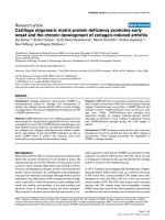
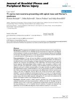
Figure 1 Compartment syndrome of both lower legs and both
thighs secondary to Systemic Capillary Leak Syndrome (SCLS).
Decompressive fasciotomy
Saugel et al. Scandinavian Journal of Trauma, Resuscitation and Emergency Medicine 2010, 18:38
/>Page 3 of 5
lin/tazobactam for another 6 days. The patient was
treated with hydrocortisone (288 mg/day) for suspected
septic shock for the first 6 days.
Over the following days the hypovolemic shock and
edema gradually subsided under volume management
(volume resuscitation with crystalloid fluid) based on
transpulmonary thermodilution data and norepinephrine
administration (fig. 2). In three surgical procedures the
fascias of both lower legs and thighs were completely
closed.
Regarding hemodynamic stabilisation, in parallel to
improving GEDVI and SVV through volume loading, the
extra-vascular lung water index (EVLWI) also increased
(20 mL/kg; n = 3-7 mL/kg), decreasing the pO2/FiO2-
ratio. There were also clinical and radiological signs of
pulmonary edema developing on hospital day 4. There-
fore a more restrictive volume balance including the
application of diuretics was initiated resulting in mark-
edly improved gas exchange. The tracheal tube was
removed on hospital day 11 and the patient was trans-
ferred to a normal ward on hospital day 14. Serum pro-
tein immunoelectrophoresis then indicated paraprotein
of the IgG kappa type. A diagnosis of idiopathic SCLS
(Clarkson's disease) was made retrospectively. Two weeks
after transfer to the normal ward the patient was dis-
charged to rehabilitation.
Conclusion
SCLS is a very rare disorder with a high mortality rate. It
is characterized by increased capillary permeability
resulting in hypovolemic shock due to a marked shift of
fluid and protein from the intravascular to the extravas-
cular space. Laboratory findings include hemoconcentra-
tion, hypoproteinemia and a monoclonal gammopathy
[2]. SCLS was first described in 1960 by Clarkson et al.
[1]. The median age for the first SCLS-manifestation is 46
years with no sex-related difference [3]. Hard physical
work several days before SCLS-symptoms and flu-like-ill-
ness at the beginning of a SCLS-episode has been
described in several case reports [3,4]. However the
pathogenesis of SCLS is still unknown. Involvement of
interleukin-2, classic pathway complement or stimulation
of 5-lipooxygenase-pathway have been suggested [5-7].
The relationship between monoclonal protein and SCLS
has also not been clarified. Plasma shift into the extravas-
cular space and muscle can result in a markedly increased
muscle compartment pressure and pressure induced
muscle damage [8-10]. Documentation of increased mus-
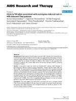
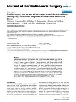
Figure 2 Time course of fluid balance, extra-vascular lung water index (EVLWI), global end-diastolic volume index (GEDVI), and norepi-
nephrine administration.
12345678910111213
EVLWI [mL/kg]
GEDVI [mL/sqm]
Fluid Balance [mL
]
Norepinephrine [µg/kg/min]
Hospital days
20
11
8
-8300
0.13
+15000
Beginning
pulmonary edema
Mechanical ventilation
459
960
0.02
Saugel et al. Scandinavian Journal of Trauma, Resuscitation and Emergency Medicine 2010, 18:38
/>Page 4 of 5
cular tension and compartment pressure can be per-
formed by manometry. Since the risk of ischemic muscle
necrosis increases markedly as compartment pressure
increases above the mean arterial pressure, fasciotomy
should be performed in cases of SCLS with hypotension
and severe compartment syndrome. Pulmonary edema,
probably induced by intravascular overloading in combi-
nation with recruitment of the initially extravasated flu-
ids, has been described in patients with SCLS [3]. In our
case report signs of pulmonary edema were present on
hospital day 4 illustrating the importance of switching
from the management of acute hypovolemia to manage-
ment of severe fluid overload using modern hemody-
namic monitoring tools.
In general optimization of intravascular volume status
under consideration of pulmonary hydration is of central
importance in the treatment of critically ill patients. Clin-
ical parameters such as filling of the jugular veins (intra-
vascular space), presence of leg edema (interstitium),
ascites or pleural effusions ("third space") are still the first
cornerstones in the estimation of hemodynamics and
pulmonary hydration. However, according to the few
studies investigating this issue, the utility of most clinical
signs for the estimation of volume status might be limited
due to poor specificity and sensitivity, when compared to
invasive procedures [11,12]. In most ICU patients CVP
can be determined easily and soon after admission. How-
ever, there is data demonstrating a poor capacity of CVP
to predict the hemodynamic response to a fluid challenge
[13]. Regarding more invasive techniques, transpulmo-
nary thermodilution and pulse contour analysis are estab-
lished for assessment of cardiac index, preload, volume
responsiveness and pulmonary hydration [14-16]: Besides
cardiac index, these techniques provide volumetric
parameters such as GEDVI as well as dynamic variables
of preload such as SVV for the assessment of volume
responsiveness. The use of dynamic variables of preload
is restricted to patients with sinus rhythm and controlled
ventilation. By contrast, transpulmonary thermodilution-
derived volumetric parameters can be used regardless of
sinus rhythm and controlled ventilation to predict fluid
responsiveness. Moreover, transpulmonary thermodilu-
tion accurately allows measurement of EVLWI to quan-
tify the degree of pulmonary edema in critically ill
patients.
The comparison between transpulmonary thermodilu-
tion and pulmonary artery catheter technology is still a
matter of debate.
Transpulmonary thermodilution is less invasive than
pulmonary thermodilution using a Swan-Ganz-catheter
because it does not require the insertion of a catheter in
the pulmonary artery but only a central venous and an
arterial catheter (that is also needed in patients moni-
tored with pulmonary thermodilution).
The pulmonary artery catheter is still considered to be
the gold standard for assessment of cardiac index and sys-
temic vascular resistance index. However, there is
increasing data that pulmonary artery wedge pressure is
not appropriate for assessment of preload and prediction
of volume responsiveness, particularly in ICU patients
with invasive mechanical ventilation and/or increased
intra-abdominal pressure [17].
In numerous studies transpulmonary thermodilution-
derived dynamic and volumetric variables of preload have
been demonstrated as superior indicators of volume
responsiveness as compared to pressures such as pulmo-
nary artery wedge pressure and central venous pressure
[14,18,19].
Regarding the presented case, in addition to cate-
cholamine administration, transpulmonary thermodilu-
tion-guided volume-management regarding decreased
GEDVI as valuable marker of volume deficiency and
increased EVLWI as "upper threshold" for further volume
resuscitation proved as very useful tool in this patient
who's hydration status was difficult to judge using clinical
criteria.
Several studies have also suggested that corticosteroid
may be useful when the capillary leak is initiated by
cytokine-mediated endothelial damage [3,20]. Treatment
with terbutalin, theophylline and immunglobulines has
been shown to be effective for decreasing the incidence
and severity of SCLS episodes [2,21,22]. Terbutalin and
theophyllin diminish the increment of bradikinin-medi-
ated capillary permeability by an increase of cyclic ade-
nosine monophosphate [9]. There are two reports
regarding patients who developed multiple myeloma
after the diagnosis of SCLS [23]. In patients with mono-
clonal gammopathy of undetermined significance, the
risk of progression to multiple myeloma at 25 year follow-
up is around 30% [24]. Therefore, annual surveillance for
multiple myeloma in patients with SCLS should be rec-
ommended.
In conclusion the reported case shows the importance
of extended hemodynamic monitoring for volume and
catecholamine management as well as the importance of
monitoring muscle compartment pressure in SCLS
patients.
Consent
Written informed consent was obtained from the patient
for publication of this case report and any accompanying
images. A copy of the written consent is available for
review by the Editor-in-Chief of this journal.
List of abbreviations
EVLWI: extra-vascular lung water index; GEDVI: global
end-diastolic volume index; ICU: intensive care unit;
Saugel et al. Scandinavian Journal of Trauma, Resuscitation and Emergency Medicine 2010, 18:38
/>Page 5 of 5
SCLS: Systemic Capillary Leak Syndrome; SVV: stroke
volume variation.
Competing interests
The authors declare that they have no competing interests.
Authors' contributions
BS, AU, FM and VP contributed to the conception and design of the case
description. They were responsible for acquisition, analysis and interpretation
of data regarding this case report. BS drafted the manuscript. RMS and WH par-
ticipated in its design and coordination and helped to draft the manuscript. All
authors read and approved the final manuscript.
Author Details
1
II. Medizinische Klinik und Poliklinik, Klinikum rechts der Isar der Technischen
Universität München, Ismaninger Str. 22, D-81675 München, Germany and
2
Klinik München Perlach, Schmidbauer Str. 44, D-81737 München, Germany
References
1. Clarkson B, Thompson D, Horwith M, Luckey EH: Cyclical edema and
shock due to increased capillary permeability. The American journal of
medicine 1960, 29:193-216.
2. Tahirkheli NK, Greipp PR: Treatment of the systemic capillary leak
syndrome with terbutaline and theophylline. A case series. Annals of
internal medicine 1999, 130(11):905-909.
3. Chihara R, Nakamoto H, Arima H, Moriwaki K, Kanno Y, Sugahara S, Okada
H, Suzuki H: Systemic capillary leak syndrome. Internal medicine (Tokyo,
Japan) 2002, 41(11):953-956.
4. Guidet B, Guerin B, Maury E, Offenstadt G, Amstutz P: Capillary leakage
complicated by compartment syndrome necessitating surgery.
Intensive care medicine 1990, 16(5):332-333.
5. Cicardi M, Gardinali M, Bisiani G, Rosti A, Allavena P, Agostoni A: The
systemic capillary leak syndrome: appearance of interleukin-2-
receptor-positive cells during attacks. Annals of internal medicine 1990,
113(6):475-477.
6. Rondeau E, Sraer J, Bens M, Doleris LM, Lacave R, Sraer JD: Production of
5-lipoxygenase pathway metabolites by peripheral leucocytes in
capillary leak syndrome (Clarkson disease). European journal of clinical
investigation 1987, 17(1):53-57.
7. Cicardi M, Berti E, Caputo V, Radice F, Gardinali M, Agostoni A: Idiopathic
capillary leak syndrome: evidence of CD8-positive lymphocytes
surrounding damaged endothelial cells. The Journal of allergy and
clinical immunology 1997, 99(3):417-419.
8. Perry J, Balasubramanian S, Imray C: Systemic capillary leak syndrome
resulting in compartment syndrome and the requirement for a surgical
airway. Anaesthesia 2009, 64(6):679-682.
9. Prieto Valderrey F, Burillo Putze G, Martinez Azario J, Santana Ramos M:
Systemic capillary leak syndrome associated with rhabdomyolysis and
compartment syndrome. The American journal of emergency medicine
1999, 17(7):743-744.
10. Sanghavi R, Aneman A, Parr M, Dunlop L, Champion D: Systemic capillary
leak syndrome associated with compartment syndrome and
rhabdomyolysis. Anaesthesia and intensive care 2006, 34(3):388-391.
11. Katz SD: Blood volume assessment in the diagnosis and treatment of
chronic heart failure. The American journal of the medical sciences 2007,
334(1):47-52.
12. McGee S, Abernethy WB, Simel DL: The rational clinical examination. Is
this patient hypovolemic? Jama 1999, 281(11):1022-1029.
13. Marik PE, Baram M, Vahid B: Does central venous pressure predict fluid
responsiveness? A systematic review of the literature and the tale of
seven mares. Chest 2008, 134(1):172-178.
14. Michard F, Alaya S, Zarka V, Bahloul M, Richard C, Teboul JL: Global end-
diastolic volume as an indicator of cardiac preload in patients with
septic shock. Chest 2003, 124(5):1900-1908.
15. Sakka SG, Reinhart K, Meier-Hellmann A: Comparison of pulmonary
artery and arterial thermodilution cardiac output in critically ill
patients. Intensive Care Med 1999, 25(8):843-846.
16. Sakka SG, Ruhl CC, Pfeiffer UJ, Beale R, McLuckie A, Reinhart K, Meier-
Hellmann A: Assessment of cardiac preload and extravascular lung
water by single transpulmonary thermodilution. Intensive Care Med
2000, 26(2):180-187.
17. Osman D, Ridel C, Ray P, Monnet X, Anguel N, Richard C, Teboul JL:
Cardiac filling pressures are not appropriate to predict hemodynamic
response to volume challenge. Crit Care Med 2007, 35(1):64-68.
18. Renner J, Gruenewald M, Brand P, Steinfath M, Scholz J, Lutter G, Bein B:
Global end-diastolic volume as a variable of fluid responsiveness
during acute changing loading conditions. Journal of cardiothoracic and
vascular anesthesia 2007, 21(5):650-654.
19. Muller L, Louart G, Bengler C, Fabbro-Peray P, Carr J, Ripart J, de La
Coussaye JE, Lefrant JY: The intrathoracic blood volume index as an
indicator of fluid responsiveness in critically ill patients with acute
circulatory failure: a comparison with central venous pressure.
Anesthesia and analgesia 2008, 107(2):607-613.
20. Takabatake T: Systemic capillary leak syndrome. Internal medicine
(Tokyo, Japan) 2002, 41(11):909-910.
21. Droder RM, Kyle RA, Greipp PR: Control of systemic capillary leak
syndrome with aminophylline and terbutaline. The American journal of
medicine 1992, 92(5):523-526.
22. Lambert M, Launay D, Hachulla E, Morell-Dubois S, Soland V, Queyrel V,
Fourrier F, Hatron PY: High-dose intravenous immunoglobulins
dramatically reverse systemic capillary leak syndrome. Crit Care Med
2008, 36(7):2184-2187.
23. Amoura Z, Papo T, Ninet J, Hatron PY, Guillaumie J, Piette AM, Bletry O,
Dequiedt P, Talasczka A, Rondeau E, Dutel JL, Wechsler B, Piette JC:
Systemic capillary leak syndrome: report on 13 patients with special
focus on course and treatment. The American journal of medicine 1997,
103(6):514-519.
24. Blade J: Clinical practice. Monoclonal gammopathy of undetermined
significance. The New England journal of medicine 2006,
355(26):2765-2770.
doi: 10.1186/1757-7241-18-38
Cite this article as: Saugel et al., Systemic Capillary Leak Syndrome associ-
ated with hypovolemic shock and compartment syndrome. Use of transpul-
monary thermodilution technique for volume management Scandinavian
Journal of Trauma, Resuscitation and Emergency Medicine 2010, 18:38
Received: 20 April 2010 Accepted: 5 July 2010
Published: 5 July 2010
This article is available from: 2010 Saugel et al; licensee BioMed Central Ltd. This is an Open Access article distributed under the terms of the Creative Commons Attribution License ( ), which permits unrestricted use, distribution, and reproduction in any medium, provided the original work is properly cited.Scandinavi an Journal of Trau ma, Resuscitatio n and Emergency Medicine 2010, 18:38