Báo cáo y học: "Blunt traumatic pericardial rupture and cardiac herniation with a penetrating twist: two case reports" ppt
Bạn đang xem bản rút gọn của tài liệu. Xem và tải ngay bản đầy đủ của tài liệu tại đây (1.72 MB, 7 trang )
BioMed Central
Page 1 of 7
(page number not for citation purposes)
Scandinavian Journal of Trauma,
Resuscitation and Emergency Medicine
Open Access
Case report
Blunt traumatic pericardial rupture and cardiac herniation with a
penetrating twist: two case reports
Peter B Sherren*, Robert Galloway and Marie Healy
Address: Department of Anaesthesia and Intensive care, The Royal London Hospital, Whitechapel, E1 1BB, UK
Email: Peter B Sherren* - ; Robert Galloway - ;
Marie Healy -
* Corresponding author
Abstract
Background: Blunt Traumatic Pericardial Rupture (BTPR) with resulting cardiac herniation
following chest trauma is an unusual and often fatal condition. Although there has been a multitude
of case reports of this condition in past literature, the recurring theme is that of a missed injury.
Its occurrence in severe blunt trauma is in the order of 0.4%. It is an injury that frequently results
in pre/early hospital death and diagnosis at autopsy, probably owing to a combination of diagnostic
difficulties, lack of familiarity and associated polytrauma. Of the patients who survive to hospital
attendance, the mortality rate is in the order of 57-64%.
Methods: We present two survivors of BTPR and cardiac herniation, one with a delayed
penetrating cardiac injury secondary to rib fractures. With these two cases and literature review,
we hope to provide a greater awareness of this injury
Conclusion: BTPR and cardiac herniation is a complex and often fatal injury that usually presents
under the umbrella of polytrauma. Clinicians must maintain a high index of suspicion for BTPR but,
even then, the diagnosis is fraught with difficulty. In blunt chest trauma, patients should be
considered high risk for BTPR when presenting with:
Cardiovascular instability with no obvious cause
Prominent or displaced cardiac silhouette and asymmetrical large volume pneumopericardium
Potentially, with increasing awareness of the injury and improved use and availability of imaging
modalities, the survival rates will improve and cardiac Herniation could even be considered the 5
th
H of reversible causes of blunt traumatic PEA arrest.
Background
Cardiac herniation is a significant and potentially fatal
complication of BTPR. This is by no means a new problem
[1,2] and its occurrence in severe blunt trauma is in the
order of 0.4% [3,4]. Despite literature experience dating
back to 1864 [5], it is an injury that frequently results in
pre/early hospital death and diagnosis at autopsy, proba-
bly owing to a combination of diagnostic difficulties, lack
of familiarity and associated polytrauma [3,6]. Of those
who make it to hospital, and are later diagnosed with
Published: 15 December 2009
Scandinavian Journal of Trauma, Resuscitation and Emergency Medicine 2009, 17:64 doi:10.1186/1757-7241-
17-64
Received: 9 November 2009
Accepted: 15 December 2009
This article is available from: />© 2009 Sherren et al; licensee BioMed Central Ltd.
This is an Open Access article distributed under the terms of the Creative Commons Attribution License ( />),
which permits unrestricted use, distribution, and reproduction in any medium, provided the original work is properly cited.
Scandinavian Journal of Trauma, Resuscitation and Emergency Medicine 2009, 17:64 />Page 2 of 7
(page number not for citation purposes)
BTPR, the survival rate is 36.4% - 42.9% [7]. The high
mortality rate is probably a reflection of not only BTPR
and cardiac herniation but also the associated injuries [3].
Here, we present two interesting cases of both left and
right pleuropericardial ruptures and cardiac herniation.
Despite the delay in initial diagnosis, both patients sur-
vived, though with varying degrees of disability secondary
to related traumatic injuries. The second patient is one of
the few reported cases of cardiac herniation and a delayed
penetrating cardiac injury secondary to rib fractures.
The common issue echoed throughout our experience
and those of others is that of missed or delayed diagnosis.
With these cases and literature review we hope to provide
further awareness of this injury and clues which can be
sought from the clinical presentation and investigations
to aid diagnosis.
Case 1
A 21-year-old male was admitted to a district general hos-
pital accident and emergency department following a
moderate speed motorbike accident with the predomi-
nant vector of force through the chest and head. Initially
when seen by the local ambulance service he was noted to
be GCS 15/15, have a high Alveolar-arterial gradient but
was cardiovascularly stable. Of note, he could not move or
feel his legs.
Management in the district general accident and emer-
gency department followed standard Advanced Trauma
Life Support (ATLS) practices. Chest radiograph showed
pulmonary contusions on the left but nothing else of sig-
nificance. He became increasingly agitated and hypoxic
and was intubated prior to transfer for computed tomog-
raphy (CT) scan.
Head CT scans showed an interventricular haemorrhage.
Spinal images showed T8/T9 fracture/dislocation with a
normal cervical CT. Initial chest CT scans were reported as
showing dextracardia and bilateral pneumothoraces; on
the left side, the pneumothorax was reported as a possible
tension pneumothorax. The possibility of a pneumoperi-
cardium was later attributed to an anterior pneumotho-
rax. Abdominal and pelvis CT scans were essentially
normal.
As time progressed, persistent hypotension developed
despite bilateral tube thoracostomies, fluid challenges
and inotropes. The initial working diagnosis of spinal
shock was made and a referral was made for further man-
agement and neurosurgical intervention for stabilisation
of the T8-9 fracture/dislocation.
On transfer to our trauma centre, the patient's condition
deteriorated; on arrival in our department, he was found
to be on a FiO
2
of 1.0 with PaO
2
around 10 kPa and
requiring high dose norepinephrine and epinephrine to
sustain his mean arterial pressure. He was re-trauma
called at this stage and plain radiographs were obtained to
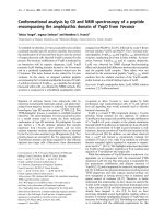
further ascertain and clarify his injuries (Figure 1).
The finding of dextracardia had been noted previously at
the district general hospital and was not thought to be
pathological at this stage. A further tube thoracostomy did
not improve the hemodynamic status of the patient. The
patient was transferred for CT scan where the following
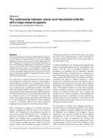
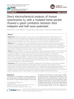
images were obtained (Figure 2 and 3).
The CT showed a multitude of head and thoracic injuries.
A number of rib fractures and bilateral haemopneumoth-
acaces as well as the aforementioned neuroaxial injuries
were all noted. The presence of pericardial air with herni-
ation of the heart into the right hemithorax was also caus-
ing concern. At this stage the patient's condition had not
improved and, on balance, it was agreed to take the
patient to theatre to investigate his thoracic injuries. The
patient underwent a clamshell thoracotomy where a 10
cm tear in the right of the pericardium (along the path of
the phrenic nerve) was noted with a cardiac herniation
through the defect. The heart was noted to be large and
dilated. The heart was relocated and the pericardium
repaired with interrupted non-absorbable sutures. An
intracranial pressure (ICP) bolt was also inserted for mon-
itoring and further management of his traumatic brain
injury.
There was an almost immediate reduction in inotrope
requirement and the patient was transferred to ICU. His
Plain supine AP chest radiograph showing a prominent, right-sided cardiac silhouette ('boot shaped'); bilateral pulmonary contusions; rib fractures; endotracheal and tube thoracosto-miesFigure 1
Plain supine AP chest radiograph showing a promi-
nent, right-sided cardiac silhouette ('boot shaped');
bilateral pulmonary contusions; rib fractures;
endotracheal and tube thoracostomies. With the bene-
fit of hindsight there is the suggestion of a left-sided pneu-
mopericardium surrounded by a faint pericardial contour.
Scandinavian Journal of Trauma, Resuscitation and Emergency Medicine 2009, 17:64 />Page 3 of 7
(page number not for citation purposes)
post-op care was complicated by a chest infection and fre-
quent episodes of fast atrial fibrillation secondary to a
myocardial contusion, requiring DC cardioversion. He
was discharged from ICU after 14 days. Although he was
left with a permanent disability from his T8/T9 fracture
dislocation, he recovered a good cognitive neurological
status and arm strength. With no ongoing cardiovascular
problems, he is currently awaiting transfer to a rehabilita-
tion centre.
Case 2
The second case is that of a 45-year-old male brought into
our regional trauma centre by air ambulance. He was the
driver in a road traffic collision in which the force vector
came through the passenger/left side of the car; unfortu-
nately, the passenger was pronounced life extinct on the
scene. On scene, the patient was agitated, moving all
limbs and complaining of difficulty in breathing. As a
result, he underwent tracheal intubation with drug assist-
ance, bilateral thoracostomies, 750 ml crystalloid and
application of a pelvic splint and usual spinal precautions.
On arrival in the department, the initial concern was that
of multiple rib fractures, left-sided flail and a large
amount of surgical emphysema. Bilateral tube thoracosto-
mies were inserted with 300 mls of blood from the left
tube; ventilation/oxygenation improved adequately. Of
note, cardiac pulsations were felt when there was a finger
sweep of the left pleural cavity.
The chest radiograph showed pneumopericardium, exten-
sive surgical emphysema and improvement in the left-
sided haemopneumothorax/right-sided pneumothorax
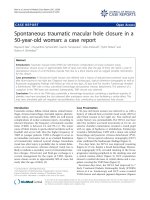
(Figure 4). The evolving problem quickly became that of
cardiovascular instability which remained fluid/packed
red blood cell responsive throughout. A Focused Assess-
ment with Sonography in Trauma (FAST) scan was nega-
tive on two occasions and a pelvic radiograph showed a
stable pelvic pubic ramus fracture. Given the negative
FAST scans and a period of stability, the patient was taken
for a full body CT. The CT chest showed a multitude of
injuries, resulting in two further tube thoracostomies
being sited (Figure 5). The CT head/spine showed multi-
ple facial and base of skull fractures but no evidence of
parencymal or extra-axial bleeds and no fracture or mal-
Axial chest CT demonstrating multiple parenchymal lung contusions; collapsed bilateral haemopneumothoraces; tube thoracostomies; surgical emphysema; large left-sided pneu-mopericardium; and displacement of the heart into the right hemithoraxFigure 2
Axial chest CT demonstrating multiple parenchymal
lung contusions; collapsed bilateral haemopneumot-
horaces; tube thoracostomies; surgical emphysema;
large left-sided pneumopericardium; and displace-
ment of the heart into the right hemithorax.
Coronal chest CT demonstrating most of the axial findings including the prominent pneumopericardium and displace-ment of the heart into the right hemithoraxFigure 3
Coronal chest CT demonstrating most of the axial
findings including the prominent pneumopericar-
dium and displacement of the heart into the right
hemithorax.
Plain supine AP chest radiograph showing extensive surgical emphysema; multitude of rib fractures and flail on the left side; bilateral pulmonary contusions and suggestion of a haemothorax on the left side; a rotated 'boot shaped' cardiac silhouette, with clear demarcation of cardiac silhouette from the diaphragm; pneumomediastinum; the pericardial contour is also distinctly visible; endotracheal tubeFigure 4
Plain supine AP chest radiograph showing extensive
surgical emphysema; multitude of rib fractures and
flail on the left side; bilateral pulmonary contusions
and suggestion of a haemothorax on the left side; a
rotated 'boot shaped' cardiac silhouette, with clear
demarcation of cardiac silhouette from the dia-
phragm; pneumomediastinum; the pericardial con-
tour is also distinctly visible; endotracheal tube.
Scandinavian Journal of Trauma, Resuscitation and Emergency Medicine 2009, 17:64 />Page 4 of 7
(page number not for citation purposes)
alignment of C/T/L spine. CT abdomen showed a small
anterior spleenic laceration with no free fluid in the peri-
toneal cavity.
Following a joint review of the CT scans and the now-sta-
ble patient, no surgical intervention was felt to be needed.
The patient had an ICP bolt inserted and was taken to the
ICU for further resuscitation and stabilisation.
Sixteen hours post-injury on the ICU, the blood output
from the left basal tube thoracostomy started to climb,
finally reaching 600 ml/Hr. This was associated with a
transfusion requirement, haemodynamic compromise
and climbing lactates. At this point, the patient was taken
to theatre for a thoracotomy to establish the cause of a
likely complex chest bleed.
The following injuries were found and repaired through a
left anterior lateral thoracotomy: left-sided longitudinal
rupture of the pericardium and cardiac herniation; left
ventricular laceration secondary to overlying rib fractures;
multiple lung lacerations; and multiple flail ribs. Follow-
ing insertion of a pericardial and further tube thoracosto-
mies (on suction), the patient was transferred back to ICU
for further care.
An eighteen day ICU admission followed, the main issues
being that of recurrent atrial flutter and a slow respiratory
wean. The respiratory wean was protracted as a result of
adult respiratory distress syndrome caused by the primary
polytrauma and ventilator-acquired pneumonia.
Over 6 weeks after his initial accident, the patient was dis-
charged home and, all things considered, was doing well
in follow up clinic one month later.
Discussion
Pathophysiology
Cardiac herniation can occur when there is a significant
defect within the pericardial sac. Pericardial tears may
involve either the superior/left/right pleuropericardium
or the diaphragmatic pericardium. The defect can allow
cardiac luxation and, in the case of diaphragmatic pericar-
dial tear, herniation of abdominal contents into the peri-
cardial sac. Clarke et al, one of the largest reviews to date,
included a review of 132 cases plus 10 further cases of
their own [3]. They found the superior/left/right pleu-
ropericardium were injured in 4%/50%/17% respectively,
with the remaining 27% of injuries originating from the
diaphragmatic pericardium [3,7]. Of these cases, the rate
of cardiac herniation was 28% [3]; however, in a more
recent literature search (since 1987), a rate of 64% of the
55 patients with BTPR had cardiac herniation [7]. Defects
of the pleuropericardium usually occur vertically along
the phrenic nerve; as in our cases [8]. If the tear is large
enough, approaching 8-12 cm, the heart can sublux
through the defect [9]. The resulting torsion of the great
vessels can lead to a form of obstructive cardiogenic shock
and cardiovascular instability [8].
Clinical presentation
As seen with our own experience and those of others there
is often a delay in diagnosis of BTPR and cardiac hernia-
tion, which is a real concern given that, once recognised,
the treatment is simple and effective [10].
The most common mechanism for BTPR are those
involved in road traffic collisions and sudden decelera-
tions; particularly those involving a vector of injury from
the left side of the chest [3]. The following pattern of asso-
ciated injuries should also arouse suspicion of BTPR [3]:
• Cardiac - contusions and dysrrthmias (28%). The
delayed penetrating cardiac injury as a result of rib
fractures, as witnessed in the second case is one of the
only reported cases of its kind.
• Chest - multiple rib fractures, haemopneumothora-
ces and pulmonary contusions almost universally
seen.
• Neurological - particularly thoracic spine fractures
and spinal cord injuries as well as traumatic brain inju-
ries (32%).
• Abdominal injuries (27%).
• Pelvic and long bone fracture indicative of a high
velocity/energy impact (49%).
Axial chest CT showing most of the pathology found on the plain radiograph but also bilateral anterior pneumothoraces; large volume anterior pneumopericardium; tube thoracosto-miesFigure 5
Axial chest CT showing most of the pathology found
on the plain radiograph but also bilateral anterior
pneumothoraces; large volume anterior pneu-
mopericardium; tube thoracostomies.
Scandinavian Journal of Trauma, Resuscitation and Emergency Medicine 2009, 17:64 />Page 5 of 7
(page number not for citation purposes)
Given the severity of associated injuries patients usually
require invasive ventilation early on. However, if the
patient is conscious, they may report symptoms of palpi-
tations, shortness of breath and chest pain as well as
angina type pains as a result of coronary obstruction fol-
lowing herniation [2,11].
The main clinical signs, which may be subtle but should
be sought, are:
• Signs similar to that of tamponade; in particular that
of hypotension, pulsus paradoxus and raised jugular
venous pressure (JVP) [2,12]. This may occur early or
late depending on the timing of herniation [13]. This
haemodynamic compromise may manifest itself
despite fluid administration and inotropic support
[13].
• Fluctuating haemodynamic parameters, sometimes
to the extent of sudden cardiac arrest (often as a result
of change in patient's position) should evoke a high
index of suspicion of BTPR [14].
• Tachycardia and dysrrthymias may also be seen [11],
such as the atrial tachyarrythmias noted in our case.
• Displaced and heaving apex beat [2,8,12].
• A splashing murmur "bruit de Moulin" as a result of
the heart moving in a haemopneumopericardium
[5,10,12].
Investigations
Identifying these symptoms and signs in a noisy and
stressful trauma environment may well prove difficult.
However, there is a multitude of investigations available
to most hospitals that can assist in the diagnosis:
• Electrocardiogram - may show a tachycardia as well
as dysrrthymias particularly those of atrial origin. Also
present could be an electrical axis deviation associated
with the cardiac herniation and rotation [2,8,12] and
a right bundle branch block [9,10]. Ischaemic changes
may be noted as a result of coronary artery occlusion
by the pericardial band [8,12]. In fact, Rippey et al [12]
reported an elevated Troponin I of 9.20 μg/L in a
patient later diagnosed with BTPR and cardiac hernia-
tion. This was thought to be multifactorial but pre-
dominantly as a result of a contusion and coronary
insufficiency.
• Chest radiograph - as a readily available imaging
modality, it is a useful screening tool for BTPR and car-
diac herniation. Given the very real chance of the chest
x-ray being completely normal, serial films may also
be of use to identify any evolving pathology [13].
Findings may include: cardiac silhouette may be unu-
sually prominent ("boot shaped") and demarcated
from the diaphragm; pneumopericardium; pneumo-
mediastinum; bowel gas/loops within pericardial sac;
prominent pulmonary artery contour; herniation and
rotation of the heart into either hemithorax with a
possible pericardial sac contour visible distinct to the
cardiac silhouette [2,8,10,13]. Associated injuries
include haemopneumothoraces, pulmonary contu-
sions, lower lobe collapse/atelectasis/consolidation,
surgical emphysema, rib/clavicle/sternal and thoracic
spine fractures [13-15].
• Transthoracic/oesophageal echocardiography and
Focused Assessment with Sonography for Trauma -
TTE/TOE have been used with varying reports of suc-
cess but the sensitivity for diagnosing even large peri-
cardial defects is thought to be low [7,15]. With the
presence of surgical emphysema and pneumopericar-
dium, the echographic windows will be poor and,
along with operator variability, cannot be relied upon
[15]. The importance of echocardiography lies in its
ability to rule out other differential diagnoses (such a
cardiac contusion or pericardial effusion and possible
tamponade) non-invasively and quickly. This can be
particularly useful in the patient who is haemodynam-
ically shocked, with a raised JVP, ± reduced heart
sounds, and is unresponsive to fluids and inotropic
support.
• Computed Tomography (CT) - along with its
increasing availability and use in the multiply injured
trauma patients, CT is also more sensitive for identify-
ing cardiac axis changes and pericardial discontinuity
than plain radiographs [13,15].
▪ Characteristic changes for a pericardial rupture
include [7,14,15]:
▪ Focal pericardial dimpling and discontinuity
▪ Pneumopericardium
▪ Interposition of lung between: aorta and pul-
monary artery; or heart and diaphragm; or
right atrium and right ventricular outflow tract
▪ Characteristic changes for a cardiac herniation
include [7,15]:
▪ "Empty pericardial sac" sign, air outlining the
empty pleuropericardium as a result of cardiac
luxution into the hemithorax.
Scandinavian Journal of Trauma, Resuscitation and Emergency Medicine 2009, 17:64 />Page 6 of 7
(page number not for citation purposes)
▪ "Collar" sign is the result of compression of
the cardiac contour as a result of constriction
by the pericardial band caused by the defect.
▪ Associated signs include dilated inferior vena
cava (IVC), reflux of contrast into IVC and
deformed ventricular silhouette, as well as, sec-
ondary signs of tamponade periportal lym-
phoedema, pericholecystic fluid and ascites.
▪ Magnetic Resonance Imaging (MRI) - In haemody-
namically stable patients with a suspected BTPR where
other imaging modalities have been suggestive but
inconclusive, cardiac MRI has been used to clarify its
presence [7].
Management
Once BTPR and cardiac herniation has been diagnosed,
treatment is simple and effective. It has even been sug-
gested that, as it is such a rapidly reversible cause of sud-
den cardiac arrest, there may be a role for post-arrest
emergency thoracotomy for select patient groups with
blunt chest trauma and positional cardiovascular instabil-
ity [14].
Video-assisted thoracoscopy has been suggested by some,
for the assessment and management of stable patients
where there is a lack of diagnostic clarity [8]. Small peri-
cardial defects where cardiac herniation is unlikely, espe-
cially those on the left side can be left alone [3,11]. The
treatment of choice for tears of the diaphragmatic pericar-
dium, right pleuropericardium, and moderate/large left
pleuropericardium defects, is surgical closure [3,10]. Clo-
sure of moderate-sized pericardial defects is best achieved
by interrupted non-absorbable sutures and larger ones
with a mesh prosthesis [3,10,11].
Conclusion
BTPR and cardiac herniation is a complex and often fatal
injury that usually presents under the umbrella of multi-
system trauma. The majority of patients will be non-sal-
vageable; where, despite best efforts, the severity of the
initial injury results in death prior to arrival in hospital. In
the polytrauma patients with severe blunt chest injuries
who survive to hospital arrival, the clinician must main-
tain a high index of suspicion for BTPR. Even with a high
index of suspicion, the diagnosis is still fraught with diffi-
culty. However, patients with blunt chest trauma and any
of the following signs are exceptionally high risk for BTPR
and the need for an urgent operative intervention should
be considered:
• Cardiovascular instability with no obvious cause.
This instability may be labile and mimic cardiac tam-
ponade, particularly with changes in patient position.
A bedside TTE in this setting is a vital tool for exclusion
of differential pathology.
• A prominent, possibly displaced cardiac silhouette
and asymmetrical large volume pneumopericardium.
These signs may show varying degrees of prominence
on the plain chest radiograph, if there is uncertainty
and the patient's condition allows, a chest CT should
be sought as it has been shown to better delineate the
injuries. In situations where the patient has a good
haemodynamic status and, despite CT, there remains
a diagnostic uncertainty, cardiac MR should be consid-
ered.
Personal experience and a review of past literature show
that, in the majority of cases, it is still an injury diagnosed
at autopsy or thoracotomy. Potentially, with increasing
awareness of the injury and improved use and availability
of imaging modalities, the survival rates will improve and
cardiac Herniation could even possibly be considered the
5
th
'H' of reversible causes of blunt traumatic PEA arrest.
Competing interests
The authors declare that they have no competing interests.
Authors' contributions
All authors were present at the conception of the project.
PBS and RG prepared the draft and all authors were
involved in revising the final manuscript. All authors have
read and approved the final manuscript.
Consent
Written informed consent was obtained from the patient
for publication of this case report and accompanying
images. A copy of the written consent is available for
review by the Editor-in-Chief of this journal.
Acknowledgements
This paper did not receive any grant or funding from any agency in the pub-
lic, commercial or not-for-profit sector.
Presented in part at - DINGLE Conference 2009 (Intensive Care Society
of Ireland)
- International Trauma conference 2009 - Manchester
References
1. Bettman RB, Tannenbaum WJ: Herniation of the heart. Ann Surg
1948, 128:1012-1014.
2. Wright MP, Nelson C, Johnson AM, Mcmillan IKR: Herniation of
the heart. Thorax 1970, 25:656-666.
3. Clark DE, Wiles CS III, Lim MK, Dunham CM, Rodriguez A: Trau-
matic rupture of the pericardium. Surgery 1983, 93:495-503.
4. Fulda G, Brathwaite CEM, Rodriguez A, Turney SZ, Dunham CM,
Cowley RA: Blunt traumatic rupture of the heart and the peri-
cardium: A ten-year experience (1979 1989). J Trauma 1991,
31:167-173.
5. Morel-Lavallee VAF: Rupture du pericarde; bruit de roue
hydraulique; bruit de moulin. Gaz Med Paris 1864, 19:695.
Publish with BioMed Central and every
scientist can read your work free of charge
"BioMed Central will be the most significant development for
disseminating the results of biomedical research in our lifetime."
Sir Paul Nurse, Cancer Research UK
Your research papers will be:
available free of charge to the entire biomedical community
peer reviewed and published immediately upon acceptance
cited in PubMed and archived on PubMed Central
yours — you keep the copyright
Submit your manuscript here:
/>BioMedcentral
Scandinavian Journal of Trauma, Resuscitation and Emergency Medicine 2009, 17:64 />Page 7 of 7
(page number not for citation purposes)
6. Farhataziz N, Landay M: Pericardial rupture after blunt chest
trauma. J Thorac Imaging 2005, 20:50-52.
7. Sohn JH, Sohn JW, Seo JB, Do KH, Lee JS, Kim DK, Song KS, Lim TH:
Pericardial rupture and cardiac herniation after blunt
trauma: a case diagnosed using cardiac MRI. The British Journal
of Radiology 2005, 78:447-449.
8. Thomas P, Saux P, Lonjon T, Viggiano M, Denis JP, Giudicelli R, Ragni
J, Gouin F, Fuentes P: Diagnosis by video assisted thoracoscopy
of traumatic pericardial rupture with delayed luxation of the
heart: case report. The Journal of Trauma Injury, Infection and Critical
care 1995, 38:967-70.
9. Carillo EH, Heniford BT, Dykes JR, McKenzie ED, Polk HC Jr, Rich-
ardson JD: Cardiac Herniation Producing Tamponade: The
Critical Role of Early Diagnosis. The Journal of Trauma Injury, Infec-
tion and Critical care 1997, 43(1):19-23.
10. Janson JT, Harris DJ, Pretorius J, Rossouw GJ: Pericardial rupture
and cardiac herniation after blunt chest trauma. Ann Thorac
Surg 2003, 75(2):581-582.
11. Chughtai T, Chiavaras MM, Sharkey P, Shulman H, Miller HA: Peri-
cardial rupture with cardiac herniation. Can J Surg 2008,
51(5):E101-E102.
12. Rippey JC, Rao S, Fatovich D: Blunt traumatic rupture of the
pericardium with cardiac herniation. CJEM 2004, 6(2):126-129.
13. Nassiri N, Yu A, Statkus N, Gosselin M: Imaging of Cardiac her-
niation in Traumatic pericardial rupture. Journal of Thoracic
Imaging 2009, 24(1):69-72.
14. Wall MJ, Mattox KL, Wolf DA: The Cardiac Pendulum: Blunt
Rupture of the Pericardium with Strangulation of the Heart.
The Journal of Trauma Injury, Infection and Critical care 2005,
59(1):136-142.
15. Wielenberg AJ, Demos TC, Luchette FA, Bova D: Cardiac Hernia-
tion Due to Blunt Trauma: Early Diagnosis Facilitated by CT.
AJR 2006, 187:W239-W240.