Advanced Techniques in Dermatologic Surgery - part 4 ppsx
Bạn đang xem bản rút gọn của tài liệu. Xem và tải ngay bản đầy đủ của tài liệu tại đây (2.28 MB, 42 trang )
fluid is removed, manual tissue stabilization (discussed below) performed
by an assistant compensates the developing laxity of the skin turgor.
Postoperatively, a thorough drainage of tumescent solution must
be achieved by leaving the incision sites open and wearing compression
garments.
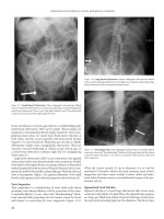
Figure 7
Endoscopic picture after complete liposuction of the lateral lower leg demon-
strating of persisting fibrous tissue without adipocytes.
Figure 8
Formation of a subcutaneous scar 12 months after liposuction. Photograph
taken during abdominoplasty.
106 Sattler
Technical Developments
To obtain an atraumatic suction technique, technical developments led to
an improvement of cannulas and liposuction-assisting devices.
Manual Liposuction, 24-Hole Cannulas
If correct tumescent local anesthesia is performed, then suction can be done
with thin, blunt-tipped cannulas. The connective tissuecan further be spared
when cannulas with multiple suction holes are used (24-hole cannulas).
After building up the suction force, a number of holes (10–12,12–16)
will be occluded by fibrous tissue. The remaining holes stay effective in lipo-
suctioning, so the cannula cannot build up a higher suction force due to
occlusion of the holes. As the suction force decreases, the holes that were
previously blocked will reopen.
When using two- or three-hole cannulas, it easily happens that all
holes are occluded simultaneously. In this case, the suction force
increases rapidly thus reinforcing the occlusion. Liposuction can be con-
tinued only after cleaning of the cannula or destruction of the blo cking
tissue.
The developed suction force in a 24-hole cannula is just strong
enough to remove the fat cells but too weak to suck in and destroy
fibrous tissue or vessels. In this way, blockage of the cannula and
destruction of the connective tissue is prevented and the treatment is
subtler.
Ultrasound-Assisted Liposuction (UAL)
To facilitate fat aspiration in difficul t areas such as the male breast or
back or in secondary sites, a number of new suction devices were devel-
oped starting in the late1980s.
In 1987, Scuderi and DeVita (12) and Zocchi (13) first described a
method of homogenizing the fat with ultrasound waves. The suction cannu-
laswereattachedtoanultrasoundgeneratorandultrasoundwavessentinto
the ti ssue supposedly destroy the ad ipocytes.
There are some severe disadvantages when using this technique.
The cannulas must have a comparatively larger volume. A large number
of seromas and skin burns and persisting hypo- or hyperaesthesias as a
result of destruction of the myelin sheath of peripheral nerves were
reported (14). There was even speculation about a potential carcinogenic
risk. Therefore, the American Society of Dermatologic Surgery rates
ultrasound assisted liposuction as an experimental method with no
extended clinical use (15,16).
Powered Liposuction/Vibrating Cannulas
In 1995 , Charles Gr oss (17), an ENT surgeo n at the University of Virginia,
described a new technique he used in liposuction of the neck called ‘‘lipo-
shaving.’’ An engine-powered cannula with an integrated rotating blade
was used to d estroy ad ipocytes under d irect vi sual o r endoscopic co ntrol.
Liposuction 107
This idea started the invent ion of a new generation of cannulas, first
with rotating blades but late r with oscillating blades.
The latest development is cannu las without blades but with a vibrat-
ing grip that leads to vibration of the cannula when passing through the
tissue (Fig. 9). One rationale behind the use of vibrating cannulas is the
different inertness of various materials wher eas, the cannula passes fibrous
tissue without damaging it, the homogenized fat can be aspirated. The
other aspect that aids this effect is the difference in velocity of the vibra-
tion and the presence of the suction force. If the vibration speed is higher
than the speed of the airflow of the suction, the suction can only withdraw
the liberated, homogenized fat. The cannula will escape and spare the
tissue structures that have tight attachments.
Vibrating cannulas facilitate the treatment of fibrous or pretreated
areas. Because they pass easily through the tissue and do not tangle with
the fibers, they make the procedure more comfortable for the patient and
the surgeon.
Severe complications have not been reported.
Further improvements of the cannulas and grips are expected,
which will lead to a wide spread usage of this suction device as it shows
greater benefits in achieving good operative outcome (18).
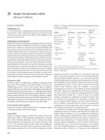
Figure 9
Demonstration of vibrating cannula technique.
Table 3
Sattler’s Tumescent Solution with Reduced Prilocaine
Prilocaine 1% 40.0 mL
Epinephrine 1:1000 1.0 mL
Sodium bicarbonate 8.4% 6.0 mL
Triamcinolon-acetonide 10 mg 1.0 mL
Physiologic saline (NaCl 0.9%)
1000.0 mL
1048.0 mL solution 0.038%
108 Sattler
Endoscopic Liposuction
Liposuction is an operation without direct visual control. Endoscopic
liposuction can be used to visualize what is happening in the subcuta-
neous space during liposuction. This method helped to control the tech-
nique and quality of liposuction and to give a further understanding of
physiodynamic processes in the adipose tissue. It is not routinely used
clinical procedure, but has helped in the development of new, useful lipo-
suction devices.
Refinements of the Tumescent Solution
In the course of time, the original Klein tumescent solution was modified
by various working groups.
We first replaced lidocaine as local anesthetic with prilocaine because of
its lower systemic plasma levels, which is relevant when using large volumes.
As a resul t of clinical observations, prilocaine could be reduced by
20% from the initial 50 mL/L to 40 mL/L, which resulted in a reduced
local anesthetic concentration of 0.038% (Table 3).
Table 4
Tumescent Solution After Schneider-Affeld and Friedrich
Prilocaine 2% 10.0 mL
Lidocaine 2% 10.0 mL
Epinephrine 1:1000 0.66 mL
Sodium bicarbonate 8.4% 6.0 mL
Triamcinolon-acetonide 40mg 0.33 mL
Physiologic saline (NaCl 0.9%)
1000.0 mL
1026,99 mL solution 0.037%
Table 5
Volumes of Tumescent Solution: Comparison of 1992 and 1997 Recommendations
1992 1997
Abdomen 800–1000 mL 5000 mL
Hips (both sides) 400–1000 mL 5000 mL
Waist (both sides) 400–1000 mL 3000 mL
Lateral thigh (both sides) 500–1200 mL 4000 mL
Ventral thigh (both sides) 600–1200 mL 4000 mL
Medial thigh (both sides) 255–700mL 3000 mL
Knee (both sides) 200–500 mL 2000 mL
Male breast (both sides) 300–800 mL 3000 mL
Neck 100–200 mL 800 mL
Liposuction 109
In clinical trials, Schneider-Affeld and Friedrich combined lido-
caine and prilocaine to decrease the side effects of a single agent. Their
solution is shown in Table 4.
As a consequence of reduction of the local anesthetic concentration
and the growing knowledge of delayed absorption, the quantities of
tumescent solution used in one session could be raised. The possibility
to use more quantities of tumescent solution widens the therapeutic
range. Today, up to 6 liters of tumescent solution are used in one session.
Figure 10
Modern liposuction equipment with infiltrating pump connected to a Stenger
distributor, a suction system, and warming devices for tumescence solution.
110 Sattler
Clinical experience showed the better effects of super-tumescence when
using large volumes; because of the reduction of tissue traumatization,
the complication rate is also reduced.
Table 5 gives a comparison of the initially recommended amounts
of solution and the amounts used in 1997 and are still used today.
The use of trimacinolone in the solution is discussed, many physi-
cians do not add it any more. The initial rationale for its use, the prevention
of postoperative inflammation, is no longer relevant. Meanwhile, other
effects like psychovegetative stabilization as well as a regulative effect on
the blood circulation play more important roles.
Over the past years, the tumescent technique has evolved from a mainly
anesthetic procedure to an essential part of successful liposuction, as it is
crucial for the described processes of physiodynamics and wound healing,
and d etermines the course of the surgery and postoperative outcome.
Improved Operating Techniques and Positioning of Patient
Besides technical and pharmacological improvements, clinical experience
led to improvements in the operation procedure.
The operative outcome can mainly be improved through active
cooperation of the patient who is awake. It helps the suction process if
the patient is able to contract the underlying muscles to build a firm base
and change positions if necessary.
Figure 11
(A) Preoperative findings in a 20-year-old patient with lipomatosis of the
thighs. (B) Postoperative results four months after liposuction via tumescence
technique with 24-hole cannulas.
Liposuction 111
Figure 12
(A) and (B) Lipomatosis of hip, medial and lateral thighs in a 42-year-old
patient, preoperative findings. (C) and (D) Postoperative result one year later.
112 Sattler
The operative outcome can significantly be improved by a correct
positioning of the patient on the operating table and an easy access to
the surgery site.
Experience has shown that it is better to treat the medial thighs not
with the patient lying on his or her back but on the side with the leg to be
treated stretched out on the operating table and the other leg in a 90
degree angle, to stabilize the position. With this positioning, there is a
far better access to the fat deposits.
When treating the back or flanks, it is better to position the patient
on the side, with the back overstretched. With this improved positioning,
the overlying skin as well as the underlying muscles are stretched, which
makes the aspiration of subcutaneous fat easier.
Manual assisted skin stabilization technique (MASST).Everyone
performing liposuction surgery in tumescent technique has experienced that
the stabilizing effect of the tumescent solution on the tissue decreases con-
stantly because it is removed along with the fat by the suctioning process.
Thus, liposuctioning gets more difficult as shearing forces on the tissue
get stronger. This can be counteracted efficiently when the tissue is bimanu-
ally stabilized by stretching it with the help of an assisting person (nurse).
Last but not least, all the minor improvements that give the patient
more comfort during the whole procedure should be provided. They
include devices to warm sheets, blankets, and the tumescent solution to
body temperature as well as a pleasant atmosphere created by music
Figure 13
(A) Marked saddle bag deformity in 52-year-old patient. (B) Result after three
liposuction sessions using tumescence anesthesia in yearly intervals, 6 years
after the last liposuction.
Liposuction 113
Figure 14
(A) and (B) Preoperative finding before liposuction of the hip, medial and
lateral thigh as well as the knee region with vibrating cannulas. (C) Postopera-
tive result after 12 months.
114 Sattler
and room furnishing. When planning the surgery suite, it is important to
include a bathroom within the easy reach of the patient (Fig. 10).
SUMMARY
The invention of the tumescent technique by Jeffrey Klein revolutionized
the history of liposuction.
This technique formed the basis on which numerous developments
in the field of liposuction took place within the last 25 years. Today, lipo-
suction in tumescent local anesthesia (a term coined by our group) is the
most commonly performed cosmetic procedure worldwide.
Owing to the improved operation techniques as well as refinements
in the tumescent solution and the cannulas used, a sub stantial reduction
of risks and side effects could be achieved. Thanks to all these improve-
ments, we have reached a point today where this operation technique can
offer a predictable and highly satisfactory cosmetic result with minimal
risk.
To show the extent of cosmetic outcomes , we include some pre- and
postoperative findings (Figs. 11–14).
Further progress can be expected through the development of more
effective but at the same time more subtle cannulas. To find the best
tumescent solution, pharmacological studies are planned.
Liposuction 115
REFERENCES
1. American Society of Liposuction Surgery (ASLSS) and American Academy of Cosmetic
Surgery (AACS): Guidelines for Liposuction Surgery, 2001.
2. Fischer A, Fischer G. Revised technique for cellulitis fat reduction in riding breeches
deformity. Bull Int Acad Cosmet Surg 1997; 2:40–41.
3. Illouz Y. Bodycontouring by lipolysis: a 5-year experience with over 3000 cases. Plast
Reconstr Surg 1983; 72:511–524.
4. Fournier P. Body Sculpturing Through Syringe Liposuction and Autologous Fat Rein-
jection. Samuel Rolf International, 1987.
5. Hanke CW, Bernstein G, Bullock BS. Safety of tumescent liposuction in 15336 patients—
national survey results. Dermatol Surg 1996; 22:459–462.
6. Klein JA. The tumescent technique for liposuction surgery. Am J Cosmet Surg 1987;
4:236–267.
7. Sommer B, Sattler G, Hanke CW. Tumeszenzlokalana
¨
sthesie. New York: Springer Berlin
Heidelberg, 1999.
8. Sattler G, Rapprich S, Hagedorn M. Tumeszenz-Lokalana
¨
sthesie- Untersuchung zur
Pharmakokinetik von Prilocain Z Hautkr. 1997; 7:522–525.
9. Sattler G, Sommer B. Tumescent Liposuction in Germany: history and new trends and
techniques. Dermatol Surg 1999; 25:221–223.
10. Lillis PJ. Liposuction surgery under local anesthesia. Limited blood loss and minimal
lidocaine absorption. J Dermatol Surg Oncol 1988; 14:1145–1148.
11. Sattler G, Hasche E, Rapprich S, Mo
¨
sler K, Hagedorn M. Neue operative Behandlungs-
mo
¨
glichkeitren bei benignen Fettgewebserkrankungen. Zeitschrift HþG 1997; 8:579–582.
12. Scuderi N, DeVita R. Nuove prospettivo nella liposuzione: La lipoemulsificazione. Giorn
Chir Plast Ricostr Este 1987; 1:33.
13. Zocchi ML. Ultrasonic liposculpturing. Aesthet Plast Surg 1992; 16:287–298.
14. Scheflan M, Tazi H. Ultrasonically assisted body contouring. Aestet Surg 1996; 16:
117–122.
15. Topaz M. Possible long-term complications in ultrasound-assisted lipoplasty induced by
sonolumiscence, sonochemistry and thermal effect. Aesthet Surg 1998; 18:19–24.
16. ASDS: Statement on ultrasonic liposuction. Dermatol Surg 1998; 24:1035.
17. Gross CW, Becker DG, Lindsey WH, Park SS, Marschall DD. The soft tissue shaving
procedure for remove of adipose tissue. Arch Otolaryngol Head Neck Surg 1995; 121:
117–1120.
18. Coleman WP III. Powered liposuction. Dermatol Surg 2000; 26(4):315–318.
116 Sattler
6
Laser-Assisted Blepharoplasty
Alina A. M. Fratila
Jungbrunnen-Klinik Dr. Fratila GmbH, Bonn, Germany
Video 6: Blepharoplasty: Lower Eye
Video 7: Skin Resurfacing
Video 8: Blepharoplasty: Upper Eye
INTRODUCTION
Signs of aging in the orbital region are the first to be noticed. Today,
aesthetic blepharoplasty is a procedure tailored for the individual.
A meticulous ophthalmologic examination of the patient and listening
to their concerns and expectations are mandatory for a good result and
patient satisfaction. The sayings ‘‘more is better’’ and ‘‘one operation fits
all’’ are no longer the general opinion on eyelid surgery. Aesthetic eyelid
surgery has a unique position among other aesthetic surgical procedures,
and the aesthetic surgeon has the major responsibility of creating a near-
perfect surgical outcome. Asymmetries of as little as 1 mm may compro-
mise the ideal result and make the patient unhappy. Using the free beam
UltraPulseT CO
2
(UPCO
2
) laser (Lumenis Inc., Santa Clara, California,
U.S.A.) as a superior incisional instrument to perform blepharoplasty,
the surgeon may better recognize the supporting structural deficiencies
(because of the ability of the ‘‘light scalpel’’ to cut and cauterize simulta-
neously) thus producing almos t perfect postoperative symmetry. The
advantages of using the UPCO
2
laser are evident: minimal intraoperative
bleeding and, thus, superior visualization, shorter operating time, and
reduced postoperative bruising and swelling.
HISTORY
Although the initial use and presentation of the laser-assisted blepharo-
plasty technique was first demonstrated by Sterl ing Baker in 1983 at
the Byron Smith Study Club during the American Academy of Ophthal-
mology Meeting in Chicago and published in the Yearbook of Ophthal-
mology in 1984, the acceptance of the CO
2
laser beam as a superior
incisional tool and the recognition of the accuracy of the laser-assisted
technique among ophthalmic plastic surgeons and surgeons performing
117
blepharoplasty in general have been very sluggish (1). There is, of course,
the learning curve and the high cost of the UltraPu lse T CO
2
laser to be
considered as well as but a step into this revolutionary innovation should
be considered by all surgeons performing blepharoplasty. As Will Rogers
said, ‘‘even if you’re on the right track, you’ll get run over if you just sit
there’’ and Sterling Baker also added, ‘‘any physician who proposes to treat
a patient with a new modality should have enough respect for both the
patient and the technology to obtain appropriate training and to develop
effective skills’’ (2). The French surgeon Bourguet was the first to describe
the transconjunctival approach for lower eyelid blepharoplasty (3) in his
1924 publication. In 1983, Baylis published the technique in the Ophthalmic
Plastic and Reconstructive Surgery Journal (4). In 1987, the dermatologist
Laurence David was the first to use CO
2
laser for transconjunctival ble-
pharoplasty (5). The advantages and disadvantages of using the CO
2
laser
as an incisional tool in blepharoplasty have been discussed in several arti-
cles (6,7), but today, there is no doubt that laser-assisted blepharoplasty of
the upper and lower eyelid (transconjunctivally) in combination with
UPCO
2
laser skin resurfacing of the periorbital skin is the state-of-the-art
technique in esthetic eyelid rejuvenation (personal communication, 1996).
SURGICAL ANATOMY OF THE ORBITAL UNIT
The upper and lower eyelids are composed of skin (the anterior lamella),
of the orbicularis muscle and the tarsus (the middle lamella or supportive
layer), and of the conjunctiva (the posterior lamella). The eyelid skin, very
thin and hairless, lies on a thin layer of subcutaneous tissue. Just beneath
the skin lies the orbicularis oculi muscle, divided into three regions, the
pretarsal and the preseptal regions (the palpebral por tion) and the orbital
region (Fig. 1). The orbicularis oculi muscle is innervated by the zygo-
matic branch of the facial nerve. The muscle fibers of the orbital portion
originate at the medial canthus and the function of these concentric loops
is to close the eyes. The palpebral portion of the orbicularis muscle is
formed by semicircular muscle fibers that extend from the medial to the
lateral canthus. Beneath the orbicularis muscle lies the orbital septum, a
fibrous structure that divides the orbit into an anterior compartment and
a posterior compartment (Fig. 2). The orbital septum of the upper eyelid
extends from the periosteum of the orbital rim down to the levator apo-
neurosis approximately 5 mm above the tarsal plate. In the lower eyelid,
the orbital septum joins the capsulopalpebral fascia, which is also approxi-
mately 5 mm below the tarsal plate. A weakened orbital septum will allow
the fat pads to prolapse forward. The preaponeurotic fat is divided into
two compartments in the upper eyelid (medial and central) and the precap-
sulopalpebral fat into three compartments in the lower eyelid (medial,
central, and lateral) (Fig. 3). The lacrimal gland is located in the lateral
preaponeurotic compartment of the upper eyelid. The medial fat pad, in
both upper and lower eyelids, is more pale and fibrous. Between the medial
and central fat pads lies the inferior oblique muscle in the lower eyelid and
118 Fratila
Figure 1
The periorbital muscles.
Figure 2
Orbital septum.
Laser-Assisted Blepharoplasty 119
fibers from Whitnall’s ligament in the upper eyelid. The fat pads are well-
vascularized and careful dissection, during blepharoplasty, is mandatory to
avoid retrobulbar hemorrhage. The upper eyelid is elevated by the levator
palpebrae superioris muscle, which arises from the orbital apex and after
turning into levator aponeurosis, inserts into the anterior surface of the tar-
sal plate. Anterior extension of the aponeurotic fibers inserts into the eyelid
skin beginning 2 mm above the superior margin of the tarsus, thereby
creating the supratarsal crease (Fig. 4). Deep below the levator palpebrae
superioris muscle lies Muller’s muscle, an extension of the levator muscle
that inserts into the superior margin of the tarsal plate. The levator palpeb-
rae superioris muscle is innervated by the oculomotor nerve (cranial nerve
III) and the Muller’s muscle by the sympathetic fibers from the superior
cervical ganglion. The equivalent of the levator aponeurosis in the lower
eyelid is the capsulopalpebral fascia extending from the inferior rectus
muscle (lower lid retractor) anterosuperiorly to the inferior border of the
tarsal plate. The fibrous band surrounding the inferior oblique muscle,
Lockwood’s ligament, is the equivalent of Whitnall’s ligament, and
the inferior tarsal (Horner’s) muscle the equivalent of Muller’s muscle.
The tarsal plates are made of dense connective tissue and are approxi-
mately 30 mm long. The upper tarsus is centrally 10 mm wide, and the
lower tarsus is only 4 to 5 mm wide. In the tarsal plates, the meibomian
glands are located. Conjunctiva, the posterior lamella, covers the sclera
Figure 3
The upper and lower eyelid fat pads.
120 Fratila
and cornea and reflects back on the upper and lower eyelids’ inner surface,
which being firmly adherent to the tarsal plates. For further details on the
surgical anatomy of the eyelids, the author recommends the following
studies (8–12).
INITIAL CONSULTATION
It is important to understand what makes the patient unhappy. The
patients have to explain what they would like to improve; if they can
point out the exact esthetic concern, the patients are good candidate
for aesthetic surgery. Their motivation, personality, and intentions
should also be evaluated. For example, do the patients have realistic
expectations? Does the patient believe that the cosmetic outcome will
improve their relationships and enhance their career? If the patient’s
answer is yes, the patient may not be a good candidate for the operation.
A safe procedure is recommended and the patient is informed about addi-
tional procedures that might be appropriate. The patient is made aware
of the side effects and complications. Other possible therapeutic proce-
dures, degree of improvement , and healing time are also explained.
Figure 4
Cross-sectional diagram of the upper and lower eyelids.
Laser-Assisted Blepharoplasty 121
The patient has to accept that additional surgery may be necessary and
that the result of the treatment is unpredictable. During the first consulta-
tion, show the patient before-and-after photographs of other patients and
a patient during dressing change. A video demonstration of the laser
procedure may be useful. Informational brochures should contain
written recommendations about preoperative skin care and postoperative
management.
At the first preoperative consultation, the patient’s medical history
and general he alth status must be assessed. Did the patient have a pre-
vious cosmetic procedure like blepharoplasty, dermabrasion, chemical
peeling, or a face-lift? Are they satisfied with the results? If the patient
has a previous cosmetic procedure and is dissatisfied with the results for
no apparent reason, there is a high probability that they will not be satis-
fied with an additional surgical procedure. Is there a history of poor heal-
ing and keloid formation or hyperpigmentation? The possible psychiatric
disturbances ha ve to be found and the patient’s acceptance of risks
and downtime has to be evaluated. The general medical history should
include specific questi oning about allergic reactions, itching, sensitive
skin, hypertension, diabetes, autoimmune disease, immunodefi ciency,
hepatitis, bleeding disorders, HIV, and thyroid function. Does the patient
smoke (number of cigarettes smoked daily and/or alcoholic drinks with
type and amount on a daily basis)? The use of aspirin-containing medica-
tions and nonsteroidal anti-inflammatory medications should be discon-
tinued for approximately two weeks preoperatively.
The ophthalmologic history includes visual acuity, use of contact
lenses or glasses to improve vision, and previous ophthalmic surgery.
Is the superior visual field decreased by the upper eyelids or does the
patient have recurrent, severe eyelid- and periorbital edema? Is there
any chronic eyelid disorder such as tearing, dryness, frequent blinking,
mucous discharge, or crusting of the lid margins? Is there facial muscle
weakness, Bell’s palsy, or trauma (13)?
RELATIVE CONTRAINDICATIONS
The relative contraindications for blepharoplasty are myxedema, hyper-
thyroidism, sarcoidosis, Sjo
¨
gren’s syndrome, pemphigoid, myasthenia
gravis, and Graves’ dysthyroid ophthalmopathy. Conditions such as the
presence of malignant lesions, malar or midface hypoplasia (polar bear
configuration), proptosis or shallow orbits, exophthalmos, tear trough
deformity, lower lid laxity, midface or suborbicularis oculi fat (SOOF ),
and descent and malar festoons require an alteration in technique (14).
PREOPERATIVE DOCUMENTATION
Preoperative photographs are obligatory. There should be slides and
polaroid pictures of the full face, close-up views of eyes—both eyes
122 Fratila
together and each eye separately, relaxed and smiling, and in upgaze. In
addition, close-up lateral views along with right and left oblique views
should be performed.
LASER-ASSISTED UPPER EYELID BLEPHAROPLASTY
Aesthetic upper eyelid blepharoplasty has become one of the most widely
accepted and frequently performed cosmetic procedures. At first sight, the
procedure seems to be simple to perform. This impr ession of simplicity is
deceiving, especially when dermatochalasis and supporting structural defi-
ciencies are simultaneously present.
In 1984, Sterling Baker’s experience with the use of the continuous
wave (CW) CO
2
laser for incisional blepharoplasty of the upper eyelid in
40 patients was published (1). Dermatochalasis, muscle hypertrophy, fat
protrusion, upper eyelid ptosis, and prolapse of the lacrimal gland are
the most common deformities associated with upper eyelid blepharoplasty.
The bloodless surgical field, when performing UPCO
2
laser-assisted upper
eyelid blepharoplasty, makes the correction of ptosis, as well as the reposi-
tioning of prolapsed lacrimal glands, easier. The quality of the scar after
laser blepharoplasty is indistinguishable from that of scalpel incisions.
Moreover, the re-creation of the upper eyelid crease, perhaps because of
heat fixation, and the bloodless dissection when performing the trans-
palpebral eyebrow lift are the unique features of laser upper eyelid
blepharoplasty. Two to three months after blepharoplasty, there are no
noticeable differences between patients operated with the CO
2
laser and
those operated with the scalpel, but the immediate postoperative period
is much more agreeable, with less swelling and bruising.
Aesthetics of Eyelid–Eyebrow Complex
The shape and configuration of the eyebrows and the eyelid–eyebrow
complex (Fig. 5), as well as the fullness of the SOOF, known to other
authors to as retroorbicularis oculi fat (ROOF) (15), should always be
analyzed before surgery. In males, the eyebrows are normally straight
and located slightly above or on the orbital rim. In females, the eyebrows
may be in a straight (seen mainly in models), curved, or arched form and
are in a higher position relative to the orbital rim. The arched configura-
tion has a peak normally located between the middle and lateral thirds
of the eyebrow and is more elevated temporally than nasally. Although
guidelines for normal eyebrow positioning are well-described in medic al
texts, each individual’s specific anatomy must be discussed with the patient
using, if necessary, old photographs. To achieve satisfactory results, the
surgeon has to be sure that the patient understands that an eyebrow ptosis
cannot be treated with blepharoplasty and a simultaneous surgical techni-
que for eyebrow elevation may be necessary (Fig. 6A and B). Together, the
surgeon and the patient must identify the specific esthetic concerns and the
patient must have realistic expectations.
Laser-Assisted Blepharoplasty 123
Figure 6
(A and B) Preoperative view and postoperative result following midforehead
brow lift and upper eyelid blepharoplasty. Note the invisible scar on the forehead
three months postoperatively hidden in a deep wrinkle. In this case, a direct
approach was preferred because the patient was over 70 years old and had a
wide forehead with deep wrinkles.
Figure 5
This patient requested upper eyelid blepharoplasty and was not aware of the
asymmetry. It is imperative to check for symmetry preoperatively and to bring
to the patient’s attention the deformity before surgery: eyelid ptosis with right
greater than left levator dehiscence and with compensatory eyebrow eleva-
tion, lateral canthus lies inferior to the medial canthus, scleral show, prolapse
of the infraorbital fat pads more accentuated on the right side.
124 Fratila
Ptosis may affect the medial or lateral aspect of the eyebrows. Analy-
zing the male patient photographs at a younger age, may help in deciding
if elevation of the eyebrow is necessary. Usually, the distance from the
eyelid crease to the inferior border of the eyebrow is twice the distance
from the eyelid margin to the eyelid crease, with smaller variations in
men as compared with women. If upper eyelid lateral fullness is present,
one should discriminate between bony prominence, prolapsed lacrimal
gland (Fig. 7D), SOOF hypertrophy, and lateral brow ptosis. If ptosis
of the eyebrow is causing the fullness of the eyelid–eyebrow complex,
an eyebrow lift should first be performed (15). Whenever possible, prefer-
ably, the eyebrow is elevated by first performing an endoscopic forehead
lift (Fig. 8A and B), a temporal brow lift, or a midforehead brow lift
(Fig. 6), a browplasty (direct brow lift) (Fig. 7), or an open forehead lift
(pretrichal).
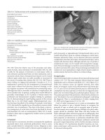
Figure 7
(A) Patient with asymmetric brow ptosis, right more than left, marked derma-
tochalasis both upper and lower eyelid, prolapse of the medial and central
fad pads but also of the lacrimal gland, scleral show, and lower lid laxity.
(B) Intraoperative view after eyebrow lift through direct approach, notice the
prolapsed lacrimal gland (right eye). (C) Intraoperative view after repositioning
of the lacrimal gland within the lacrimal fossa (horizontal mattress suture is
placed through the periosteum of the inner aspect of the superior orbital
rim—the lacrimal fossa—and the outer pole of the lacrimal gland) and lateral
canthopexy (left eye). (D) Immediate postoperative result.
Laser-Assisted Blepharoplasty 125
Upper Eyelid Examination
The preoperative ophthalmological examination must include analysis of
the palpebral aperture. The normal palpebral aperture measures 10 to
12 mm vertically and approximately 30 mm horizontally. Normally, the
upper eyelid covers 1 to 2 mm of the superior, and the lower eyelid just
touches the inferior corneal limbus with no ‘‘scleral show’’ (sclera between
the inferior limbus and the lower eyelid margin is visible) (15). If the super-
ior limbus is visible, a thyroid disease may be present. If there is a scleral
show, lower eyelid laxity or retraction may be a concern. Ptosis is present
when the upper lid droops more than 2 mm over the iris in prim ary gaze.
Ptosis can be congenital or acquired and may be present asymmetrically.
It can be classified as mild: 1 to 2 mm, moderate: 2 to 3 mm, and severe:
greater than equal to 4 mm (13).
Shape and configuration of the eyelid fold should be analyzed.
Normally, the upper eyelid f old is p resent 8 to 10 m m a b ove the lid margin
(in males somewhat lower than in females). Asymmetry of the upper-eyelid
crease shou ld be demonstrated to the patient prior to surgery. If the superior
sulcus is relatively deep, conservative removal of the orbital fat is recom-
mended to avoid skeletonization of the e ye (Fig. 9C).
Analyze the location of fat pad protrusion—usually medial and
centrolateral in the upper eyelid and lateral, centra l, and medial in the
lower eyelid. Gentle pressure on the globe through a closed eyelid will
show the location and the size of the individual fat pads.
The patient is referred to an ophthalmologist, prior to the opera-
tion, for examination; visual acuity, visual field, presence or absence of
corneal scars or injury, corneal diseases, heterophoria, and strabismus
to exclude an enophthalmos are noted and the status of the ocular media,
macula, and optic nerve is checked. The levator excursion is normally
15 to 18 mm but levator excursion of 10 to 14 mm is acceptable. Tear
production, should also be analyzed.
Figure 8
(A and B) Preoperative view and postoperative result 17 days after endo-
scopic forehead lift and upper eyelid blepharoplasty.
126 Fratila
Marking Surgical Incision Lines
It is imperative that the surgical incision lines are marked prior to the local
infiltration of anesthetic solutions with a marking material resistant to the
surgical preparations. The preapon eurotic fat pads should be marked
preoperatively with the patient sitting upright and gazing up. The surgical
lid crease in the upper eyelids is marked 9 to 10 mm above the lash line in
the pupillary axis. At the medial and lateral aspects, this line should never
be closer than 5 mm to the superior punctum or to the lateral canthal angle
(Fig. 10). It should extend medially to a line drawn vertically through
the superior punctum. The superior incision line is placed no closer than
1 cm from the inferior border of the eyebrow—the transition zone between
the thicker skin of the eyebrow and the thinner skin of the eyelid (15).
Preferably, the lines are marked with the patient lying down while we do
our final check of the marking symmetry in a sitting position.
Anesthesia
Local anesthesia and IV sedation are generally preferred. Inject approxi-
mately 2 mL of lidocaine 1% with epinephrine and hyaluronidase
Figure 9
Preoperative view (A), one day postoperative (B) and four weeks after upper
eyelid blepharoplasty and lateral canthopexy (C). Note the deep-set upper
eyelid crease on the right side with asymmetry of the dermatochalasis and
the scleral show preoperatively (A). In (B) (one day postoperative), note the
typical blepharoptosis because of the supratarsal fixation made for a better
definition of the upper eyelid crease. Preaponeurotic fat was not removed.
Laser-Assisted Blepharoplasty 127
subcutaneously, making a ‘‘bleb’’ at the lateral canthus (Fig. 11). Mas-
sage it gently through the entire upper eyelid in both directions. Thi s very
much reduces the risk of postoperative ecchymosis by reducing the
necessity of multiple injections.
Figure 10
Marking the surgical incision lines on the upper eyelid.
Figure 11
A ‘‘bleb’’ of local anesthesia at the lateral canthus is gently massaged through
the entire upper eyelid in both directions.
128 Fratila
The globe ha s to be anesthetized with a topical local anesthesia for it
to be able to tolerate the David–Baker retractor or the metal eye shields.
The undersurface of the protective shields is lubricated with MethocelT gel.
In the author’s experience, the use of general anesthesia is more
comfortable for the patient.
Surgical Procedure
Protect the ope rated eye with the David-Baker retrac tor (16) while the
skin incision is performed with the UPCO
2
laser using the 0.2-mm hand
piece in UltraPulseT mode at 15 mJ and 4 W. Som e surgeons prefer
25 mJ/pulse at a power of 5 W (15) or even CW CO
2
laser at 6 W. Incise
the skin flap at a constant speed (usually about 1 cm/sec). If slowed
down, the incision will be deeper. One should stay in focus and should
not use multiple passes. If the patient requires early suture removal, the
skin incision may be performed with a scalpel, but the risk of bleeding
is higher. The temporal portion of the incision should extend only
through skin to avoid cu tting terminal branches of the lacrimal artery,
which pass between skin and orbicularis muscle at the level of the lateral
orbital rim (15). A skin orbicularis muscle flap, is raised centrally and
nasally.
Grasp the skin flap with a forceps at the temporal end of the marked
ellipse and perform the excision of the skin flap with the UPCO
2
laser
using the 0.2-mm beam diameter defocused in CW mode and with
a power setting of 6 W. With gentle traction on the flap, once the orbital
rim has been crossed, the plane of dissection is deepened to excise a sk in-
muscle flap (Fig. 12A). Protection of the nose with the Jaeger stainless
steel plate is mandatory. The surgical apparatus is kept anterior to the
orbital septum at all times.
Defocus the beam and vaporize any bleeding vessels. Always have
a cautery unit available in case a vessel gets away.
At this surgical step, the David–Baker retractor is replaced with
stainless steel shields to avoid pressure trauma and prolonged edema
of the pretarsal skin. This permits better ballottement of the globe and
exposure of the preaponeurotic fat pads (Fig. 12B).
The septum is opened superiorly over the fat pa d to avoid injury
to the levator aponeurosis, using the Rabkin spatula as a backstop
(Fig. 12C). The spatula is passed beneath the septum and this is incised
across its entire extent (Fig. 12D). First, the middle (Fig. 12E) then med-
ial fat pads (Fig. 12F) are resected using a wet cotton-tipped applicator or
the Rabkin spatula as a backstop. Remove only the fat herniating ante-
rior to the orbital rim; avoid removal of excessive fat as this leads to ske-
letonization or an ‘‘A’’ shape deformity of the eyelid.
If fullness of the lateral brow is because of the descent of the SOOF,
sculpt this region with the CO
2
laser defocused beam (Fig. 12G).
To create a more defined lid crease, remove the metal eye shield.
This facilitates visualization of the iris ensuring symmetric placement of
crease-defining sutures (supratarsal fixation). Place three interrupted
Laser-Assisted Blepharoplasty 129
Figure 12
(Caption on facing page)
130 Fratila