Pacing Options in the Adult Patient with Congenital Heart Disease - part 9 doc
Bạn đang xem bản rút gọn của tài liệu. Xem và tải ngay bản đầy đủ của tài liệu tại đây (187.47 KB, 15 trang )
CHAPTER 23
Univentricular heart
The univentricular heart represents a broad spectrum of congenital abnor-
malities of the heart and great vessels, where the common abnormality is a
single ventricle. This concept is typically associated with any of six possible
anatomical variations of tricuspid atresia, most of which are associated
with a non-existant or rudimentary venous ventricle (Figure 23.1).
The Fontan procedure, to separate and redirect venous blood flow,
presents the most challenging pacing options for the adult with congenital
Figure 23.1 Schematic of tricuspid atresia (univentricular heart) (type 1B). The right
ventricle and outflow pulmonary artery are rudimentary and effectively non-existent. In this
defect, survival depends on an effective atrial septal communication (broken ring).
111
112 Chapter 23
Figure 23.2 Schematic of tricuspid atresia (univentricular heart) with Fontan repair. In the
more classic “Fontan” surgical repair, the atrial septal defect is closed and a direct right atrial
(RA) - pulmonary artery (PA) anastomosis created. The ultimately elevated atrial pressures
(often in the range of 20mmHg) eventually cause severe atrial dilatation and wall thickening.
As expected, sinus node dysfunction and atrial arrhythmias are common.
heart disease. The operation and its many modifications is performed in
up to four surgical procedures in order to separate the systemic and pul-
monary circulations. This is accomplished by either a direct anastomosis
of the right atrium to the pulmonary artery (Figure 23.2) or any variations
of anastomoses involving the superior and inferior venae cavae to the pul-
monary artery using an intra-atrial tunnel or extra-cardiac conduit. These
latter techniques are referred to as total cavopulmonary connection. As might
be expected, a lateral tunnel or external conduit repair may preclude use
of transvenous atrial pacing as the vena cava may no longer communic-
ate with the atrial chamber. Thus, it is essential that the operation notes
be reviewed before consideration of a transvenous atrial lead placement.
Early reported procedures, describe a direct connection between the
right atrium and the pulmonary artery causing extensive dilatation and
damage to the right atrium (Figure 23.3) [238]. The right atrioventricular
(tricuspid) valveorificeand pulmonaryvalve orifice, ifpresent, were closed
denying access by the transvenous route to the univentricular chamber.
Because of the extensive and cumulative atrial damage with each opera-
tion, there is a high incidence of postoperative arrhythmias with primarily
loss of sinus rhythm [239–243]. Thus, it is not unusual for patients who
have undergone the Fontan procedure in childhood to present for cardiac
Univentricular heart 113
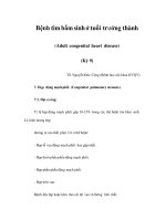
PA
Figure 23.3 Tricuspid atresia (univentricular heart). Chest cine fluoroscopic postero-anterior
(PA) view demonstrating a very dilated right atrium following a classic Fontan procedure.
The 6Fr quadripolar pacing catheter demarcates the extent of the atrial dimensions.
pacing as a teenager or adult with atrial bradyarrhythmias and intact
atrioventricular conduction [238].
The conventional method of atrial pacing following the Fontan pro-
cedure is right atrial epimyocardial [244]. However, because of multiple
previous cardiac operations and atrial scarring, extensive dissection is
required to obtain satisfactory pacing and sensing and the left atrium
has been suggested as an alternative site [245, 246]. Despite the per-
ceived difficulties, good results havebeendocumented using the epicardial
approach [247]. Transmural placement of the lead into the right atrium at
thoracotomy has also been reported [248].
If there is a venous passageway to the right atrium, traditional
single chamber transvenous atrial pacing can be successfully performed
[63, 238, 244, 249, 250] Because of the theoretical risk of obstructing venous
flow into the pulmonary artery, small diameter leads are recommended
and in particular, the SelectSecure
®
lead inserted with a steerable catheter,
the SelectSite
®
(Figure 7.5) [63]. In such situations, the question arises as
to the value of long-term oral anticoagulants such as coumadin. Seeing
that there is such a high incidence of atrial tachyarrhythmias such as atrial
flutter as well, it seems prudent to make such a recommendation.
Atrioventricular block tends to occur following the Fontan procedure in
older children or young adults undergoing the surgery [63, 250]. Because,
there is no connection to the single ventricle, ventricular pacing cannot
be accomplished theoretically by the transvenous route. Consequently,
the ventricular lead should must be positioned on the epimyocardial sur-
face [247]. Because of the difficulties obtaining satisfactory long-term atrial
114 Chapter 23
PA L Lat
Figure 23.4 Tricuspid atresia (univentricular heart). Chest radiographs, postero-anterior
(PA) and left lateral (L Lat), showing dual chamber pacing in a patient with a univentricular
heart who had previously undergone a Fontan procedure. In the PA view, a transvenous
active-fixation lead is attached to the antero-lateral right atrial wall. This lead is then brought
down to the anterior abdominal wall using a connector (white oval) buried behind the breast.
For ventricular pacing, two screw-in epimyocardial leads are attached to the lowermost
portion of the single ventricle. The two epimyocardial leads are on top of each other in the
PA view which has been highlighted with a box. In the L Lat view, a black arrow points to the
two epimyocardial leads, one behind the other.
I aVR V1 V4
II aVL V2 V5
III aVF V3 V6
II
Figure 23.5 Tricuspid atresia (univentricular heart). Resting 12-lead ECG from the same
patient in Figure 23.4, demonstrating dual chamber pacing. There is both sensing and
pacing in the atrium. Ventricular pacing demonstrates a right bundle branch block
configuration and a left axis deviation suggesting left ventricular pacing from the apical
region. The QRS complexes probably show fusion.
pacing, it is best where possible to perform dual chamber pacing using a
two stage hybrid procedure if possible. The atrial lead can be implanted
via the transvenous route. Following the second stage attachment of the
epicardial/epimyocardial lead the pulse generator can be inserted in the
subclavicular fossa or the atrial lead can be extended and brought down to
Univentricular heart 115
the anterior abdominal wall. The pulse generator can then be attached in
the abdomen (Figure 23.4). In this situation, the ventricular lead is attached
anatomically and physiologically to the left ventricle giving rise to a right
bundle branch block appearance on the ECG (Figure 23.5).
Despite the assumed lack of ventricular access, there have been cases of
successful transvenously positioning of ventricular leads in patients hav-
ing undergone a Fontan procedure. This can been achieved by the coronary
sinus route if accessible [251] puncturing the dacron graft covering the tri-
cuspid orifice [252] or puncturing the intra-atrial tunnel with a trans-septal
needle [63, 253]. There have been successful cases of transvenous lead posi-
tioning in patients with univentricular hearts, who have not had the Fontan
procedure [241, 250, 254].
Because of the high incidence of atrial tachyarrhythmias it is worth con-
sidering implanting a pulse generator with antitachycardia capabilities.
This should include atrial overdrive pacing and maybe atrial reversion
therapies.
Concluding remarks
As children born with congenital heart disease continue to age, physicians
caring for adults will be exposed to this increasing population of patients.
At the time of this publication, only the “tip of the iceberg” is visible.
With an incidence of approximately 1% of live births and ever-improving
surgical and device technologies, congenital heart patients will continue
to survive to adulthood in increasing numbers. Based on the US National
Center for Health Statistics, by 2020 the number of children in the United
States born with congenital heart disease in 1990 alone will approximate
760,000 individuals [255]. Other countries may expect similar numbers.
The authors of this text have attempted to provide the reader with a
glimpse into some of the technical challenges associated with pacemaker
and ICD device implantation in these patients. By no means is this text
inclusive of all congenital heart defects and all problems and pitfalls. As
newer technologies evolve, the implanting physician will continue to face
new and diverse challenges and will always require ingenuity and tricks
to overcome them.
117
References
1 Giudici MC. Experience with a cosmetic approach to device implantation. PACE
2001; 24: 1679–1680.
2 Mond HG. The Cardiac Pacemaker. Function and Malfunction. Harcourt Brace
Jovanovich, New York 1983: 199.
3 Jacobs DM, Fink AS, Miller RP et al. Anatomical and Morphologic evaluation of
pacemaker lead compression. PACE 1993; 16: 434–444.
4 Ong LS, Barold S,Lederman M et al. Cephalicvein guide technique for implantation
of permanent pacemakers. Am Heart J 1987; 114: 753–756.
5 Rao G. Letter to the Editor. PACE 1981; 4: 39.
6 Camous JP, Raybaud F, Lesto I et al. Introduction of permanent cardiac stimula-
tion/defibrillation leads via the retro-pectoral veins. PACE 2005; 28: 324–325.
7 Arnold AG. Permanent cardiac pacing using anterior pectoral veins. Br Heart J 1980;
43: 321–323.
8 Gosalbez F, Cofino J, Llorente A et al. Retroclavicular route for electrode placement
in endocardial pacemakers. Chest 1976; 70: 679–683.
9 Belott PH. Blind axillary venous access. PACE 1999; 22: 1085–1089.
10 Burri H, Sunthorn H, Dorsaz PA et al. Prospective study of axillary vein punc-
ture with or without contrast venography for pacemaker and defibrillator lead
implantation. PACE 2005; 28: S280–S283.
11 Byrd CL. Clinical experience with the extrathoracic introducer insertion technique.
PACE 1993; 16: 1781–1784.
12 Belott PH, Reynolds DW. Permanent pacemaker and implantable cardioverter-
defibrillator implantation. In: Ellenbogen, Kay, Wilkoff, eds. Clinical Cardiac
Pacing and Defibrillation, 2nd edn. WB Saunders Co. Philadelphia 2000:
573–644.
13 Mond HG. The Cardiac Pacemaker. Function and Malfunction. Harcourt Brace
Jovanovich, New York 1983: 201.
14 Ellestad MH, French J. Iliac vein approach to permanent pacemaker implantation.
PACE 1989; 12: 1030–1033.
15 Antonelli D,Freeberg NA,RosenfeldT.Transiliac veinapproachto arate-responsive
permanent pacemaker implantation. PACE 1993; 16: 1751–1752.
16 Barakat K, Hill J, Kelly P. Permanent transfemoral pacemaker implantation is the
technique of choice for patients in whom the super vena cava is inaccessible. PACE
2000; 23: 446–449.
119
120 References
17 Giudici MC, Paul DL, Meierbachtol CJ. Active-can implantable cardioverter-
defibrillator placement from a femoral approach. PACE 2003; 26: 1297–1298.
18 Garcia Guerrero JJ, De La Concha Castaneda JF, Mora GF et al. Permanent trans-
femoral pacemaker: A single-center series performed with an easier and safer
surgical technique. PACE 2005; 28: 675–679.
19 Fishberger SB, Camunas J, Rodriguez-Fernandez H et al. Permanent pacemaker
lead implantation via the transhepatic route. PACE 1996; 19: 1124–1125.
20 West JNW, Shearmann CP, Gammage MD. Permanent pacemaker positioning via
the inferior vena cava in a case of single ventricle with loss of right atrial-vena cava
continuity. PACE 1993; 16: 1753–1755.
21 Bayliss CE, Beanlands DS, Baird RJ. The pacemaker-twiddlers syndrome: A new
complication of implantable transvenous pacemakers. Can Med Assoc J 1968; 99:
371–373.
22 Ventri EP, Mower MM, Reid PR. Twiddler’s syndrome. A new twist. PACE 1984; 7:
1004–1009.
23 Anderson MH, Nathan AW. Ventricular pacing from the atrial channel of a VDD
pacemaker: A consequence of pacemaker twiddling? PACE 1990; 13: 1567–1570.
24 Roberts JS, Wenger NK. Pacemaker twiddler’s syndrome. Am J Cardiol 1989; 63:
1013–1016.
25 Robinson LA, Windle JR. Defibrillator twiddler’s syndrome. Ann Thorac Surg 1994;
58: 247–249.
26 Beauregard LM, Russo AM, Heim J et al. Twiddler’s syndrome complicating
automatic defibrillator function. PACE 1995; 18: 735–737.
27 Mehta D, Lipsius M, Suri RS et al. Twiddler’s syndrome with the implantable
cardioverter-defibrillator. Am Heart J 1992; 123: 1079–1082.
28 Higgins SL, Suh BD, Stein JB et al. Recurrent twiddler’s syndrome in a nonthora-
cotomy ICD system despite a Dacron pouch. PACE 1998; 21: 130–133.
29 Bohm A, Komaromy K, Pinter A et al. Pacemaker lead fracture due to twiddler’s
syndrome. PACE 1998; 21: 1162–1163.
30 Saliba BC, Ghantous AE, Schoenfeld MH et al. Twiddler’s syndrome with trans-
venous defibrillators in the pectoral region. PACE 1999; 22: 1419–1421.
31 Kistler P, Eizenberg N, Fynn SP et al. The subpectoral pacemaker implant: It isn’t
what it seems! PACE 2004; 27: 361–364.
32 Crossly GH, Gayle DD, Bailey JR et al. Defibrillator twiddler’s syndrome causing
device failure in a subpectoral transvenous system. PACE 1996; 19: 376–377.
33 Wilkoff BL, Shimokochi DD, Schaal SF. Pacing rate increase due to application of
steady external pressure on an activity-sensing pacemaker (Abstract). PACE 1987;
10: 423.
34 Horenstein MS, Karpawich PP. Chronic performance of steroid-eluting epicardial
leads in a growing pediatric population: a 10-year comparison. PACE 2003; 26:
1467–1471.
35 Walker F, Siu SC, Woods S et al. Long-term outcomes of cardiac pacing in adults
with congenital heart disease. J Am Coll Cardiol 2004; 43: 1894–1901.
36 Karpawich PP, Hakami M, Arciniegas et al. Improved chronic epicardial pacing
in children: Steroid contribution to porous platinized electrodes. PACE 1992; 15:
1151–1157.
References 121
37 Karpawich P, Walters H, Hakimi M. Chronic performance of a transvenous steroid
pacing lead used as an epi-intramyocardial electrode. PACE 1998; 21: 1486–1488.
38 Hayes DL, Vlietstra RE Puga FJ et al. A novel approach to atrial endocardial pacing.
PACE 1989; 12: 125–130.
39 Juneja R, Saxena A, Choudhary S et al. Transatrial permanent pacing lead implant-
ation in a patient of Ebstein’s anomaly after one and half repair. Indian Heart J 2004;
56: 670–672.
40 Byrd CL, Schwartz SJ. Transatrial implantation of transvenous pacing leads as an
alternative to implantation of epicardial leads. PACE 1990; 13: 1856–1859.
41 Westerman GR, Van Devanter SH. Transthoracic trans-atrial endocardial lead
placement for permanent pacing. Ann Thorac Surg 1987; 43: 445–446.
42 Karpawich PP. Chronic right ventricular pacing and cardiac performance: The
pediatric perspective. PACE 2004; 27: 844–849.
43 Karpawich PP, Justice CD, Cavitt DL et al. Developmental sequelae of fixed-
rate ventricular pacing in the immature canine heart: An electrophysiologic,
hemodynamic and histopathologic evaluation. Am Heart J 1991; 121 : 827–833.
44 Tantengco MV, Thomas RL, Karpawich PP. Left ventricular dysfunction following
chronic right ventricular apical pacing in young patients. J Am Coll Cardiol 2001; 37:
2093–2100.
45 Thambo JB, Bordachar P, Garrigue S et al. Detrimental ventricular remodelling in
patients with congenital complete heart block and chronic right ventricular apical
pacing. Circulation 2004; 110: 3766–3772.
46 Sweeney MO, Hellkamp AS, Ellenbogen KA et al. Adverse effect of ventricular
pacing on heart failure and atrial fibrillation among patients with normal baseline
QRS duration in a clinical trial of pacemaker therapy for sinus node dysfunction.
Circulation 2003; 107: 2932–2937.
47 Mond HG, Gammage MD. Selective site pacing: The future of cardiac pacing? PACE
2004; 27: 835–836.
48 Karpawich PP, Gates J, Stokes K. Septal His-Purkinje ventricular pacing in canines:
A new endocardial electrode approach. PACE 1992; 15: 2011–2015.
49 Deshmukh P, Romanyshyn M. Direct His-bundle pacing: Present and future. PACE
2004; 27: 862–887.
50 Giudici MC, Karpawich PP. Alternative site pacing: It’s time to define terms. PACE
1999; 22: 551–553.
51 Karpawich PP, Mital S. Comparative left ventricular function following atrial,
septal and apical single chamber right heart pacing in the young. PACE 1997; 20:
1983–1988.
52 Lieberman R, Grenz D, Mond H et al. Selective site pacing: Defining and reaching
the selected site. PACE 2004; 27: 883–886.
53 Karpawich PP, Horenstein MS, Webster P. Site-specific right ventricular implant
pacing to optimize paced left ventricular function in the young with and without
congenital heart (Abstract). PACE 2002; 25: 566.
54 Riedlbauchova L, Kautzner J, Hatala R et al. Is right ventricular outflow tract pacing
an alternative to left ventricular/biventricular pacing? PACE 2004; 27: 871–877.
55 Mond HG, Stokes KB. The electrode-tissue interface: The revolutionary role of
steroid elution. PACE 1992; 15: 95–107.
122 References
56 Hua W, Mond HG, Strathmore N. Chronic steroid-eluting lead performance:
A comparison of atrial and ventricular pacing. PACE 1997; 20: 17–24.
57 Mond HG, Hua W, Wang CC. Atrial pacingleads: Theclinical contributionof steroid
elution. PACE 1995; 18: 1601–1608.
58 Hua W, Mond HG, Sparks P. The clinical performance of three designs of atrial
pacing leads from a single manufacturer: The value of steroid elution. Eur JCPE
1996; 6: 99–103.
59 Hidden-Lucet F, Halimi F, Gallais Y et al. Low chronic pacing thresholds of steroid-
eluting active-fixation ventricular pacemaker leads: A useful alternative to passive-
fixation leads. PACE 2000; 23: 1978–1800.
60 Kistler PM, Liew G, Mond HG. Long-term performance of active-fixation pacing
leads: A prospective study PACE 2006; 29: 226–230.
61 Crossley G, Reynolds D, Brinkler JA et al. Permanent ventricular pacing from the
right ventricular outflow tract or septum with an active fixation lead provides for
excellent long term results (Abstract). PACE 1997; 20: 1210.
62 Ward DE, Clarke B, Schofield PM et al. Long term transvenous ventricular pacing
in adults with congenital abnormalities of the heart and great arteries. Br Heart J
1983; 50: 325–329.
63 Hansky B, Blanz U, Peuster M et al. Endocardial pacing after Fontan-type
procedures. PACE 2005; 28: 140–148.
64 Chintala K, Forbes T, Karpawich PP. Effectiveness of transvenous pacemaker leads
placed through intravascular stents in patients with congenital heart disease. Am J
Cardiol 2005; 95: 424–427.
65 Kistler PM, Sanders P, Davidson NC et al. The challenge of endocardial right
ventricular pacing in patients with a tricuspid annuloplasty ring and severe
tricuspid regurgitation. PACE 2002; 25: 201–205.
66 Byrd C, Wilkoff B. Techniques and devices for extraction of pacemaker and
implantable cardioverter-defibrillator leads. In: Ellenbogen K, Kay G, Wilkoff, eds.
Clinical Cardiac Pacing and Defibrillation. WB Saunders, Philadelphia, PA, 2000:
695–709.
67 Mond Harry G. The Cardiac Pacemaker. Function and Malfunction. Harcourt Brace
Jovanovich, New York 1983: 206.
68 Bai Y, Strathmore N, Mond H et al. Permanent ventricular pacing via the great
cardiac vein. PACE 1994; 17: 678–683.
69 Faerestrand S, Ohm Ole-Jørgen. Alternate pacing sites for patients with tricuspid
valve prosthesis. PACE 2002; 25: 234–238.
70 Curnis A, MascioliG, Bianchetti F et al.Implantation of a single chamberpacemaker
in patients with triple mechanical valve prosthesis: Utilization of coronary sinus
distal branches to stimulate the left ventricle. PACE 2002; 25: 239–240.
71 Garrigue S, Barold SS, Hocini M et al. Transvenous left atrial and left ventricular
pacing in Ebstein’s anomaly with severe interatrial conduction block. PACE 2001;
24: 1032–1035.
72 Nguyen LS, Swaroop S, Prejean CA. Pacing in the middle cardiac vein in a patient
with tricuspid prosthesis. PACE 2002; 25: 243–244.
73 Bos HS, Pop GAM, Stel EA et al. Dual site coronary sinus pacing in a patient with
an artificial tricuspid valve prosthesis. PACE 2004; 27: 1451–1452.
References 123
74 Lucas R, Krabill K. Abnormal systemic venous connections. In: Emmanouilides
et al., ed. Heart Disease in Infants, Children and Adolescents, 5th edn. Baltimore,
Williams & Wilkins, 1995: 874–902.
75 Van Gelder BM, Elders J Bracke FA et al. Implantation of a biventricular pacing
system in a patient with a coronary sinus not communicating with the right atrium.
PACE 2003; 26: 1294–1296
76 Tada H, Ito S, Naito S et al. Longitudinally partitioned coronary sinus: An unusual
anomaly of the coronary venous system. PACE 2005; 28: 352–353.
77 Kantoch MJ, McKay R, Tyrrell MJ. Left ventricular transvenous electrode dislodge-
ment after Mustard repair for transposition of the great arteries. PACE 1993; 16:
1887–1891.
78 Morquio L. Sur une maladie infantile et familiale caracterisee par des modifications
permanentes du pouls des attaques syncopale et epileptiformes et la morte subite.
Arch Med Enf 1901; 4: 467–474.
79 Lev Maurice. Pathogenesis of congenital atrioventricular block. Prog Cardiovasc Dis
1972; 15: 145–157.
80 Benson DW, Wang DW, Dyment M et al. Congenital sick sinus syndrome caused
by recessive mutations in the cardiac sodium channel gene (SCN5A). J Clin Invest
2003; 112: 1019–1028.
81 Schott JJ, Alshinawi C, Kyndt F, et al. Cardiac conduction defects associated with
mutations in SCN5A. Nat Genet 1999; 23: 201–221.
82 Groenewegen WA, Firouzi M, Bezzina CR et al. A cardiac sodium channel mutation
cosegregates with a rare connexin 40 genotype in familial atrial standstill. Circ Res
2002; 92: 14–22.
83 Kurita T, Ohe T, Marui N et al. Bradycardia-induced abnormal QT prolongation in
patients with complete atrioventricular block with torsade de pointes. Am J Cardiol
1992; 69: 628–633.
84 Anderson RH, Wenick ACG, Losekoot TG et al. Congenitally complete heart block.
Developmental aspects. Circulation 1977; 56: 90–101.
85 Bharati S, McCue CM, Tingelstad JB et al. Lack of connection between the atria
and the peripheral conduction system in a case of corrected transposition with
congenital atrioventricular block. Am J Cardiol 1978; 42: 147–153.
86 Gerlis LM, Anderson RH, Becker AE. Complete heart block as a consequence of
atrionodal discontinuity. Br Heart J 1975; 37: 345–356.
87 Lev M, Cuadros H, Paul MH. Interruption of the atrioventricular bundle with
congenital atrioventricular block. Circulation 1971; 43: 703–710.
88 Bharati S, Lev M. Congenital abnormalities of the conducting system in sudden
death in young adults. J Am Coll Cardiol 1986; 8: 1096–1104.
89 Husson GS, Blackman MS, Rogers MC et al. Familial congenital bundle branch
system disease. Am J Cardiol 1973; 32: 365–369.
90 Rosen KM, Mehta A, Rahimtoola SH et al. Sites of congenital and surgical heart
block as defined by His bundle electrocardiography. Circulation 1971; 44: 833–841.
91 Kelly DT, Brodsky SJ, Mirowski M et al. Bundle of His recordings in congenital
complete heart block. Circulation 1972; 45: 277–280.
92 Nasrallah AT, Gillette PC, Mullins CE. Congenital and surgical atrioventricular
block within the His bundle. Am J Cardiol 1975; 36: 914–920.
124 References
93 James TN, Spencer MS, Kloepfer JC. De Subitaneis Mortibus. XXI. Adult onset
syncope, with comments on the nature of congenital heart block and the
morphogenesis of the human atrioventricular septal junction. Circulation 1976; 54:
1001–1009.
94 Sarachek NS, Leonard JJ. Familial heart block and sinus bradycardia. Classification
and natural history. Am J Cardiol 1972; 29: 451–458.
95 Stephan E. Hereditary bundle branch system defect. Am Heart J 1978; 95: 89–95.
96 Morgans CM, Gray KE, Robb GH. A survey of familial heart block. Br Heart J 1974;
36: 693–696.
97 Lynch HT, Mohiuddin S, Moran J et al. Hereditary progressive atrioventricular
conduction defect. Am J Cardiol 1975; 36: 297–301.
98 Greenspahn BR, Denes P, Daniel W et al. Chronic bifascicular block: Evaluation of
familial factors. Ann Intern Med 1976; 84: 521–525.
99 Graber HL, Unverferth DV, Baker PBet al. Evolutionof a hereditary cardiac conduc-
tion and muscle disorder: A study involving a family with six generations affected.
Circulation 1986; 74: 21–35.
100 Tucker KJ, Murphy J, Conti JB et al. Syncope and sinus arrest associated with the
upper limb-cardiovascular (Holt-Oram) syndrome. PACE 1994; 17: 1678–1680.
101 Silverman ME, Copeland AJ, Hurst JW. The Holt-Oram syndrome: The long and
the short of it. Am J Cardiol 1970; 25: 11–17.
102 Kearns TP, Sayre GP. Retinitis pigmentosa, external ophthalmoplegia and complete
heart block. Arch Ophthalmol 1958; 60: 280–289.
103 Charles R, Holt S, Kay JM et al. Myocardial ultrastructure and the development of
atrioventricular block in Kearns-Sayre syndrome. Circulation 1981; 63: 214–219.
104 Young TJ, Shah AK, Lee MH etal. Kearns-Sayre syndrome: A case report and review
of cardiovascular complications. PACE 2005; 28: 454–457.
105 Ulicny KS, Detterbeck FC, Hall CD. Sinus dysrhythmia in Kearns-Sayre syndrome.
PACE 1994; 17: 991–994.
106 Ross BA. Congenital complete atrioventricular block. Pediatr Clin North Am 1990;
37: 69–78.
107 Lee LA, Coulter S, Erner S et al. Cardiac immunoglobulin deposition in congenital
heart block associated with maternal anti-Ro autoantibodies. Am J Med 1987; 83:
793–796.
108 TaylorPV, Scott JS,Gerlis LMet al.Maternal antibodies against fetal cardiac antigens
in congenital complete heart block. N Engl J Med 1986; 315: 667–672.
109 Goble MM, Dick M II, McCune et al. Atrioventricular conduction in children of
women with systemic lupus erythematosus. Am J Cardiol 1993; 71: 94–98.
110 Schmidt KG, Ulmer HE, Silverman NH et al. Perinatal outcome of fetal com-
plete atrioventricular block: A multicenter experience. J Am Coll Cardiol 1991; 17:
1360–1366.
111 Smeenk RJT. Immunological aspects of congenital atrioventricular block. PACE
1007; 20: 2093–2097.
112 O’Connor BK, Gillette P. Congenital heart block: Natural history and management.
ACC Current J Rev 1995; Mar/Apr: 63–65.
113 Buyon J, Roubey R, Swersky S et al. Complete congenital heart block: Risk of
occurrence andtherapeutic approach to prevention. JRheumatol 1988; 15: 1104–1105.
References 125
114 Lewman LV, Demany MA, Zimmerman HA. Congenital tumor of atrioventricu-
lar node with complete heart block and sudden death. Am J Cardiol 1972; 29:
554–557.
115 Moak J, Barron KS, Hougen TJ et al. Congenital heart block: Development of late-
onset cardiomyopathy, a previously underappreciated sequela. J Am Coll Cardiol
2001; 37: 238–242.
116 Thery CL, Lekieffre J, Dupuis CL. Atrioventricular block secondary to a congen-
ital aneurysm of the membranous septum. Histological examination of conduction
system. Br Heart J 1975; 37: 1097–1100.
117 Esscher EB. Congenital complete heart block in adolescenceand adult life. A follow-
up study. Eur Heart J 1981; 2: 281–288.
118 Karpawich PP, Gillette PC, Garson A et al. Congenital complete atrioventricular
block: Clinical and electrophysiologic predictors of need for pacemaker insertion.
Am J Cardiol 1981; 48: 1098–1102.
119 Winkler RB, Freed MD, Nadas AS. Exercise-induced ventricular ectopy in
children and young adults with complete heart block. Am Heart J 1980; 99:
87–92.
120 Molthan ME, Miller RA, Hastreiter AR et al. Congenital heart block with fatal
Adams-Stokes attacks in childhood. Pediatrics 1962; 30: 32–35.
121 Michaelsson M, Jonzon A, Riesenfeld T. Isolated congenital complete atrioventricu-
lar block in adult life: A prospective study. Circulation 1995; 92: 442–449.
122 Michaelsson M, Riesenfeld T, Jonzon A. Natural history of congenital complete
atrioventricular block. PACE 1997; 20: 2098–2101.
123 Walsh CA, McAlister HF, Andrews CA et al. Pacemaker implantation in children:
A 21-year experience. PACE 1988; 11: 1940–1944.
124 Campbell M, Emanuel R. Six cases of congenital heart block followed for
30–40 years. Br Heart J 1967; 29: 577–587.
125 Corne RA, MathewsonAL. Congenitalcomplete atrioventricularblock. Am J Cardiol
1972: 29: 12–421.
126 Bharati S, Lev M. Congenital abnormalities of the conduction system in sudden
death in young adults. J Am Coll Cardiol 1986; 8: 1096–1104.
127 Ngarmukos T, Werres R. Normal sinus rhythm in a patient with corrected trans-
position of great arteries after 30 years of complete heart block. PACE 1999; 22:
1116–1117.
128 Strieper M. Karpawich P, Frias P et al. Initial experience with cardiac resynchroniz-
ation therapy in young patients with ventricular dysfunction and congenital heart
disease. Am J Cardiol 2004; 94: 1352–1354.
129 Dubin A, Cecchin F, Law, I et al. Resynchronization therapy in pediatric and con-
genital heart disease patients: A multicenter study. Heart Rhythm (Abstract) 2004;
1: S50.
130 Horenstein MS, Karpawich PP. Pacemaker syndrome in the young: Do children
need dual chamber as the initial pacing mode? PACE 2004; 27: 600–605.
131 Friedman RA, Fenrich AL, Kertesz NJ. Congenital complete atrioventricular block.
PACE 2001; 24: 1681–1688.
132 Griffiths SP. Congenital complete heart block (Editorial). Circulation 1971; 43:
615–617.