Báo cáo y học: "Mouse maps of gene expression in the brain" pot
Bạn đang xem bản rút gọn của tài liệu. Xem và tải ngay bản đầy đủ của tài liệu tại đây (631.93 KB, 4 trang )
Genome Biology 2007, 8:212
Minireview
Mouse maps of gene expression in the brain
Susan E Koester* and Thomas R Insel
†
Addresses: *Division of Neuroscience and Basic Behavioral Science, National Institute of Mental Health, Executive Blvd, Bethesda, MD
20892-9645, USA.
†
National Institute of Mental Health, Executive Blvd, Bethesda, MD 20892-9645, USA.
Correspondence: Thomas R Insel. Email:
Abstract
The completion of the Allen Brain Atlas generated a great deal of press interest and enthusiasm
from the research community. What does it do, and what other complementary resources
increase its functionality?
Published: 18 May 2007
Genome Biology 2007, 8:212 (doi:10.1186/gb-2007-8-5-212)
The electronic version of this article is the complete one and can be
found online at />© 2007 BioMed Central Ltd
Since the 19th century, neuroscientists have struggled to
categorize the types of cells in the brain using anatomical or
physiological markers as an indication of cell function [1].
Subdivisions of the brain are defined by clusters of neurons
related by cellular architecture, connectional specificity or
physiological properties. In the past 20 years, neuroscientists
have added molecular biology to their arsenal of tools for
categorizing brain cells. With the complete sequencing of the
genomes of many organisms, neuroscientists have for the
first time an opportunity to understand the full range of gene
expression across the brain. Until recently, individual
researchers have been assembling this information in stages
appropriate to individual laboratories (for example, an
expression map of all known transcription factors in the
mouse brain [2]). Now three large-scale projects are working
toward a map of all gene expression in the mouse brain,
bringing us a giant step closer to understanding the classifica-
tion and function of different types of neurons in the brain.
A complete neuroanatomical atlas of brain
expression
Arguably the most complete of the molecular brain atlas
efforts (in terms of coverage of the genome) is the Allen
Brain Atlas [3], which was recently described by Ed Lein and
colleagues [4]. The Allen Brain Institute (ABI), established
in 2003 by Paul Allen (one of the original founders of
Microsoft), set out to describe the expression of all known
genes in the adult mouse brain. The atlas project used
high-throughput, semi-automated in situ hybridization
methods developed by collaborators Gregor Eichele and Axel
Visel [5] on rigorously controlled coronal sections through
the entire postnatal day (P) 56 (adult) male mouse brain.
Colorimetric in situ hybridization data for 21,000 genes are
now posted online with open access [4]. The data are
searchable by gene names or symbols, as well as by large
anatomical regions (for example, cortex, midbrain,
cerebellum) or by a growing number of smaller-scale brain
nuclei (such as the substantia nigra and the ventral
tegmental area).
A major advantage of the atlas is the near-saturation
coverage of the genome in the brain-expression data. This
allows researchers to understand brain gene expression at a
genomic level: surprisingly, nearly 80% of all genes are
expressed in the brain of the adult mouse. In addition, the
vast majority of these show regionally or cell-type restricted
patterns of expression. ABI staff members have developed a
reference anatomical atlas that can be directly compared
side by side with the in situ expression data. An add-on
module also allows three-dimensional reconstruction of
gene-expression patterns. Another useful feature is that the
atlas links directly from images of particular gene-
expression patterns to the corresponding gene entry in a
variety of databases of gene structure and function,
including the developmental GenePaint database described
below. This significantly increases the versatility of the atlas.
While the recent paper by Lein et al. [4] is a landmark for
molecular neuroanatomy, the Allen Brain Atlas needs to be
understood as a work in progress. As with any effort of this
size, there are errors that will need to be corrected, hopefully
in response to feedback from users. The limitation to one age
and one gender will no doubt be addressed in future efforts.
And, at this point, the annotation of the anatomical location
of expression is only semi-automated and requires expert
human validation. This is currently being done by a small
number of expert collaborators, but the addition of new
subregions will clearly be incredibly laborious and time-
consuming in the absence of automated methods.
Collections of mouse knockouts for
brain-specific genes
Meanwhile, the National Institutes of Health (NIH) has been
working in parallel on the Gene Expression Nervous System
Atlas (GENSAT) to establish a resource of targeted
knockouts of brain-specific mouse genes with interesting
expression patterns in development as well as in the adult
brain [6,7]. There are two arms to the GENSAT project. A
group at St Jude Children’s Research hospital, Tennessee,
led by Thomas Curran (now at the Children’s Hospital of
Philadelphia, Pennsylvania), has carried out radiometric in
situ hybridization in mouse brains at four stages of
development. A separate group led by Nathaniel Heintz at
Rockefeller University, New York, cloned the DNA flanking
the genes with restricted expression patterns in brain into
bacterial artificial chromosomes (BACs) with the gene-
coding sequence replaced by an enhanced green fluorescent
protein (EGFP) reporter gene. The BAC is then used to
create a transgenic mouse line that ideally expresses EGFP
in the cells that normally express the gene of interest. The
advantage of using the BAC as a cloning vector is that it can
carry as much as 200 kb of genomic DNA - typically enough
to include most, if not all, of the appropriate regulatory
elements of the gene. The mice can then express the EGFP
reporter construct in the same anatomical and cell-type-
specific locations as the original gene. Although the
subcellular localization of the native protein cannot be
inferred from the EGFP location, the reporter fills the entire
cell, allowing a clear characterization of the anatomical cell
type in which the gene is expressed. Multiple lines are
analyzed to ensure that the expression pattern is consistent,
indicating that the pattern is due to the promoter elements
of the gene of interest rather than the genomic insertion
point. The anatomical annotators of the GENSAT data have
embraced the variability between their in situ data and the
expression patterns seen in the BAC transgenic mice, noting
where there are differences for each gene catalogued.
GENSAT is quite different from the Allen Brain Atlas in that
it is designed to yield abundant information about the
expression patterns of a subset of genes, not to be a
comprehensive atlas. The GENSAT project produces mouse
lines for physiological as well as anatomical analysis and it
includes developmental patterns of expression. Currently
436 lines [6] are distributed through the Mutant Mouse
Research Resource Centers [8,9]. In addition, for some of
the most interesting expression patterns, the project is
developing lines where the BAC containing the gene
promoter elements drive expression of the bacteriophage,
Cre recombinase (Figure 1). These lines can then be bred
with other lines of mice whose genomes are engineered to
contain genes surrounded by loxP sites, resulting in a
recombinant strain of mouse that eliminates a gene’s
expression only in the cells that express the Cre. These
lines will be a valuable resource for investigators looking to
understand the role of individual genes in specific subsets
of cells in the nervous system.
212.2 Genome Biology 2007, Volume 8, Issue 5, Article 212 Koester and Insel />Genome Biology 2007, 8:212
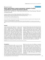
Figure 1
An example of Cre expression in mouse forebrain circuits, with labeling
of specific neuronal projection systems in the cerebral cortex and
striatum. (a) The ETS domain transcription factor (etv1) BAC drives Cre
recombinase expression in layer 5 corticostriatal nuerons. (b) The
neurotensin receptor (ntsr1) BAC drives Cre expression in layer 6
corticothalamic neurons. The projection axons of these neurons, which
terminate in the dorsal thalamic nuclei, are clearly labeled. In the striatum,
the majority of neurons are medium spiny projection neurons, which are
evenly divided into striatopallidal (indirect pathway) and striatonigral
(direct pathway) neurons, which selectively express the dopamine
receptors Drd2 and Drd1a, respectively. Cre expression produced in
drd2 BAC-Cre lines (c) is directed to striatopallidal neurons. In this line,
labeled neurons in the striatum extend axons that terminate in the globus
pallidus external segment (GPe). (d) Expression produced in drd1a BAC-
Cre lines, is directed to striatonigral neurons, which have axons that
extend through the globus pallidus to terminate in the internal segment of
the globus pallidus (GPi) and substantia nigra (not shown). Images
courtesy of Charles Gerfen, National Institute of Mental Health.
(a) (etv1) layer 5 cerebral cortex
corticostriatal neurons
Striatum
(b) (ntsr1) layer 6 cerebral cortex
corticothalamic neurons
Cerebal
cortex
Thalamus
(c) (drd2) striatopallidal neurons
(indirect pathway)
(d) (drd1a) striatonigral neurons
(direct pathway)
Striatum
Striatum
GPe
GPe
GPi
GPi
The third project mapping gene expression in the mouse
brain is the GenePaint atlas [10], a large-scale European
effort led by Gregor Eichele at the Max-Planck-Institute of
Biophysical Chemistry in Göttingen, Germany. In approach,
this is a hybrid of the Allen Brain Atlas and GENSAT. The
GenePaint atlas [10] catalogs in situ hybridization
expression data in selected sections of whole mouse embryos
for several thousand genes at embryonic day (E) 14.5 and
augments these data with additional data for some genes in
E10.5 embryos, E15.5 head, P7 and adult (day 56) brain,
often in both coronal and sagittal sections. New data are
posted regularly, with a list of new genes updated weekly on
the home page. Researchers can search gene expression by
gross anatomical area using the E14.5 dataset and can then
link to views of the same gene expressed at different ages.
GenePaint is a useful addition to the Allen Brain Atlas,
giving researchers a flavor of dynamic gene-expression
patterns during development. Although it does not have the
same coverage of the genome as the Allen Brain Atlas,
investigators can submit requests for additional genes to be
tested and expression pattern data will be made available
within a few weeks.
Mouse genetic resources
Once an investigator identifies a gene with an interesting
expression pattern or some information on its function,
many would welcome ready access to a targeted knockout
mouse. The NIH Knockout Mouse Project (KOMP) [11] is an
NIH effort to generate null mutations of all genes in
C57BL/6 mice and distribute them to the research community.
This effort is proceeding in several parallel directions. First,
NIH have licensed 250 knockout lines from the companies
Deltagen and Lexicon to make them widely available to the
academic community; second, NIH issued contracts to
create new knockouts of a list of priority gene candidates;
and third, NIH is also ‘repatriating’ knockout mouse lines for
distribution that have been generated by academic
researchers, many using NIH funds. The KOMP project will
manage the necessary husbandry and record-keeping for the
distribution, which is an important advantage to investi-
gators submitting popular mouse strains. The priorities for
gene targeting or knockouts for repatriation are derived
from public input from the research community, which is
actively solicited [12]. The list of genes scheduled for
targeting and the current list of knockout lines available are
online [13]. In addition, genetically modified mice that were
generated with other funding can be nominated to the
KOMP for redistribution by the Mutant Mouse Regional
Resource Centers, supported by NIH [8].
The European Union has launched a complementary effort
to generate 20,000 lines of mice with conditional mutations
with the ultimate goal of functionally characterizing all the
genes in the mouse genome [14]. The European Conditional
Mouse Mutagenesis Program (EUCOMM) brings together
several different European projects to generate genetically
modified mouse resources under one coordinating group.
While the group is collaborating with the NIH KOMP as well
as with a related Canadian effort, their emphasis is comple-
mentary. EUCOMM will use conditional gene-trap strategies
to generate embryonic stem cell lines for eventual worldwide
distribution. The project is in the early stages, but its
progress can be followed at the EUCOMM website [15].
Future challenges for gene-expression databases
The philosophy of open access has guided the development
of all of these resources, which is what makes them such a
remarkable boon to neuroscientists and other biomedical
researchers. Researchers are already finding the atlases
useful in augmenting their data from unbiased screens for
gene function [16]. The atlases provide critical reference data
that allow investigators to include or exclude candidate
genes on the basis of their expression patterns, and provide
initial insights leading to a more complete investigation of
expression patterns [17,18]. As the developers consider the
evolution of the databases, it would be useful to expand that
spirit to include data sources from the community.
Leveraging the huge body of work by experts and
incorporating new discoveries will be key to keeping these
resources on the cutting edge.
A problem faced by the gene-expression databases is the lack
of complete neuroanatomical annotation. This is not unex-
pected, as this type of annotation is time consuming,
requires great expertise and is not easily automated. The
task is made more difficult by the lack of a common
neuroanatomical nomenclature. The Allen Brain Atlas
devised a broad-scale nomenclature for its own reference
atlas, rather than choose sides in the ongoing debate about
nomenclature in individual brain areas. GenePaint points
the user to several standard published adult and develop-
mental brain atlases for both mice and rats. The GENSAT
project goes farthest in describing the anatomy of gene
expression and compares data from numbers of individual
animals using different methods to assay gene expression.
However, the number of genes covered remains small by
comparison to the Allen Brain atlas. Furthermore, searching
any of the databases for genes based on their expression in a
particular cell type remains an elusive goal.
To be sustainable and useful in the long term, all these
atlases will need to grow and evolve to incorporate
additional data about subregional and cell-type-specific
expression. The NIH databases that have become a mainstay
for biomedical research, such as GenBank, rely on
continuous update by users following a specified submission
policy and curated by an in-house staff. In contrast, brain
atlases have followed an older, proprietary model, alluding
to their tradition of publication as books. A user-annotation
model that allows addition of references to peer-reviewed
Genome Biology 2007, Volume 8, Issue 5, Article 212 Koester and Insel 212.3
Genome Biology 2007, 8:212
publications would ensure the atlases continue to support
user needs and the current state of the science. This
approach has been successfully followed in other model
organisms such as the zebrafish, with their Zebrafish Infor-
mation Network [19].
Ultimately, the subdivision of specific regions will need to be
based on functional differences among nuclei or brain areas.
This work has already begun in a number of species and cell
types, correlating gene expression, anatomical and electro-
physiological characteristics (reviewed in [20]). For example,
Arlotta et al. [21] used a combination of axon tracing and
gene expression data to categorize pyramidal neurons in
layer 5 of the neocortex in mice, demonstrating that the
anatomical distinctions also have gene-expression corre-
lates. Sugino et al. [22] combined microarray analysis with
electrophysiology and neurotransmitter immunocyto-
chemistry to describe the variability within and between 12
known classes of forebrain neurons in mice. Data such as
these will be key to understanding the function and
development of circuits in the brain and for genetic
manipulation of circuit elements. This level of analysis will
be critical to the next stages of understanding how changes
in gene function affect neuronal circuits and eventually
complex behaviors.
Acknowledgements
We thank Andrea Beckel-Mitchner, Michael Huerta and Laura Mamounas
for helpful discussions.
References
1. Ramon y Cajal S: Les Nouvelles Idées sur la Structure du Système
Nerveux chez l’Homme et chez Vertébrés. Paris: Reinwald; 1894.
2. Gray PA, Fu H, Luo P, Zhao Q, Yu J, Ferrari A, Tenzen T, Yuk DI,
Tsung EF, Cai Z, et al.: Mouse brain organization revealed
through direct genome-scale TF expression analysis. Science
2004, 306:2255-2257.
3. Allen Brain Atlas []
4. Lein ES, Hawrylycz MJ, Ao N, Ayres M, Bensinger A, Bernard A, Boe
AF, Boguski MS, Brockway KS, Byrnes EJ, et al.: Genome-wide
atlas of gene expression in the adult mouse brain. Nature
2007, 445:168-176.
5. Visel A, Thaller C, Eichele G: GenePaint.org: an atlas of gene
expression patterns in the mouse embryo. Nucleic Acids Res
2004, 32(Database issue):D552-D556.
6. NINDS GENSAT BAC Transgenic Project [http://www.
gensat.org]
7. Gong S, Zheng C, Doughty ML, Losos K, Didkovsky N, Schambra
UB, Nowak NJ, Joyner A, Leblanc G, Hatten ME, et al.: A gene
expression atlas of the central nervous system based on
bacterial artificial chromosomes. Nature 2003, 425:917-925.
8. Mutant Mouse Regional Resource Centers [http://www.
mmrrc.org]
9. NINDS GENSAT BAC Transgenics Project: MMRC mouse
catalog [ />10. Gene Paint []
11. NIH Knockout Mouse Project [ />models/mouse/knockout]
12. Request for Community Input for the Nomination and Pri-
oritization of Genes to be Targeted in the Knockout Mouse
Project (KOMP): NOT-HG-07-004 [ />guide/notice-files/NOT-HG-07-004.html]
13. Knockout Mouse Project Data Coordination Center
[]
14. Auwerx J, Avner P, Baldock R, Ballabio A, Balling R, Barbacid M,
Berns A, Bradley A, Brown S, Carmeliet P, et al.: The European
dimension for the mouse genome mutagenesis program.
Nat Genet 2004, 36:925-927.
15. European Conditional Mouse Mutagenesis Program
[]
16. Dugas JC, Tai YC, Speed TP, Ngai J, Barres BA: Functional
genomic analysis of oligodendrocyte differentiation. J Neu-
rosci 2006, 26:10967-10983.
17. Papassotiropoulos A, Stephan DA, Huentelman MJ, Hoerndli FJ,
Craig DW, Pearson JV, Huynh KD, Brunner F, Corneveaux J,
Osborne D, et al.: Common Kibra alleles are associated with
human memory performance. Science 2006, 314:475-478.
18. Ponomarev I, Maiya R, Harnett MT, Schafer GL, Ryabinin AE,
Blednov YA, Morikawa H, Boehm SL 2nd, Homanics GE, Berman AE,
et al.: Transcriptional signatures of cellular plasticity in mice
lacking the alpha1 subunit of GABAA receptors. J Neurosci
2006, 26:5673-5683.
19. ZFIN: the Zebrafish Model Organism Database
[]
20. Nelson SB, Hempel C, Sugino K: Probing the transcriptome of
neuronal cell types. Curr Opin Neurobiol 2006, 16:571-576.
21. Arlotta P, Molyneaux BJ, Chen J, Inoue J, Kominami R, Macklis JD:
Neuronal subtype-specific genes that control corticospinal
motor neuron development in vivo. Neuron 2005, 45:207-221.
22. Sugino K, Hempel CM, Miller MN, Hattox AM, Shapiro P, Wu C,
Huang ZJ, Nelson SB: Molecular taxonomy of major neuronal
classes in the adult mouse forebrain. Nat Neurosci 2006,
9:99-107.
212.4 Genome Biology 2007, Volume 8, Issue 5, Article 212 Koester and Insel />Genome Biology 2007, 8:212