Báo cáo y học: " Adipose tissue transcriptomic signature highlights the pathological relevance of extracellular matrix in human obesity" pot
Bạn đang xem bản rút gọn của tài liệu. Xem và tải ngay bản đầy đủ của tài liệu tại đây (3.63 MB, 32 trang )
Genome Biology 2008, 9:R14
Open Access
2008Henegaret al.Volume 9, Issue 1, Article R14
Research
Adipose tissue transcriptomic signature highlights the pathological
relevance of extracellular matrix in human obesity
Corneliu Henegar
*†
, Joan Tordjman
*†
, Vincent Achard
*†
, Danièle Lacasa
*†
,
Isabelle Cremer
*†‡
, Michèle Guerre-Millo
*†
, Christine Poitou
*†§
,
Arnaud Basdevant
*†§
, Vladimir Stich
¶
, Nathalie Viguerie
¶¥#**
,
Dominique Langin
¶¥#**
, Pierre Bedossa
††‡‡
, Jean-Daniel Zucker
*§§
and
Karine Clement
*†§
Addresses:
*
INSERM, UMR-S 872, Les Cordeliers, Eq. 7 Nutriomique and Eq. 13, Paris, F-75006 France.
†
Pierre et Marie Curie-Paris 6
University, Cordeliers Research Center, UMR-S 872, Paris, F-75006 France.
‡
Paris Descartes University, UMR-S 872, Paris, F-75006 France.
§
Assistance Publique-Hôpitaux de Paris (AP-HP), Pitié Salpêtrière Hospital, Nutrition and Endocrinology department, Paris, F-75013 France.
¶
Franco-Czech Laboratory for Clinical Research on Obesity, INSERM and 3rd Faculty of Medicine, Charles University, Prague, CZ-10000,
Czech Republic.
¥
INSERM, U858, Obesity Research Laboratory, I2MR, Toulouse, F-31432 France.
#
Paul Sabatier University, Louis Bugnard
Institute IFR31, Toulouse, F-31432 France.
**
Centre Hospitalier Universitaire de Toulouse, Toulouse, F-31059 France.
††
Assistance Publique-
Hôpitaux de Paris (AP-HP), Beaujon Hospital, Pathology department, Clichy, F-92110 France.
‡‡
CNRS, UMR 8149, Clichy, F-92110 France.
§§
IRD UR Géodes, Centre IRD de l'Ile de France, Bondy, F-93143 France.
Correspondence: Corneliu Henegar. Email:
© 2008 Henegar et al.; licensee BioMed Central Ltd.
This is an open access article distributed under the terms of the Creative Commons Attribution License ( which
permits unrestricted use, distribution, and reproduction in any medium, provided the original work is properly cited.
Extracellular matrix in obesity<p>Analysis of the transcriptomic signature of white adipose tissue in obese human subjects revealed increased interstitial fibrosis and an infiltration of inflammatory cells into the tissue. </p>
Abstract
Background: Investigations performed in mice and humans have acknowledged obesity as a low-
grade inflammatory disease. Several molecular mechanisms have been convincingly shown to be
involved in activating inflammatory processes and altering cell composition in white adipose tissue
(WAT). However, the overall importance of these alterations, and their long-term impact on the
metabolic functions of the WAT and on its morphology, remain unclear.
Results: Here, we analyzed the transcriptomic signature of the subcutaneous WAT in obese
human subjects, in stable weight conditions and after weight loss following bariatric surgery. An
original integrative functional genomics approach was applied to quantify relations between
relevant structural and functional themes annotating differentially expressed genes in order to
construct a comprehensive map of transcriptional interactions defining the obese WAT. These
analyses highlighted a significant up-regulation of genes and biological themes related to
extracellular matrix (ECM) constituents, including members of the integrin family, and suggested
that these elements could play a major mediating role in a chain of interactions that connect local
inflammatory phenomena to the alteration of WAT metabolic functions in obese subjects. Tissue
and cellular investigations, driven by the analysis of transcriptional interactions, revealed an
increased amount of interstitial fibrosis in obese WAT, associated with an infiltration of different
types of inflammatory cells, and suggest that phenotypic alterations of human pre-adipocytes,
Published: 21 January 2008
Genome Biology 2008, 9:R14 (doi:10.1186/gb-2008-9-1-r14)
Received: 6 July 2007
Revised: 29 September 2007
Accepted: 21 January 2008
The electronic version of this article is the complete one and can be
found online at />Genome Biology 2008, 9:R14
Genome Biology 2008, Volume 9, Issue 1, Article R14 Henegar et al. R14.2
induced by a pro-inflammatory environment, may lead to an excessive synthesis of ECM
components.
Conclusion: This study opens new perspectives in understanding the biology of human WAT and
its pathologic changes indicative of tissue deterioration associated with the development of obesity.
Background
Investigations performed in mice and humans have led to a
pathophysiological paradigm that acknowledges obesity as a
low-grade inflammatory disease. Elevated inflammatory pro-
teins in obese individuals [1] suggest that inflammation may
play a determinant role in connecting obesity to metabolic,
hepatic and cardiovascular diseases [2], and to some cancers
[3]. In such chronic pathologies, in which obesity appears as
a well established risk factor, a prominent role for the
immuno-inflammatory processes has been put forward as
contributing to disease progression and tissue deterioration
[4]. However, in spite of substantial evidence demonstrating
the existence of a low-grade inflammatory component in
obesity [5], the molecular mechanisms that link inflamma-
tory changes to the development, aggravation, maintenance,
and resistance to treatment that characterize obesity states
remain poorly understood.
White adipose tissue (WAT), now considered as a pivotal
endocrine organ, contributes to the systemic inflammation by
producing biomolecules, including pro-inflammatory media-
tors, whose estimated number grows constantly and whose
synthesis is altered along with the expansion of the adipose
tissue [6,7]. These molecules are delivered into the blood
stream and exert metabolic and immune functions, as illus-
trated by the extensively studied adipose hormones leptin
and adiponectin. Their functions are essential for inter-organ
cross-talk, body weight homeostasis and probably in linking
adipose tissue to the downstream complications associated
with obesity [8]. Cellular types composing WAT include
mature adipocytes, the specialized metabolic cells, and a vari-
ety of other cells grouped in the 'stroma vascular fraction'
(SVF), which are not well characterized in humans. Although
some molecules secreted by WAT, such as leptin and adi-
ponectin, are synthesized by mature adipocytes [8], the non-
adipose SVF, comprising infiltrated macrophages among
other cellular types, is a source of inflammation-related mol-
ecules that may exert a local action on adipose tissue biology,
particularly within the enlarged WAT [9-11]. The possible
infiltration of the obese WAT by other inflammatory cells is
also suggested by recent analyses in mice showing the modu-
lation of T and natural killer (NK) cell subtypes in animals fed
with a high fat diet [12]. Adipose loss leads to the improve-
ment of the inflammatory profile [11], with a concomitant
reduction of infiltrating macrophages [13].
In obese human subjects, large-scale transcriptomic analyses
of WAT, in stable weight conditions or during weight loss, led
mostly to the description of inflammatory changes and pro-
duced extensive lists of regulated genes involved in a number
of biological functions [14]. However, the relationship
between these genes, the cellular processes in which they are
involved, and the tissue structure as a whole remains poorly
understood. To address this question, we took advantage of
increasing progress in the analysis of complex biological
interactions, which has attracted a great amount of interest in
various fields. An important motivation for the study of such
networks of biological interactions resides in their ability to
formally characterize the roles played by various interacting
elements comprising cellular environments, thus helping pri-
oritize further mechanistic investigations. In particular, the
study of gene interaction networks, constructed by relating
co-expressed genes (that is, genes sharing similar expression
profiles), contributed to the characterization of several key
properties of biological networks, such as the scale-free distri-
bution of their connectivity [15], their hierarchical architec-
ture built from modules of functionally related components
(that is, genes, enzymes, metabolites) [15], the various types
of net hubs [16], or the small-world aspect of their fast syn-
chronizability [17]. Along with the development of interac-
tions analysis, the biological interpretation of large-scale gene
expression profiling data has evolved gradually into a highly
standardized and powerful analytical framework. Available
exploratory tools rely on curated gene annotation resources
and standardized statistical evaluation techniques to identify
significantly over-represented biological themes in high-
throughput gene expression datasets [18].
The objective of our study was to construct a full-scale map of
the biological interactions defining the transcriptomic signa-
ture of WAT in obese subjects. For this purpose we devised an
original analytical approach, which further extended the con-
ventional gene co-expression network analysis to include the
evaluation of transcriptomic interactions between relevant
biological themes, including cellular components, biological
processes and regulatory or metabolic pathways. This
approach was applied to the analysis of two sets of microarray
gene expression profiles obtained previously from human
WAT of obese subjects in stable weight conditions [11,19] and
three months after significant weight loss induced by gastric
surgery [13]. Our analysis revealed major and interrelated
changes of WAT transcriptomic signature in obese human
subjects, involving extracellular matrix (ECM), and inflam-
matory and adipose metabolic processes. Tissue and cellular
investigations, directed by the hypotheses raised by the anal-
ysis of gene and functional interactions, show that
Genome Biology 2008, Volume 9, Issue 1, Article R14 Henegar et al. R14.3
Genome Biology 2008, 9:R14
subcutaneous adipose tissue of obese subjects is character-
ized by an excessive amount of interstitial fibrosis and suggest
that the phenotypic changes in human pre-adipocytes,
induced by a pro-inflammatory environment, are associated
with excessive synthesis of ECM components, which may
contribute to tissue deterioration.
Results
The transcriptomic signature of the subcutaneous
WAT in obese subjects
Thirty five cDNA microarray experiments were performed in
25 weight-stable obese subjects (body mass index (BMI)
40.58 ± 1.58 kg/m
2
, range 32.6-60.5 kg/m
2
) and 10 healthy
lean controls (BMI 23.67 ± 0.48 kg/m
2
, range 21.4-26.2 kg/
m
2
) to characterize the transcriptomic signature of the subcu-
taneous WAT associated with chronic obesity. The overall
clinical and biochemical parameters of the studied popula-
tion are presented in Table 1, and on the companion website
as online supplementary data [20]. The analysis of the differ-
ential gene expression with the Significance analysis of
microarrays (SAM) procedure [21], performed on the cDNA
measurements with signals recovered in at least 80% of the
microarray experiments, detected 366 up- and 474 down-reg-
ulated genes, corresponding to a 5% false discovery rate
(FDR). The functional analysis of these genes identified 704
genes (307 up- and 397 down-regulated) annotated with
Gene Ontology (GO) categories [22], and 253 genes (101 up-
Table 1
Overall clinical and biological parameters of 55 obese subjects and 15 lean controls
Phenotype Obese subjects Lean controls
n5515
Female/Male 52/3 15/0
Age (years) 40.13 ± 11.67 34.2 ± 8.52
BMI (kg/m
2
) 44.07 ± 9.06* 23.67 ± 1.51
Glucose homeostasis
Glucose (mmol/l) 5.56 ± 1.70 4.82 ± 1.01
Insulin (μU/ml) 13.51 ± 8.57 7.20 ± 3.49
QUICKI 0.33 ± 0.05 0.36 ± 0.04
Type 2 diabetes
Glycemia > 7 mmol/l or treatment 6 (11%) 0
Lipid homeostasis
Cholesterol (mmol/l) 5.22 ± 1.04 4.36 ± 1.05
HDL cholesterol (mmol/l) 1.27 ± 0.34 1.43 ± 0.23
Triglycerides (mmol/l) 1.40 ± 0.62
†
0.45 ± 0.10
Adipokines
Leptin (ng/ml) 54.55 ± 19.92 11.24 ± 1.12
Adiponectin (μg/ml) 7.14 ± 2.87 -
Risk factors
HDL < 1.03 mmol/l (M), < 1.29 mmol/l (F) 26 (47%)* 1 (6%)
Hypertension ≥ 130/85 mmHg 11 (20%) 0
Glucose ≥ 5.6 mmol/l 17 (31%) 1 (6%)
Triglycerides ≥ 1.7 mmol/l 11 (20%) 0
Inflammatory factors
TNF-α (pg/ml) 1.77 ± 0.62 -
IL6 (pg/ml) 2.24 ± 1.17 -
hsCRP (mg/dl) 8.62 ± 10.38 -
Orosomucoid (g/l) 0.99 ± 0.18 -
Serum amyloid A (μg/ml) 21.35 ± 22.39 -
Hepatic factors
Aspartate aminotransferase (IU/l) 22.66 ± 6.87 -
Alanine aminotransferase (IU/l) 35.72 ± 18.56 -
γGT (mg/dl) 45.91 ± 46.43 -
*Bilateral significance p value < 0.05 for the difference between the two groups.
†
Bilateral significance p value < 0.001 for the difference between the
two groups. Hyphens indicate parameters that were not available for the lean controls group. F, female; HDL, high-density lipoproteins; M, male.
Genome Biology 2008, 9:R14
Genome Biology 2008, Volume 9, Issue 1, Article R14 Henegar et al. R14.4
and 152 down-regulated) annotated with categories of the
Kyoto Encyclopedia of Genes and Genomes (KEGG) [23].
Figures 1a, 2a and 3a illustrate the biological themes charac-
terizing the transcriptomic signature of the subcutaneous
WAT in obese subjects. Relevant biological themes, annotat-
ing genes differentially expressed in the obese WAT com-
pared to lean controls, are indicated by significantly over-
represented categories from the GO Cellular Component and
Biological Process ontologies and from KEGG. While the
genes up-regulated in the obese WAT were annotated mainly
by structural and functional themes associated with the cellu-
lar membrane and the extracellular space, the down-regu-
lated genes were annotated mostly by themes related to the
intracellular domain. We relied on our in-house analytical
approach to quantify transcriptomic interactions between
these themes by aggregating the similarities of their anno-
tated gene expression profiles (see Materials and methods for
details), and then related them to build biological interaction
maps. This analysis uncovered a highly segregated transcrip-
tomic interaction pattern, regardless of the system used to
annotate differentially expressed genes (Figures 1b, 2b and
3b). Two distinct types of biological interaction modules
(indicated hereafter as module 1 and module 2) have been
identified, one associating structural components, processes
and regulatory pathways related to cellular membranes and
the extracellular space (module 1), while the other groups
components, processes and pathways associated with the
intracellular domain (module 2).
GO Cellular Component categories annotating up-regulated
genes (Figure 1a) formed a first module (Figure 1b, module 1)
composed from themes primarily related to membrane com-
ponents ('integral to membrane', 'plasma membrane part',
'intrinsic to plasma membrane') and to the extracellular
region ('extracellular region', 'extracellular region part').
'Lysosome' and 'endoplasmic reticulum' were the only catego-
ries designating intracellular organelles in this module. The
biological processes designated by GO Biological Process cat-
egories annotating up-regulated genes (Figure 2a,b) were
related to immune, inflammatory, and stress responses
('immunoglobulin mediated immune response', 'antimicro-
bial humoral response', 'immune response', 'response to
stress'), as well as to cell adhesion and signaling processes
('cell adhesion', 'cell surface receptor linked signal transduc-
tion'). The KEGG pathways annotating genes up-regulated in
the obese WAT (Figure 3a) formed a strong interaction mod-
ule associating categories related to immunological and
inflammatory responses as well as to cellular adhesion and
signaling mechanisms (Figure 3b, module 1).
A very distinctive biological pattern was observed for themes
associated with the genes down-regulated in the obese WAT.
GO Cellular Component structural categories annotating
these genes (Figure 1a) formed a second module (Figure 1b,
module 2), grouping themes associated with intracellular
components, among which are the nucleus, the cytoplasm,
the ribosome and the mitochondrion ('intracellular',
'nucleus', 'cytoplasmic part', 'ribosome', 'intracellular
organelle part', 'cytosolic part', 'intracellular part', 'mitochon-
drion', 'mitochondrial membrane part'). GO Biological Proc-
ess categories annotating down-regulated genes (Figure 2a,b)
were essentially related to lipid, protein and energy metabo-
lism ('lipid metabolism', 'fatty acid metabolism', 'protein bio-
synthesis', 'generation of precursor metabolites and energy'),
as well as to the regulation of the apoptotic machinery
('induction of apoptosis'). The examination of KEGG path-
ways revealed a similar interaction pattern associating a
number of key adipocyte metabolic and regulatory pathways
(Figure 3a,b, module 2)
Since the analysis of transcriptomic interactions in the obese
WAT revealed a neat segregated pattern, we sought to deter-
mine the tissular fraction specificity of the two types of inter-
action modules. Taking advantage of our previous large-scale
transcriptomic analysis [11], we explored the specific enrich-
ment of isolated WAT cellular fractions in genes annotated
with categories belonging to one of the two types of modules.
This analysis showed that biological themes related to the
extracellular space (module 1) were annotating genes pre-
dominantly expressed in the SVF of WAT, while the genes
annotated with themes related to the intracellular domain
(module 2) were expressed predominantly in mature adi-
pocytes (Figures 1b, 2b and 3b).
ECM remodeling and inflammation related genes
We then examined the similarity between the expression pro-
files of individual genes to build the co-expression network
underlying the described functional interactions. Among the
genes annotated with significantly over-represented GO cate-
gories, 40 genes (12.5%, among which 24 genes were up-reg-
ulated and 16 genes down-regulated) were found to encode
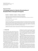
GO Cellular Component enriched themes and their interaction map, illustrating the transcriptomic signature of obese WATFigure 1 (see following page)
GO Cellular Component enriched themes and their interaction map, illustrating the transcriptomic signature of obese WAT. (a) The GO Cellular
Component annotation categories showing a significant enrichment in genes up- or down-regulated in WAT of obese subjects. (b) These categories were
related to construct a biological interaction map after quantifying their proximity based on the expression similarity of their annotated genes. Continuous
lines indicate the strongest interactions (that is, superior to the upper quartile of their distribution), while dashed lines depict medium strength
interactions (that is, superior to the median of the distribution but inferior to its upper quartile). The enrichment in genes expressed preferentially in one
of the two main cellular fractions of WAT, illustrated in a percentage scale (mature adipocytes in light gray versus SVF in black), was significantly different
in the two modules (p value < 0.001).
Genome Biology 2008, Volume 9, Issue 1, Article R14 Henegar et al. R14.5
Genome Biology 2008, 9:R14
Figure 1 (see legend on previous page)
Up-regulated Transcripts
integral to membrane
intrinsic to plasma membrane
endoplasmic reticulum
extracellular region
extracellular region part
plasma membrane part
lysosome
1
00 80 60 40 20 0
Down-regulated Transcripts
nucleus
intracellular
mitochondrion
cytoplasmic part
ribosome
intracellular organelle part
cytosolic part
intracellular part
mitochondrial membrane part
0 2040608010
0
GO Cellular Component
Transcript space coverage (% )
SVF
0 20406080100
Adipocytes
M1 M2
Module 1
Module 2
Up-regulated
Up-regulated
Down-regulated
Down-regulated
(a)
(b)
Genome Biology 2008, 9:R14
Genome Biology 2008, Volume 9, Issue 1, Article R14 Henegar et al. R14.6
various structural components of the ECM or molecules
involved in ECM remodeling and regulation (Additional data
file 2 and supplementary online table 1).
Figure 4 depicts a bi-modular co-expression network relating
genes annotated with significantly over-represented GO Bio-
logical Process categories in the obese WAT (Figure 2a). The
first co-expression module (Figure 4, module 1) groups up-
regulated genes associated with processes constituting the
first functional interaction module (Figure 2b, module 1).
This module includes representatives from all major classes
of ECM components, namely structural proteins such as
members of the collagen family, adherent proteins such as
fibronectin and laminin family members, glycosaminogly-
cans and proteoglycans, and specialized glycoproteins such as
integrins, as well as several enzymes involved in ECM remod-
eling (Additional data file 2 and supplementary online table
1). A sub-network grouping all ECM related genes, showing
significant differential expression in obese WAT, is presented
in Figure 5.
Among various ECM components, several genes coding for
members of the integrin family were found to be significantly
induced and co-expressed in obese WAT, occupying central
positions in the first co-expression module (Figure 4, module
1). This module included integrins alpha V (ITGAV), referred
to as the vitronectin receptor, and alpha M (ITGAM), as well
as integrins beta 1 (ITGB1; also named fibronectin receptor or
beta polypeptide), beta 2 (ITGB2) and beta 3 (ITGB5). These
integrins displayed strong co-expression with other key
components of the ECM (Figures 4 and 5; Additional data file
2 and supplementary online table 1), such as members of the
collagen family, including the major type IV alpha collagen
chain of basement membranes (COL4A1), and members of
the fibril associated collagen (COL5A2 and COL12A1). They
were also co-expressed with members of the glycosaminogly-
can and proteoglycan family (syndecan binding protein
(SDCBP), lumican (LUM)), known to play an important role
in the initiation of inflammatory phenomena, as well as in the
recruitment, rolling, and subsequent extravasation of lym-
phocytes [24], the laminin beta 1 (LAMB1), and with several
proteases and other enzymes involved in ECM remodeling
and cell-cell or cell-matrix interactions. Some of the genes
coding for these enzymes were significantly induced in the
obese WAT. Among them, metalloproteinases domain 12
(ADAM12) and domain 9 (ADAM9), which belong to the dis-
integrin family, are known to modulate the communication
between the fibronectin-rich ECM and the actin cytoskeleton,
and are also involved in the early stages of pre-adipocyte dif-
ferentiation [25]. Lysyl oxidase (LOX) is involved in cross-
linking extracellular matrix proteins, while chondroitin sul-
fate GalNAcT-2 (GALNACT-2
) plays a central role in the syn-
thesis of some members of the glycosaminoglycan and
proteoglycan family. Other ECM related genes were signifi-
cantly under-expressed in WAT of obese subjects, such as
metallopeptidases domain 17 (ADAM17) and domain 15
(ADAM15), or the collagen type I alpha 1 (COL1A1).
Interestingly, the first co-expression module (Figure 4, mod-
ule 1) grouped not only genes related to ECM components,
but also a number of genes coding for cytokines and surface
markers secreted by immune cells possibly infiltrating WAT
in obese subjects. A number of these genes showed significant
co-expression with members of the integrin family and are
known to be involved in the recruitment and activation of
immune circulating cells, such as monocytes, lymphocytes or
neutrophils. Among them were markers of the alternative
pathway of macrophage activation, as the CC chemokine lig-
and 18 (CCL18) and the macrophage scavenger receptor
(CD163), which showed strong co-expression with the
integrin alpha V (ITGAV) and the macrophage receptor 1
(Mac-1) complex formed by integrins alpha M (ITGAM) and
beta 2 (ITGB2). Available data demonstrate that the synthesis
of CCL18 by alternatively activated macrophages is induced
by Th2 cytokines, integrin beta 2 (ITGB2) and the scavenger
receptor (CD163) [26]. CCL18 is also known to be involved in
the recruitment and activation of CD4+ and CD8+ T cells and,
more remarkably, is credited with playing a central role in
perpetuating fibrotic processes through its involvement in a
positive feedback loop that links activated macrophages to
fibroblasts [26]. Moreover, expression of the Mac-1 complex
is increased by conditions such as diabetes, being overweight
and tissular hypoxia [27,28], and plays an important role in
the recruitment, adhesion, and activation of circulating
monocytes and neutrophils, and in the phagocytosis of com-
plement coated particles [28,29]. Co-expressed with Mac-1
components, the hypoxia-inducible factor 1 (HIF1A) is a well
characterized transcription factor that performs an essential
role in cellular responses to hypoxia. HIF1A is also involved in
the regulation of macrophage migration, and modulates the
metabolism of immune cells exposed to low oxygen tensions
in hypoxic areas of inflamed tissues [30].
To the same group of pro-inflammatory molecules belong
also interleukin (IL)1 receptor type I (IL1R1), which modu-
lates many cytokine induced immune and inflammatory
responses, and IL15 (IL15), which regulates T and natural
killer cell activation and proliferation [31,32]. Both of them
were strongly co-expressed with the Mac-1 complex and with
C-type lectin domain family 4 member A (CLEC4A), known to
play an important role in mediating the immune and inflam-
matory responses, especially in neutrophils [33].
Several molecules demonstrated strong co-expression with
IL1R1, among which are the CD53 (CD53) and CD9 (CD9
)
markers, known to complex with integrins, and annexin I
(ANXA1), credited with a potential anti-inflammatory activ-
ity, all of them performing important homeostatic roles by
modulating innate immunity [34-36]. In the same spectrum,
integrin alpha V (ITGAV) displayed strong co-expression
with CD163, a well known macrophage-specific marker medi-
Genome Biology 2008, Volume 9, Issue 1, Article R14 Henegar et al. R14.7
Genome Biology 2008, 9:R14
ating an anti-inflammatory pathway that includes IL10, and
whose synthesis was shown to be well correlated with local
and systemic inflammatory phenomena [37,38]. Also, the
heat shock protein 8 (HSPA8), a surface marker for the undif-
ferentiated cellular state expressed on the surface of human
embryonic stem cells [39], performs an important role in the
GO Biological Process enriched themes and their interaction map, illustrating the transcriptomic signature of obese WATFigure 2
GO Biological Process enriched themes and their interaction map, illustrating the transcriptomic signature of obese WAT. (a) The GO Biological Process
annotation categories showing a significant enrichment in genes up- or down-regulated in WAT of obese subjects. (b) These categories were related to
construct a functional interaction map after quantifying their proximity based on the expression similarity of their annotated genes. Continuous lines
indicate the strongest interactions (that is, superior to the upper quartile of their distribution), while dashed lines depict medium strength interactions
(that is, superior to the median of the distribution but inferior to its upper quartile). The enrichment in genes expressed preferentially in one of the two
main cellular fractions of WAT, illustrated in a percentage scale (mature adipocytes in light gray versus SVF in black), was significantly different in the two
modules (p value < 0.05).
Module 1
Module 2
Up-regulated
Down-regulated
SVF
0 20406080100
Adipocytes
M1 M2
Up-regulated Transcripts
cell adhesion
immune response
cell surface receptor linked signal transduction
response to stress
immunoglobulin mediated immune response
antimicrobial humoral response
100 80 60 40 20 0
Down-regulated Transcripts
protein biosynthesis
lipid metabolism
induction of apoptosis
fatty acid metabolism
generation of precursor metabolites and energy
0 20406080100
GO Biological Process
Transcript space coverage (% )
(a)
(b)
Genome Biology 2008, 9:R14
Genome Biology 2008, Volume 9, Issue 1, Article R14 Henegar et al. R14.8
KEGG enriched themes and their interaction map, illustrating the transcriptomic signature of obese WATFigure 3
KEGG enriched themes and their interaction map, illustrating the transcriptomic signature of obese WAT. (a) The KEGG annotating categories showing a
significant enrichment in genes up- or down-regulated in the WAT of obese subjects. (b) These categories were related to construct a functional
interaction map after quantifying their proximity based on the expression similarity of their annotated genes. Continuous lines indicate the strongest
interactions (that is, superior to the upper quartile of their distribution), while dashed lines depict medium strength interactions (that is, superior to the
median of the distribution but inferior to its upper quartile). The enrichment in genes expressed preferentially in one of the two main cellular fractions of
WAT, illustrated in a percentage scale (mature adipocytes in light gray versus SVF in black), was significantly different in the two modules (p value < 0.001).
Module 1
Module 1
Module 2
Module 2
Up-regulated
Up-regulated
Down-regulated
Down-regulated
(a)
(b)
Up-regulated Transcripts
Regulation of actin cytoskeleton
Cell adhesion molecules (CAMs)
ECM-receptor interaction
Antigen processing and presentation
Natural killer cell mediated cytotoxicity
Hematopoietic cell lineage
100 80 60 40 20 0
Down-regulated Transcripts
Insulin signaling pathway
Fatty acid metabolism
Adipocytokine signaling pathway
Lysine degradation
0 20406080100
KEGG
Transcript space coverage (% )
SVF
0 20406080100
Adipocytes
M1 M2
Genome Biology 2008, Volume 9, Issue 1, Article R14 Henegar et al. R14.9
Genome Biology 2008, 9:R14
repair processes following harmful tissular assaults (for
example, hemorrhage or local ischemia) [40], and was found
to be significantly co-expressed with integrin alpha V,
annexin I and other ECM components.
A panel of the genes clustered in module 1 of the co-expres-
sion network (Figure 4) displayed significant positive
correlations between their expression levels in WAT of obese
and non-obese subjects and the BMI of these subjects (Figure
6 and Table 2). Among the genes showing the strongest asso-
ciation with the BMI were cathepsin S (CTSS), involved in the
degradation of several components of the extracellular matrix
[19], lymphocyte cytosolic protein 2 (LCP2) and CD247
(CD247), both related to T cell development and activation, as
well as the hypoxia-inducible factor 1 (HIF1A).
The adipose metabolism related genes
The second co-expression module (Figure 4, module 2)
grouped several genes encoding proteins involved in lipolysis
pathways, which were down-regulated in the obese WAT,
including hormone-sensitive lipase (LIPE), perilipin (PLIN),
and monoglyceride lipase (MGLL). The insulin receptor
(INSR) and antilipolytic adenosine A1 receptor (ADORA1)
were also located in this module, together with a number of
genes encoding mitochondrial enzymes, including NADH
dehydrogenase 1 alpha subcomplex (NDUFA1) and cyto-
chrome c oxidase assembly homolog (COX17). The NDUFA1
gene encodes a component of respiratory chain complex I that
transfers electrons from NADH to ubiquinone, while COX17
might contribute in the mitochondrial terminal complex to
the functioning of cytochrome c oxidase, which catalyzes elec-
tron transfer from the reduced cytochrome c to oxygen. Sev-
eral genes of module 2 (Figure 4; online supplementary data
[20]) are involved in the synthesis, transport and oxidation of
a variety of fatty acids. Among them, some genes are known
to code for proteins intervening in the initial step (acyl-coen-
zyme A dehydrogenase (ACADS)), and the processing (3-
hydroxyacyl-CoA dehydrogenase type II (HADH), 3,2 trans-
enoyl-CoA isomerase (DC1)) and the termination (acyl-CoA
thioesterase 4 (ACOT4)) of the mitochondrial fatty acid β-oxi-
dation pathway. The β-oxidation of long-chain fatty acids
usually implicates the sequential action of carnitine
palmitoyltransferase I and carnitine palmitoyltransferase II
together with a carnitine-acylcarnitine translocase. The
expression levels of two members of the carnitine/choline
acetyltransferase family (CPT1A and CPT1B) involved in this
rate limiting step across the mitochondrial inner membrane
were decreased as well as that of the CRAT gene, which cata-
lyzes the reversible transfer of acyl groups from an acyl-CoA
thioester to carnitine and regulates the ratio of acylCoA/CoA
in the mitochondrial compartments. Interestingly, module 2
also gathered several genes involved in the induction of apop-
tosis, such as the death-associated protein (DAP), the death-
associated protein kinase 2 (DAPK2), and the serine/threo-
nine kinase 17a (STK17A), a member of the DAP kinase-
related apoptosis-inducing protein kinase family, as well as
the apoptosis-inducing factor (SIVA1), TNFRSF1A-associ-
ated via death domain (TRADD
) and programmed cell death
5 (PDCD5), some being strongly co-expressed with mitochon-
drial enzymes described above. Protein kinase C epsilon
(PRKCE), involved in several intracellular signaling pathways
and particularly in apoptosis, was linked to DAPK2, CRAT,
and ACADS in this module. Other down-regulated genes
encode components of cytoplasmic or mitochondrial ribos-
omal subunits, which are part of ribosomal proteins, and sev-
eral eukaryotic translation elongation factors implicated in
protein synthesis.
In contrast with the genes comprising the co-expression mod-
ule 1, the expression profiles of the majority of the genes com-
prising module 2 demonstrated significant negative
correlations with BMI (Figure 7 and Table 2). Among them,
some of the strongest negative correlations were observed for
the insulin receptor (INSR), molecules of the adipocyte lipol-
ytic pathway (LIPE, PLIN), some mitochondrial components
(CRAT, ACADS, NDUFA1, COX17), and some members of
apoptotic pathways (DAPK2, SIVA1, DAP). Also, the expres-
sion profiles of numerous components of cytoplasmic or
mitochondrial ribosomal subunits showed significant nega-
tive correlations with the BMI (RPL28, RPS12, RPL35, RPS2,
and RPS21 among others).
Since at the functional level the processes related to immune,
inflammatory and stress responses, as well as to cell adhesion
and signaling (Figures 2b and 3b, module 1), displayed an
opposite regulation pattern to that of the metabolic functions
(Figures 2b and 3b, module 2), we examined the links that
may connect these two functional modules at the gene level,
and searched for which genes could play a mediating role by
linking the ECM to intracellular pathways. As shown in Fig-
ure 4, some ECM related genes were co-expressed with a set
of inflammatory genes (module 1), while showing a signifi-
cant inverse expression pattern to that of genes belonging to
the metabolic module (module 2). Among them, integrin
alpha V (ITGAV), CD163 and CCL18, two markers of the alter-
native pathway of macrophage activation, heat shock protein
8 (HSPA8), and contactin associated protein 1 (CNTNAP1
),
involved in the activation of intracellular signaling pathways,
were strongly related to several genes encoding enzymes of
the lipolytic pathway, including hormone-sensitive lipase
(LIPE) and perilipin (PLIN), phosphatidic acid phosphatase
type 2B (PAP2B), a member of the lipid phosphate phos-
phatases family, and to genes related to apoptosis, such as
death-associated protein kinase 2 (DAPK2) and non-meta-
static cells 3 protein (NME3).
A shift in the functional profile of the WAT transcriptomic signature
three months after bariatric surgery
We have shown previously that weight loss is associated with
improvement in the inflammatory profile, together with
regression of macrophage infiltration in WAT [11]. To better
characterize the association between adipose mass variation,
Genome Biology 2008, 9:R14
Genome Biology 2008, Volume 9, Issue 1, Article R14 Henegar et al. R14.10
local inflammatory phenomena and ECM remodeling, we fur-
ther examined the functional profile of the transcriptomic sig-
nature of the obese WAT after a significant weight loss
induced by bariatric surgery. Ten cDNA microarray experi-
ments were performed from subcutaneous WAT biopsies car-
ried out in morbidly obese subjects (BMI 47.65 ± 4.4 kg/m
2
,
range 42.5-57 kg/m
2
), before and three months after
undergoing a laparoscopic gastric bypass [41]. The detailed
clinical and biochemical parameters of these subjects were
presented elsewhere [13], and are provided as online supple-
mentary data [20]. The analysis of differential gene expres-
sion with the SAM procedure [21], performed on the cDNA
measurements with signals recovered in at least 80% of the
microarray experiments, detected 1,744 up- and 1,627 down-
regulated genes, corresponding to a 5% FDR. Functional
analysis of these genes identified 2,687 genes (1,390 up- and
1,297 down-regulated) annotated with GO categories, and
868 genes (450 up- and 418 down-regulated) annotated with
KEGG categories.
Figures 8a, 9a and 10a illustrate the biological themes charac-
terizing the transcriptomic signature of the obese WAT three
months after gastric surgery, as indicated by significantly
over-represented categories from GO Cellular Component
(Figure 8a) and GO Biological Process ontologies (Figure 9a),
and from KEGG (Figure 10a). This analysis shows a
diametrical shift in the functional profile of the obese WAT
associated with weight loss. Indeed, the majority of the genes
up-regulated in WAT after gastric bypass were associated
with structural themes (GO Cellular Component) related to
the intracellular domain and organelles ('protein complex',
'cytoplasm', 'mitochondrion', 'endoplasmic reticulum', 'lyso-
some', 'actin cytoskeleton', 'cytosolic part'), while the down-
regulated genes (Figure 8a) were mostly associated with cel-
lular membrane and extracellular space specific themes
('integral to membrane', 'plasma membrane', 'extracellular
region', 'extracellular matrix part'). The cellular processes
(GO Biological Process) associated with the WAT up-regu-
lated genes (Figure 9a) were related primarily to carbohy-
drate and protein metabolisms, including ubiquitin-
dependent protein catabolism ('cellular protein metabolism',
'carbohydrate metabolism', 'ubiquitin-dependent protein
catabolism'), to energy metabolism ('oxidative
phosphorylation') and to transcriptional, translational and
transport processes ('RNA processing', 'tRNA metabolism',
'translation', 'protein transport'). In contrast, down-regulated
genes were mainly associated with processes related to cell
adhesion and signaling (Figure 9a), notably via G-protein
coupled receptor proteins ('signal transduction', 'cell adhe-
sion', 'G-protein coupled receptor protein signaling pathway',
'cell surface receptor linked signal transduction'), as well as to
the immune response and apoptosis ('immune response',
'apoptosis'). Finally, the KEGG pathways involving WAT
genes up-regulated after weight loss (Figure 10a) were related
to energy and nucleotides metabolisms ('oxidative phosphor-
ylation', 'purine metabolism'), as well as to the degradation of
some key ECM constituents, namely the glycosaminoglycans
('glycan structures - degradation', 'glycosaminoglycan degra-
dation'). In accordance with GO annotations, the down-regu-
lated KEGG pathways were related mostly to signaling
processes and immune and inflammatory responses (Figure
10a), including complement and coagulation cascades and
signaling of T and B cell receptors ('MAPK signaling pathway',
'Wnt signaling pathway', 'Complement and coagulation cas-
cades', 'T cell receptor signaling pathway', 'B cell receptor sig-
naling pathway', 'mTOR signaling pathway', and so on).
The quantification of the transcriptomic interactions relating
biological themes associated with various structures, proc-
esses or regulatory pathways identified a very distinct interac-
tion pattern from that observed in the previous condition.
Figures 8, 9 and 10 illustrate a very dense interaction pattern
relating up- and down-regulated processes in a strongly inter-
connected network. Figure 9c depicts the two most represent-
ative functional interaction modules (GO Biological Process)
in this condition; this illustrates the strong interactions that
connect the up-regulated themes composing the first func-
tional module, mostly related to carbohydrate, energy and
protein metabolism, with the down-regulated themes
grouped in the second interaction module and related essen-
tially to immune and inflammatory responses, signaling, cel-
lular proliferation and apoptotic processes.
Co-expression networks underlying these functional modules
(see the online supplementary data [20]) confirmed the dense
interaction pattern associating genes related to the ECM and
inflammatory and metabolic processes. A number of ECM
components showed opposite expression patterns to those
noted in the previous condition, some being induced by
weight loss while others were down-regulated (online supple-
mentary Table 2 [20]). Among others, several genes coding
Gene co-expression network underlying the GO Biological Process interaction map in obese WATFigure 4 (see following page)
Gene co-expression network underlying the GO Biological Process interaction map in obese WAT. The relationships of differentially expressed genes
annotated with over-represented categories of the GO Biological Process ontology were determined in order to build a co-expression network. The
absolute value of a Spearman's correlation coefficient Rs ≥ 0.8 between expression profiles was used as a co-expression threshold to relate co- or
inversely expressed genes. Red lines indicate co-expression relationships while blue lines illustrate inverse expression relationships. Genes with a yellow
border code for known ECM components, while genes with a blue border are related to mitochondrial components. The enrichment in genes expressed
preferentially in one of the two main cellular fractions of WAT, illustrated in a percentage scale (mature adipocytes in light gray versus SVF in black), was
significantly different in the two modules (p value < 0.05). The shapes indicate the module to which the analyzed genes belong: a triangle for Module 1 and
a lozenge for Module 2.
Genome Biology 2008, Volume 9, Issue 1, Article R14 Henegar et al. R14.11
Genome Biology 2008, 9:R14
Figure 4 (see legend on previous page)
Module 1
Module 2
SVF
0 20406080100
Adipocytes
M1 M2
Up-regulated
Down-regulated
ECM
Mitochondrion
Genome Biology 2008, 9:R14
Genome Biology 2008, Volume 9, Issue 1, Article R14 Henegar et al. R14.12
Figure 5 (see legend on next page)
Up-regulated
Down-regulated
Genome Biology 2008, Volume 9, Issue 1, Article R14 Henegar et al. R14.13
Genome Biology 2008, 9:R14
for structural proteins were significantly down-regulated
after weight loss (online supplementary Table 2 [20]), includ-
ing members of the integrin family, such as integrin alpha V
(ITGAV), integrin beta 4 (ITGB4), and integrin beta 6
(ITGB6). Enzymes involved in the degradation of
glycosaminoglycans and proteoglycans were also significantly
up-regulated after weight loss, as shown by the induction of
the related KEGG pathways (online supplementary data
[20]). In addition, some metallopeptidases implicated in the
degradation of other ECM components were equally induced,
such as the matrix metallopeptidase 2 (MMP2), concomi-
tantly with several metallopeptidase inhibitors from the tis-
sue inhibitor of metalloproteinase family (TIMP1, TIMP2).
Finally, a remarkable number of genes related to mitochon-
drial enzymes involved in the oxidative phosphorylation
pathway (Figure 10; online supplementary data [20]) were
significantly up-regulated after weight loss, including genes
coding for NADH dehydrogenases (NDUFA3, NDUFA5,
NDUFA6, NDUFA9, NDUFA11, NDUFA4L2, NDUFB7,
NDUFB11, NDUFS2, NDUFS8), ATP synthases (ATP5G1,
ATP5G2, ATP5H, ATP5I, ATP5O, ATP6AP1) and cytochrome
c-1 (CYC1).
Morphological characterization of the subcutaneous
WAT in obese subjects
Analysis of functional and gene co-expression networks sug-
gested a link between ECM remodeling, inflammatory
changes and deregulation of adipocyte metabolism in relation
to the degree of obesity. In chronic low-grade inflammatory
diseases, prolonged inflammation stimuli result in tissue
injuries that can lead to excessive synthesis of ECM elements
and their progressive deposition. Examination of the func-
tional interaction networks indicated that a similar phenom-
enon may occur in the obese WAT, involving the presence of
inflammatory cells and a possible contribution by fibroblast
derived pre-adipocytes in producing ECM components. We
therefore combined series of optical, electron microscopy and
immunohistochemistry analyses to examine the extracellular
space of obese WAT and to quantify fibrosis in WAT of lean
and obese subjects, in weight stable conditions and after
weight loss.
Macrophages, lymphocytes and NK cells in adipose tissue of
massively obese subjects
Functional analysis using KEGG annotations showed that the
pathway of NK cell mediated cytotoxicity was significantly
enriched in genes up-regulated in obese WAT (Figure 3a),
while the T cell receptor signaling pathway was enriched in
genes down-regulated after gastric bypass (Figure 10a).
Immunostaining for T lymphocytes and NK cells using CD3
and NKp46 antibodies confirmed the presence of these cells
in the adipose tissue of morbidly obese subjects (Figure 11a-
d), although at low abundance. Macrophages, demonstrating
cytoplasmic extensions, and lymphocytes were detected by
electron microscopy in the vicinity of adipocytes and near
vessel walls (Figure 11e-g).
Increased fibrosis in the obese adipose tissue
We quantified fibrosis in the WAT of ten morbidly obese sub-
jects before and three months after undergoing bariatric sur-
gery, and ten age-matched lean controls (Figure 12a-d). The
percentage of fibrosis in the subcutaneous WAT was signifi-
cantly increased in obese subjects compared to lean controls
(6.29% ± 2 versus 2.19% ± 0.25, p value < 0.05; Figure
12a,b,d), and remained high three months after bariatric sur-
gery (5.7% ± 1.63; Figure 12b-d). Examination of WAT
fibrotic zones in obese subjects revealed areas of swirling
picrosirius stained fibers distributed in between adipocyte
Co-expression network of ECM related genes showing significant differential expression in obese WATFigure 5 (see previous page)
Co-expression network of ECM related genes showing significant differential expression in obese WAT. The relationships of differentially expressed genes
annotated with structural or functional GO categories related to ECM were determined in order to build a co-expression network. The absolute value of
a Spearman's correlation coefficient Rs ≥ 0.8 between expression profiles was used as co-expression threshold to relate co- or inversely expressed genes.
Red lines indicate co-expression relationships while blue lines illustrate inverse expression relationships. Genes with a yellow border are annotated with
significantly over-represented GO Biological Process categories (Figures 2 and 4). The shapes illustrate the membership of those genes in different families
of ECM components among those listed in the online supplementary table 1 and the Additional file 2.
Significant correlations between the BMI and the expression profiles of the genes annotated with themes composing the first GO Biological Process interaction module in obese WATFigure 6
Significant correlations between the BMI and the expression profiles of the
genes annotated with themes composing the first GO Biological Process
interaction module in obese WAT. Significant Spearman's rank
correlations between BMI and the WAT expression profiles of the genes
annotated with themes composing the first interaction module (GO
Biological Process) were selected in relation to a 5% FDR. The expression
levels of these genes in each of the analyzed subjects are represented as
green (down-regulated) or red (up-regulated) dots.
21
30
61
EBI2 0.62
PTPRC 0.61
CTSS 0.54
APH1A 0.50
CSF2RB 0.49
LCP2 0.49
CD247 0.49
DNAJA1 0.48
DCBLD2 0.47
MAP4K4 0.47
HIF1A 0.46
MAP4K5 0.44
SGK3 0.44
DST 0.44
PDIA5 0.43
ETS1 0.41
CNTNAP1 0.41
IFI30 0.40
ARPC1B 0.39
Genes Rs
Expression measurements
Down-regulated Up-regulated
Module 1
GO Biological Process
BMI
kg m
2
10 lean controls
25 obese subject s
Genome Biology 2008, 9:R14
Genome Biology 2008, Volume 9, Issue 1, Article R14 Henegar et al. R14.14
Table 2
Significant correlations between BMI and expression profiles of genes annotated with themes composing the GO Biological Process
interaction modules in obese WAT
EntrezGene gene ID Gene symbol Gene name Fold* Rs
†
FDR
‡
Tissular fraction
§
Module 1
1880 EBI2 Epstein-Barr virus induced gene 2 (lymphocyte-specific G protein-coupled receptor) 1.63 0.62 0.00 SVF
5788 PTPRC protein tyrosine phosphatase,receptor type,C 1.74 0.61 0.00 SVF
1520 CTSS cathepsin S 1.53 0.54 0.00 SVF
51107 APH1A anterior pharynx defective 1 homolog A 1.40 0.50 0.01 SVF
1439 CSF2RB colony stimulating factor 2 receptor,beta,low-affinity (granulocyte-macrophage) 2.03 0.49 0.01 SVF
3937 LCP2 lymphocyte cytosolic protein 2 (SH2 domain containing leukocyte protein of 76 kDa) 1.87 0.49 0.01 SVF
919 CD247 CD247 molecule 1.36 0.49 0.01 SVF
3301 DNAJA1 DnaJ (Hsp40)homolog,subfamily A,member 1 1.46 0.48 0.02 SVF
131566 DCBLD2 discoidin,CUB and LCCL domain containing 2 2.00 0.47 0.02 -
9448 MAP4K4 mitogen-activated protein kinase kinase kinase kinase 4 1.40 0.47 0.02 -
3091 HIF1A hypoxia-inducible factor 1,alpha subunit (basic helix-loop-helix transcription factor) 1.32 0.46 0.02 SVF
11183 MAP4K5 mitogen-activated protein kinase kinase kinase kinase 5 1.35 0.44 0.03 A
23678 SGK3 serum/glucocorticoid regulated kinase family,member 3 1.48 0.44 0.03 -
667 DST dystonin 1.66 0.44 0.03 SVF
10954 PDIA5 protein disulfide isomerase family A,member 5 1.34 0.43 0.03 -
2113 ETS1 v-ets erythroblastosis virus E26 oncogene homolog 1 1.65 0.41 0.03 -
8506 CNTNAP1 contactin associated protein 1 1.24 0.41 0.03 SVF
10437 IFI30 interferon,gamma-inducible protein 30 2.35 0.40 0.04 SVF
10095 ARPC1B actin related protein 2/3 complex,subunit 1B,41 kDa 1.74 0.39 0.04 SVF
Module 2
5256 PHKA2 phosphorylase kinase,alpha 2 0.67 -0.67 0.00 A
3643 INSR insulin receptor 0.67 -0.66 0.00 -
23604 DAPK2 death-associated protein kinase 2 0.44 -0.61 0.00 A
1384 CRAT carnitine acetyltransferase 0.62 -0.61 0.00 A
1968 EIF2S3 eukaryotic translation initiation factor 2,subunit 3 gamma,52 kDa 0.63 -0.58 0.00 -
11000 SLC27A3 solute carrier family 27 (fatty acid transporter),member 3 0.76 -0.56 0.01 SVF
10572 SIVA1 CD27-binding (Siva)protein 0.69 -0.54 0.01 A
6158 RPL28 ribosomal protein L28 0.66 -0.53 0.01 SVF
6206 RPS12 ribosomal protein S12 0.53 -0.52 0.01 SVF
11224 RPL35 ribosomal protein L35 0.65 -0.51 0.01 SVF
6187 RPS2 ribosomal protein S2 0.77 -0.51 0.01 -
51069 MRPL2 mitochondrial ribosomal protein L2 0.59 -0.50 0.01 A
93974 ATPIF1 ATPase inhibitory factor 1 0.79 -0.49 0.01 -
134 ADORA1 adenosine A1 receptor 0.75 -0.48 0.01 A
51023 MRPS18C mitochondrial ribosomal protein S18C 0.74 -0.48 0.01 A
35 ACADS acyl-CoA dehydrogenase,C-2 to C-3 short chain 0.69 -0.48 0.01 A
6227 RPS21 ribosomal protein S21 0.62 -0.48 0.02 SVF
4694 NDUFA1 NADH dehydrogenase (ubiquinone)1 alpha subcomplex,1,7.5 kDa 0.86 -0.48 0.02 A
1936 EEF1D eukaryotic translation elongation factor 1 delta (guanine nucleotide exchange protein) 0.75 -0.47 0.02 SVF
3991 LIPE lipase,hormone-sensitive 0.76 -0.46 0.02 A
10063 COX17 COX17 cytochrome c oxidase assembly homolog 0.71 -0.45 0.02 -
5346 PLIN perilipin 0.71 -0.45 0.02 A
27335 EIF3S12 eukaryotic translation initiation factor 3,subunit 12 0.73 -0.43 0.03 A
84545
MRPL43 mitochondrial ribosomal protein L43 0.78 -0.43 0.03 A
Genome Biology 2008, Volume 9, Issue 1, Article R14 Henegar et al. R14.15
Genome Biology 2008, 9:R14
lobules (Figure 12e). Electron microscopy study of a similar
fibrotic region showed layers of cell-free amorphous
structures characteristic of extracellular matrix (Figure 12f).
Additionally, we scored liver fibrosis in the same obese
subjects and analyzed its relation to the amount of fibrosis in
the WAT. This analysis showed that patients having the high-
est hepatic fibrosis score (fibrosis = 2) have also more WAT
fibrosis than those with a lower hepatic fibrosis score (fibrosis
= 0 or 1) (p value < 0.05).
Macrophage secretions promote ECM component expression and
secretion by pre-adipocytes
Cellular studies were further performed to examine the possi-
bility that pre-adipocytes may produce ECM components and
cytokines with fibrotic properties when submitted to an
inflammatory stimulus. To address this question, we used our
previously described cell culture system in which human pre-
adipocytes are cultured with activated macrophage (AcMC)
conditioned media [42]. A transcriptomic analysis was per-
formed on these cells to identify the genes and functions
induced by this pro-inflammatory stimulus. More than 5,200
genes were significantly up-regulated in pre-adipocytes
treated by AcMC medium (Additional data file 1). The
functional analysis, using either GO or KEGG annotations,
revealed that most over-expressed genes were involved in
inflammatory, immune and stress responses, as well as in cell
adhesion related processes, as shown in Figure 13. The
examination of the genes grouped in these functions retrieved
representatives from all classes of ECM components, such as
structural proteins, including members of the collagen family
and several precursors of collagen formation, adherent pro-
teins, such as fibronectin 1 and its receptor, as well as laminin
family members, glycosaminoglycans and proteoglycans
(lumican (LUM)), and specialized glycoproteins, including
several integrins. ECM remodeling enzymes (metallopro-
teases and hydroxylases involved in collagen synthesis and
degradation), but also TIMP1, a natural inhibitor of the
matrix metalloproteinases, were also induced (online supple-
mentary Table 3 [20]). Among the ECM-related genes show-
ing significant differential expression in the obese WAT
compared to lean controls, 71.4% registered also significant
8613 PPAP2B phosphatidic acid phosphatase type 2B 0.50 -0.42 0.03 SVF
1983 EIF5 eukaryotic translation initiation factor 5 0.76 -0.42 0.03 SVF
1611 DAP death-associated protein 0.70 -0.40 0.04 SVF
6166 RPL36AL ribosomal protein L36a-like 0.73 -0.40 0.04 -
6152 RPL24 ribosomal protein L24 0.74 -0.40 0.04 SVF
122970 ACOT4 acyl-CoA thioesterase 4 0.65 -0.40 0.04 A
5255 PHKA1 phosphorylase kinase,alpha 1 0.81 -0.39 0.04 -
6165 RPL35A ribosomal protein L35a 0.72 -0.39 0.04 SVF
*Gene expression fold change in the obese versus lean condition.
†
Spearman's correlation coefficients between gene expression profiles and the BMI
of analyzed subjects.
‡
The q-values obtained by applying the Storey (2002) FDR method to adjust the p values computed with the Spearman's
correlation test.
§
Genes expressed predominantly in one of the two main cellular fractions of the adipose tissue: mature adipocytes (A) or the
stroma vascular fraction (SVF). Hyphens indicate genes for which no significantly predominant expression in one of the two main cellular fractions of
the adipose tissue could be detected.
Table 2 (Continued)
Significant correlations between BMI and expression profiles of genes annotated with themes composing the GO Biological Process
interaction modules in obese WAT
Significant correlations between the BMI and the expression profiles of the genes annotated with themes composing the second GO Biological Process interaction module in obese WATFigure 7
Significant correlations between the BMI and the expression profiles of the
genes annotated with themes composing the second GO Biological
Process interaction module in obese WAT. Significant Spearman's rank
correlations between the BMI and the WAT expression profiles of the
genes annotated with themes composing the second interaction module
(GO Biological Process) were selected in relation to a 5% FDR. The
expression levels of these genes in each of the analyzed subjects are
represented as green (down-regulated) or red (up-regulated) dots.
21
30
61
PHKA2 -0.67
INSR -0.66
DAPK2 -0.61
CRAT -0.61
EIF2S3 -0.58
SLC27A3 -0.56
SIVA1 -0.54
RPL28 -0.53
RPS12 -0.52
RPL35 -0.51
RPS2 -0.51
MRPL2 -0.50
AT PI F1 -0.49
ADORA1 -0.48
MRPS18C -0.48
ACADS -0.48
RPS21 -0.48
NDUFA1 -0.48
EEF1D -0.47
LIPE -0.46
COX17 -0.45
PLIN -0.45
QPRT -0.44
EIF3S12 -0.43
MRPL43 -0.43
PPAP2B -0.42
EIF5 -0.42
DAP -0.40
RPL36AL -0.40
RPL24 -0.40
ACOT 4 -0.40
RPS19 -0.40
PHKA1 -0.39
RPL35A -0.39
Genes Rs
Expression measurements
Down-regulated Up-regulated
Module 2
GO Biological Process
BMI
kg m
2
10 lean controls
25 obese subject s
Genome Biology 2008, 9:R14
Genome Biology 2008, Volume 9, Issue 1, Article R14 Henegar et al. R14.16
expression changes in pre-adipocytes cultured with AcMC
medium (Additional data file 2). Sixty percent of these ECM-
related genes demonstrated a similar variation of their
expression patterns in both in vivo and in vitro conditions, a
proportion significantly greater (p value < 0.05) than the
overall percentage of genes sharing similar expression pat-
terns among those demonstrating a significant differential
expression in the human and cell studies.
Additionally, we also observed in the cell culture study that a
panel of inflammatory cytokines, including interleukins and
their inducers (members of the interferon family), acute
phase proteins (SAA), and chemokines (CCL5) and their
receptors, were up-regulated (online supplementary Table 3
[20]). Among them, we noted the induction of IL13RA1, a
subunit of the IL13 receptor complex reported to play a role in
the internalization of IL13, and a major profibrotic protein
known to induce transforming growth factor beta, and also of
the IL4 receptor, which binds IL13 and IL4 and represents
another well recognized profibrotic cytokine. It was indeed
suggested that IL4 could be involved in the regulation of
profibrotic events [43]. CCL5/rantes, known to stimulate
liver fibrogenesis [43,44], was also induced. Also, real time
quantitative PCR (RTqPCR) analysis of the gene encoding
transforming growth factor beta in this set of experiments
showed a 2.5-fold increase in pre-adipocytes treated by
AcMC-conditioned media (p value < 0.05).
To find whether this change in gene expression pattern could
be associated with an increase in the secretion of ECM pro-
teins, we used the same cell culture system and performed
immunofluorescence experiments using anti-collagen type I,
the most abundant component of the ECM, and anti-
fibronectin antibodies after ten days of culturing pre-adi-
pocytes in the presence of AcMC-conditioned media. Colla-
gen type I and fibronectin were over-expressed in AcMC-
conditioned media and organized in a fiber network structure
(Figure 14a-d). Electron microscopy of this ECM area illus-
trates macrophages in close contact with collagen type I fibers
(Figure 14e).
Discussion
The transcriptomic signature of obese WAT illustrates
the central role of ECM components in linking
inflammatory and adipose metabolic anomalies
In the present study we relied on an original strategy that
combined the two conventional frameworks of functional
genomic profiling and gene co-expression network analysis
into an integrated analytical approach. This strategy enabled
us to evaluate transcriptomic interactions between relevant
functional themes and to quantify their overall significance
within the global transcriptomic profile of obese WAT. The
bioinformatic analysis of gene expression data identified rel-
evant biological themes, including structural components,
cellular processes and regulatory pathways, significantly
enriched in up- or down-regulated genes, and compiled them
into a comprehensive map of interactions illustrating the
transcriptomic signature of obese WAT (Figure 15). This sys-
tematic approach provides significant advantages over con-
ventional methods of functional profiling or transcriptomic
network analysis, since it allows the extraction of robust and
reliable information about the transcriptomic proximity of
biological themes from the expression similarity (that is, co-
expression) of their related genes. The advantage of analyzing
transcriptomic interactions between biological themes is par-
ticularly well illustrated by the 'weight loss' condition, where
the gene co-expression networks (online supplementary data
[20]) are very dense and do not provide an immediate com-
prehensive view of interacting genes and related functions in
the adipose tissue.
Our full-scale exploratory analysis of the obese WAT tran-
scriptomic signature highlights the central place occupied by
inflammatory and immune processes and shows the strong
interaction with ECM components grouped in the same mod-
ule (module 1). More precise examination of this module also
suggests the involvement of several inflammatory cell types,
among them T lymphocytes and NK cells, in addition to
macrophages. This analysis also highlighted a segregated
transcriptomic interaction pattern in obese WAT, distin-
guishing two interaction modules: one (module 1) grouping
inflammatory and ECM related processes and another (mod-
ule 2) associating adipose metabolic functions and other
themes related to apoptosis and protein synthesis processes.
This segregated interaction pattern was also confirmed by the
observation that a significant fraction of the genes composing
module 1 were positively correlated with BMI, while most of
the genes grouped in module 2 showed negative correlation
with the degree of obesity.
In spite of the segregated interaction pattern, the analysis of
gene co-expression networks underlying the two functional
interaction modules identified several candidate genes as
having a mediator role in relating inflammatory phenomena
and ECM remodeling to adipocyte biology. A number of up-
GO Cellular Component enriched themes and their interaction map, illustrating the transcriptomic signature of WAT in obese subjects three months after gastric bypassFigure 8 (see following page)
GO Cellular Component enriched themes and their interaction map, illustrating the transcriptomic signature of WAT in obese subjects three months
after gastric bypass. (a,b) Structural themes, represented by enriched annotation categories of GO Cellular Component (a), were correlated in an
interaction network after quantifying their proximity based on the expression similarity of their annotated genes (a). Continuous lines indicate the
strongest interactions superior to the upper quartile of their distribution, while dashed lines depict medium strength interactions superior to the median
of the distribution but inferior to its upper quartile.
Genome Biology 2008, Volume 9, Issue 1, Article R14 Henegar et al. R14.17
Genome Biology 2008, 9:R14
Figure 8 (see legend on previous page)
Up-regulated Transcripts
protein complex
cytoplasm
mitochondrion
endoplasmic reticulum
cytoplasmic part
lysosome
actin cytoskeleton
cytosolic part
intracellular organelle part
intracellular part
100 80 60 40 20 0
Down-regulated Transcripts
integral to membrane
plasma membrane
extracellular region
extracellular region part
extracellular matrix part
cell projection part
0 20406080100
GO Cellular Component
Transcript space coverage (% )
Up-regulated
Down-regulated
(a)
Module 1
Module 2
(b)
Genome Biology 2008, 9:R14
Genome Biology 2008, Volume 9, Issue 1, Article R14 Henegar et al. R14.18
regulated genes coding for ECM components belonging to the
integrin family showed a significant inverse expression
pattern with down-regulated genes coding for enzymes
related to lipid and energy metabolism. It is well known that
ECM modulations are transmitted to integrin complexes that
regulate cytoskeleton dynamics and intracellular pathways.
These phenomena have to be better understood in the context
of adipocyte biology and, in particular, the links that connect
ECM changes and integrin mediated signaling to processes
such as cell apoptosis, protein synthesis and fatty acid oxida-
tion in mitochondria. This latter process appeared recently to
be more important than initially thought in human WAT [45].
Interestingly, the surgery induced weight loss was associated
with a major shift of the WAT regulatory and interaction pat-
terns, which reversed the functional genomic profile of the
obese WAT and dramatically increased the intensity of the
interactions between up-regulated adipose metabolic proc-
esses and down-regulated inflammatory and immune
responses. Associated with the down-regulation of genes
coding for inflammation mediators, an important number of
genes related to oxidative phosphorylation and various other
mitochondrial enzymes, as well as genes coding for enzymes
involved in the degradation of glycosaminoglycans and
proteoglycans, registered a significantly increased expression
after weight loss.
Inflammatory cells in human adipose tissue
Our analytical strategy raises several pathophysiological
hypotheses that propose that an excessive synthesis of ECM
components plays a mechanistic role in the constellation of
anomalies characterizing obese WAT. The functional themes
grouped in module 1 are enriched in genes expressed predom-
inantly in the SVF, suggesting that several immune cell types
may provide a local chronic inflammatory stimulus. Among
them, we confirmed the significant presence of macrophage
cells in human WAT [46]. In obese mice, a shift in the activa-
tion state of WAT macrophages from an M2 'alternatively
activated' state to an M1 'pro-inflammatory state' was
observed in response to diet-induced obesity [47]. The precise
phenotype of macrophages in the human WAT is still
unknown. Our analysis, showing the up-regulation of several
genes known to be induced by Th2 cytokines, such as CCL18
and CD163, suggests that M2-polarized macrophages infil-
trate the WAT of severely obese subjects. This may be
associated with the presence of M1 macrophages, since genes
encoding pro-inflammatory factors were also induced.
In addition to macrophages, several other lymphoid cells may
synthesize families of cytokines, promoting a local
inflammatory state and, thus, affecting the fibrotic response.
Several genes of module 1, known to be markers of lym-
phocytes and NK cell activation, were strongly co-expressed
with ECM components. We observed the presence of NK and
T lymphocytes in obese WAT, although they appeared to be
less abundant than macrophage cells. NK and natural killer T
cells (NKT), as well as subclasses of T lymphocytes, have been
previously described in obese WAT in animal models. A rela-
tionship between lymphocyte count and the weight of visceral
and subcutaneous fat pads was also noted [48]. To date, only
a few comparative studies have described the lymphoid accu-
mulation in WAT of obese subjects [49].
Interstitial fibrosis in human adipose tissue
Fibrosis, studied in several common diseases [50-54], is usu-
ally defined by the modification of the amount and the com-
position of a wide panel of ECM proteins, including collagen
types (notably fibrillar collagens I and III) and glycoproteins
(laminin, fibronectin, elastins). The persistence of tissue
injuries can lead over time to an excessive production of ECM
components, which accumulate progressively and may result
eventually in impaired tissular function. Both our functional
analysis and cellular studies indicate that such a pathological
process might occur in obese WAT. Histological examination
confirmed that the subcutaneous WAT of obese subjects had
a significant increase of interstitial fibrosis, as suggested pre-
viously by a more limited assessment performed in obese
children [55]. The fibrotic material was located around
adipocytes, forming amorphous zones in electronic micros-
copy, possibly indicative of tissue deterioration. Ffibrosis
quantification in the same subjects three months after bariat-
ric surgery found no significant decrease of interstitial fibro-
sis, in spite of a significant down-regulation of the genes
related to inflammatory and immune responses and
extensive variations in the expression of genes involved in
ECM remodeling. One possibility is that there is a degree of
irreversibility of WAT interstitial fibrosis, consistent with
processes previously described in the liver [56]. The irrevers-
ibility of hepatic fibrosis has been challenged since some
authors hypothesize a potential resolution step involving the
activation of ECM degradation enzymes from the matrix met-
alloproteinase family [57]. The co-expression network
analysis showed a concomitant up-regulation of genes related
to both matrix metalloproteinase and tissue inhibitor of met-
alloproteinase families (online supplementary Table 2 [20]),
GO Biological Process enriched themes and their interaction map, illustrating the transcriptomic signature of WAT in obese subjects three months after gastric bypassFigure 9 (see following page)
GO Biological Process enriched themes and their interaction map, illustrating the transcriptomic signature of WAT in obese subjects three months after
gastric bypass. (a,b) Functional themes, represented by enriched annotation categories of GO Biological Process (a), were correlated in an interaction
network after quantifying their proximity based on the expression similarity of their annotated genes (b). Continuous lines indicate the strongest
interactions superior to the upper quartile of their distribution, while dashed lines depict medium strength interactions superior to the median of the
distribution but inferior to its upper quartile. (c) A close-up view of the two most important functional interaction modules.
Genome Biology 2008, Volume 9, Issue 1, Article R14 Henegar et al. R14.19
Genome Biology 2008, 9:R14
Figure 9 (see legend on previous page)
Up-regulated Transcripts
protein transport
cellular protein metabolism
carbohydrate metabolism
intracellular protein transport
RNA processing
tRNA metabolism
ubiquitin?dependent protein catabolism
coenzyme biosynthesis
translation
oxidative phosphorylation
100 80 60 40 20 0
Down-regulated Transcripts
signal transduction
cell adhesion
protein amino acid phosphorylation
immune response
development
G?protein coupled receptor protein signaling pathway
apoptosis
regulation of transcription from RNA polymerase II promote
r
cell surface receptor linked signal transduction
cell differentiation
0 20406080100
GO Biological Process
Transcript space coverage (%)
Up-regulated
Down-regulated
(a)
Module 1
Module 2
(b)
Module 1
Module 2
(c)
Module 3
Genome Biology 2008, 9:R14
Genome Biology 2008, Volume 9, Issue 1, Article R14 Henegar et al. R14.20
Figure 10 (see legend on next page)
Up-regulated Transcripts
Oxidative phosphorylation
Purine metabolism
Glycan structures - degradation
Glycosaminoglycan degradation
100 80 60 40 20 0
Down-regulated Transcripts
MAPK signaling pathway
Wnt signaling pathway
Colorectal cancer
Complement and coagulation cascades
T cell receptor signaling pathway
B cell receptor signaling pathway
Chronic myeloid leukemia
Pancreatic cancer
Tyrosine metabolism
mTOR signaling pathway
0 20406080100
KEGG
Transcript space coverage (% )
Up-regulated
Down-regulated
(a)
Module 1
Module 2
(b)
Genome Biology 2008, Volume 9, Issue 1, Article R14 Henegar et al. R14.21
Genome Biology 2008, 9:R14
which could explain the reduced degradation of ECM compo-
nents after weight loss. Another possibility could be that
fibrosis may take more than three months to resolve, lagging
behind the amelioration of WAT inflammatory status
observed after bariatric surgery. It is noteworthy to mention
that some dissociation between mechanisms regulating fibro-
sis and inflammatory processes have also been proposed [58].
Cell types producing ECM components in adipose
tissue: the role of pre-adipocytes
In fibrotic diseases it has been shown that the accumulation
of ECM components can be driven primarily by inflammatory
processes [59]. Several cell types in adipose tissue may have
the capacity to synthesize ECM components, particularly in a
pro-inflammatory environment characterized by an excessive
production of a wide panel of cytokines and chemokines,
some with well-described profibrotic properties. The
transcriptomic profile of the two main cellular fractions of
adipose tissue (adipocytes and SVF cells) obtained from over-
weight human subjects [13] showed that genes encoding ECM
components or related to inflammatory processes were pre-
dominantly expressed in the SVF. This observation is sup-
ported by the RTqPCR quantification of a panel of ECM
related genes, performed separately in the two cellular frac-
tions of obese and lean subjects. Indeed, this quantification
showed that, for most of the analyzed genes, the increase in
their expression level in the obese state occurs more
predominantly in the SVF cells than in mature adipocytes
[20], thus supporting the predominant role of these cells in
the excessive production of ECM components affecting the
adipose tissue of obese subjects. However, these results do
not exclude the role of mature adipocytes in the excessive syn-
thesis of some ECM components.
We formulated the hypothesis that pre-adipocytes in the
presence of inflammatory stimuli might contribute to the syn-
thesis of ECM components. Recent data provided by our team
showed that human pre-adipocytes in contact with activated
macrophage media display a fibroblastic-like appearance,
significantly proliferate and acquire pro-inflammatory prop-
erties [42]. Microarray analysis confirmed that pre-adi-
pocytes treated by AcMC-conditioned media displayed an
increased expression of a panel of genes related to ECM com-
ponents or involved in inflammatory processes. In agree-
ment, human pre-adipocytes cultured in the presence of
AcMCs increased their production of fibronectin and collagen
type I, which formed a fibrous network around the pre-adi-
pocytes. Whether different factors produced by other adipose
SVF cells in obese subjects could contribute to modify the pre-
adipocyte phenotype in a similar manner as the one observed
with AcMC media needs to be further explored, as well as the
participation of other cell types, such as myofibroblasts or
fibroblasts derived from blood-borne mesenchymal progeni-
tors. A more precise characterization of the cells composing
the adipose SVF is necessary to determine if such cell types
are also components of the human WAT.
Conclusion
From a temporal perspective, human obesity can be consid-
ered as a set of phenotypes that develop successively over
time. In this sequence one can distinguish: a 'pre-obese static
phase' when the individual at risk of obesity has a stable
weight and energy balance status; a 'dynamic weight gain
phase' during which weight increases as a result of a positive
energy balance with intakes exceeding expenditures; and an
'obese static phase' when the individual stabilizes their weight
status at a higher level and the energy balance is re-estab-
lished [60]. Once the obese phase is attained, the new weight
status appears to be strongly defended by both biological and
psychological regulatory mechanisms. In the initial phase,
behavioral and environmental factors could play a key role in
the constitution of adipose tissue excess on a genetically
predisposed background [61]. Progressive biological altera-
tions of adipose tissue metabolism could also lead to some
degree of irreversibility and contribute to the development of
obesity-linked metabolic and cardiovascular complications.
As suggested by studies in mice and, to a lesser degree, in
humans, inflammation characterized by the infiltration of
various types of circulating immune cells appears to follow
the different phases of fat mass accumulation, but the mech-
anisms and roles of these inflammatory phenomena in the
different stages of human obesity remain to be established.
Our study of functional profiles and transcriptomic
interactions characterizing the adipose tissue of subjects in
the obese static phase confirm the strong relationship linking
inflammatory processes and ECM remodeling, associated
with different inflammatory cell types and to some degree of
interstitial fibrosis in WAT. Fibrosis may be more than a
passive witness of the pathologic state of the tissue, possibly
indicating a degree of irreversibility in the evolution of obes-
ity, as seems to be suggested by its persistence after a drastic
decrease of the adipose mass, in spite of the regression of the
local inflammatory phenomena. More needs to be understood
about the dynamics of WAT fibrosis in the different stages of
obesity, its role in the perturbation of pre-adipocyte and
KEGG enriched themes and their interaction map, illustrating the transcriptomic signature of WAT in obese subjects three months after gastric bypassFigure 10 (see previous page)
KEGG enriched themes and their interaction map, illustrating the transcriptomic signature of WAT in obese subjects three months after gastric bypass.
(a,b) Functional themes, represented by enriched annotation categories of KEGG (a), were correlated in an interaction network after quantifying their
proximity based on the expression similarity of their annotated genes (b). Continuous lines indicate the strongest interactions superior to the upper
quartile of their distribution, while dashed lines depict medium strength interactions superior to the median of the distribution but inferior to its upper
quartile.
Genome Biology 2008, 9:R14
Genome Biology 2008, Volume 9, Issue 1, Article R14 Henegar et al. R14.22
Figure 11 (see legend on next page)
(g)
m
X10000
L
X10000
(f)
m
a
a
X10000
(e)
m
V
a
a
a
(c)
X100
(a)
X100
(b)
X40
(d)
X40
Genome Biology 2008, Volume 9, Issue 1, Article R14 Henegar et al. R14.23
Genome Biology 2008, 9:R14
adipocyte biology, and in the resistance to weight loss.
Regardless of the actual mechanism explaining its persist-
ence, the increase of interstitial fibrosis in the adipose tissue
could impair cell-cell contact and, therefore, interfere with
cellular signaling mechanisms that regulate adipogenesis and
metabolic functions of WAT. Our work opens new perspec-
tives on the molecular mechanisms involved in fibrosis devel-
opment and its possible consequences for adipose tissue
function.
Materials and methods
Subjects and study design
Fifty five obese subjects (BMI 44.07 ± 9.06 kg/m
2
, aged 40.13
± 11.67 years) and 15 lean controls (BMI 23.67 ± 1.51 kg/m
2
,
aged 34.2 ± 8.52 years) were prospectively recruited in the
nutrition department at the Hôtel-Dieu hospital (Paris,
France) between 2002 and 2006. We excluded subjects with
associated acute or chronic inflammatory diseases, infection
and/or cancer. All obese subjects had a stable weight status
for at least three months before inclusion. Ten of the obese
subjects underwent gastric bypass, which was performed in
the surgery department of Hôtel-Dieu hospital (Paris,
France). Clinical and biochemical parameters were assessed
and recorded at their peak weight before and three months
after bariatric surgery. Blood samples were withdrawn after
overnight fasting for biochemical testing of circulating pro-
teins (such as serum leptin, adiponectin, IL6, tumor necrosis
factor (TNF)α and high-sensitivity C reactive protein
(hsCRP)). Controls were healthy lean subjects with no per-
sonal history of obesity undergoing esthetic surgery proce-
dures. The overall clinical and biochemical parameters of the
analyzed subjects are shown in Table 1. Further details for
each subgroup of subjects are provided as online supplemen-
tary data [20]. There was no significant difference in terms of
age between obese subjects and lean controls. All clinical
investigations were performed according to the Declaration
of Helsinki and were approved by the Ethics Committees of
Hôtel-Dieu hospital (Paris, France). Signed informed con-
sents were obtained for all subjects involved in the study.
Laboratory tests
Blood samples were collected after an overnight fast of 12
hours. Glycemia was measured by enzymatic methods. Serum
insulin concentrations were measured using a commercial
IRMA kit (Bi-INSULINE IRMA, CisBio International, Saclay
France). Serum leptin and adiponectin were determined
using a radioimmunoassay kit from Linco research (Saint
Louis, MI, USA), according to the manufacturer's recommen-
dations. The sensitivity of these assays was 0.5 ng/ml and 0.8
ng/ml for leptin and adiponectin, respectively. Serum levels
of IL6 and TNFα were measured by an ultrasensitive ELISA
system (QuantikineUS, R&D Systems Europe Ltd, Abingdon
UK). The sensitivity of this assay was < 0.04 pg/ml and 0.12
pg/ml for IL-6 and TNFα, respectively. Intra-assay and inter-
assay coefficient of variation (CV) were below 8% for IL6 and
8.8% and 16%, respectively, for TNFα. Orosomucoid and
hsCRP were measured using an IMMAGE automatic immu-
noassay system (Beckman-Coulter, Fullerton, CA, USA). The
sensitivity was 35 mg/dl and 0.02 mg/dl, respectively. Intra-
assay and inter-assay CV were below 4% and 6%, respectively,
for orosomucoid and below 5% and 7.5%, respectively, for
hsCRP.
Insulin sensitivity of subjects was evaluated using the quanti-
tative insulin sensitivity check index (QUICKI) method,
which was shown to be well correlated with the hyperin-
sulinemic euglycemic clamp method, considered as the refer-
ence method. The calculation was performed for fasting
glucose and insulin as described previously [62].
Microarray experiments
Samples of subcutaneous WAT were obtained from the peri-
umbilical region of obese and lean subjects through a needle
aspiration procedure. Total RNA was prepared using the
RNeasy total RNA Mini kit (Qiagen, Courtaboeuf, France),
according to the manufacturer's protocol. The concentration
of total RNA was determined using a Ultrospec 2000 spectro-
photometer (Pharmacia Biotech, Piscataway, NJ, USA) and
the integrity of the RNA was assessed using a 2100 Bioana-
lyzer (Agilent Technologies, Massy, France). One microgram
of total RNA from each sample preparation was amplified
using the MessageAmp RNA kit (Ambion, Austin, TX), and 3
μg of amplified RNA (aRNA) was Cy-dye labeled using the
CyScribe first-strand cDNA labeling kit (Amersham Bio-
sciences, Orsay, France) [63,64].
To compare microarrray experiments performed in obese and
lean subjects, we used a common reference pool generated by
mixing equal amounts of total RNA extracted from adipose
tissue samples of all analyzed patients. aRNA from the refer-
ence pool was labeled with Cy3, while the aRNA from the test-
ing samples was labeled with Cy5. A total of 35 individual
cDNA microarrays were performed in this condition.
Presence of macrophages, T lymphocytes and NK cells in the subcutaneous WAT of morbidly obese subjectsFigure 11 (see previous page)
Presence of macrophages, T lymphocytes and NK cells in the subcutaneous WAT of morbidly obese subjects. (a-d) Immunohistochemistry on paraffin-
embedded adipose tissue and nuclei staining with haematoxylin (blue) shows CD3 positive cells between adipocytes (f) and in vessel walls (b), as well as
NK cells (anti NKp46) (c,d). (e-g) Electron microscopy of adipose tissue shows macrophages ('m') with cytoplasmic expansions (arrows) in stromal areas
between adipocytes ('a') and sometimes close to lymphocytes ('L'). (a,b) Representative images of ten independent slides taken from ten obese or ten lean
patients; (c,d) representative images of five slides taken from five patients.
Genome Biology 2008, 9:R14
Genome Biology 2008, Volume 9, Issue 1, Article R14 Henegar et al. R14.24
Figure 12 (see legend on next page)
Lean scWAT Obese scWAT
Obese scWAT 3M
after gastric bypass
Fibrosis (%)
024681012
2.07
7.91
5.71
*
*
p-value <0.05
Control
Obese scWAT
Obese scWAT 3M
X20
X100
X5000
(b)(a)
(c)
(d)
(f)
(e)
X20
X20
Genome Biology 2008, Volume 9, Issue 1, Article R14 Henegar et al. R14.25
Genome Biology 2008, 9:R14
In the gastric surgery condition the aRNA extracted from
each WAT sample before surgery was labeled with Cy3 dye,
while the aRNA from the WAT samples obtained three
months after surgery was labeled with Cy5 dye. A total of ten
individual cDNA microarrays were performed for this
condition.
Differences in gene expression between isolated adipocytes
and SVF cells were examined from subcutaneous WAT speci-
mens of previously described overweight subjects (women, n
= 9, BMI 27.9 ± 6.8 kg/m
2
) [11]. This group of subjects was
different from the subjects who participated in the clinical
investigation protocols. We compared total RNA extracted
separately from adipocytes and from SVF cells, obtained after
enzymatic digestion of WAT specimens and separation of the
two cellular fractions as previously described [11]. It cannot
be excluded that this enzymatic digestion technique may have
a potential influence on the expression profiles of separated
cells, as noted previously in mouse studies [65]. In this condi-
tion, the cDNA microarray experiments were performed after
pooling an equal amount of total RNA from adipocyte and
from SVF cell preparations, repeated six times. aRNA from
SVF cells was labeled with Cy3, whereas aRNA from adi-
pocytes was labeled with Cy5.
Finally, eight cDNA microarray experiments were performed
to evaluate gene expression changes in cultured human pre-
adipocytes induced by inflammatory cytokines secreted by
lipopolysaccharide-activated circulating monocytes (AcMCs).
The aRNA extracted from cultured pre-adipocytes incubated
with control RPMI medium was labeled with Cy3 dye, while
the aRNA obtained from pre-adipocytes incubated with
AcMC-conditioned media was labeled with Cy5 dye.
For all these conditions, the hybridization, washing, and
scanning procedures were performed as previously described
[11]. Several quality cross-checks (for total RNA quality,
aRNA quality, dye incorporation efficiency, and so on) and
microarray 'dye swap' experiments were also performed. The
raw microarray data relating to all these conditions has been
deposited in the Gene Expression Omnibus [66] public repos-
itory (accession number: GSE9157).
Real time quantitative PCR
We validated the gene expression changes by reverse tran-
scription and RTqPCR, performed as described in [63]. These
results are presented as online supplementary data [20]. We
used 18S ribosomal RNA (Ribosomal RNA Control TaqMan
Assay kit, Applied Biosystems, Foster City, CA, USA) as a nor-
malization control. The primers and TaqMan probes for
mRNA were obtained from Applied Biosystems. These probes
were labeled with a reporter dye (FAM) on the 5' end. The
probe for 18S ribosomal RNA was labeled with the reporter
dyes VIC and TAMRA on the 5' end and the 3' end, respec-
tively. For each primer and probe pair, a standard curve was
obtained using serial dilutions of human adipose tissue cDNA
prior to mRNA quantification.
Statistical analyses
A print-tip loess normalization of the microarray experiments
was performed after the log-transformation of the back-
ground-corrected expression measurements, as indicated in
[67]. Transcripts with significant expression changes were
identified by applying the SAM procedure [21]. Significant
differential expression was established by imposing a 5% FDR
threshold in the SAM selection procedure for all conditions. A
Wilcoxon test was further used to evaluate differential
expression of the genes analyzed by RTqPCR (for example,
obese versus lean, before versus after bariatric surgery). Cor-
relations between gene expression measurements, and clini-
cal and biochemical parameters were examined with the
Spearman's rank test. In all analyses the threshold for statis-
tical significance was considered as corresponding to a p
value < 0.05. In all conditions in which multiple testing errors
were expected, due to the high number of consecutive statis-
tical computations, the p values computed from the afore-
mentioned tests were adjusted by applying the Storey (2002)
correction approach [68], corresponding to an estimated
FDR of 5%. All statistical analyses were performed with the R
software environment for statistical computing [69].
Analysis of the biological interactions characterizing
the transcriptomic signature of obese WAT
The integrative strategy, applied to analyze differentially reg-
ulated genes, consisted of three consecutive steps: first,
identification of contextually relevant biological themes
through an automated annotation of differentially regulated
genes; second, quantification of the transcriptomic interac-
tions relating relevant biological themes and construction of
functional interaction maps characterizing the
transcriptomic signature of obese WAT in the two analyzed
clinical situations; and third, analysis of the gene co-expres-
sion networks underlying the biological interaction modules
and computation of network centrality measures for related
gene nodes.
Quantification and characterization of interstitial fibrosis in subcutaneous WAT (scWAT) of morbidly obese subjectsFigure 12 (see previous page)
Quantification and characterization of interstitial fibrosis in subcutaneous WAT (scWAT) of morbidly obese subjects. (a-c) Low magnification pictures of
adipose tissue connective areas (stained with picrosirius, red) in a lean control and an obese patient before and three months after bariatric surgery.
Pictures are representative of ten analyzed subjects. (d) Automated software quantification of picrosirius areas. Error bars indicate the upper limit of the
95% confidence interval of the mean percentage of fibrosis (e,f) Appearance of stromal connective tissue at higher magnification (e) and by electron
microscopy (f) showing layer shaped fibers.