Báo cáo y học: "Thrombomodulin phenotype of a distinct monocyte subtype is an independent prognostic marker for disseminated intravascular coagulation" ppsx
Bạn đang xem bản rút gọn của tài liệu. Xem và tải ngay bản đầy đủ của tài liệu tại đây (649.07 KB, 11 trang )
RESEARCH Open Access
Thrombomodulin phenotype of a distinct
monocyte subtype is an independent prognostic
marker for disseminated intravascular coagulation
Sang Mee Hwang
1,2†
, Ji-Eun Kim
1,2†
, Kyou-Sup Han
1,2
and Hyun Kyung Kim
1,2*
Abstract
Introduction: Thrombomodulin, which is expressed solely on monocytes, along with tissue factor (TF), takes part
in coagulation and inflammation. Circulating blood monocytes can be divided into 3 major subtypes on the basis
of their receptor phenotype: classical (CD14
bright
CD16
negative
, CMs), inflammatory (CD14
bright
CD16
positive
; IMs), and
dendritic cell-like (CD14
dim
CD16
positive
DMs). Monocyte subtype is strongly regulated, and the balance may
influence the clinical outcomes of disseminated intravascular coagulation (DIC). Therefore, we investigated the
phenotypic difference in thrombomodulin and TF expression between different monocyte subtypes in
coagulopathy severity and prognosis in patients suspected of having DIC.
Methods: In total, 98 patients suspected of having DIC were enrol led. The subtypes of circulating monocytes were
identified using CD14 and CD16 and the thrombomodulin and TF expression in each subtype, expressed as mean
fluorescence intensity, was measured by flow cytometry. Plasma level of tissue factor was measured by ELISA. In
cultures of microbead-selected, CD14-positive peripheral monocytes, lipopolysaccharide (LPS)- or interleukin-10-
induced expression profiles were analyzed, using flow cytometry.
Results: The proportion of monocyte subtypes did not significantly differ between the overt and non-overt DIC
groups. The IM thrombomodulin expression level was prominent in the overt DIC group and was well correlated
with other coagulation markers. Of note, IM thrombomodulin expression was found to be an independent
prognostic marker in multivariate Cox regression analysis. In addition, in vitro culture of peripheral monocytes
showed that LPS stimulation upregulated thrombomodulin expression and TF expression in distinct populations of
monocytes.
Conclusions: These findings suggest that the IM thrombomodulin phenotype is a potential independent
prognostic marker for DIC, and that thrombomodulin-induced upregulation of monocytes is a vestige of the
physiological defense mechanism against hypercoagulopathy.
Introduction
Thrombomodulin (TM) is a transmembrane glycopro-
tein that blo cks the interaction between thrombin and
procoagulant protein substrates and acts as a vascular
endothelial cell receptor for thrombin to activate protein
C. Activated protein C inact ivates factors Va and VIIIa
and inhibits further thrombin generat ion and thus plays
an important role in the antico agulant stat e of the
endothelium [1]. Tissue factor (TF) is an essential cofac-
tor for the initiation of the extrinsic coagulation path-
way. TF complexes with factors VII and VIIa and
activates factors IX and X, an d these activated factors
contribute to the generation of thrombin on cell sur-
faces [2].
Disseminated intravascular coagulation (DIC) is char-
acterized by systemic fibrin formation, resulting from
increased generation of thrombin, simultaneous suppres-
sion of physiological anticoagulants, and impaired fibri-
nolysis [3]. A marked impairment in the protein C
system worsens coagulopathy because the protein C
pathway plays a role in the major regulatory loop that
* Correspondence:
† Contributed equally
1
Department of Laboratory Medicine, Seoul National University College of
Medicine, 101, Daehak-ro Jongno-gu, Seoul 110-744, Republic of Korea
Full list of author information is available at the end of the article
Hwang et al. Critical Care 2011, 15:R113
/>© 2011 Hwang et al.; licensee BioMed Central Ltd. This is an open access article distribu ted under the terms of the Creative Commons
Attribution License ( which permits unrestricted use, distribution, and reproduction in
any medium, provided the original work is properly cited.
limits thrombin generation. This reduction in the pro-
tein C system is caused, in part, by the cytokine-induced
decrement in TM activity and free protein S levels and
impaired protein synthesis [3,4].
Monocytes play an important role in the coagulation
system [5]. Endothelial cells and circulating monocytes
express TF and TM within the vasculature [6]. Dysregu-
lation of TF and TM expressions on cell surfaces may
affect intravascular coagulati on status. For example,
inflammato ry cytokines induce monocyte TF expression,
which would yield procoagulant diathesis [5]. Also, in
numerous pathophysiological conditions, monocyte TM
expression was shown to be alte red [7-9]. Therefore,
one may speculate that the imbalance of the surface
molecule expression of monocytes plays a role in the
pathophysiology of DIC. In addition, monocytes, as key
components of the humoral and c ellular immune sys-
tem, have been studied for subpopulation changes dur-
ing infection and inflammatory conditions [10,11].
Whereas some inflammatory cytokines were known to
increase TF of monocytes [12], anti-inflammatory cyto-
kines such as IL-10 and IL-4 could suppress TF expres-
sion [13]. Because both inflammatory and anti-
inflammatory cytokines are usually elevated in DIC,
these cytokines may affect the expression of TF and TM
in monocytes.
Monocytes subcategorized by the surface molecules
CD14 and CD16 have b een classified into three g roups:
CD14
bright
CD16
negative
classical monocytes (CMs), which
constitute the majority of circulating monocytes;
CD14
bright
CD16
positive
inflammatory monocytes (IMs),
which produce proinflammatory cytokines; and
CD14
dim
CD16
positive
dendritic cell-like monocytes
(DMs), which have features of differentiated monocytes
or tissue macrophages, such as increased migration into
tissues [14-16]. Many studies reported increases in the
levels of IMs during inflammatory conditions such as in
sepsis, rheumatoid arthritis, and hemolytic uremic syn-
drome [10,11,17]; however, changes in the DMs were
variable [17-19].
In experimental models of sepsis, TF and TM mRNA
upregulations through thrombin generation have been
reported [7]. Monocyte subtype is strongly regulated,
and the modulation of TF and TM expressions on
monocyte subtype may influence the clinical outcomes
of coagulopathy. Because the number of IMs are
increased during inflammatory conditions [10], it can be
hypothesized that the expression status of TF and TM
on IMs may be a reflection of ongoing coagulopathy.
Therefore, we investigated the phenotypic difference in
TM and TF expressions among different monocyte sub-
types associated with coagulopathy severity and prog-
nosis in patients suspected of having DIC. Furthermore,
to explore the changing pattern in expression phenotype
of each monocyte subtype induced by both inflamma-
tory stimuli and anti-inflammatory stimuli, the surface
expression of TF and TM was investigated in monocytes
derived from the in vitro culture of peripheral blood
monocytes stimulated with lipopolysaccharide (LPS) and
IL-10.
Materials and methods
Study population
A total of 98 patients who were clinically suspected of
having DIC and who u nderwent screening battery tests
of DIC were recruited for this study. This study was
approved by the institutional review board of Seoul
National University Hospital. Individual patient consent
was not obtained, since all data used in this study were
acquired retrospectively and anonymously from the
laboratory information system without any additional
blood sampling. Demographic and clinical data, includ-
ing illness severity scores, were obtained from medical
records (Table 1). Patients were labeled as having ‘overt
DIC’ when their scores were at least 5 according to the
International Society on Thrombosis and Haemostasis
(ISTH) subcommittee scoring system [20,21]. Patients
having a cumulative score of less than 5 were arbitrarily
labeled as having ‘non-overt DIC’.
Blood samples and plasma assays
Peripheral blood was collected in sodium citrate tubes
(Becton, Dickinson and Company, Franklin Lakes, NJ,
USA). The whole blood samples were centrifuged for 15
minutes at 1,550g within 2 hours of blood sampling.
Prothrombin time (PT) and fibrinogen were assayed in
accordance with a standard clotting assay on a STA-R
analyzer (Diagnostica Stago, Asnières-sur-Seine, France).
D-dimer was measured by immunoturbidimetric assay
and pr otein C and antithrombin were measured by
chromogenic assay on an ACL TOP (Beckman Coulter
Inc.,Fullerton,CA,USA).PlasmaTFwasmeasured
with an Imubind Tissue Factor ELISA kit (American
Diagnostica Inc., Stamford, CT, USA).
Flow cytometric analysis
From ethylenediaminetetraacetic acid-treated whole
blood that remained after measurement of complete
blood cell count, peripheral blood mononuclear cells
(PBMCs) were obta ined by density gradient c entrifuga-
tion over Ficoll-Paque (GE Healthcare Bio-Science AB,
Uppsala, Sweden). Cell surface staining was performed
on whole blood by using allophycocyanin-conjugated
mouse anti-human CD14 (BD Biosciences, San Jose,
CA, USA), fluorescein isothiocyanate-conjugated
mouse anti-human CD16 (BD Biosciences), phycoery-
thrin-conjugated mouse anti-human tissue factor (BD
Biosciences), and phycoerythrin-conjugated mouse
Hwang et al. Critical Care 2011, 15:R113
/>Page 2 of 11
anti-human TM (BD Biosciences). Appropriate isotype
controls were used. On the basis of the scatter profile,
monocytes were gated upon the lymphocyte tail on a
FACSCalibur flow cytometer (Becton, Dickinson and
Company, Franklin Lakes, NJ, USA). In total, 5,000
monocytes were acquired for each sample. Isotype-
matched control antibodies were used to determine
the cutoff between negative and positive CD14, CD16,
TM, and TF. Once the monocyte population was eval-
uated with CD14 and CD16, each population was ana-
lyzed for the surface expression of TM and TF. Data
were analyzed with Flow Jo version 7.6.1 software (Tree
Star, Inc., Ashland, OR, USA).
In vitro phenotype of monocytes
Peripheral blood was collected from four healthy volun-
teers (one man and three women; mean age of 33.5 years)
who provided informed consent. PBMCs were obtained by
the above density gradient centrifugation method. Mono-
cytes were purified from the PBMCs by using CD14
microbeads (Miltenyi Biotec Inc., Auburn, CA, USA) in
accordance with the instructions of the manufacturer.
More than 90% of the purified monocytes expressed sur-
face CD14. The monocytes were suspended in RPMI 1640
medium containing 10% heat-inactivated fetal bovine
serum (Invitrogen Corp oration, Carlsbad , CA, USA) and
stimulated with vehicle (phosphate-buffered saline),
100 ng/mL LPS (Sigma-Aldrich, St. Louis, MO, USA), or
10 ng/mL IL-10 (Pierce Endogen, Rockford, IL, USA).
After 24 hours of incubation, the cells were stained for
flow cytometric analysis.
Statistical analysis
All statistical analyses were performed with SPSS 12.0 K
for Windows (SPSS Inc., Chicago, IL, USA). Continuous
data comparisons were performed by using the Mann-
Whitney U rank sum test and Kruskal-Wallis tests, and
the correlations were analyzed by using the Spe arman’s
correlation coefficient. Comparison of categorical variables
was performed by using the chi-square test. Kaplan-Meier
survival analysis by the log-rank method was carried out
for survival analysis of 28-day survival. Univariate and
multivariate Cox regression analyses were performed to
identify parameters to predict 28-day hospital mortality.
The optimal cutoff values and diagnostic value of each
parameter were determined with receiver operating char-
acteristic (ROC) curve analysis by using MedCalc (Med-
Calc Software, Mariakerke, Belgium). A P value of less
than 0.05 was set for statistical significance.
Results
Monocyte population according to overt disseminated
intravascular coagulation status and mortality
Overt DIC status was diagnosed in 31 of 98 patients by
using the ISTH diagnostic criteria (Table 1). There were
no differences in age or gender b etween overt and non-
overt DIC patients. Overt DIC patients showed lower pla-
telet counts and fibrinogen, antithr ombin, and protein C
Table 1 Characteristics of the study population
Non-overt DIC Overt DIC Survivors Non-survivors
Number 67 31 76 22
Age in years, mean (SD) 53.9 (17.4) 53.7 (12.6) 52.8 (16.8) 57.3 (12.7)
Gender, n (%)
Male 40 (59.7) 21 (64.5) 46 (60.5) 15 (68.2)
Female 27 (40.3) 10 (35.5) 30 (39.5) 7 (31.8)
Clinical diagnosis, n (%)
Sepsis/severe infection 10 (14.9) 8 (25.8) 11 (14.5) 7 (31.8)
Malignancies 21 (31.3) 12 (38.7) 22 (28.9) 10 (45.5)
Hepatic failure 14 (20.9) 11 (35.5) 23 (30.3) 2 (9.1)
Others
a
22 (32.8) 0 (0.0) 19 (25.0) 3 (13.6)
SOFA score 3.0 (0.0-4.0) 7.0 (5.0-8.0)
b
3.0 (0.0-5.0) 8.0 (5.0-8.8)
c
SAPS II 22.0 (11.0-35.0) 44.0 (25.3-66.5)
b
22.0 (12.0-37.0) 61.5 (27.5-74.5)
c
Platelets, × 10
3
/μL 164.0 (60.0-236.0) 51.0 (33-67.5)
b
133.5 (54.5-227.5) 56.5 (31.5-88.3)
c
Prothrombin time, seconds 15.0 (13.7-15.9) 22.0 (19.5-24.3)
b
15.0 (13.8-17.1) 21.6 (17.2-23.1)
c
D-dimer, μg/mL 2.0 (0.9-4.6) 7.0 (4.6-12.4)
b
2.0 (0.9-6.3) 5.5 (2.8-17.0)
c
Fibrinogen, mg/dL 338 (260-451) 199 (127-272)
b
303 (223-413) 272 (100-386)
Antithrombin, % 85 (60-112) 64 (32.5-81.5)
b
79.5 (59-107.5) 54.5 (32.0-83.8)
c
Protein C, % 67 (49-89) 27 (20-37.5)
b
59.0 (38.5-85.3) 34.5 (22.0-73.5)
c
Soluble tissue factor, pg/mL 68 (39-100) 98 (69-130)
b
68.7 (41.1-96.8) 116.5 (93.2-138.1)
c
Values are presented as median (interqua rtile range).
a
’Others’ refers to obstetric complications (n = 7), surgery (n = 6), aortic aneurysm (n = 3), and others (n =
6).
b
P < 0.05 between non-overt disseminated intravascular coagulation (DIC) and overt DIC.
c
P < 0.05 between 28-day survivors and 28-day non-survivors. SAPS
II, Simplified Acute Physiology Score II; SD, standard deviation; SOFA, Sequential Organ Failure Assessment.
Hwang et al. Critical Care 2011, 15:R113
/>Page 3 of 11
levels than non-overt DIC patients, and prothrombin
time, D-dimer level, Sequential Organ Failure Assess-
ment (SOFA) score, Simplified Acute Physiology Score II
(SAPS II), and plasma TF level were significantly higher
in the overt DIC patients. When divided into two groups
by 28-day hospital mortality, clinical and laboratory para-
meters were also significantly different between the two
groups.
The median percentage of monocyte subpopulation
phenotype according to overt DIC st atus and mortality is
shown in Table 2. The expression levels of TF and TM
were significantly higher in IMs and DMs than in CMs in
all patient groups (P < 0.001). The absolute monocyte
count and the percentages of CMs, IMs, and DMs did
not differ b etween the overt and non-overt DIC groups.
In the overt DIC group, the TF expression level
expressed by mean fluorescence intensity on CMs was
lower than that in the non-overt DIC group, whereas the
TM expression level of the IMs was significantly greater
in the overt DIC group. T he TF and TM expression
levels of the DMs did not differ between the overt and
non-overt DIC groups. In terms of hospital mortality,
increased absolute monocyte count and increased expres-
sion of TM in the CMs were observed in the non-survival
group. Of note, the marked ly increased level of TM in
the IMs was noted in the non-survival group. In addition,
the TF and TM expressions on each monocyte subtype
had positive correlations (CMs: P < 0.001, r = 0.497; IMs:
P = 0.044, r = 0.205; DMs: P < 0.001, r = 0.362). However,
there were no differences of TM and TF expressions on
each monocyte s ubpopulation between the disease cate-
gories (data not shown).
Diagnostic performance of the thrombomodulin
phenotype of the inflammatory monocytes
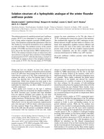
Because the difference in the IM TM expression level
between the overt and non-overt DIC groups was
significant, we focused on the TM expression level of
IMsasapotentialmarkerofDIC.Toinvestigate
whether the IM TM level correlated with coagulopathy,
we divided the patients into three tertile groups accord-
ing to PT, TF, antithrombin, and protein C levels. Inter-
estingly, the IM TM level gradually increased as PT and
TF increased (Figure 1a, b). In addition, the IM TM
level correlated with levels of both antithrombin and
protein C (Figure 1c, d). In regard to the linear relation-
ship between IM TM level and DIC markers, IM TM
level was si gnificantly correlated with PT (P < 0.001, r =
0.428), TF (P = 0.003, r = 0.307), antithrombin (P <
0.001, r = 0.451), and protein C (P <0.001,r = -0.431)
by Spearman’s correlation analysis. The TM expression
on IM was separately analyzed for the subgroups by dis-
ease categories. The correlation of TM expression on
IM with coagulation markers was observed in the sepsis
group with PT (P=0.009, r = 0.609), TF (P = 0.023, r =
0.565), antithrombin (P = 0.004, r = -0.662), and protein
C(P =0.010,r = -0.603). In the hepatic failure group,
there was a correlation with PT (P=0.002, r =0.580),
antithrombin (P = 0.001, r = -0.606), and protein C (P =
0.002, r = -0.580). However, other subpopulations did
not show correlations of TM expression on IM with
coagulation markers individually.
The diagnostic value of IM TM level was evaluated by
using the a rea under the ROC curve (AUC). The AUC
of antithrombin and protein C, well-known DIC mar-
kers, showed significantly good discriminative power
(Figure 2). The AUC of IM TM level was also significant
but showed less discriminative power than that of
antithrombin or protein C.
Prognostic performance of the inflammatory monocyte
thrombomodulin phenotype
Twenty-eight-day hospital mortality was used as a para-
meter of clinical prognosis. The cutoff values of different
Table 2 Percentage and phenotype of monocyte subpopulations according to overt disseminated intravascular
coagulation status and mortality
Non-overt DIC Overt DIC Survivors Non-survivors
Number 67 31 76 22
Absolute monocyte count, × 10
6
/L 510 (336-752) 699 (351-1,260) 496 (337-743) 883 (452-1,913)
a
CD14
bright
CD16
negative
classic monocytes Percentage 62.0 (48.3-70.9) 55.0 (48.4-65.6) 62.4 (51.1-70.7) 50.0 (39.2-54.5)
a
Thrombomodulin 32.0 (23.9-41.9) 29.0 (23.2-52.1) 31.1 (22.5-40.6) 35.9 (24.9-75.8)
a
Tissue factor 4.0 (3.4-4.5) 3.4 (2.6-4.4)
b
4.0 (3.3-4.4) 3.5 (2.7-4.3)
CD14
bright
CD16
positive
inflammatory monocytes Percentage 13.0 (7.7-18.9) 11.0 (7.1-19.0) 12.8 (7.7-18.8) 10.7 (5.9-18.9)
Thrombomodulin 55.0 (42.5-75.1) 70.0 (54.5-117.5)
b
54.7 (43.1-71.9) 73.7 (60.5-125.5)
a
Tissue factor 5.4 (4.2-7.1) 5.6 (4.7-6.5) 5.5 (4.2-7.1) 5.3 (4.7-6.3)
CD14
dim
CD16
positive
dendritic monocytes Percentage 1.8 (0.8-4.4) 1.6 (1.0-3.2) 1.7 (0.8-3.6) 2.7 (1.0-6.2)
Thrombomodulin 92.5 (49.9-114.8) 71.6 (47.7-115.0) 85.2 (46.7-114.5) 71.9 (55.2-115.8)
Tissue factor 9.5 (5.1-20.7) 8.6 (6.1-16.2) 10.0 (5.2-19.4) 7.0 (5.9-17.0)
a
P < 0.05 between survivors and non-survivors.
b
P < 0.05 between non-overt disseminated intravascular coagulation (DIC) and overt DIC. The expression levels of
thrombomodulin and tissue factor were scaled by an arbitrary unit of mean fluorescence intensity.
Hwang et al. Critical Care 2011, 15:R113
/>Page 4 of 11
markers for DIC were defined as the value at which the
ROC curves showed optimal prognostic power. Patient
groups with higher CM percentages (>57.9%) and lower
TM expression levels of CMs (≤60.9) and IMs (≤63.2)
showed better survival compared with those with lower
CM percentages and higher TM expression levels of
CMs and IMs (Figure 3). However, there were no signif-
icant differences in survival of the groups divided by the
characteristics (the percentages or TM or TF expres-
sion) of DM.
(B)
(A)
(C)
(D)
Figure 1 Thrombomodulin expression level of inflammatory monocytes (CD14
bright
CD16
positive
). Levels are based on the prothrombin
time (PT) (a) and plasma levels of tissue factor (b), antithrombin (c), and protein C (d). The expression level of thrombomodulin was scaled by an
arbitrary unit of mean fluorescence intensity. The upper limit of each box represents the median value, and the bar represents the value of the
25th-75th percentile.
†
P < 0.05,
‡
P < 0.001.
Hwang et al. Critical Care 2011, 15:R113
/>Page 5 of 11
Cox univariate analysis showed that decreased platelet
count and prolonged PT, elevated D-dimer, low fibrino-
gen, low antithrombin, low protein C, and high p lasma
TF levels were significant predictors of 28-day mortality
(Table 3). As for the monocyte phenotypes, high absolute
monocyte count, low CM percentage, and high CM and
IM TM expressi on were significant predictors for 28-day
mortality in Cox univariate analysis. The TF expression
levels of CM and IM were not statistically significant in
univariate analysis, but in Cox multivariate analysis, low
CM TF expression was an independent predictor of mor-
tality along with fibrinogen and IM TM level.
Monocyte subtype proportion and expression phenotype
patterns in an in vitro culture system
Purified monocytes from PBMCs of healthy donors were
cultured in vitro for 24 hours. In vitro monocyte cul-
tures showed decreasing CM and DM percentages and
an increasing IM percentage (Figure 4). The IL-10-trea-
ted group revealed a further CM decrease and a corre-
sponding IM increase compared with the control and
LPS-treated groups (Figure 4a). The DM proportion
decreased in the LPS- and IL-10-treated groups com-
pared with the control group. The LPS-treated group
showed markedly high TF expression in all monocyte
subpopulations. The IL-10-treated group tended to exhi-
bit slightly low TF expression, but the difference was
not significant. TM expression levels increased the most
in DMs, followed by IMs, and then finally CMs. In the
LPS-treated group, CMs showed high TM expression at
2 hours, whereas IMs showed higher TM expression
from 12 to 24 hours of c ulture in comparison with that
of the control. In all monocyte subpopulations, IL-10
treatment tended to slightly decrease TM expression.
Discussion
Tightly controlled TF and TM expressions maintain
normal rheological properties of the blood. However,
various stimuli such as infection and inflammation can
induce inflamm atory cytokines that increase TF expres-
sion and suppress anticoagulant protein expression
[22-24]. This imbalance would eventually yield to the
procoagulant diathesis of DIC. Therefore, the changed
pattern of TF and TM expressions plays an important
role in various pathophysiological conditions. Although
the vascular endothelium is known to express TF and
TM [6], circul ating monocytes are also important cellu-
lar sources of TF and TM expressions within vessels [5].
The existence of different populations of monocytes
(CMs, IMs, and DMs) is well established, and each
population has a distinct antigen phenotype and func-
tion [11]. To date, there are no data on the express ion
pattern of TF and TM in any of these monocyte subpo-
pulations. This study was the first to demonstrate the
phenotypic changes of TF and TM i n each monocyte
subpopulation during DIC.
Interestingly, IM TM expression was prominent in the
overt DIC group and had good correlation with other
coagulation markers. Of note, IM TM expression was
found to be an independent prognostic marker for DIC,
which has bee n the focus of this study. Other phenoty-
pic changes of the monocytes also showed differences
between the overt and non-overt DIC, such as the lower
TF expression of CMs in the overt DIC group. TF
expression of CM was significant in multivariate analy-
sis, but the correlations with other coagulation markers
were weak and the differences between the survivor/
non-survivor groups were minimal, and this needs to be
studied further. When the survivors and non-survivors
were compared, the p ercentage of CM was lower and
TM expression on CMs and IMs was higher in the non-
survivors. The TM expression on CM was significant in
the univariat e analysis but was not found to be an inde-
pendent prognostic factor. In addition, the TM and TF
expressions of DMs were higher than those of the IMs,
but the mean differences of the TM and TF express ions
of DMs between survivors and non-survivor were not
sig nificant and the phenotype of DMs was not found to
be significant in multivariate analysis. These findings
support the clinical relevance and importance of TM
rather than TF expression in IMs.
02040608010
0
0
20
40
60
80
1
00
100-S
p
ecificit
y
(
%
)
Protein C
(AUC=0.870, SE<0.001)
Antithrombin
(AUC=0.764, SE<0.001)
IM-Thrombomodulin
(AUC=0.672, SE=0.007)
Figure 2 Receiver operating characteristic (ROC) curves and
the area under the ROC curves (AUC) for antithrombin, protein
C, and thrombomodulin levels of CD14
bright
CD16
positive
inflammatory monocytes (IM). Curves were used for the diagnosis
of overt disseminated intravascular coagulation. SE, standard error.
Hwang et al. Critical Care 2011, 15:R113
/>Page 6 of 11
Evaluation of the TF and TM expressions on each
monocyte subtype showed positive correlation within
each subpopulation of the monocytes. TF is a well-
known initiator of coagulation and an important modu-
lator of inflammation induced by proinflammatory
cytokines [12], but the TM functions as both an anticoa-
gulant and an anti-inflammatory molecule [25], so it is
necessary to understand how TM expression is inte-
grated to maintain homeostasis under hypercoagulable
and proinflammatory conditions. TM is known to be
C
B
A
Figure 3 Kaplan-Meier survival analysis according to p roportions and expression levels of thrombomodulin and tissue factor.
Proportions and expression levels of (a) classical monocytes (CM), (b) inflammatory monocytes (IM), and (c) dendritic monocytes (DM) are
shown. The cutoff values were determined as the values at which the prognostic power to predict 28-day mortality were the highest.
Hwang et al. Critical Care 2011, 15:R113
/>Page 7 of 11
transcriptionally upregulated by thrombin, vascular
endothelial growth factor, histamine, dibutyryl cAMP,
retinoic acid, theophylline, and statin, whereas shear
stress, hemodynamic forces, hypoxia, and oxidized low-
density lipoprotein suppress its expression [25]. In our
study, TM expression tended to increase in hypercoa-
gulable conditions. This finding is consistent with that
of the previous in vitro experiment, which showed that
viral stimulation increased TM expression in m onocytes
and endothelial cells [8]. This is also in agreement with
the study that showed thrombin-induced upregulation
of TM mRNA levels [7] an d with the study that showed
increased a mounts of surface TM on monocytes during
meningococcal disease [9]. All of these findings support
the general notion that infection or inflammation shifts
the hemostatic balance to thrombosis.
Although IM expansion was shown in inflammatory
conditions [17-19], it is currently unclear how to change
the TM phenotype of IMs. In our study, the IM TM
expression level was highly associated with severe coagu-
lopathy and poor prognosis, but those of CMs and DMs
werenot.ThisfindingsuggeststhatIMsplayarolein
maintai ning the hemostati c balance of the active anticoa-
gulant system by enhancing TM expre ssion. The vivid
reaction of IMs can be speculated from that of a previous
study, which states that IMs produce proinflammatory
cytokines [11]. The surface-bound TM is theoretically
considered to be a regulator of the coagulation cascade in
monocytes. However, it re mains unclear whether IM TM
expression exerts functional activity to dampen hyper-
coagulation. In our study, coagulopathy was severe in
patients with high levels of TM, suggesting that the
enhanced expression of TM in IMs plays an insufficient
role in regulating the inflammatory sequelae. This change
mightjustbetheresultofaphysiologicaldefense
mechanism against hypercoagulopathy [26].
In our result, the percentage of monocyte subpopula-
tions did not significantly differ between the overt and
the non-overt DIC groups. Most related studies have
compared the monocyte subpopulations between control
and sepsis patients [17-19]. However, our study focused
on patients suspected of having DIC (some with a
recent inflammatory insult, others with overlaying sti-
muli in chronic conditions, and others in recovery);
thus, the result may not show a clear-cut difference
between the overt and the non-overt groups. This het-
erogeneity within each subgroup may have created a
less dramatic difference between the expression level of
TF or TM on monocytes as well.
To evaluate the diagnostic value of the IM TM pheno-
type, we analyzed the AUC value and compared it with
that of well-known DIC markers. The AUC for the TM
Table 3 Univariate and multivariate analyses for predictors of 28-day mortality
Univariate Multivariate
Variables HR 95% CI P value HR 95% CI P value
Platelet (>112 vs. ≤112 × 10
9
/L) 5.54 1.64-18.75 0.012 1.30 0.18-9.50 0.797
Prothrombin time (≤18.4 vs. >18.4 s) 7.25 2.94-17.85 <0.001 2.20 0.17-29.07 0.548
D-dimer (≤2.0 vs. >2.0 μg/mL) 8.57 2.00-36.69 0.004 3.48 0.45-27.11 0.233
Fibrinogen (>118 vs. ≤118 mg/dL) 7.35 2.92-18.48 <0.001 22.35 2.25-221.81 0.008
Antithrombin (>35% vs. ≤35%) 7.50 3.18-17.65 <0.001 2.15 0.13-36.70 0.598
Protein C (>27% vs. ≤27%) 4.04 1.74-9.37 0.001 1.63 0.06-47.58 0.777
Soluble tissue factor (≤106.1 vs. >106.1 pg/mL) 3.59 2.71-18.47 <0.001 1.20 1.73-8.36 0.852
Absolute monocyte count (≤755 vs. >755 × 10
6
/L) 3.76 1.61-8.81 0.002 2.31 0.39-13.71 0.359
CD14
bright
CD16
negative
classical monocytes
Percentage (>57.9% vs. ≤57.9%) 8.16 2.41-27.61 0.001 4.94 0.66-37.01 0.120
Thrombomodulin (≤60.9 vs. >60.9) 4.93 2.06-11.81 <0.001 1.36 0.36-5.18 0.649
Tissue factor (>3.8 vs. ≤3.8) 2.14 0.90-5.11 0.086 5.27 1.14-24.47 0.034
CD14
bright
CD16
positive
inflammatory monocytes
Percentage (≤10.7% vs. >10.7%) 1.80 0.78-4.17 0.171 1.36 0.25-7.25 0.722
Thrombomodulin (≤63.2 vs. >63.2) 4.67 1.82-11.94 0.001 19.11 1.51-241.47 0.023
Tissue factor (≤4.3 vs. >4.3) 3.03 0.71-12.98 0.135 1.36 0.07-25.34 0.836
CD14
dim
CD16
positive
dendritic monocytes
Percentage (>4.1% vs. ≤4.1%) 2.17 0.93-5.08 0.074 5.14 0.81-32.40 0.082
Thrombomodulin (>83.4 vs. ≤83.4) 1.67 0.40-3.98 0.249 1.12 0.13-9.85 0.918
Tissue factor (>7.0 vs. ≤7.0) 2.21 0.93-5.24 0.073 1.28 0.26-6.22 0.762
The cutoff values were determined as the values at which the best prognostic value was produced.
The expression levels of thrombomodulin and tissue factor were scaled by an arbitrary unit of mean fluorescence intensity. CI, confidence interval; HR, hazard
ratio.
Hwang et al. Critical Care 2011, 15:R113
/>Page 8 of 11
$
%
&
Figure 4 Changes in the proportion and expression phenotype of a monocyte subtype cultured in vitro. Purified monocytes from
healthy donors (n = 4) were cultured in vitro for 24 hours with vehicle, 100 mg/dL lipopolysaccharide (LPS), or 10 ng/mL interleukin-10 (IL-10).
(a) Changes in the proportion and phenotype of (b) tissue factor and (c) thrombomodulin expression among three monocyte subtypes -
classical monocytes (CM), inflammatory monocytes (IM), and dendritic monocytes (DM) - are shown over culture time. MFI, mean fluorescence
intensity.
Hwang et al. Critical Care 2011, 15:R113
/>Page 9 of 11
phenotype was significant (0.672) but was lower than
that of protein C and antithrombin, suggesting that the
IM TM phenotype is not a good diagnostic marker of
overt DIC. On the other hand, it was useful for estimat-
ing prognosis. IM TM expression remained a significant
prognostic factor in multivariate Cox analysis, with a
hazard ratio of 19.11 after adjustment for the effect of
other coagulation markers. Given that most of the DIC
markers are dependent on each other, the IM TM phe-
notype is expected to be a useful potential marker of
prognosis. A future prospective study is needed to verify
the prognostic value of this marker.
In vitro cu lture results showed that the IM proportion
increased with culture time in both control and stimu-
lated monocytes. Interestingly, IL-10 induced a high
proportion of IMs and a correspondingly low proportion
of CMs in comparison with LPS or no treatment. More-
over, IL-10 treatment tended to decrease TF and
increase TM, although the difference was minimal.
Given that IL-10 is an anti-infla mmatory cytokine, these
actions are thought to be counter-responsive to the
inflammatory stimuli. Our suggestion is in good agree-
ment with a previous report in which the alternative
activation of monocytes by IL-10 induced a phenotype
that promoted tissue repair and suppressed inflamma-
tion [14]. On the other hand, TF expression in all
monocyte subpopulations increa sed in the LPS-treated
group, as o bserved in other studies [13,24,27]. An ele-
gant study reported that TF mRNA levels in l eukocytes
increased during DIC [28]. In our clinical results, TF
expression was not a significant marker except in CM,
in which low TF expression predicted poor prognosis. It
is currently unclear why low TF expression represents
poor prognosis. In our data, the TF expression bet ween
overt and non-overt DIC was not different, although in
vitro culture suggested that LPS induced the expression
of both TF and TM. In the in vitro experiment, mono-
cytes from healthy individuals were stimulated with an
inflammatory stimulus (LPS), reflecting the basic modu-
lation of TF and TM expressions by an inflammatory
insult. However, the studiedpopulationisaheteroge-
neous group even in the overt or non-overt DIC group;
thus, the result may not show a clear-cut difference
between the overt and the non-overt group s. TM
expression did not differ significantly between the three
monocyte subpopulations, but LPS treatment upregu-
latedTMat2hoursinCMsandat12to24hoursin
IMs.We[29]andanothergroup[30]previously
reported that LPS downregulated TM expression in
monocytes. However, we could not demonstrate LPS-
induced TM downregulat ion. We speculate that the di f-
ference in expression may be a result of different culture
conditions. Previous experiments used a culture of
PBMCs that included high numbers of lymphocytes
[29,30], and this potentially produces amounts of
inflammatory cytokines that can affect the TM level. In
this experiment, we used purified monocytes that con-
tained low numbers of lymphocytes. Upregulation of
TM may contribute to the regulation of coagulation by
promoting activated protein C, thus suggesting a defense
mechanism against the development of extensive micro-
vascular fibrin deposition during DIC. However, as
shown in our clinical study, insufficient TM function is
expected in monocytes.
Conclusions
The peripheral monocytes of patients suspected of
having DIC were categorized into three subtypes and
studiedforTMandTFexpressions.TheIMTM
expression level showed a significant correlation with
the known DIC m arkers and had diagnostic value for
overt DIC. Furthermore, the IM TM expression level
was found to b e an independent prognostic factor for
28-day mortality in DIC. In addition, in vitro culture
of peripheral monocytes showed that LPS stimulation
upregulated TM and TF expressions in a distinct sub-
type of monocytes. These findings suggest that IM
TM upregulation is a vestige of the physiological
defense mechanism against hypercoagulopathy and is
a good potential independent prognostic marker for
DIC.
Key messages
• Thrombomodulin expressi on level of inflammatory
monocytes shows a significant correlation with the
known disseminated intravascular coagulation (DIC)
markers and had diagnostic value for overt DIC.
• Thrombomodulin exp ression of inflammatory
monocytes is an independent prognostic marker in
patients suspected of having DIC.
• Lipopolysaccharide stimulation upregulates throm-
bomodulin and tissue factor expression in a distinct
subtype of monocytes in in vitro culture of periph-
eral monocytes.
Abbreviations
AUC: area under the receiver operating characteristics curve; CM: classical
monocyte; DIC: disseminated intravascular coagulation; DM: dendritic cell-like
monocyte; IL: interleukin; IM: inflammatory monocyte; ISTH: International
Society on Thrombosis and Haemostasis; LPS: lipopolysaccharide; PBMC:
peripheral blood mononuclear cell; PT: prothrombin time; ROC: receiver
operating characteristic; TF: tissue factor; TM: thrombomodulin.
Acknowledgements
This research was supported by the Basic Science Research Program through
the National Research Foundation of Korea (NRF) funded by the Ministry of
Education, Science and Technology (2010-0004215).
Author details
1
Department of Laboratory Medicine, Seoul National University College of
Medicine, 101, Daehak-ro Jongno-gu, Seoul 110-744, Republic of Korea.
Hwang et al. Critical Care 2011, 15:R113
/>Page 10 of 11
2
Cancer Research Institute, Seoul National University College of Medicine,
101, Daehak-ro Jongno-gu, Seoul 110-744, Republic of Korea.
Authors’ contributions
HKK designed the study, shared responsibility for the study design and for
data management and statistical analysis, and helped to write the
manuscript. JEK performed the experiments and shared responsibility for
data management and statistical analysis. SMH shared responsi bility for data
management and statistical analysis and helped to write the manuscript.
KSH shared responsibility for the study design, data interpretation, and
manuscript revision for important intellectual content. All authors read and
approved the final manuscript.
Competing interests
The authors declare that they have no competing interests.
Received: 22 November 2010 Revised: 18 February 2011
Accepted: 14 April 2011 Published: 14 April 2011
References
1. Sarangi P, Lee H, Kim M: Activated protein C action in inflammation. Brit J
Haematol 2010, 148:817-833.
2. Persson E, Olsen O: Current status on tissue factor activation of factor
VIIa. Thromb Res 2010, 125:S11-S12.
3. Levi M, Ten Cate H: Disseminated intravascular coagulation. N Engl J Med
1999, 341:586-592.
4. Conway EM, Rosenberg RD: Tumor necrosis factor suppresses
transcription of the thrombomodulin gene in endothelial cells. Mol Cell
Biol 1988, 8:5588-5592.
5. Moore K, Andreoli S, Esmon N, Esmon C, Bang N: Endotoxin enhances
tissue factor and suppresses thrombomodulin expression of human
vascular endothelium in vitro. J Clin Invest 1987, 79:124-130.
6. McCachren S, Diggs J, Weinberg J, Dittman W: Thrombomodulin
expression by human blood monocytes and by human synovial tissue
lining macrophages. Blood 1991, 78:3128-3132.
7. Bartha K, Brisson C, Archipoff G, de la Salle C, Lanza F, Cazenave J, Beretz A:
Thrombin regulates tissue factor and thrombomodulin mRNA levels and
activities in human saphenous vein endothelial cells by distinct
mechanisms. J Biol Chem 1993, 268:421-429.
8. Chen LC, Shyu HW, Lin HM, Lei HY, Lin YS, Liu HS, Yeh TM: Dengue virus
induces thrombomodulin expression in human endothelial cells and
monocytes in vitro. J Infection 2009, 58:368-374.
9. Faust S, Heyderman R, Levin M: Coagulation in severe sepsis: a central
role for thrombomodulin and activated protein C. Crit Care Med 2001, 29:
S62-67.
10. Fingerle G, Pforte A, Passlick B, Blumenstein M, Strobel M, Ziegler-
Heitbrock H: The novel subset of CD14+/CD16+ blood monocytes is
expanded in sepsis patients. Blood 1993, 82:3170-3176.
11. Ziegler-Heitbrock L: The CD14+ CD16+ blood monocytes: their role in
infection and inflammation. J Leukocyte Biol 2007, 81:584-592.
12. Levi M, van der Poll T, ten Cate H: Tissue factor in infection and severe
inflammation. Semin Thromb Hemost 2006, 32:33-39.
13. Lindmark T, Chen S: IL-10 inhibits LPS-induced human monocyte tissue
factor expression in whole blood. Brit J Haematol 1998, 102:597-604.
14. Gordon S, Taylor P: Monocyte and macrophage heterogeneity. Nat Rev
Immunol 2005, 5:953-964.
15. McPherson R, Pincus M: Henry’s Clinical Diagnosis and Management by
Laboratory Methods. 21 edition. Philadelphia: Saunders; 2006.
16. Thomas R, Lipsky P: Human peripheral blood dendritic cell subsets.
Isolation and characterization of precursor and mature antigen-
presenting cells. J Immunol 1994, 153:4016-4028.
17. Skrzeczynska J, Kobylarz K, Hartwich Z, Zembala M, Pryjma J: CD14+ CD16+
monocytes in the course of sepsis in neonates and small children:
monitoring and functional studies. Scand J Immunol 2002, 55:629-638.
18. Poehlmann H, Schefold J, Zuckermann-Becker H, Volk H, Meisel C:
Phenotype changes and impaired function of dendritic cell subsets in
patients with sepsis: a prospective observational analysis. Crit Care 2009,
13:R119.
19. Skinner N, MacIsaac C, Hamilton J, Visvanathan K: Regulation of Toll like
receptor (TLR) 2 and TLR4 on CD14dimCD16+ monocytes in response to
sepsis related antigens. Clin Exp Immunol 2005, 141:270-278.
20. Toh C, Hoots W: The scoring system of the Scientific and Standardisation
Committee on Disseminated Intravascular Coagulation of the
International Society on Thrombosis and Haemostasis: a 5-year
overview. J Thromb Haemost 2007, 5:604-606.
21. Taylor F, Toh C, Hoots W, Wada H, Levi M: Towards definition, clinical and
laboratory criteria, and a scoring system for disseminated intravascular
coagulation. Thromb Haemost 2001, 86:1327-1330.
22. Osterud B, Bjorklid E: The tissue factor pathway in disseminated
intravascular coagulation. Semin Thromb Haemost 2001, 27:605-618.
23. Celi A, Pellegrini G, Lorenzet R, De Blasi A, Ready N, Furie B, Furie B: P-
selectin induces the expression of tissue factor on monocytes. Proc Natl
Acad Sci USA 1994, 91:8767-8771.
24. Osterud B: Tissue factor expression in monocytes: in vitro compared to
ex vivo. Thromb Haemostasis 2000, 84:521-522.
25. Van de Wouwer M, Collen D, Conway EM: Thrombomodulin-protein C-
EPCR system: integrated to regulate coagulation and inflammation.
Arterioscl Thromb Vas 2004, 24:1374-1383.
26. Tsai C, Tsai Y, Lin C, Lin T, Huang G, Hong G, Lin F: Expression of
thrombomodulin on monocytes is associated with early outcomes in
patients with coronary artery bypass graft surgery. Shock 2010, 34:31-39.
27. Herbert J, Savi P, Laplace M, Lale A: IL-4 inhibits LPS-, IL-1 [beta]-and TNF
[alpha]-induced expression of tissue factor in endothelial cells and
monocytes. FEBS Lett 1992, 310:31-33.
28. Sase T, Wada H, Nishioka J, Abe Y, Gabazza EC, Shiku H, Suzuki K,
Nakamura S, Nobori T: Measurement of tissue factor messenger RNA
levels in leukocytes from patients in hypercoagulable state caused by
several underlying diseases. Thromb Haemost 2003, 89:660-665.
29. Kim H, Kim J, Chung J, Kim Y, Kang S, Han K, Cho H:
Lipopolysaccharide
down-regulates the thrombomodulin expression of peripheral blood
monocytes: effect of serum on thrombomodulin expression in the THP-1
monocytic cell line. Blood Coagul Fibrin 2007, 18:157-164.
30. Satta N, Freyssinet J, Toti F: The significance of human monocyte
thrombomodulin during membrane vesiculation and after stimulation
by lipopolysaccharide. Brit J Haematol 1997, 96:534-542.
doi:10.1186/cc10139
Cite this article as: Hwang et al.: Thrombomodulin phenotype of a
distinct monocyte subtype is an independent prognostic marker for
disseminated intravascular coagulation. Critical Care 2011 15:R113.
Submit your next manuscript to BioMed Central
and take full advantage of:
• Convenient online submission
• Thorough peer review
• No space constraints or color figure charges
• Immediate publication on acceptance
• Inclusion in PubMed, CAS, Scopus and Google Scholar
• Research which is freely available for redistribution
Submit your manuscript at
www.biomedcentral.com/submit
Hwang et al. Critical Care 2011, 15:R113
/>Page 11 of 11