Báo cáo y học: " Positive end-expiratory pressure optimization with forced oscillation technique reduces ventilator induced lung injury: a controlled experimental study in pigs with saline lavage lung injury" ppt
Bạn đang xem bản rút gọn của tài liệu. Xem và tải ngay bản đầy đủ của tài liệu tại đây (1.11 MB, 9 trang )
RESEARCH Open Access
Positive end-expiratory pressure optimization
with forced oscillation technique reduces
ventilator induced lung injury: a controlled
experimental study in pigs with saline lavage
lung injury
Peter Kostic
1
, Emanuela Zannin
2
, Marie Andersson Olerud
1
, Pasquale P Pompilio
2
, Göran Hedenstierna
3
,
Antonio Pedotti
2
, Anders Larsson
1
, Peter Frykholm
1
and Raffaele L Dellaca
2*
Abstract
Introduction: Protocols using high levels of positive end-expiratory pressure (PEEP) in combi nation with low tidal
volumes have been shown to reduce mortality in patients with severe acute respiratory distress syndrome (ARDS).
However, the optimal method for setting PEEP is yet to be defined. It has been shown that respiratory system
reactance (Xrs), measured by the forced oscillation technique (FOT) at 5 Hz, may be used to identify the minimal
PEEP level required to maintain lung recruitment. The aim of the present study was to evaluate if using Xrs for
setting PEEP would improve lung mechanics and reduce lung injury compared to an oxygenation-based approach.
Methods: 17 pigs, in which acute lung injury (ALI) was induced by saline lavage, were studied. Animals were
randomized into two groups: in the first PEEP was titrated according to Xrs (FOT group), in the control group PEEP
was set according to the ARDSNet protocol (ARDSNet group). The duration of the trial was 12 hours. In both
groups recruitment maneuvers (RM) were performed every 2 hours, increasing PEEP to 20 cmH
2
O. In the FOT
group PEEP was titrated by monitoring Xrs while PEEP was reduced from 20 cmH
2
O in steps of 2 cmH
2
O. PEEP
was considered optimal at the step before which Xrs started to decrease. Ventilatory parameters, lung mechanics,
blood gases and hemodynamic parameters were recorded hourly. Lung injury was evaluated by histopathological
analysis.
Results: The PEEP levels set in the FOT group were significantly higher compared to those set in the ARDSNet
group during the whole trial. These higher values of PEEP resulted in improved lung mechanics, reduced driving
pressure, improved oxygenation, with a trend for higher PaCO
2
and lower systemic and pulmonary pressure. After
12 hours of ventilation, histopathological analysis showed a significantly lower score of lung injury in the FOT
group compared to the ARDSNet group.
Conclusions: In a lavage model of lung injury a PEEP optimiza tion strategy based on maximizing Xrs attenuated
the signs of ventilator induced lung injury. The respiratory system reactance measured by FOT could thus be an
important component in a strategy for delivering protective ventilation to patients with ARDS/acute lung injury.
* Correspondence:
2
Dipartimento di Bioingegneria, Politecnico di Milano University, P.zza
Leonardo da Vinci 32, 20133 Milano, Italy
Full list of author information is available at the end of the article
Kostic et al. Critical Care 2011, 15:R126
/>© 2011 Kostic et al.; licensee BioMed Central Ltd. This is an open access article distributed under the terms of the Creative Commons
Attribution License (http://creativecomm ons.org/licenses/by/2.0), which permits unrestricted use, distribution, and reproduction in
any medium, provided the original work is properly cited.
Introduction
Mechanical ventilation is a mainstay of intensive care
for patients with acute lung i njury (ALI) and the acute
respiratory distress syndrome (ARDS). A ventilation
strategy based on tidal volumes of 6 ml.kg
-1
and pre-
defined positive end-expiratory pressure (PEEP) set-
tings has been shown to reduce morbidity and mortal-
ity probably due to less ventilation-induced lung
injury (VILI) [1-3]. Various protocols using higher
levels of PEEP in combinati on with low tidal volumes
(Vt) have also been shown to reduce mortality in
patients with ARDS [4], which was corroborated in a
recent me ta-analysis [5,6 ]. Meanwhile, experimental
studies have been designed to define the optimal PEEP
level based on lung compliance or elastance recorded
during a recruitment maneuver (RM) with decremen-
tal PEEP [7,8].
We have recently shown that respiratory system reac-
tance (Xrs) obtained by the forced oscillation technique
(FOT) at 5 Hz is mo re reliable than dynamic compli-
ance for assessing lung collapse and the effects of lung
RMs in a porcine ALI model [9,10]. Specifically, Xrs
(an d its deriv ed variable C
X5
, the oscillatory compliance
at 5 Hz) identifies the minimum PEEP level required to
maintain lung recruitment with high sensitivity and spe-
cificity. The advantages of this non-invasive appro ach
are that it can be easily integrated in mechanical ventila-
tors, it is suitable for bedside continuous monitoring,
and it can also be used in the presence of spontaneous
breaths.
During long-term ventilatory treatments, the opt imal
PEEP level is likely to change wit h time due to the
developing disease process as well as various interven-
tions in the ICU. Hence, a strategy d esigned to reduce
VILI should probably include repeated assessment of
lung mechanics, with su bsequent changes in the ventila-
tor settings.
Theaimofthepresentstudywastoevaluatethe
effects of repeated PEEP optimization based on Xrs
on oxygenation, lung mechanics, and histologic mar-
kers of lung injury, and compare them to the results
obtained by applying the ARDSNet protocol based on
oxygenation alone, in a porcine surfactant-depletion
lung injury model over a 12-hour ventilation period.
The hypothesis was that repeated PEEP optimization
by FOT could improve lung mechanics and reduce
VILI.
Materials and methods
Seventeen healthy pigs (weight 26.6 ± 2.2 kg, Swedish
mixed country breed) were studied at the Hedenstierna
laboratory, Department of Surgical Sciences of the Uni-
versity Hospital of Uppsala, Sweden. The study was
approved by the local animal ethics committee.
Animal preparation
Anesthesia was induced by tiletamine 6 mg.kg
-1
,zolaze-
pam 6 mg.kg
-1
, xylazine 2.2 mg.kg
-1
intramuscularly,
and maintained with an intravenous (iv) infusion of phe-
nobarbital 1 mg/ml, pancuronium 0.032 mg/ml, and
morphine 0.06 mg·ml
-1
at a rate of 8 ml·kg
-1
·h
-1
. After a
bolus injection of fentanyl 10 μ.kg
-1
iv a tracheotomy
was performed and the lungs were ventilated through a
shortened 8 mm inner diameter endotracheal tube (Mal-
linckrodt, Athlone, Ireland) in a volume-controlled
mode (Servo i ventilator, Maquet, Solna, Sweden) with a
Vt 6 ml/kg, a PEEP 5 cmH
2
O, and respiratory rate
titrated to obtain normocapnea (35 < partial pressure of
carbon dioxide (pCO
2
) < 45 mmHg). Lung injury was
induced by repeated broncho -alveolar lavage with instil-
lation of approximately 25 ml/kg warm saline solution
per lavage. The end-point of the lavage was a sustained
reduction in the partial pressure of o xygen (pO
2
)/frac-
tion of inspired oxygen (FiO
2
) less than 100 mmHg dur-
ing a period of 60 minutes.
Measurements
Systemic and pulmonary arterial pressures, heart rate,
mixed venous saturation, and body temperature were
continuously monitored (CCombo 7.5-Fr, Edwards Life
Sciences LLC, Irvine, CA, USA). Arterial blood gases
were sampled every hour to measure partial pressure of
arterial oxygen (PaO
2
), partial pressure of arterial carbon
dioxide (PaCO
2
), pH and oxygen saturat ion (SpO
2
;ABL
500, Radiometer, Copenhagen, Denmark).
FOT was applied by using a system that has been
described elsewhere [9]. Briefly, low amplitude sinusoi-
dal pressure oscillations (about 1.5 cmH
2
O peak-to-
peak)at5Hzweregeneratedbyaloudspeakercon-
nected to the inspirato ry line of the mechanical ventila-
tor. Flow at the airway opening (Vao) was measured by
a differential pressure transducer (PXLA02X5DN, Sen-
sym, Milpitas, CA, USA) connected to a mesh-type
heated pneumotachograph. Tracheal pressure was mea-
sured at the tip of the endotracheal tube by a differential
pressure transducer (PXLA0075DN, Sensym, Milpitas,
CA, USA). Signals were sampled at 200 Hz by the same
A/D-D/A board used to control the loudspeaker and
recorded on a personal computer.
Experimental protocol
This study was the second part of a two-study protocol,
designed to spare animals. Part 1 included a stepwise
RM and computed tomography (CT)-scanning. For this
reason, the ventilation trial started about five hours after
the induction of lung injury.
The animals were randomized into two groups. One
was treated with optimal PEEP (PEEPol) according to
Xrs (FOT group), t he other was treated with PEEP
Kostic et al. Critical Care 2011, 15:R126
/>Page 2 of 9
adjusted according to the ARDSNet protocol [1] (ARDS-
Net group). All animals underwent identical treatment
before random ization, and there were no significant dif-
ferences between the groups with re gards to PaO
2
/FiO
2
,
PaCO
2
, dynamic compliance (Cdyn), mean arterial pres-
sure (MAP), and mean pulmonary arterial pressure
(MPAP) before the intervention trial.
The duration of the protocol was 12 hours, with every
experimental session involving two animals, one from
each group, studied in parallel with a time shift of one
hour to avoid the overlap of RM performed by the
researchers. In both groups RMs were performed every
two hours by increasing PEEP to 20 cmH
2
Ofortwo
minutes, preceded by tracheal suctioning for five sec-
onds, to simulate a clinical situation in which a RM is
performed to counteract derecruitment due to suction-
ing. Arterial blood gases were sampled and recorded five
minutes after RMs and hourly. PEEP was adjusted after
the RM every two hours in both groups.
In the FOT group, PEEPol according to Xrs was iden-
tified as shown in Figure 1. Briefly, a decremental PEEP
trial was performed immediately after the RM by a step-
wise reduction of PEEP from 20 cmH
2
O in one minute-
stepsof2cmH
2
O until Xrs reached its maximum and
started to decrease. PEEPol was defined as the PEEP
level at the step preceding the first reduction of Xrs.
Immediately after obtaining PEEPol, PEEP was increased
again u p to 20 cmH
2
O for one minute in order to
restore lung volume and then it was brought back to
PEEPol, which was maintained for the next two hours
until the next scheduled optimization procedure.
In the ARDSNet group the optimization has been per-
formed by following ARDSNet indications [1]. More-
over, in the ARDSNet group, PEEP was also adjusted
between RMs whenever indicated.
By using this protocol, both groups (ARDSNet and
FOT) received the same amount of RMs.
RespiratoryrateandFiO
2
were adjusted accordin g to
the ARDSNet protocol in both groups.
Leaks from the tracheal tube and ventilator circuits
were continuously monitored for all the duration of the
study.
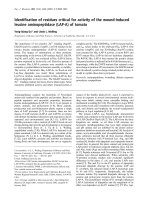
Figure 1 PEE P optimization procedure according to optimal Xrs. The upper panel shows tracheal pressure and the lower panel shows
respiratory system reactance (Xrs) measured at end-expiration over time during a representative positive end-expiratory pressure (PEEP)
optimization procedure. PEEP was increased up to 20 cmH
2
O, and then decreased in one-minute steps of 2 cmH
2
O while Xrs was continuously
monitored. When Xrs started to decrease, PEEP was increased back to 20 cmH
2
O and finally set to the PEEP level corresponding to the
maximum Xrs.
Kostic et al. Critical Care 2011, 15:R126
/>Page 3 of 9
At the end of the experiment, the animals were sacri-
ficed by iv injections of potassium chloride (KCl). Thor-
acotomy was performed, and sections from the left lung
(the lingula and the left lower lobe, two sections from
each lobe) were fixed in buffered formalin solution and
subsequently embedded in paraffin, sectioned at a thick-
ness of 6 μm, and stained with H&E.
Data analysis
Histopathology
The histopathological analysis was performed by a
pathologist who was blinded to the outcome of rando-
mization. Four fields for each pig (two from the lingula
and two from the left lower lobe) were evaluated ran-
domly. A grading scale (0 to 4) for four diff erent histo-
pathological markers of lung injury was used: presence
of alveolar edema, hyaline membranes, inflammatory
cells in alveoli, and inflammatory cells in septa, respec-
tively (modified from [11]). Alveolar edema and hyaline
membranes were graded according to the following cri-
teria: 0-none, 1-focal in one to two fields, 2-focal in
three to four fields, 3-widespread, 4-whole lung. Inflam-
matory cells in alveoli and inflammatory cells in septa
were graded according to the following criteria: 0-none,
1-focal, a few cells, 2-widespread, a few cells, 3-all
alveoli/sept a, few cells, 4-brisk in all alveoli and septa.
The evaluation scores for these markers were averaged
to obtain a cumulative histopathology score for each
animal.
Lung mechanics
The estimation of total respiratory system impedance
(Zrs) was obtained from the flow and pressure signals
by a l east squares algorithm [12]. Zrs was expressed as
real part, respiratory system resistance (Rrs), and ima-
ginary part, respiratory reactance (Xrs).
Comparison between groups
The behavior of the two groups along the ventilation
trial was compared in terms of ventilatory parameters
(PEEP, Cdyn, plateau pressure (Pplat), and driving pres-
sure (ΔP)), gas exchange (PaO
2
/FIO
2
and PaCO
2
), and
hemodynamics (MAP and MPAP). Cdyn values were
provided by the ventilator using multiple regression ana-
lysis. The time of each measurement was referred to the
first optimization procedure performed on the animal
(time 0).
Statistical analysis
Data are expressed as mean (standard deviation). Aft er
testing normality by the Kolmogorov-Smirnov test, sig-
nificance of differences between baseline parameters in
the two groups was tested by unpaired t-test, when nor-
malit y test succeeded, and by Mann-Whitney test, when
normality test failed. Significa nce of differences between
the two groups was tested by two-way analysis of var-
iance (ANOVA) for repeated measurements using group
and protocol step as factors. Multiple comparison after
ANOVA was performed using Holm-Sidak test. Signifi-
cance of differences between the histopathological
scoresgiventothetwogroupswastestedbyMann-
Whitney test. Differences were considered statistically
significant for P < 0.05.
Results
The protocol could be followed without interruption in
both groups, and it was possible to identify an optimal
PEEP value after each RM in the FOT group. During
RMs, moderate decreases in MAP and increases in
MPAP were observed.
The experimental tracings recorded during a represen-
tative optimization procedure are reported in Figure 1.
The pressure tracing shows the breathing cycles, the
stepwise reduction of P EEP, and the end-expiratory
pauses performed in order to establish the values of Xrs
at end-expiration. The values of Xrs measured during
the pauses are reported in the lower panel, where the
expected increasing-decreasing pattern is evident. An
optimal PEEP of 12 cmH
2
O was identified during this
procedure, with a maximum Xrs value of -0.52
cmH
2
O*s/l.
Figure 2 shows the values of the maximal Xrs and the
optimal PEEP identified in all animals at the different
optimization steps. The increase in Xrs clearly shows
that there was an average improvement in the oscillatory
mechanics with time, and this led to a progressively
lower PEEP applied to the FOT group.
The relevant parameters measured every hour during
the ventilation trial were averaged for all animals. The
values of PEEP, Cdyn, Pplat, and ΔP are reported in
Figure 2 Time course of PEEP and Xrs in the FOT group.Mean
and standard deviations of maximum respiratory system reactance
(Xrs) values (closed symbols) and optimized positive end-expiratory
pressure (PEEP; open symbols) assessed during the optimization
procedure performed every two hours in the FOT group. In average,
there was an improvement of oscillatory mechanics, which resulted
in a reduction of optimal PEEP with time.
Kostic et al. Critical Care 2011, 15:R126
/>Page 4 of 9
Figure 3, and the values related to gas exchange and
hemodynamics are reported in Figure 4. Cdyn, Pplat,
and ΔP present an oscillatory pattern, likely due to the
fact that the data were recorded every hour, while RMs
and PEEP optimizations were performed every second
hour. These data suggest that one hour after the PEEP
optimization, the mechanical conditions of the lung
were not as good as immediately after RM.
At the beginning of the trial, the optimization based
on Xrs resulted in a significantly higher PEEP compared
with that set in t he ARDSNet group. T hese settings led
to a significantly lower ΔP, a better oxygenation, and
lower MPAP and MAP in the FOT group.
Overthecourseofthe12-hourexperiment,PEEP
decreased in both groups-from 10.4 (1.7) to 8.9 (1.8)
cmH
2
O in the FOT group, and from 7.4 (2.1) to 5.0 (0)
cmH
2
O in the ARDSNet group. These higher values of
PEEP in the FOT group were associated with improved
respiratory mechanics, as indicated by the significantly
lower ΔP (decreasing from 9.88 (1.78) to 10.1 (2.05)
Figure 3 Ventilatory and respiratory mechanics parameters over time. Positive end-expiratory pressure (PEEP), plateau pressure (Pplat),
driving pressure (ΔP), and dynamic compliance (Cdyn) for the forced oscillation technique (FOT) group (closed symbols) and for the acute
respiratory distress syndrome (ARDS)Net group (open symbols). Data are presented as mean ± standard deviation. Significance of differences
between the two groups at any protocol step are also reported. *, P < 0.01; +, P < 0.05.
Kostic et al. Critical Care 2011, 15:R126
/>Page 5 of 9
compared with from 16.9 (5.2) to 13.4 (4.4) in the ARDS-
Net group) and the higher Cdyn for most of the course
of the experiment (15.1 (4.4) to 15.7 (4.5) compared with
10.8 (4.2) to 13.7 (5.3) ml/cmH
2
O, respectively). There
was a trend for lower Pplat in the FOT group, but the
differences between the groups were not significant. At
the end of the experiment, only changes in oxygenation
and PEEP were still significantly different.
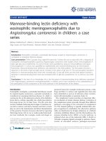
Qualitative and semi-quantitative analysis of histo-
pathologic sections showed significant differences
between the groups, as displayed in Table 1. Inflamma-
tory exudation with hyaline membranes and signs of
massive acute inflammation were found in both groups,
but with a lower injury score in the FOT group. This is
illustrated in Figure 5, with representative sections from
both groups.
Discussion
The main result of this study was that during a 12-hour
ventilation trial, the optimization of PEEP according to
Figure 4 Blood gases and hemodynamic parameters over time. Partial pressure of arterial oxygen (PaO
2
)/fraction of inspired oxygen (FiO
2
),
partial pressure of arterial carbon dioxide (PaCO
2
), mean arterial pressure (MAP), and mean pulmonary arterial pressure (MPAP) for forced
oscillation technique (FOT) group (closed symbols) and for acute respiratory distress syndrome (ARDS)Net group (open symbols). Data are
presented as mean ± standard deviation. Significance of differences between the two groups at any protocol step are also reported. *, P < 0.01;
+, P < 0.05.
Kostic et al. Critical Care 2011, 15:R126
/>Page 6 of 9
Xrs resulted in improved lung mechanics (assessed by
conventi onal methods), a greater PaO
2
/FiO
2
ratio and a
reduced histopathologic evidence of VILI. The PEEP
optimization procedure based on Xrs that we used in
this study requires a RM followed by a decremental
PEEP trial to identify PEEPol. The ARDSNet group was
thus ventilated according t o the ARDSNet protocol,
with the addition of RMs performed at two-hour inter-
vals to allow comparison between the two different
PEEP strate gies with all other interventions being equal.
Previous animal studies have usually focused on short-
term changes. To our knowledge, this is the first study
to follow lung mecha nics and ventilation pa rameters
throughout the course of 12 hours, which more closely
resembles a clinical situation with time enough for the
more subtle mechanisms of VILI to have effect.
Even if the gold standard to assess lung volume
rec ruitment is still CT scanning, there is increasing evi-
dence that lung mechanics is a better surrogate than gas
exchange variations for the assessment of lung recruit-
ment at the bedside [13]. Starting from the pioneering
work of Suter et al. [14], several studies suggested that
the use of dynamic compliance [7,8,14,15] may guide in
the identification of the optimal PEEP. A recent study in
ALI/ARDS patients used a combinatio n of oxygenation
data (venous admixture) and lung mechanics obtained
by electrical impedance tomography [16]. They reported
that volume-de pendent compliance seemed to be super-
ior to dynamic compliance over the whole breath for
monitoring lung recruitment and defining optimal
PEEP. However, this method is labor intensive and
expensive. Moreover, we have recently demonstrated
that the volume-dependent component of compliance
can only partially account for the non-linear behavior of
the respiratory system during mechanical ventilation for
ALI [10]. Also, the monitoring of esophageal pressure in
order to maintain positive trans-pulmonary pressure has
recently been suggested for PEEP optimization [17].
However, the necessity of an appropriate positioning of
the esophageal balloon and the intrinsic difficulties in
such a measurement implicate problems with the imple-
mentation of this technique in clinical applications.
Conversely, utili zing FOT, the peripheral lung
mechanics can be continuously monitored via the venti-
lator circuit, and this could therefore be a preferable
technique. Bellardine et al. applied FOT using the
enhanced ventilation waveform approach on an animal
model of ARDS to study changes in lung mechanics at
different PEEP levels [18]. The authors found that opti-
mal PEEP identified by CT scans minimizes mechanical
heterogeneity, defined as the frequency dependence of
Rrs and low-frequen cy elast ance. However, this
approach requires the assessment of mechanical impe-
dances on a frequency range of 0.2 to 8 Hz and, there-
fore, is not suitable for patients w ith spontaneous
breathing activity. In two previous studies we have
shown that single f requency FOT at 5 Hz can be used
to accurately evaluate lung volume de-recruitment over-
coming several limitations of Cdyn, such as the effects
of non-linearities in the respirator y system and the need
for deep sedation or paralysis of the patients [9,10]. The
results of these studies also suggested that by
Table 1 Histopathological analysis
ARSDNet group FOT group P
Alveolar edema 0.88 ± 0.92 0.06 ± 0.18 0.03
Hyaline membrane 0.94 ± 0.73 0.69 ± 0.70 0.50
Alveolar infl. Cells 1.31 ± 0.80 0.81 ± 0.46 0.15
Septal infl. Cells 2.69 ± 0.80 2.25 ± 0.65 0.25
mean ± SD 1.45 ± 0.47 0.95 ± 0.37 0.03
Lung injury scores for the forced oscillation technique (FOT) and the acute
respiratory distress syndrome (ARDS)Net groups. Data are reported as mean ±
standard deviation (SD).
ARDSNet group, group of animals in whi ch PEEP was adjusted using the
ARDSNet protocol; FOT group, group in which positive end-expiratory
pressure (PEEP) was adjusted according to FOT measurement; infl.,
inflammatory.
Figure 5 Representative tissue samples. Representative histopathology images of lung samples from the forced oscillation techn ique (FOT)
group (right) and the acute respiratory distress syndrome (ARDS)Net group (left). There is more alveolar edema and inflammatory cells in the
septa as well as in the alveoli in the animal ventilated with the positive end-expiratory pressure (PEEP) suggested by ARDSNet.
Kostic et al. Critical Care 2011, 15:R126
/>Page 7 of 9
monitoring Xrs it is possible to continuously assess the
development of lung collapse and to evaluate the effi-
cacy of RMs, allowing bedside characterization of lung
recruitability. In the present study, we implemented
these findings in designing a strict protocol based on
PEEP optimization according to Xrs performed every
two hours. What we found is that optimal PEEP set on
the basis of Xrs changes was clearly advantag eous com-
pared with PEEP settings according to the ARDSNet
protocol, which only uses oxygenation data.
However, in the FOT group in which Xrs was con-
tinuously monitored, we occas ionally observed decreases
in Xrs during the two-hour intervals between the sched-
uled RMs, but no ad justments were made. Thus we did
not fully use the information provided by FOT. An
improved clinical protocol could perhaps be developed,
including RMs coupled with Xrs monitoring for P EEP
optimization, with the addition of using Xrs triggers for
performing subsequent RMs immediately when de-
recruitment occurs.
Hemodynamically, there were no differences between
the FOT and ARDSNet groups except for the pulmon-
ary artery pressure. The lower pulmonary arterial pres-
sure in the FOT group could be due to successful lung
recruitment-the optimal PEEP keeping the lung open
and thus decreasing pulmonary vascular resistance. Dur-
ing the latter half of the experimental period, this differ-
ence was no longer significant. This may have been due
to the long duration of the experiment, with attenuation
of the lung injury in b oth gro ups explained by the
recovery of the lung often seen in the lavage model.
Limitations of the study
We performed histopathologic analysis of several sec-
tions of lung tissue after sacrifice. We chose not to
excise whole lungs, precluding true quantitative analysis
of histopathologic changes, but the qualitative and semi-
quantitative multi-parameter score based on several pre-
vious studies showed clear and significant differences
between the groups.
The saline lavage model of lung injury causes surfac-
tant depletion and atelectasis that is easily recruitable,
in contrast to the heterogeneous inflammatory cha nges
of long-lasting nature that characterize the human
ARDS. This could explain the relatively low v entilatory
pressures and PEEP settings that adherence to the
ARDSNet protocol dictated in the present study. It
could also account for the clinical improvement
through the course of the experiment, including both
blood gases and ventilatory settings. An advantage of
this was that the final damage seen in the histopatho-
logic sections could most likely be attributed to
mechanical ventilation-the focus of the study-rather
than the initial lavage injury.
In this study we compared PEEP optimization per-
formed by FOT with the one based on oxygenation data
as suggested by the ARDSNet. With this experimental
protocol, we could not compare our results with the
ones that would have been obtained by using other opti-
mization procedures based on the assessment o f
mechanical properties (such as Cdyn). However, in a
previousstudywehaveshownthatPEEPoldefinedby
Cdyn is similar but not equal to the one identified by
FOT. Moreover, given that FOT is not affected by the
non-linearities of th e respiratory system, nor by th e
spontaneous breathing of the patient, and it can be
easily integrated in mechanical ventilators, we think that
single-frequency FOT could be easier than other techni-
ques to be applied in clinical practice.
Finally, in the present study the esophageal pressure
was not measured, and thus changes in Xrs include
both changes in lung and chest wall mechanics. How-
ever, we have previously shown, by using mathematical
models, that the contribution of the changes in chest
wall compliance to Xrs is negligible compared with the
contribution of lung volume recruitment/derecruitment
and, therefore, it does not affect the estimation of PEE-
Pol [10].
Conclusions
The results indicate that there is a scientific basis for
implementing an open-lung strategy that includes RMs
and PEEP optimization using FOT. Future studies
should aim to confirm these observations in ALI/ARDS
patients, possibly taking the protocol one step further by
utilizing the continuous monitoring of reactance and
investigating the feasibility of a reactance based trigger
for RMs. Considering that the optimization procedure
based on Xrs can be easily integrated in commercial
mechanical ventilators and tha t it provides continuous
monitoring of the mechanical properties of the periph-
eral airways, we conclude that FOT could be an impor-
tant component in a strategy for delivering protective
ventilation to patients with ALI.
Key messages
• In a surfactant-depletion model of ALI, during a
decremental PEEP trial following a RM there was
always a PEEP level at which the respira tory system
reactance measured by FOT reached a maximum.
• When PEEP was set to the value that maximized
the reactance , higher PEEP levels, improved lung
mechanics, and better oxygenation were observed
compared with those measured when PEEP was set
following standard clinical protocols based on oxyge-
nation (ARDSNet).
• A PEEP setting strategy based on the optimization
of respiratory reactance produced less histologic
Kostic et al. Critical Care 2011, 15:R126
/>Page 8 of 9
signs of lung injury compared with the oxygenation-
based ARDSNet protocol after a 12-hour ventil ation
trial.
Abbreviations
ALI: acute lung injury; ANOVA: analysis of variance; ARDS: acute respiratory
distress syndrome; Cdyn: dynamic compliance; CT: computed tomography;
FiO
2
: fraction of inspired oxygen; FOT: forced oscillation technique; H&E:
hematoxylin and eosin; MAP: mean arterial pressure; MPAP: mean pulmonary
arterial pressure; PaCO
2
: partial pressure of arterial carbon dioxide; PaO
2
:
partial pressure of arterial oxygen; pCO
2
: partial pressure of carbon dioxide;
PEEP: positive end-expiratory pressure; PEEPol: open lung PEEP; Pplat:
plateau pressure; pO
2
: partial pressure of oxygen; RM: recruitment maneuver;
Rrs: respiratory system resistance; SpO2: oxygen saturation; VILI: ventilator-
induced lung injury; Vt: tidal volume; Xrs: respiratory system reactance; Zrs:
total respiratory system impedance; ΔP: driving pressure.
Acknowledgements
The authors gratefully acknowledge Agneta Roneus and Karin Fagerbrink of
the Clinical Physiology Laboratory and Monica Segelsjö of the Radiology
Department of the University Hospital of Uppsala for their precious help
during the experimental activity. The authors are very grateful also to doctor
Valeria Lucchini and doctor Peter Hlavcak for the histopathological analysis.
This study was funded by Uppsala University Hospital Clinical Research
Grants, the Tore Nilsson Research Foundation, the Swedish Heart-Lung
Foundation and by grants from Politecnico di Milano, from the Istituto
Italiano di Tecnologie, IIT, Politecnico di Milano unit.
Author details
1
Department of Surgical Sciences, Anaesthesia and Intensive Care, Uppsala
University, S 751 85 Uppsala, Sweden.
2
Dipartimento di Bioingegneria,
Politecnico di Milano University, P.zza Leonardo da Vinci 32, 20133 Milano,
Italy.
3
Department of Medical Sciences, Clinical Physiology, Uppsala
University, 751 85 Uppsala, Sweden.
Authors’ contributions
PK contributed to the study design, participated in the experimental activity
and drafting the manuscript. EZ contributed to the study design,
participated in the experimental activity , performed the data processing, and
contributed to the data interpretation and drafting the manuscript. MAO
participated in the experimental activity . PP designed the experimental set-
up, participated in the experimental activity, and contributed to data
processing. GH contributed to the study design, and critically revised the
manuscript. AP contributed to the study design. AL critically revised the
manuscript. PF contributed to the study design, participated in the
experimental activity, and in the interpretation of the results and contributed
to drafting the manuscript. RD contributed to the study design, designed
the experimental set-up, participated in the experimental activity, and in the
interpretation of the results and contributed to drafting the manuscript.
Competing interests
Politecnico di Milano University, the institution of EZ, PP, AP and RD, owns a
pending patent on the detection of lung recruitment by FOT, which to date
has not been licensed to any company.
Received: 20 February 2011 Revised: 9 April 2011
Accepted: 28 April 2011 Published: 28 April 2011
References
1. Ventilation with lower tidal volumes as compared with traditional tidal
volumes for acute lung injury and the acute respiratory distress
syndrome. The Acute Respiratory Distress Syndrome Network. N Engl J
Med 2000, 342:1301-1308.
2. Slutsky AS, Ranieri VM: Mechanical ventilation: lessons from the ARDSNet
trial. Respir Res 2000, 1:73-77.
3. Burns KE, Adhikari NK, Slutsky AS, Guyatt GH, Villar J, Zhang H, Zhou Q,
Cook DJ, Stewart TE, Meade MO: Pressure and volume limited ventilation
for the ventilatory management of patients with acute lung injury: a
systematic review and meta-analysis. PLoS One 2011, 6:e14623.
4. Villar J, Kacmarek RM, Perez-Mendez L, guirre-Jaime A: A high positive end-
expiratory pressure, low tidal volume ventilatory strategy improves
outcome in persistent acute respiratory distress syndrome: a
randomized, controlled trial. Crit Care Med 2006, 34:1311-1318.
5. Meade MO, Cook DJ, Guyatt GH, Slutsky AS, Arabi YM, Cooper DJ,
Davies AR, Hand LE, Zhou Q, Thabane L, Austin P, Lapinsky S, Baxter A,
Russell J, Skrobik Y, Ronco JJ, Stewart TE: Ventilation strategy using low
tidal volumes, recruitment maneuvers, and high positive end-expiratory
pressure for acute lung injury and acute respiratory distress syndrome: a
randomized controlled trial. JAMA 2008, 299:637-645.
6. Mercat A, Richard JC, Vielle B, Jaber S, Osman D, Diehl JL, Lefrant JY, Prat G,
Richecoeur J, Nieszkowska A, Gervais C, Baudot J, Bouadma L, Brochard L:
Positive end-expiratory pressure setting in adults with acute lung injury
and acute respiratory distress syndrome: a randomized controlled trial.
JAMA 2008, 299:646-655.
7. Carvalho AR, Jandre FC, Pino AV, Bozza FA, Salluh J, Rodrigues R, Ascoli FO,
Giannella-Neto A: Positive end-expiratory pressure at minimal respiratory
elastance represents the best compromise between mechanical stress
and lung aeration in oleic acid induced lung injury. Crit Care 2007, 11:
R86.
8. Suarez-Sipmann F, Bohm SH, Tusman G, Pesch T, Thamm O, Reissmann H,
Reske A, Magnusson A, Hedenstierna G: Use of dynamic compliance for
open lung positive end-expiratory pressure titration in an experimental
study. Crit Care Med 2007, 35:214-221.
9. Dellaca RL, Andersson OM, Zannin E, Kostic P, Pompilio PP, Hedenstierna G,
Pedotti A, Frykholm P: Lung recruitment assessed by total respiratory
system input reactance. Intensive Care Med 2009, 35:2164-2172.
10. Dellaca RL, Zannin E, Kostic P, Andersson OM, Pompilio PP, Hedenstierna G,
Pedotti A, Frykholm P: Optimisation of positive end-expiratory pressure
by forced oscillation technique in a lavage model of acute lung injury.
Intensive Care Med 2011, 37:1021-1030.
11. Akinci OI, Celik M, Mutlu GM, Martino JM, Tugrul S, Ozcan PE,
Yilmazbayhan D, Yeldandi AV, Turkoz KH, Kiran B, Telci L, Cakar N: Effects of
body temperature on ventilator-induced lung injury. J Crit Care 2005,
20:66-73.
12. Kaczka DW, Ingenito EP, Lutchen KR: Technique to determine inspiratory
impedance during mechanical ventilation: implications for flow limited
patients. Ann Biomed Eng 1999, 27:340-355.
13. Gattinoni L, Carlesso E, Brazzi L, Caironi P: Positive end-expiratory pressure.
Curr Opin Crit Care 2010,
16:39-44.
14. Suter PM, Fairley B, Isenberg MD: Optimum end-expiratory airway
pressure in patients with acute pulmonary failure. N Engl J Med 1975,
292:284-289.
15. Carvalho AR, Spieth PM, Pelosi P, Vidal Melo MF, Koch T, Jandre FC,
Giannella-Neto A, de Abreu MG: Ability of dynamic airway pressure curve
profile and elastance for positive end-expiratory pressure titration.
Intensive Care Med 2008, 34:2291-2299.
16. Lowhagen K, Lindgren S, Odenstedt H, Stenqvist O, Lundin S: Prolonged
moderate pressure recruitment manoeuvre results in lower optimal
positive end-expiratory pressure and plateau pressure. Acta Anaesthesiol
Scand 2011, 55:175-184.
17. Talmor D, Sarge T, Malhotra A, O’Donnell CR, Ritz R, Lisbon A, Novack V,
Loring SH: Mechanical ventilation guided by esophageal pressure in
acute lung injury. N Engl J Med 2008, 359:2095-2104.
18. Bellardine Black CL, Hoffman AM, Tsai LW, Ingenito EP, Suki B, Kaczka DW,
Simon BA, Lutchen KR: Relationship between dynamic respiratory
mechanics and disease heterogeneity in sheep lavage injury. Crit Care
Med 2007, 35:870-878.
doi:10.1186/cc10236
Cite this article as: Kostic et al.: Positive end-expiratory pressure
optimization with forced oscillation technique reduces ventilator
induced lung injury: a controlled experimental study in pigs with saline
lavage lung injury. Critical Care 2011 15:R126.
Kostic et al. Critical Care 2011, 15:R126
/>Page 9 of 9