Báo cáo sinh học: "Induction of revertant fibres in the mdx mouse using antisense oligonucleotides" pptx
Bạn đang xem bản rút gọn của tài liệu. Xem và tải ngay bản đầy đủ của tài liệu tại đây (1.42 MB, 12 trang )
BioMed Central
Page 1 of 12
(page number not for citation purposes)
Genetic Vaccines and Therapy
Open Access
Research
Induction of revertant fibres in the mdx mouse using antisense
oligonucleotides
Abbie M Fall
1
, Russell Johnsen
1
, Kaite Honeyman
1
, Pat Iversen
2
,
Susan Fletcher
1
and Stephen D Wilton*
1
Address:
1
Experimental Molecular Medicine Group, Centre for Neuromuscular and Neurological Disorders, University of Western Australia,
Nedlands, Perth, 6009, Western Australia and
2
AVI BioPharma, Corvallis, Oregon, USA
Email: Abbie M Fall - ; Russell Johnsen - ;
Kaite Honeyman - ; Pat Iversen - ; Susan Fletcher - ;
Stephen D Wilton* -
* Corresponding author
Abstract
Background: Duchenne muscular dystrophy is a fatal genetic disorder caused by dystrophin gene
mutations that result in premature termination of translation and the absence of functional protein.
Despite the primary dystrophin gene lesion, immunostaining studies have shown that at least 50%
of DMD patients, mdx mice and a canine model of DMD have rare dystrophin-positive or
'revertant' fibres. Fine epitope mapping has shown that the majority of transcripts responsible for
revertant fibres exclude multiple exons, one of which includes the dystrophin mutation.
Methods: The mdx mouse model of muscular dystrophy has a nonsense mutation in exon 23 of
the dystrophin gene. We have shown that antisense oligonucleotides (AOs) can induce the removal
of this exon, resulting in an in-frame mRNA transcript encoding a shortened but functional
dystrophin protein. To emulate one exonic combination associated with revertant fibres, we target
multiple exons for removal by the application of a group of AOs combined as a "cocktail".
Results: Exons 19–25 were consistently excluded from the dystrophin gene transcript using a
cocktail of AOs. This corresponds to an alternatively processed gene transcript that has been
sporadically detected in untreated dystrophic mouse muscle, and is presumed to give rise to a
revertant dystrophin isoform. The transcript and the resultant correctly localised smaller protein
were confirmed by RT-PCR, immunohistochemistry and western blot analysis.
Conclusion: This work demonstrates the feasibility of AO cocktails to by-pass dystrophin
mutation hotspots through multi-exon skipping. Multi-exon skipping could be important in
expediting an exon skipping therapy to treat DMD, so that the same AO formulations may be
applied to several different mutations within particular domains of the dystrophin gene.
Background
Duchenne muscular dystrophy (DMD), the most well-
known of the nine major types of muscular dystrophy, is
a severe muscle-wasting disease that arises from muta-
tions in the dystrophin gene (Xp21.2) (review [1,2]). Dys-
trophin provides structural support to the muscle fibre,
Published: 24 May 2006
Genetic Vaccines and Therapy 2006, 4:3 doi:10.1186/1479-0556-4-3
Received: 01 February 2006
Accepted: 24 May 2006
This article is available from: />© 2006 Fall et al; licensee BioMed Central Ltd.
This is an Open Access article distributed under the terms of the Creative Commons Attribution License ( />),
which permits unrestricted use, distribution, and reproduction in any medium, provided the original work is properly cited.
Genetic Vaccines and Therapy 2006, 4:3 />Page 2 of 12
(page number not for citation purposes)
and without this protein and its associated protein com-
plex, cell membrane stability becomes compromised,
leading to degeneration of the muscle fibres [3]. The sig-
nificance of dystrophin is demonstrated by the acute
pathology resulting from the absence of a functional pro-
tein [4,5].
Immunohistochemical analysis of sections of dystrophic
muscle using anti-dystrophin antibodies reveals single or
clusters of dystrophin-positive fibres called revertant
fibres [6,7]. Revertant fibres were observed in at least 50%
of DMD patients [8,9], with the incidence in patients'
muscles ranging from 0–70% [8-11] and typically less
than 1% of muscle fibres in the mdx mouse [6,12]. Rever-
tant fibres tend to increase in frequency with age [6,13] in
both human and animal models of DMD, possibly indi-
cating a selective advantage over dystrophin negative
fibres.
Revertant fibres, observed in DMD patients [9], mdx
mouse [6] and Golden Retriever Muscular Dystrophy
(GRMD) muscle [14], arise from some naturally occurring
mechanism where the splicing machinery has been redi-
rected to by-pass the disease-causing mutation and a vari-
able number of flanking exons. These revertant fibres do
not elicit an immune response and therefore represent
potential templates for functional dystrophins. Studies by
Lu et al [15] indicated that although a variety of exon skip-
ping combinations were involved in by-passing the mdx
nonsense mutation, the gene appeared structurally intact
in the majority of revertant fibres. Antibody epitope map-
ping revealed that the loss of 20 exons or more was found
in >65% of revertant fibres [15]. Two of the shorter more
commonly encountered transcripts detected by RT-PCR
arose from the splicing of exon 18 to 35 and exon 13 to 48
[15].
The mdx mouse has a nonsense mutation in exon 23 of the
dystrophin gene [16] and has been used as an animal
model of dystrophin mutations and muscular dystrophy.
This model has, and continues to improve our under-
standing of both the normal function of dystrophin and
the dystrophinopathy, as well as aiding in the develop-
ment of potential therapies. An alternative to replacing or
repairing the faulty dystrophin gene is to modify its
expression by applying antisense oligonucleotides (AOs)
to alter pre-mRNA splicing [17-19] in order to produce a
functional protein. In recent years, AOs have been shown
to be an effective tool to alter dystrophin pre-mRNA splic-
ing in mdx mouse [18-21] and human cell lines [22].
Rather than redirecting splicing by targeting one or two
exons to by-pass each specific dystrophin mutation, it
may be more effective to induce multiple exon skipping to
emulate the mechanism resulting in revertant fibres. Elim-
ination of several exons and introns from the pre-messen-
ger RNA would also enable one cocktail of AOs to treat a
variety of mutations clustered within a target region. Fur-
thermore, on the hypothesis that naturally occurring
revertant fibres have some selective survival advantage
over the neighbouring dystrophic cells and do not appear
to induce an immune response, the naturally occurring
revertant dystrophin protein may be more functional than
that produced by the exclusion of only one exon.
In this report we describe the in vitro evaluation and opti-
misation of an AO cocktail designed to remove exons 19–
25, first using 2'-O-methyl phosphorothioate (2OMe)
AOs and subsequently phosphorodiamidite morpholino
oligonucleotides (PMOs), to induce multiple dystrophin
exon skipping in the mdx mouse.
Table 1: Primer sequences used for nested PCR analysis
Primer set No. PCR Primer Sequence (5'-3') Full length product (bp)
1 Outer Exon:13F
Exon:27R
GCT TCA AGA AGA TCT AGA ACA GGA GC
CTA TTT ACA GTA TCA GTA AGG
Inner Exon 18F
Exon 26R
GAA GCT GTA TTA CAG AGT TCT G
CCT GCC TTT AAG GCT TCC TT
1250 bp
2 Outer Exon:13F
Exon:27R
GCT TCA AGA AGA TCT AGA ACA GGA GC
CTA TTT ACA GTA TCA GTA AGG
Inner Exon 18F
Exon 26 R
GAT ATA ACT GAA CTT CAC AG
TTC TTC AGC TTG TGT CAT CC
1357 bp
3 Outer Exon:13F
Exon:35R
GCT TCA AGA AGA TCT AGA ACA GGA GC
GGT GAC AGC TAT CCA GTT ACT GTT
Inner Exon 13F
Exon 35R
CTC GCT CAC TCA CAT GGT AGT AGT G
GCC CAA CAC CAT TTT CAA AGA CTC
3406 bp
4 Outer Exon:13F
Exon:50R
GCT TCA AGA AGA TCT AGA ACA GGA GC
CCA GTA GTG CTC AGT CCA GGG
Inner Exon 13F
Exon 50R
CTC GCT CAC TCA CAT GGT AGT AGT G
GGT TTA CAG CCT CCC ACT CAG
5720 bp
Genetic Vaccines and Therapy 2006, 4:3 />Page 3 of 12
(page number not for citation purposes)
Methods
Animals
All procedures were approved by the Animal Experimen-
tation Ethics Committee (Approval ID 4/100/373). Nor-
mal control C57BL/10ScSn (C57) mice and mutant
C57BL/10ScSn-(Dmd
mdx
) (mdx) mice were housed in
cages, in temperature controlled rooms (22°C) with a
humidity of 50% and a 12:12 hr light-dark cycle. Mice
were obtained from the Animal Resources Centre (ARC),
Murdoch, Western Australia.
Amplification of alternatively processed dystrophin gene
transcripts
Superscript III one-tube RT-PCR was used with ~50 ng of
total RNA as template, in a 12.5 µl reaction using the outer
primer sets. 1 µl of RT-PCR solution was used in a second-
ary nested PCR using AmpliTaq Gold (Applied Biosystems
Inc, California). Nested PCR was used throughout and
primer sequences are shown in Table 1.
Design and synthesis of antisense oligonucleotides
2'-O-methyl phosphorothioate AOs were prepared on an
Expedite 8909 Nucleic Acid Synthesizer (Applied Biosys-
tems Inc) as described previously [23]. The PMOs were
supplied by AVI BioPharma (Corvallis, Oregon). To facil-
itate a direct comparison between the PMO and 2OMe
chemistries, both AO chemistries were of the same
sequence with nomenclature based on that previously
described [19]. Details of AO sequences are shown in
Table 2.
Cell culture and transfection
H-2Kb-tsA58 (H-2K) mdx myoblasts [24] were cultured as
described previously [18]. Briefly, when 60–80% conflu-
ent, H-2K myoblast cultures were treated with trypsin
(Life Technologies) and seeded at a density of 2 × 10
4
per
well into 24 well plates, pre-treated with 50 µg/ml poly-D-
lysine (Sigma) and 100 µg/ml Matrigel (Becton Dickin-
son). Cultures were induced to differentiate into myo-
tubes 24 hours prior to transfection by incubation at
37°C, 5% CO
2
in DMEM containing 5% horse serum.
2OMe AO cocktails were complexed with Lipofectin (Life
Technologies) at the ratio of 2:1 lipofectin:AO and used in
a final transfection volume of 500 µl/well of a 24-well
plate as per the manufacturer's instructions, except that
the solution was not removed after 3 hours. Morpholino
cocktails were delivered uncomplexed in normal saline at
concentrations specified, in a final transfection volume of
500 µl/well.
Bandstab and direct DNA sequencing
PCR products of interest were isolated from agarose gel
and re-amplified using the bandstab technique described
previously [25]. A pipette tip was used to stab the desired
band which was visualized on a UV transilluminator after
staining in ethidum bromide. This was then used to inoc-
ulate a PCR reaction and a further 25 cycles of amplifica-
tion were performed under identical conditions to the
previous secondary PCR, except that the annealing tem-
perature was lowered by 5°C. The re-amplified products
were purified using spin columns (MoBio) as per the
manufacturer's instructions. Direct sequencing was per-
formed using the prism Big Dye-terminator chemistry
(V3.1) and a 377A DNA sequencer (Applied Biosystems
Inc).
Intramuscular AO injection and tissue preparation
Equal amounts of each PMO were combined in normal
saline to prepare dosages of 2 and 10 µg per 15 µl injec-
tion. One tibialis anterior muscle of each mdx mouse was
injected with 15 µl of the AO preparation, the contra-lat-
eral muscle was injected with an equal volume of saline.
Two age groups were treated, 11 days (pups) and 16 weeks
(adults) and the animals were sacrificed at 2, 4 and 8
weeks after injection (n = 4). The muscles were removed
and frozen in iso-pentane cooled in liquid nitrogen,
Table 2: Antisense oligonucleotides used to induce the targeted skipping of murine dystrophin exons 19–25. The optimised cocktail
consisting of nine individual AOs (No. 1–9) was used in all experiments to induce exon 19–25 removal. Skipping of single exons resulted
in either in-frame (IF) or out-of-frame (OF) transcripts.
No. Nomenclature Antisense sequence (5'-3') Length (bp) G/C% No. of AO
evaluated
Transcript
1 M19A(+35+65) GCC UGA GCU GAU CUG CUG GCA UCU UGC AGU U 31 52 12 OF
2 M20A(+23+47) GUU CAG UUG UUC UGA AGC UUG UCU G 25 44 5 OF
3 M20A(+140+164) AGU AGU UGU CAU CUG UUC CAA UUG U 25 36 OF
4 M21D(+04-16) AAG UGU UUU UAC UUA CUU GU 20 25 1 OF
5 M22D(+08-12) AUG UCC ACA GAC CUG UAA UU 20 40 1 OF
6 M23D(+07-18) GGC CAA ACC UCG GCU UAC CUG AAA U 25 52 1 IF
7 M24A(+16+40) CAA CUU CAG CCA UCC AUU UCU GUA A 25 40 6 IF
8 M24A(+78+102) GAG CUG UUU UUU CAG GAU UUC AGC A 25 40 IF
9 M25D(+06-14) UAA ACU AGU CAU ACC UGG CG 20 45 1 IF
10 M23D(+02-18) * GGC CAA ACC UCG GCU UAC CU 20 60 IF
*M23D(+02-18) has been described previously [18, 21]
Genetic Vaccines and Therapy 2006, 4:3 />Page 4 of 12
(page number not for citation purposes)
before being cryosectioned and prepared for RNA, protein
and immunofluorescence studies.
Dystrophin immuno-fluorescence
Dystrophin was detected in 6 µm unfixed cryostat sections
using the Novacastra NCL-DYS2 monoclonal antibody
that reacts with the C-terminus of dystrophin. Immun-
ofluorescence was performed using the Zenon Alexa Fluor
488 labelling kit (Invitrogen), as per the manufacturer's
protocol with minor modifications. The initial fixation
step was omitted and the primary antibody was used at a
dilution of 1:10 with a molar ratio of 4.5:1 [23]. Sections
were viewed with an Olympus IX 70 inverted microscope
and the images were captured on an Olympus DP 70 dig-
ital camera.
RNA preparation, RT-PCR analysis and western blotting
RNA was extracted from 2–4 mg of cryosections from fro-
zen tissue blocks, using Trizol (Invitrogen) according to
the manufacturer's protocol. RT-PCR and secondary
amplification were performed across dystrophin exons
18–26, 13–35 and 13–50 using primers detailed in Table
1. Products were fractionated on 2% agarose gels, stained
with ethidium bromide and images were captured by a
Chemi-Smart 3000 gel documentation system (Vilber
Lourmat, Marne La Vallee).
Protein extracts were prepared by adding 120 µl of treat-
ment buffer (125 mM Tris/HCl pH 6.8, 4% SDS, 40%
glycerol, 0.5 mM PMSF, 50 mM dithiothreitol,
bromophenol blue (Sigma) and protease inhibitor cock-
tail) per 4 mg mdx mouse muscle cryostat sections. Sam-
ples were briefly vortexed, sonicated for 2 seconds 4–8
times and heated at 95°C for 5 minutes, before being frac-
tionated at 16°C on a 3–10% SDS gradient gel at pH 8.8
with a 3% stacking gel at pH 6.8. Five or 10 µl of extracts
from C57 and 55 µl of extract from treated mdx muscle
was added to each well. Proteins were transferred from the
gel to Hybond nitrocellulose (Amersham Biosciences,
Castle Hill) overnight at 18°C, at 290 mA. Dystrophin
was visualised using NCL-DYS2 monoclonal anti-dys-
trophin (Novacastra, Newcastle-upon-Tyne, UK) at a dilu-
tion of 1:100 for 2 hours at room temperature, with
subsequent detection using the Western Breeze protein
detection kit (Invitrogen). Images were captured by a
Chemi-Smart 3000 gel documentation system using
Chemi-Capt software for image acquisition and Bio-1D
software for image analysis (Vilber Lourmat) [23]. One
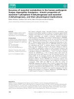
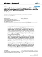
Dystrophin revertant fibres and transcripts in mdx mouse muscleFigure 1
Dystrophin revertant fibres and transcripts in mdx mouse muscle. (A) A cluster of revertant fibres surrounded by a
few single positive fibres in the mdx mouse. Tissue was immunostained for dystrophin using NCL-Dys 2. (B-D)Nested RT-PCR
was carried out using Primer set 3 inner (Table 1) to study alternative splicing arrangements between exons 13–35. The bar (-
) indicates the novel junctions in these transcripts. Unmatched boundary colours identify out-of-frame transcripts. The identity
of all transcripts was confirmed by direct sequencing. (B)-Normal dystrophin, (C)-out-of-frame dystrophin transcripts and (D)-
in-frame dystrophin transcripts. *Previously reported revertant transcripts *[26] and **[15].
1212
1313 1414
1515 1616 1717
1818
2323 2424 2525
2626
19
19
2020
2121
2222
2727 2828 2929 3030 3131 3232 3333 3434
1212
1313
3535
3030 3131 3232 3333 3434 3535
1212
1313
1212
1313 1414
1515 1616 1717
1818
2424 2525
2626
2727 2828 2929 3030 3131 3232 3333 3434 3535
1212
1313 1414
1515 1616 1717
1818
2626
2727 2828 2929 3030 3131 3232 3333 3434 3535
1212
1313 1414
1515 1616 1717
1818
3030 3131 3232 3333 3434 3535
1212
1313 1414
1515 1616 1717
1818
3434 3535
1212
1313 1414
1515 1616 1717
1818
3535
1212
1313 1414
1515 1616 1717
1818
2626
19
19
2020 2727 2828 2929 3030 3131 3232 3333 3434 3535
1212
1313 1414
1515 1616 1717
1818
19
19
2020 3030 3131 3232 3333 3434 3535
1212
1313 1414
1515 1616 1717
1818
19
19
2020
2121
22
1212
1313 1414
1515 1616 1717
1818
19
19
3030 3131 3232 3333 3434 3535
1212
1313 1414
1515 1616 1717
1818
19
19
2020
2121
3030 3131 3232 3333 3434
3535
1212
1313 1414
1515 1616 1717
1818
19
19
2020
2121
3535
Naturally occurring out-of-frame dystrophin transcripts
Naturally occurring in-frame dystrophin transcripts
Normal dystrophin transcript
A
3535
3535
**
*
*
*
*
*
10x
D
B
C
Genetic Vaccines and Therapy 2006, 4:3 />Page 5 of 12
(page number not for citation purposes)
mdx muscle extract was mixed with 10 µl of C57 protein
to allow normal dystrophin detection in the presence of
protein from the mdx mouse.
Results
Naturally occurring revertant fibre transcripts
The occurrence of revertant fibres is inconsistent and rare
and occurs in dystrophic tissue as either single fibres or
small clusters of fibres, observed after immunohistochem-
ical analysis of untreated mdx muscle sections (Figure 1A).
The diameter of muscle fibres in mdx skeletal muscle is
less uniform than those in normal (C57BLl/10ScSn) mus-
cle (data not shown). Figure 1B–D indicates the exonic
combinations representing alternatively processed dys-
trophin transcripts detected in mdx and normal mouse
muscle after RT-PCR amplification of exons 13–35 (Table
1 primer set 3). Over 100 in vitro and in vivo samples were
subjected to this RT-PCR assay, with only thirteen differ-
ent alternatively spliced transcripts identified, and all but
3 representing in-frame mRNAs. Six of the thirteen tran-
scripts found, indicated by an asterisk in Figure 1A, have
been reported previously [15,26].
Development of 2OMe AOs to induce skipping of exons
19–25
Despite targeting the obvious donor and acceptor splice
sites of individual exons, consistent induction of exon
exclusion was not guaranteed. Intra-exonic splicing
enhancer (ESE) motifs were targeted and some of these
were found to be amenable to redirection of splicing. The
likelihood of successful exon skipping after targeting any
particular ESE region increased when more than one ser-
ine/arginine-rich (SR) binding site was covered by the AO.
ESE finder Release 2.0 [27] was used to predict potential
binding sites. ESE finder is a human based program, and
since it is recognised that there are splicing differences
between the human and mouse, the program was used
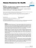
Induction of multiple exon skipping comparing two different AO cocktailsFigure 3
Induction of multiple exon skipping comparing two different AO cocktails. The cocktail used in (A) contains
M23D(+02–18) and (B) contains M23D(+07–18), other AOs indicated in Table 2. Primer set 2 inner (Table 1) was used for
nested PCR amplification where the intact product was 1357 bp long. Both (A) and (B) show the presence of generated multi-
ple bands when the AO cocktail was used in vitro. Products were sequenced and transcripts identified. The major induced tran-
script of 217 bp, corresponds to the deletion of exons 19–25.
A
B
217 bp
100 bp
600 nM
500 nM
400 nM
300 nM
200 nM
UT
-ve
100 bp
100 bp
600 nM
500 nM
400 nM
300 nM
200 nM
UT
-ve
100 bp
969 bp
1357 bp
373 bp
1818
2525
2626
1818
2323 2525
2626
21
21
1818
2626
1818
2323 2424 2525
2626
19
19
2020
2121
2222
1818
2323
2626
21
21
611 bp
767 bp
1818
2323 2424 2525
2626
19
19 2121
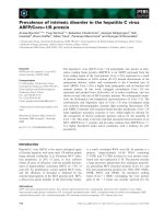
Exon skipping in cultured mdx cells after transfection with AOs directed at targeted exonsFigure 2
Exon skipping in cultured mdx cells after transfection
with AOs directed at targeted exons. Primer set 2 inner
(Table 1) was used for nested PCR amplification to generate
the full length product of 1357 bp (indicated). Lane numbers
correspond to targeted exons. The induced exon skipping
products are 1263 bp (∆ exon 19), 1115 bp (∆ exon 20),
1176 bp (∆ exon 21), 1211 bp (∆ exon 22), 1144 bp (∆ exon
23 A06), 1243 bp (∆ exon 24), 1201 bp (∆ exon 25), (∆
exon19), 1144 bp (∆ exon 23 AO10, Table 2). The 998 bp
product corresponds to the removal of exons 22 and 23, a
common product of exon 23 targeting.
1357bp
100 bp
19
20
21
22
23
24
25
23
Lipo
UT
-ve
100 bp
Exons
Genetic Vaccines and Therapy 2006, 4:3 />Page 6 of 12
(page number not for citation purposes)
only as a guide. Several AOs were evaluated individually
for each exon before selecting the compounds listed in
Table 2. All AOs listed in Table 2 induced skipping of the
targeted single exon to varying degrees (Figure 2). Exons
20 and 24 were more difficult to remove from the mature
mRNA than others, and consistent removal was not
achieved with any single AO (data not shown). However,
when two apparently ineffective AOs were used in combi-
nation, strong and consistent exon 20 and 24 skipping
occurred (Figure 2).
AO refinement and optimisation for multiple exon skip-
ping was clearly influenced by the composition of the
individual components. Two different AOs targeting exon
23 were evaluated during the optimisation of the 19–25
cocktail. Individually, both AOs induced similarly high
levels of exon 23 skipping (Figure 2) but when combined
in cocktails, different efficiencies were consistently
observed (Figure 3). The inclusion of the 20 mer
M23D(+2–18) in the cocktail, did not result in reproduc-
ible multiple exon removal (Figure 3a), whereas the inclu-
sion of the 25 mer, M23D(+7–18) in the AO mix, induced
consistent skipping over a range of concentrations (Figure
3b). This pattern of exon removal resulting from transfec-
tion of the two different AO cocktails was highly repro-
ducible and the AO cocktail containing M23D(+07–18)
was used for subsequent studies.
Evaluation of the 2OMe 19–25 cocktail
Amplification across exons 18 to 26 generated a full
length product of 1357 bp that was visible only in the
treated samples at AO cocktail transfection concentrations
of 5 and 10 nM and in the untreated control (Figure 4). In
the majority of the treated samples the full length ampli-
con was missing. Products representing transcripts miss-
ing combinations of other exons were observed and their
identity was determined by DNA sequencing (Figure 4).
Titration studies of the 2OMe 19–25 AO cocktail showed
consistent induced exon skipping after transfection with a
total AO concentration of 200 nM, (approximately 20nM
of each AO) (Figure 4a). However, different mdx myoblast
cultures (both H2K-mdx and primary mdx myoblasts)
demonstrated variable responsiveness to the AO cocktail.
Cell densities were kept consistent but exon 19–25 skip-
ping could be induced at lower transfection concentra-
tions in some experiments where revertant transcripts
were detected in untreated cells (Figure 4b). Generally,
when naturally occurring revertant transcripts were
detected in the untreated samples, inducible exon 19–25
skipping was observed after application of an AO cocktail,
at concentrations lower than 200 nM. This trend was com-
mon to both conditionally immortalised cells and pri-
mary mdx cells.
The duration of exon skipping after a single in vitro AO
delivery was assessed. The 2OMe AO cocktail targeting
exons 19–25 induced sustained and strong skipping up to
5 days after transfection. However, significant cell death
was caused by the transfection reagent (Lipofectin) and
results were not consistent after the five day time point
(data not shown).
RT-PCR of shortened transcripts induced in the presence of background alternative splicingFigure 4
RT-PCR of shortened transcripts induced in the presence of background alternative splicing. RT-PCR pattern of
exon 19–25 skipping induced in an immortalized culture where there was (A) no evidence of revertant transcripts in the
untreated samples, (B) low levels of endogenous alternative splicing visible in the untreated cells. Primer set 2 inner (Table 1)
was used for amplification.
A
B
305 bp
217 bp
554 bp
767 bp
1357 bp
373 bp
100 bp
600 nM
500 nM
400 nM
300 nM
200 nM
100 nM
50 nM
25 nM
10 nM
5 nM
Lipo
UT
-ve
100 bp
100 bp
600 nM
500 nM
400 nM
300 nM
200 nM
100 nM
50 nM
25 nM
10 nM
5 nM
Lipo
UT
-ve
100 bp
1818
2525
2626
1818
2323 2525
2626
21
21
1818
2525
2626
21
21
1818
2626
1818
2323 2424 2525
2626
19
19
2020
2121
2222
1818
2626
19
19
Genetic Vaccines and Therapy 2006, 4:3 />Page 7 of 12
(page number not for citation purposes)
The AO sequences used in the 2OMe cocktail were re-syn-
thesized as PMO compounds to compare the effectiveness
of these chemistries. The ratios of individual compounds
were the same for both chemistries, however the PMO
cocktail was transfected at substantially higher concentra-
tion because of the poor uptake of these uncharged com-
pounds in vitro. The RT-PCR product representing exon
skipping was detectable at low levels (after 7 days) at
transfection concentrations above 5 µM (data not
shown). Subsequent intramuscular injections of the AO
cocktail in the mdx mouse were conducted using only the
PMO chemistry as we recently reported that PMOs have
substantial advantages over the 2OMe chemistry in vivo
[23].
In vivo studies
Total RNA, extracted from mdx mouse muscle sections (2–
3 mg) at 2, 4 and 8 weeks after a single intramuscular
injection of 2 or 10 µg of the PMO cocktail, was analysed
by nested RT-PCR amplification across dystrophin exons
18–26 (Table 1 primer set 1). Amplification products rep-
resenting the shortened transcript missing exons 19–25
were observed in all samples from muscles injected with
10 µg of the PMO cocktail. Muscle from mice injected at
11 days or 16 weeks of age contained the shortened tran-
script at 2 and 4 weeks after a single injection (Figure 5a).
A more efficient set of inner primers was used to amplify
a full-length transcript of 1250 bp, with the induced tran-
script represented by a 110 bp product. The expected 110
bp product was not detected in any muscle injected with
only the 2 µg dose even though tissue sections had stained
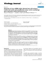
positive for dystrophin (data not shown). Immunohisto-
chemical staining with NCL-Dys2 confirmed that the 10
µg injection of the 19–25 PMO cocktail induced wide-
spread dystrophin expression at 2, 4 and 8 weeks after
injection in the pups and adult mdx mice (Figure 5b). Dys-
trophin expression appeared maximal at 4 weeks post
treatment. Dystrophin staining was localized along the
needle track in the sections from the pups, whereas the
dystrophin staining in the older mdx mice was patchy and
more widespread. Dystrophin immunostaining did not
appear to differ substantially with the age of the animal.
Immunofluorescent staining patterns are similar to those
reported with the removal of the single exon 23 [23]. RT-
PCR results (Figure 5a) showed the removal of multiple
exons with only minor products representing trace
amounts of alternatively processed transcripts.
Consistent with the observation that the19–25 transcript
was the major induced dystrophin mRNA, western blot-
ting of extracts from treated muscle demonstrated a faint
band of induced protein in the samples from pups and
adult mice 4 weeks after treatment (Figure 5c). The dys-
trophin detected in muscles injected with the 19–25 cock-
tail appeared to be of a lower molecular weight than the
C57BL/10ScSn and the exon 23 deleted products. To con-
firm that the apparent difference in molecular weight was
not an artefact of protein loading, 15% normal muscle
protein extract was mixed with 85% untreated mdx muscle
protein in the loading buffer. Levels of protein were too
low to be detected at 2 or 8 weeks, even though dys-
trophin was observed by immunofluorescent staining of
muscle sections.
Induced revertant fibres
To determine if other multiple skipping events occurred as
a consequence of treatment with AO cocktails, long range
RT-PCR across exons 13–50 was performed on untreated
and treated samples from both pups and adult mdx mice
at the 2 and 4 week time points (Figure 6). The frequency
of revertant transcripts appeared to be higher in PMO
cocktail-treated samples than in sham-injected muscle.
Only one of the 4 untreated samples contained a naturally
occurring in-frame revertant transcript missing exons 20–
49, whereas all eight of the treated samples contained
additional shorter transcripts. The transcript skipping
exons 20–49 was also found in one of the PMO cocktail
treated samples, with most of the induced shortened tran-
scripts found to be in-frame (Figure 6). Due to either its
size, the quality of the RNA, or the efficiency of the cDNA
synthesis and amplification, the full length product of
5720 bp was not generated by this assay and the reaction
was biased towards the amplification of shorter alterna-
tively processed products.
Discussion
A study of BMD dystrophin gene rearrangements that
result in an altered but partially functional protein, readily
identifies dispensable domains within the dystrophin
protein [28]. There are many cases of asymptomatic BMD,
where patients have only been diagnosed late in life. Eng-
land et al [29] reported a BMD case identified at 60 years
of age, with a deletion of exons 17–48 that encompassed
46% of the gene. The reading frame rule proposed by
Monaco et al [4,5] holds true for over 90% of dystrophin
mutations. However, there are exceptions and it has been
reported that in-frame deletions exceeding 36 exons are
generally associated with a severe clinical phenotype [30-
32]. Nevertheless, other reports of milder, though variable
phenotypes with large in-frame deletions, involve the loss
of up to 66% of the dystrophin gene [33]. It would appear
that most exons encoding the rod domain may be deleted
without substantial loss of function. The loss of a single
exon that either codes for a crucial binding domain or dis-
rupts the reading frame will have catastrophic conse-
quences on dystrophin function [29,33,34]. AOs must be
designed and optimised to remove the exon containing
the mutation, and/or surrounding exons, to restore or
maintain the reading frame. While DMD and most BMD
patients lack full-length dystrophin, expression of shorter
Genetic Vaccines and Therapy 2006, 4:3 />Page 8 of 12
(page number not for citation purposes)
In vivo skipping of exons 19–25 induced with a PMO cocktailFigure 5
In vivo skipping of exons 19–25 induced with a PMO cocktail. As indicated by the 110 bp product, the PMO cocktail
induced the removal of exons 19–25 at 2 and 4 weeks after a single 10 µg intramuscular injection in pups and adult mdx mice
(A). Primer set 1 inner (Table 1) was used for nested PCR amplification. (B) Dystrophin expression 2, 4 and 8 weeks after
intramuscular injection of PMO cocktail in both adults and pups. (C) Western blot analysis was performed one month after
injection. A faint protein band of lower than normal dystrophin molecular weight is visible in lanes 6 and 7.
Genetic Vaccines and Therapy 2006, 4:3 />Page 9 of 12
(page number not for citation purposes)
dystrophin isoforms (Dp260, Dp140, Dp116 and Dp71)
occurs in different muscle and non-muscle tissues, with
inter-patient variation depending on the position of the
primary lesion in the dystrophin gene [35].
The aim of multiple exon skipping was to induce a previ-
ously identified, naturally occurring revertant dystrophin
transcript [26]. Revertant fibres arise from spontaneous
exon skipping events that occur at low levels in both nor-
mal and dystrophic muscle [17]. We focussed on identify-
ing revertant fibre transcripts between exons 13 and 35
(~29% of the dystrophin gene) in the mdx mouse model.
It has been proposed that the dystrophin in revertant
fibres arises from alternatively spliced transcripts that lack
both the mutant exon and a variable number of adjacent
exons [15,36]. Splicing is a very complex process with
many cis and trans elements contributing to the selection
of, and efficiency with which splice sites are recognised
and exons are joined during pre-mRNA processing. The
strength of the 3' and 5' splice sites, branch point
sequences, exonic splicing enhancers and silencers, exon
length, and secondary structure all play a role in pre-
mRNA processing [37-40]. Alternative splicing has now
been recognised as a major mechanism for generating
protein diversity in higher eukaryotes [41]. Alternative
splicing does occur naturally in the dystrophin gene tran-
script, but the role of many isoforms has yet to be eluci-
dated [42].
Amplification of dystrophin exons 13–50, RNA extracted from treated and untreated mdx muscleFigure 6
Amplification of dystrophin exons 13–50, RNA extracted from treated and untreated mdx muscle. Primer set 4
inner (Table 1) was used for nested PCR amplification. The identity of the alternatively spliced products is shown with the
reading frame indicated. Lanes 3, 4, 7 and 9 correspond to the samples used in Figure 5a lanes 2–5.
Treated Pups
Control
1 2 3 4 5 6 7 8 9 10 11 12 13 14
2 weeks 2 weeks
4 weeks
4 weeks
Treated Adults
2 weeks
4 weeks
1515
4545
1313
4444
1818
4949
20
20
4949
1313
4545
1515
4949
20
49
1094 bp
914 bp
1032 bp
1040 bp
884 bp
434 bp
1 100 bp
2 2231 bp
3
4
5
6
7
8 splicing artefact
9
10 No skipping
11 No skipping
12
13 -ve
14 100 bp
Legend
1040 bp
1000 bp
2222 4545
Genetic Vaccines and Therapy 2006, 4:3 />Page 10 of 12
(page number not for citation purposes)
It appears that revertant fibres arise from some alternative
splicing mechanism, as evidenced by the presence of the
shorter dystrophin isoforms, however the low frequency
would suggest some error in splicing, where a localised
event within fibres rescues dystrophin expression.
Although too few in number to be of any therapeutic ben-
efit, the presence and persistence of these dystrophin pos-
itive fibres implies that they do not elicit an immune
response.
The concept of a localised change in the general splicing
machinery or an altered ratio of SR proteins leading to
alternative splicing to by-pass the dystrophin gene lesion
seems unlikely. It is tempting to speculate that perhaps
some novel microRNA is expressed in the revertant fibres,
leading to natural exon skipping in a single cell, with clus-
ters of dystrophin positive fibres suggesting a clonal ori-
gin. MicroRNAs have been implicated in a variety of cell
processes, including apoptosis, translation andsplicing
[43,44]. Regardless of the mechanism used to induce
revertant dystrophin, it is possible that the addition of
AOs is somehow enhancing the production or action of
some microRNAs.
Substantial optimisation was performed in assembling
the AO cocktail, with a number of AOs evaluated before
selecting the combination shown in Table 2. Although
there was no common pre-mRNA motif that could be tar-
geted to induce reliable and sustained exon skipping, in
general longer AOs were found to be more effective [45].
Mouse dystrophin exon 19 has been studied previously
and may be regarded as an easy exon to remove from the
dystrophin mRNA [46]. Ten AOs directed at the acceptor,
ESE and donor splice sites all induced exon 19 skipping.
In contrast exons 20 and 24 proved much harder to dis-
place from the mature mRNA. Five AOs were directed at
exon 20, and six at exon 24. Individually, these AOs
proved ineffective at inducing skipping of the target exon
but the combinations described here were most effective.
One could speculate that this indicates some exons have
multiple motifs necessary for exon recognition by the
splicing machinery and more than one target must be
masked to redirect splicing. In these cases it may be neces-
sary to apply multiple AOs to target a single exon for
mRNA excision.
The AOs evaluated for exon 23 removal proved to be cru-
cial to the 19–25 cocktail. Individually both AOs,
M23D(+02–18) and M23D(+07–18), removed exon 23
but the 25mer was the more effective of the two AOs. It
was noted that a single AO could substantially influence
the efficiency of the AO cocktail with respect to multiple
exon skipping. In order to induce exon 19–25 skipping
with the AO cocktail containing M23D(+02–18), it was
necessary to adjust the ratios of each AO in the mixture
(data not shown). However, if equal molar amounts of all
AOs were used then the M23D(+02–18) cocktail did not
reliably induce exon 19–25 skipping. Upon inclusion of
M23D(+07–18) in the mixture, consistent and reliable
multiple exon skipping was induced (Figure 3). Further
optimisation of all other AOs in the mixture could be
undertaken, but since consistent generation of the desired
transcript was achieved, it was decided to undertake sub-
sequent in vitro and in vivo experiments with the cocktail
containing M23D(+07–18).
Administration of the AO cocktail containing
M23D(+07–18) appeared to increase the incidence of
dystrophin revertant transcripts in mdx myogenic cells and
tissue. RNA extracted from treated and untreated muscle
was subjected to RT-PCR across exons 13–50. Each treated
sample had one or more alternatively processed dys-
trophin gene transcripts, whereas shorter products were
rare in untreated samples.
The subtle variations in protein band migration observed
on the western blot (Figure 5c) indicates the size differ-
ences between dystrophin of normal length (427 kD),
exon 23 deleted (420 kD) and the removal of exons 19–
25 (389 kD). The concept of multiple transcripts is con-
sistent with observation of "fuzzy" bands on western blots
of treated muscle that were fractionated specifically to
enhance resolution and size differentiation. Multiple
exon skipping events could occur at many positions in the
mdx dystrophin gene transcript and still by-pass the non-
sense mutation.
We have recently shown that PMOs are more effective
than 2OMe AOs at inducing exon 23 skipping in the dys-
trophin gene transcript [23]. PMOs exhibit very low toxic-
ity in treated cells [47] and have been reported to have
minimal non-antisense effects [48]. Intramuscular injec-
tions of PMOs produced no obvious adverse reactions at
or around the injection site (data not shown). RT-PCR,
immunohistochemistry and western blot all confirmed
that induced skipping of exons 19–25 was far more effi-
cient in vivo with PMOs than with 2OMe AOs (data not
shown).
Dystrophin was still detectable 8 weeks after a single intra-
muscular injection of the PMO cocktail into 11 day old
pups and 16 week old adult mice, although differences in
immunostaining patterns were apparent. The dystrophin
staining pattern in the pups appeared strongly localised at
the injection site and consistently positive, whereas the
pattern in adult mice was more patchy and widespread.
This may have been due to the amount of degenera-
tion:regeneration that had occurred in the adult mdx
mouse, and to the fact that the pups were injected prior to
Genetic Vaccines and Therapy 2006, 4:3 />Page 11 of 12
(page number not for citation purposes)
the extensive necrosis that occurs at around 18 days of age
[49].
In some cases, the removal of a single exon would not be
sufficient to address the disease-causing mutation and a
cocktail of two or more AOs would be required to restore
the reading frame. For example, the intron 6 splice site
mutation in the GRMD canine model of DMD leads to the
skipping of exon 7, with a subsequent frame-shift in the
dystrophin mRNA [50]. The minimum change to restore
the reading frame requires the removal of exons 6 and 8
and was recently reported by McClorey et al [51]. Simi-
larly, any nonsense mutation in exons 6, 7 or 8 of the
human dystrophin gene would require removal of all 3
exons to by-pass the mutation and still maintain the read-
ing frame. For these reasons, multiple exon skipping and
the application of AO cocktails will be an absolute
requirement to address some DMD mutations
Conclusion
The removal of exons 19–25 in the mdx mouse provides
evidence that multiple exon skipping is feasible and that
clusters of mutations in the dystrophin gene could be cor-
rected with a cocktail of AOs. Once a comprehensive set of
AOs are designed these could theoretically benefit >75%
of all DMD patients. One of the major limitations is to
gain regulatory approval for the clinical use of so many
different compounds [52,53]. The cost of safety and toxi-
cology testing alone could render exon-skipping a non-
viable approach for all amenable dystrophin mutations,
in particular, those defects occurring outside the recog-
nised deletion hot-spots. AO cocktails to induce multiple
exon skipping could significantly lower the number of
preparations required to address clustered dystrophin
mutations in different families [53].
Competing interests
The author(s) declare that they have no competing inter-
ests.
Authors' contributions
AF carried out the molecular genetic studies, immunohis-
tochemistry, participated in its design and coordination
and helped to draft the study, RJ carried out the Western
Blots, KH helped to draft the study, PI designed and sup-
plied the PMOs, SF and SW conceived the study, partici-
pated in its design and coordination and manuscript
preparation. All authors read and approved the final man-
uscript.
Acknowledgements
The authors would like to acknowledge funding from National Medical &
Health Research Council of Australia (303216), National Institute of Health
USA (RO1 NS044146–02), Muscular Dystrophy Association of USA
(MDA3718), Parent Project Muscular Dystrophy USA, Medical and Health
Research Infrastructure Fund of Western Australia and Aktion Benni and
Co.
References
1. Ahn AH, Kunkel LM: The structural and functional diversity of
dystrophin. Nat Genet 1993, 3:283-291.
2. Love DR, Byth BC, Tinsley JM, Blake DJ, Davies KE: Dystrophin and
dystrophin-related proteins: a review of protein and RNA
studies. Neuromuscul Disord 1993, 3:5-21.
3. Hoffman EP, Brown RHJ, Kunkel LM: Dystrophin: the protein
product of the Duchenne muscular dystrophy locus. Cell
1987, 51:919-928.
4. Monaco AP, Bertelson CJ, Liechti-Gallati S, Moser H, Kunkel LM: An
explanation for the phenotypic differences between patients
bearing partial deletions of the DMD locus. Genomics 1988,
2:90-95.
5. Collins CA, Morgan JE: Duchenne's muscular dystrophy: animal
models used to investigate pathogenesis and develop thera-
peutic strategies. Int J Exp Pathol 2003, 84:165-172.
6. Hoffman EP, Morgan JE, Watkins SC, Partridge TA: Somatic rever-
sion/suppression of the mouse mdx phenotype in vivo. J Neu-
rol Sci 1990, 99:9-25.
7. Sherratt TG, Vulliamy T, Dubowitz V, Sewry CA, Strong PN: Exon
skipping and translation in patients with frameshift deletions
in the dystrophin gene. Am J Hum Genet 1993, 53:1007-1015.
8. Klein CJ, Coovert DD, Bulman DE, Ray PN, Mendell JR, Burghes AH:
Somatic reversion/suppression in Duchenne muscular dys-
trophy (DMD): evidence supporting a frame-restoring mech-
anism in rare dystrophin-positive fibers. Am J Hum Genet 1992,
50:950-959.
9. Fanin M, Danieli GA, Cadaldini M, Miorin M, Vitiello L, Angelini C:
Dystrophin-positive fibers in Duchenne dystrophy: origin
and correlation to clinical course. Muscle Nerve 1995,
18:1115-1120.
10. Burrow KL, Coovert DD, Klein CJ, Bulman DE, Kissel JT, Rammohan
KW, Burghes AH, Mendell JR: Dystrophin expression and
somatic reversion in prednisone-treated and untreated
Duchenne dystrophy. CIDD Study Group. Neurology 1991,
41:661-666.
11. Uchino M, Tokunaga M, Mita S, Uyama E, Ando Y, Teramoto H, Miike
T, Ando M: PCR and immunocytochemical analyses of dys-
trophin-positive fibers in Duchenne muscular dystrophy. J
Neurol Sci 1995, 129:44-50.
12. Nicholson LV, Johnson MA, Bushby KM, Gardner-Medwin D, Curtis
A, Ginjaar IB, den Dunnen JT, Welch JL, Butler TJ, Bakker E: Inte-
grated study of 100 patients with Xp21 linked muscular dys-
trophy using clinical, genetic, immunochemical, and
histopathological data. Part 3. Differential diagnosis and
prognosis. J Med Genet 1993, 30:745-751.
13. Danko I, Chapman V, Wolff JA: The frequency of revertants in
mdx mouse genetic models for Duchenne muscular dystro-
phy. Pediatr Res 1992, 32:128-131.
14. Schatzberg SJ, Anderson LV, Wilton SD, Kornegay JN, Mann CJ, Solo-
mon GG, Sharp NJ: Alternative dystrophin gene transcripts in
golden retriever muscular dystrophy. Muscle Nerve 1998,
21:991-998.
15. Lu QL, Morris GE, Wilton SD, Ly T, Artem'yeva OV, Strong P, Par-
tridge TA: Massive idiosyncratic exon skipping corrects the
nonsense mutation in dystrophic mouse muscle and pro-
duces functional revertant fibers by clonal expansion. J Cell
Biol 2000, 148:985-996.
16. Bulfield G, Siller WG, Wight PA, Moore KJ: X chromosome-linked
muscular dystrophy (mdx) in the mouse. Proc Natl Acad Sci U S
A 1984, 81:1189-1192.
17. Wilton SD, Dye DE, Blechynden LM, Laing NG: Revertant fibres: a
possible genetic therapy for Duchenne muscular dystrophy?
Neuromuscul Disord 1997, 7:329-335.
18. Mann CJ, Honeyman K, Cheng AJ, Ly T, Lloyd F, Fletcher S, Morgan
JE, Partridge TA, Wilton SD: Antisense-induced exon skipping
and synthesis of dystrophin in the mdx mouse. Proc Natl Acad
Sci U S A 2001, 98:42-47.
19. Mann CJ, Honeyman K, McClorey G, Fletcher S, Wilton SD:
Improved antisense oligonucleotide induced exon skipping
in the mdx mouse model of muscular dystrophy. J Gene Med
2002, 4:644-654.
Publish with BioMed Central and every
scientist can read your work free of charge
"BioMed Central will be the most significant development for
disseminating the results of biomedical research in our lifetime."
Sir Paul Nurse, Cancer Research UK
Your research papers will be:
available free of charge to the entire biomedical community
peer reviewed and published immediately upon acceptance
cited in PubMed and archived on PubMed Central
yours — you keep the copyright
Submit your manuscript here:
/>BioMedcentral
Genetic Vaccines and Therapy 2006, 4:3 />Page 12 of 12
(page number not for citation purposes)
20. Wilton SD, Lloyd F, Carville K, Fletcher S, Honeyman K, Agrawal S,
Kole R: Specific removal of the nonsense mutation from the
mdx dystrophin mRNA using antisense oligonucleotides.
Neuromuscul Disord 1999, 9:330-338.
21. Lu QL, Mann CJ, Lou F, Bou-Gharios G, Morris GE, Xue SA, Fletcher
S, Partridge TA, Wilton SD: Functional amounts of dystrophin
produced by skipping the mutated exon in the mdx dys-
trophic mouse. Nat Med 2003, 9:1009-1014.
22. van Deutekom JC, Bremmer-Bout M, Janson AA, Ginjaar IB, Baas F,
den Dunnen JT, van Ommen GJ: Antisense-induced exon skip-
ping restores dystrophin expression in DMD patient derived
muscle cells. Hum Mol Genet 2001, 10:1547-1554.
23. Fletcher S, Honeyman K, Fall AM, Harding PL, Johnsen RD, Wilton
SD: Dystrophin expression in the mdx mouse after localised
and systemic administration of a morpholino antisense oli-
gonucleotide. J Gene Med 2006, 8:207-216.
24. Morgan JE, Beauchamp JR, Pagel CN, Peckham M, Ataliotis P, Jat PS,
Noble MD, Farmer K, Partridge TA: Myogenic cell lines derived
from transgenic mice carrying a thermolabile T antigen: a
model system for the derivation of tissue-specific and muta-
tion-specific cell lines. Dev Biol 1994, 162:486-498.
25. Wilton SD, Lim L, Dye D, Laing N: Bandstab: a PCR-based alter-
native to cloning PCR products. Biotechniques 1997, 22:642-645.
26. Wilton SD, Dye DE, Laing NG: Dystrophin gene transcripts skip-
ping the mdx mutation. Muscle Nerve 1997, 20:728-734.
27. Cartegni L, Wang J, Zhu Z, Zhang MQ, Krainer AR: ESEfinder: a
web resource to identify exonic splicing enhancers. Nucleic
Acid Research 2003, Vol. 31:3568-3571.
28. Beggs AH, Hoffman EP, Snyder JR, Arahata K, Specht L, Shapiro F,
Angelini C, Sugita H, Kunkel LM: Exploring the molecular basis
for variability among patients with Becker muscular dystro-
phy: dystrophin gene and protein studies. Am J Hum Genet
1991, 49:54-67.
29. England SB, Nicholson LV, Johnson MA, Forrest SM, Love DR,
Zubrzycka-Gaarn EE, Bulman DE, Harris JB, Davies KE: Very mild
muscular dystrophy associated with the deletion of 46% of
dystrophin. Nature 1990, 343:180-182.
30. Takeshima Y, Nishio H, Narita N, Wada H, Ishikawa Y, Minami R,
Nakamura H, Matsuo M: Amino-terminal deletion of 53% of dys-
trophin results in an intermediate Duchenne-Becker muscu-
lar dystrophy phenotype. Neurology 1994, 44:1648-1651.
31. Winnard AV, Klein CJ, Coovert DD, Prior T, Papp A, Snyder P, Bul-
man DE, Ray PN, McAndrew P, King W, Burghes AH: Characteri-
zation of translational frame exception patients in
Duchenne/Becker muscular dystrophy. Hum Mol Genet 1993,
2:737-744.
32. Fanin M, Freda MP, Vitiello L, Danieli GA, Pegoraro E, Angelini C:
Duchenne phenotype with in-frame deletion removing
major portion of dystrophin rod: threshold effect for dele-
tion size? Muscle Nerve 1996, 19:1154-1160.
33. Passos-Bueno MR, Vainzof M, Marie SK, Zatz M: Half the dys-
trophin gene is apparently enough for a mild clinical course:
confirmation of its potential use for gene therapy. Hum Mol
Genet 1994, 3:919-922.
34. Heald A, Anderson LV, Bushby KM, Shaw PJ: Becker muscular dys-
trophy with onset after 60 years. Neurology 1994, 44:2388-2390.
35. Tokarz SA, Duncan NM, Rash SM, Sadeghi A, Dewan AK, Pillers DA:
Redefinition of dystrophin isoform distribution in mouse tis-
sue by RT-PCR implies role in nonmuscle manifestations of
duchenne muscular dystrophy. Mol Genet Metab 1998,
65:272-281.
36. Thanh LT, Nguyen TM, Helliwell TR, Morris GE: Characterization
of revertant muscle fibers in Duchenne muscular dystrophy,
using exon-specific monoclonal antibodies against dys-
trophin. Am J Hum Genet 1995, 56:725-731.
37. Lopez AJ: Alternative splicing of pre-mRNA: developmental
consequences and mechanisms of regulation. Annu Rev Genet
1998, 32:279-305.
38. Shapiro MB, Senapathy P: RNA splice junctions of different
classes of eukaryotes: sequence statistics and functional
implications in gene expression. Nucleic Acids Res 1987,
15:7155-7174.
39. Dominski Z, Kole R: Selection of splice sites in pre-mRNAs
with short internal exons. Mol Cell Biol 1991, 11:6075-6083.
40. Balvay L, Libri D, Fiszman MY: Pre-mRNA secondary structure
and the regulation of splicing. Bioessays 1993, 15:165-169.
41. Neverov AD, Artamonova II, Nurtdinov RN, Frishman D, Gelfand
MS, Mironov AA: Alternative splicing and protein function.
BMC Bioinformatics 2005, 6:266.
42. Sironi M, Cagliani R, Pozzoli U, Bardoni A, Comi GP, Giorda R, Bre-
solin N: The dystrophin gene is alternatively spliced through-
out its coding sequence. FEBS Lett 2002, 517:163-166.
43. Hastings ML, Milcarek C, Martincic K, Peterson ML, Munroe SH:
Expression of the thyroid hormone receptor gene, erbAal-
pha, in B lymphocytes: alternative mRNA processing is inde-
pendent of differentiation but correlates with antisense RNA
levels. Nucleic Acids Res 1997, 25:4296-4300.
44. Cheng AM, Byrom MW, Shelton J, Ford LP: Antisense inhibition of
human miRNAs and indications for an involvement of
miRNA in cell growth and apoptosis. Nucleic Acids Res 2005,
33:1290-1297.
45. Harding PL, Fall AM, Honeyman K, Fletcher S, Wilton S: AO design
for splicing blockade of dystrophin pre-mRNA processing:
size does matter. Molecular Therapy 2006, Accepted:.
46. Errington SJ, Mann CJ, Fletcher S, Wilton SD: Target selection for
antisense oligonucleotide induced exon skipping in the dys-
trophin gene. J Gene Med 2003, 5:518-527.
47. Iversen PL: Phosphorodiamidate morpholino oligomers: favo-
rable properties for sequence-specific gene inactivation. Curr
Opin Mol Ther 2001, 3:235-238.
48. Summerton J: Morpholino antisense oligomers: the case for an
RNase H-independent structural type. Biochim Biophys Acta
1999, 1489:141-158.
49. Muntoni F, Mateddu A, Marchei F, Clerk A, Serra G: Muscular
weakness in the mdx mouse. J Neurol Sci 1993, 120:71-77.
50. Sharp NJ, Kornegay JN, Van Camp SD, Herbstreith MH, Secore SL,
Kettle S, Hung WY, Constantinou CD, Dykstra MJ, Roses AD, Bar-
tlett RJ: An error in dystrophin mRNA processing in golden
retriever muscular dystrophy, an animal homologue of
Duchenne muscular dystrophy. Genomics 1992, 13:115-121.
51. McClorey G, Moulton HM, Iversen PL, and SF, Wilton SD: Antisense
oligonucleotide induced exon skipping restores dystrophin
expression in a canine model of DMD. Gene Therapy 2006,
Accepted:.
52. van Deutekom JC, van Ommen GJ: Advances in Duchenne mus-
cular dystrophy gene therapy. Nat Rev Genet 2003, 4:774-783.
53. Aartsma-Rus A, Janson AA, Kaman WE, Bremmer-Bout M, van
Ommen GJ, den Dunnen JT, van Deutekom JC: Antisense-induced
multiexon skipping for Duchenne muscular dystrophy makes
more sense. Am J Hum Genet 2004, 74:83-92.