Báo cáo y học: "Mobilization of pro-inflammatory lipids in obese Plscr3-deficient mice" ppt
Bạn đang xem bản rút gọn của tài liệu. Xem và tải ngay bản đầy đủ của tài liệu tại đây (460.31 KB, 8 trang )
Genome Biology 2007, 8:R38
comment reviews reports deposited research refereed research interactions information
Open Access
2007Mutchet al.Volume 8, Issue 3, Article R38
Research
Mobilization of pro-inflammatory lipids in obese Plscr3-deficient
mice
David M Mutch
¤
*‡
, Grace O'Maille
¤
†
, William R Wikoff
†
,
Therese Wiedmer
*§
, Peter J Sims
*§
and Gary Siuzdak
†
Addresses:
*
The Scripps Research Institute, Department of Molecular and Experimental Medicine, North Torrey Pines Road, La Jolla, CA
92037, USA.
†
The Scripps Research Institute, Department of Molecular Biology and the Center for Mass Spectrometry, North Torrey Pines
Road, La Jolla, CA 92037, USA.
‡
Current address: INSERM U755 Nutriomique, Paris, F-75004 France; Pierre and Marie Curie - Paris 6
University, Faculty of Medicine, Les Cordeliers, 75004 Paris, France.
§
Current address: Department of Pathology and Laboratory of Medicine,
University of Rochester Medical Center, Rochester, NY 14642, USA.
¤ These authors contributed equally to this work.
Correspondence: Gary Siuzdak. Email:
© 2007 Mutch et al.; licensee BioMed Central Ltd.
This is an open access article distributed under the terms of the Creative Commons Attribution License ( which
permits unrestricted use, distribution, and reproduction in any medium, provided the original work is properly cited.
Lipid mobilization in obese mice<p>Metabolic profiling of mice deficient in phospholipid scramblase 3 reveals a possible molecular link between obesity and inflamma-tion.</p>
Abstract
Background: The obesity epidemic has prompted the search for candidate genes capable of
influencing adipose function. One such candidate, that encoding phospholipid scramblase 3
(PLSCR3), was recently identified, as genetic deletion of it led to lipid accumulation in abdominal
fat pads and changes characteristic of metabolic syndrome. Because adipose tissue is increasingly
recognized as an endocrine organ, capable of releasing small molecules that modulate disparate
physiological processes, we examined the plasma from wild-type, Plscr1-/-, Plscr3-/- and Plscr1&3-/-
mice. Using an untargeted comprehensive metabolite profiling approach coupled with targeted
gene expression analyses, the perturbed biochemistry and functional redundancy of PLSCR
proteins was assessed.
Results: Nineteen metabolites were differentially and similarly regulated in both Plscr3-/- and
Plscr1&3-/- animals, of which five were characterized from accurate mass, tandem mass
spectrometry data and their correlation to the Metlin database as lysophosphatidylcholine (LPC)
species enriched with C16:1, C18:1, C20:3, C20:5 and C22:5 fatty acids. No significant changes in
the plasma metabolome were detected upon elimination of PLSCR1, indicating that increases in
pro-inflammatory lipids are specifically associated with the obese state of Plscr3-deficient animals.
Correspondingly, increases in white adipose lipogenic gene expression confirm a role for PLSCR3
in adipose lipid metabolism.
Conclusion: The untargeted profiling of circulating metabolites suggests no detectable functional
redundancies between PLSCR proteins; however, this approach simultaneously identified
previously unrecognized lipid metabolites that suggest a novel molecular link between obesity,
inflammation and the downstream consequences associated with PLSCR3-deficiency.
Published: 13 March 2007
Genome Biology 2007, 8:R38 (doi:10.1186/gb-2007-8-3-r38)
Received: 12 January 2007
Revised: 23 January 2007
Accepted: 13 March 2007
The electronic version of this article is the complete one and can be
found online at />R38.2 Genome Biology 2007, Volume 8, Issue 3, Article R38 Mutch et al. />Genome Biology 2007, 8:R38
Background
Despite the overt recognition of the taxing effects of obesity
on both medical and social programs throughout the world,
the estimated 300 million adults currently considered clini-
cally obese in addition to the universal increase in childhood
obesity indicates we are still succumbing to this global epi-
demic. Indeed, the poorly understood gene-environment
interactions have revealed the complexity of this metabolic
disease; however, with each passing year an increasing
number of genetic candidates are discovered that help to fur-
ther unravel the perturbed metabolism underlying the obese
phenotype [1,2]. Recently, phospholipid scramblase (Plscr) 3
was identified as a genetic candidate capable of influencing
adipose function and, ultimately, the obese phenotype. Mice
deficient in PLSCR3 were found to accumulate lipid in
abdominal fat pads and were characterized with insulin
resistance, dyslipidemia, and glucose intolerance, classic tell-
tale markers for metabolic syndrome [3]. While these obser-
vations suggest a role for PLSCR3 in adipose function, much
work remains if the obese phenotype and the downstream
consequences stemming from a dysfunctional PLSCR3 are to
be understood.
PLSCR3 is one of four structurally related members (termed
PLSCR1 through 4) in the phospholipid scramblase family
[4]. The first of these plasma membrane proteins to be cloned
and characterized (PLSCR1) implicated this protein family in
the trans-bilayer migration of membrane phospholipids in
platelets, erythrocytes, and other cell types in response to an
elevation in intracellular calcium. As such, these proteins
were thought to have roles in platelet procoagulent activity,
cell injury by complement, and apoptosis. Since their initial
discovery, phospholipid scramblases are hypothesized to
have a more complex biology than previously thought. Stud-
ies aimed at defining the biological functions of Plscr1, the
most widely studied member of the phospholipid scramblase
family, have demonstrated that it is transcriptionally up-reg-
ulated by interferon-α and other factors [5,6], and that a por-
tion of the newly synthesized PLSCR1 protein can translocate
into the cell's nucleus and interact with genomic DNA, sug-
gesting it has a potential role in the regulation of gene expres-
sion [7,8].
The development of murine models deficient in PLSCR pro-
teins provides a means to elucidate the biochemistry underly-
ing Plscr3-mediated obesity. As previously observed with
members of a protein family, a degree of redundancy exists in
order for an organism to maintain physiological homeostasis
and preserve the full gamut of biological functions required
for survival [9,10]. With regards to the phospholipid scram-
blases, adipocytes accumulate neutral lipid in Plscr3-defi-
cient mice; however, Plscr1-deficient animals also have a
small increase in adipose lipid and the Plscr1&3-deficient
mice have an even greater accumulation of lipid than the
Plscr3-null mice [3]. This would suggest that, to some extent,
a redundancy in the adipose functions of these two proteins
may exist.
To begin to unravel the perturbed biochemistry associated
with Plscr3 deficiency, we employed comprehensive metabo-
lite screening technologies to determine whether the biologi-
cal abnormalities stemming from the lack of PLSCR3 protein
are reflected in plasma. As metabolites represent a metabolic
endpoint of gene and protein function, their analysis provides
insight into the cellular function of genetically modified mice
[11,12]. As such, untargeted metabolomics offers a powerful
method to further define the obese phenotype in organisms
characterized by genetic modifications [13,14]. Furthermore,
an additional inherent advantage of untargeted metabo-
lomics (which can also be extended to alternative functional
genomic strategies) versus a more targeted analysis (that is,
the lipidome [15]) is its ability to generate novel hypotheses
through the identification of previously unrecognized signal-
ing pathways [2]. As demonstrated in the present manuscript,
the characterized metabolites identified in animals lacking
Plscr3 suggest a novel molecular link between the chronic
low-level inflammation characteristic of an obese state and
the heightened downstream risk of cardiovascular disease.
Results and discussion
Plasma from male mice of each genotype (n = 4) were
obtained, separated into two groups of two, and analyzed in
triplicate at two separate run dates. As such, only metabolites
that were consistently identified in all four animals of a spe-
cific genotype, irrespective of date of analysis, were consid-
ered 'true' peaks. Using XCMS software, we were able to
confidently examine the data generated in the multiple anal-
yses by first performing nonlinear alignment compensating
for minor differences in ion retention times between the runs,
and then identifying and matching peaks for further analysis
(Figure 1). In this regard, correction of retention times per-
mits the relative metabolite ion intensities to be statistically
compared between the various genotypes in order to identify
unique and/or shared ions associated with deficiencies in
PLSCR proteins. For this to be accomplished with confidence,
a number of criteria were used prior to considering an ion as
significantly different between the various phenotypes. Ini-
tially, an intensity threshold was selected to ensure that ions
could be subsequently enriched and analyzed for their accu-
rate mass by electrospray ionization coupled with time of
flight analysis (ESI-TOF). Subsequent to this analysis, ions
were only deemed significantly different between the various
genotypes if they had a p value ≤ 0.01. Furthermore, only
those metabolites simultaneously identified in both a single
knock-out model and the double knock-out model (that is,
KO1 and DKO, or KO3 and DKO) were considered as 'candi-
date metabolites'. This approach meant that all metabolites
discussed in this manuscript were identified in eight mice
(each in triplicate liquid chromatography (LC) runs), thereby
strengthening the biological relevance and statistical
Genome Biology 2007, Volume 8, Issue 3, Article R38 Mutch et al. R38.3
comment reviews reports refereed researchdeposited research interactions information
Genome Biology 2007, 8:R38
significance of our results. Finally, data generated on the indi-
vidual ions were manually inspected to validate the changes
and identify the most important molecules for future
isolation.
All possible comparisons were performed, with the goal of
identifying metabolites both unique to a specific genotype
and shared between two or more genotypes. XCMS detected
over 10,000 peaks across the 48 LC/mass spectrometry (MS)
runs performed (corresponding to 16 mice, each run in tripli-
cate; see Additional data file 1 for all raw data); however, to
minimize spurious results, we considered only those peaks
that were consistently identified in a genotype (that is, found
in 12 LC/MS runs). The comparison between wild type (WT)
and the three knock-out models did not identify common
metabolites amongst the various mutant models, suggesting
that Plscr1 and Plscr3 affect the plasma metabolome differ-
ently. Pair-wise comparisons (WT and KO1 versus KO3 and
DKO, WT and KO3 versus KO1 and DKO, and WT and DKO
versus KO1 and KO3) revealed that eliminating Plscr3 gave
rise to an increase in the abundance of metabolites common
to both KO3 and DKO models. This was not unexpected, as
DKO animals have a phenotype similar to that of KO3 ani-
mals. The XCMS software identified 19 metabolites that were
significantly different from WT and KO1 mice (Additional
data file 2), of which 5 were characterized using the Metlin
database (Figure 2) [16]. While our initial statistical cut-off
for significance was arbitrarily set at a p value of 0.01, it is
noteworthy to mention that, for all 19 identified metabolites,
the p values were less than 0.0005, and the 5 characterized
metabolites were of even greater statistical significance (p ≤
0.00001). It is expected that the 14 currently non-identifiable
metabolites, once characterized and incorporated into public
databases by the greater metabolomic community, will pro-
vide further information into the dysfunctional biology
underlying the Plscr3-/- mouse. The five identifiable metab-
olites, with exact masses (m/z) of 494.3241, 522.3554,
542.3241, 546.3570, and 570.3543, correspond to lysophos-
phatidylcholine (LPC) species containing palmitoleic
(C16:1n-7; PO), oleic (C18:1n-9; OA), dihomo-γ-linolenic
(C20:3n-6; DGLA), eicosapentaenoic (C20:5n-3; EPA) and
osbond (C22:5n-6; ObA) acids, respectively (Figure 2).
Changes in all 19 metabolites were uniquely associated with
Plscr3 deficiency, since these metabolites did not vary in the
KO1 model. By contrast, we did not observe significant differ-
ences in metabolite profiles between the WT and KO1
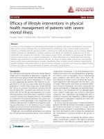
Graphical depiction of workflow for the identification of metabolitesFigure 1
Graphical depiction of workflow for the identification of metabolites. (a) Mass spectral data collection and processing using XCMS. LC/MS data were first
analyzed with XCMS software to produce a list of metabolite features, where each feature is defined by both a specific retention time and m/z value. The
XCMS software then applies a non-linear retention time correction to align the same metabolite features found in different biological samples. The final
XCMS output lists the t-test results based on the intensity variations of common metabolite features found among the defined sample classes. (b)
Metabolite selection, characterization and identification. Metabolite features meeting statistical criteria for significance, based on XCMS processing, were
further characterized by accurate mass measurement, identified using mass spectral databases such as the Metlin database and the LIPID MAPS database,
and further confirmed by tandem (MS/MS) mass spectral data.
(a)
LC/MS data Extracted ions Nonlinear alignment Ions aligned
XCMS t-test and
manual evaluation
TimeTime (min)
080
Time
XCMS output
Accurate m/z measurement
and database evaluation
Tandem (MS/MS) mass spectral data
m/z
WT KO1 KO3 DKOWT KO1 KO3 DKO
m/z 546 546.3546
MS
(b)
m/z 600100
MS/MS
MS/MSMS/MS
R38.4 Genome Biology 2007, Volume 8, Issue 3, Article R38 Mutch et al. />Genome Biology 2007, 8:R38
animals, which was further reinforced by revealing no com-
mon, significantly modulated metabolites between the KO1
and DKO models.
Increasing evidence has positioned adipose tissue as not
merely a reservoir for lipid storage, but also as a major endo-
crine and secretory organ. The recognition that obesity is
characterized by chronic mild inflammation led to the discov-
ery of factors, termed adipokines, that are released from adi-
pose tissue and critical in regulating such physiological
processes as inflammation, lipid metabolism, insulin sensi-
tivity, angiogenesis, and eating behavior [17,18]. Further-
more, several factors have been shown to regulate
atherogenic processes, such as hypertension and vascular
remodeling [19,20]. It is interesting to find that plasma from
Plscr3-deficient mice show increases in pro-inflammatory
LPC molecules that have been linked to certain pathophysio-
logical conditions, including atherosclerosis, cancer and
rheumatoid arthritis [21-23]. While these bioactive lysophos-
pholipids have not been previously described in obesity, their
identification suggests a novel pro-inflammatory class linking
obesity and atherosclerosis that merits further examination.
The characterization of normal human serum revealed that
four LPC metabolites (LPC containing 16:0, 18:0, 18:1, or 18:2
fatty acids) are among the most abundant circulating metab-
olites found [24]. The genetic deletion of murine Plscr3 led to
significant changes in only one of these highly abundant LPC
species (18:1). Additionally, as changes in the abundance of
other lysophospholipid classes were not observed, this indi-
cates that increases in LPC species are specific to Plscr3 defi-
ciency. Although to our knowledge LPC molecules have not
been previously examined in obese models (either rodent or
human), this finding may be a general characteristic of the
obese state and requires confirmation in other models of
obesity.
Conformational and positional characterization of the acyl
portion of lipid metabolites was not performed; therefore, the
discussion herein is based on the previous reports of the most
common fatty acids produced in eukaryotes [25]. The LPC
species found to be most abundant in plasma was oleoyl-LPC
(Figure 2). Due to this abundance, we were able to further
confirm the presence of this molecule by converting it to the
corresponding fatty acid methyl ester, followed by gas
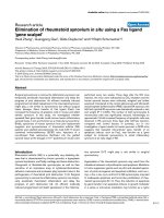
Exact masses and chemical structures of metabolites characterized with the Metlin databaseFigure 2
Exact masses and chemical structures of metabolites characterized with the Metlin database. The significance of fold changes between the KO1 versus
WT, KO3 versus WT, and DKO versus WT are indicated with an asterisk where p < 0.005. The significance for all 5 metabolites is further increased to p
< 0.00001 when comparing KO3 and DKO to KO1 and WT. Average intensity values ± relative standard deviation values are indicated in parentheses
below the fold changes, and demonstrate the large difference in metabolite abundance between the five LPC molecules. Structures were obtained from the
LipidMaps website [34].
Fold change
Observed
m/z
Calculated
m/z
ppm
error
Major
CID
ions
Molecular
formula
[M+H]
Lysophosphocholine
KO1
vs.
WT
KO3
vs.
WT
DKO
vs.
WT
494.3227
494.3241
-4.0
494 ->
476 +
184 +
104
C
24
H
49
NO
7
P
GPCho 16:1
0.7
(245026
±
39%)
3.0*
(1063557
±
39%)
1.7*
(608502
±
39%)
522.3547
522.3554
-2.4
522->
504 +
184 +
104
C
26
H
53
NO
7
P
GPCho 18:1
1.0
(1303528
±
38%)
2.1*
(2708499
±
31%)
1.6*
(2091021
±
23%)
542.3233
542.3241
-2.5
542 ->
524 +
184 +
104
C
28
H
49
NO
7
P
GPCho 20:5
0.6
(32547
±
37%)
4.7*
(264092
±
42%)
1.8*
(100890
±
27%)
546.3546
546.3554
-2.5
546 ->
528 +
184 +
104
C
28
H
53
NO
7
P
GPCho 20:3
0.9
(562354
±
46%)
2.6*
(1623847
±
44%)
2.2*
(1369986
±
27%)
570.3545
570.3554
-2.6
570 ->
552 +
184 +
104
C
30
H
53
NO
7
P
GPCho 22:5
0.7
(64245
±
42%)
2.6*
(242052
±
41%)
1.9*
(171225
±
23%)
Genome Biology 2007, Volume 8, Issue 3, Article R38 Mutch et al. R38.5
comment reviews reports refereed researchdeposited research interactions information
Genome Biology 2007, 8:R38
chromatography (GC)/MS analysis. After production of the
methyl esters, the GC/MS experiment confirmed the
presence of the expected fatty acid C18:1 (C
18
H
34
O
2
) in an
LPC molecule. More specifically, the hypothesized molecular
ion for the 18:1 methyl ester (m/z 296, M+H) and the frag-
mentation pattern were found, which matched the correct
model spectrum in the NIST 2002 spectral database. Both
palmitoleic (C16:1n7) and oleic acids (C18:1n9) are synthe-
sized via stearoyl-CoA desaturase (SCD1), the rate-limiting
enzyme regulating the introduction of a cis-double bond in
the Δ9 position of its fatty acyl-CoA substrates palmitate
(C16:0) and stearate (C18:0). SCD1 has a pivotal role in
whole-body lipid metabolism, as exemplified by the finding
that Scd1-deficient mice are resistant to diet-induced obesity
[26]. However, the physiological complexity underlying the
obese state suggests that interpreting plasma metabolite pro-
files has the power to identify biomarkers without indicating
their originating tissue source.
Thus, to begin to elucidate the tissue responsible for altering
lipid metabolism in Plscr3-/- mice, the expression of a subset
of lipogenic genes (those encoding Ppar-γ, Lxr-α, Srebp-1c,
Fasn, and Scd1) was examined in the liver and white adipose
tissue (WAT) of the various genotypes. While a similar profile
for Ppar-γ expression was observed in both tissues - WAT
(KO1, 2.0-fold; KO3, 5.5-fold; DKO, 9.4-fold) and liver (KO1,
4.4-fold; KO3, 12.1-fold; DKO, 39.5-fold) - the expression pat-
tern for the remaining genes were tissue-specific. Hepatic
gene expression in the three knock-out models indicated sim-
ilar fold-increases in Lxr-α, Srebp-1c, and Fasn, with the
exception of Scd1, which was not significantly changed in the
liver (Figure 3a). In contrast, gene expression in WAT
revealed marked differences between the various genotypes
(Figure 3b). For all genes examined, induction was minimal
and less significant in KO1 mice, whereas changes in KO3 and
DKO mice were highly significant and of greater importance
(p ≤ 0.001). Of particular interest was the expression of Lxr-
α and Srebp-1c, transcriptional regulators of Fasn and Scd1.
KO1 animals had only a slight 1.7-fold increase in Srebp-1c
expression, while KO3 and DKO mice had far more promi-
nent increases of 6.5- and 10.8-fold, respectively. Similar
changes were seen with Lxr-α (KO1, 2.1-fold; KO3, 9.8-fold;
DKO, 16.8-fold), Fasn (KO1, 2.1-fold; KO3, 3.6-fold; DKO,
4.7-fold) and Scd1 expression (KO1, 3.0-fold; KO3, 5.9-fold;
DKO, 4.4-fold). As Plscr3 expression is very low in liver [27]
and Plscr1 expression is nearly absent in WAT [3], the tran-
scriptional and lipid metabolite changes observed with Plscr3
deficiency would suggest they originate from the WAT and
may be directly related to the SREBP-1c-mediated transcrip-
tional cascade. Indeed, if the plasma lipid profile reflected
hepatic transcriptional events, one could hypothesize that all
genotypes would have similar changes in plasma lipids; how-
ever, since this was not the case, we propose that the differen-
tial plasma lipid profile arises from transcriptional events
occurring in WAT.
As described above, Plscr3-/- mice have an increase in Δ9-
desaturase expression similar to that previously noted in var-
ious obese models and animals fed diets promoting weight
gain (that is, obesity-inducing diets increased Scd1 expres-
sion and activity, while diets reducing adiposity decrease it)
[11]. The identification of DGLA, EPA, and ObA fatty acid spe-
cies may reflect additional modulation of adipose Δ5 and Δ6
desaturase enzyme activities, as both enzymes have roles in
the metabolism of long-chain polyunsaturated fatty acid (LC-
PUFA) species [28,29]; however, in the absence of adipose
metabolite data this hypothesis remains to be proven. It has
recently been proposed by Das [30] that defects in Δ5 and Δ6
desaturases may play a role in the development of insulin
resistance by reducing the synthesis of beneficial LC-PUFA
arachidonic acid (AA) and docosahexaenoic acid (DHA).
Plscr3-/- mice are insulin resistant [3] and the increase in
22:5-containing LPC would further support Das's hypothesis,
as increases in the abundance of 22:5 fatty acids are consid-
ered an indicator of DHA deficiency [31]. These findings posi-
tion Plscr3 as a regulator of adipose lipid metabolism.
Of additional interest, both DGLA and EPA are purported to
have anti-inflammatory properties via their conversion into
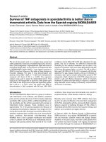
Assessingn (a) liver and (b) WATFigure 3
Assessing gene expression in (a) liver and (b) WAT. A subset of genes
under the transcriptional regulation of Srebp-1c were examined by real-
time RT-PCR and, in WAT, were more significantly and strongly up-
regulated in the absence of Plscr3. As the goal was to address genotype
differences and not individual animal variation, total RNA from three
animals/genotype was pooled and technical triplicates were run. *p < 0.01,
**p < 0.001.
-4
-2
0
2
4
6
8
10
Fold change (relative to WT)
Srebp1c Lxr-a Scd1 Fasn
WT
KO1
KO3
DKO
**
**
**
**
**
**
**
**
**
(a)
0
2
4
6
8
10
12
14
16
18
Fold change (relative to WT)
Srebp1c Lxr-a Scd1 Fasn
WT
KO1
KO3
DKO
*
*
*
**
**
**
**
**
**
**
**
**
(b)
R38.6 Genome Biology 2007, Volume 8, Issue 3, Article R38 Mutch et al. />Genome Biology 2007, 8:R38
the 1 series and 3 series of prostaglandins, respectively [32].
Although the precise contribution of ω-3 and ω-6 LC-PUFA to
the inflammatory state has yet to be resolved, both classes are
precursors to inflammatory eicosanoids; however, it appears
that ω-3 LC-PUFA derived eicosanoids are associated with a
less severe inflammatory profile than those derived from ω-6
LC-PUFA [33]. Thus, it is conceivable that plasmatic
increases in LPC containing DGLA and EPA reflect altered
biochemical pathways in WAT eicosanoid metabolism associ-
ated with a deficiency in Plscr3; however, no changes in the
eicosanoids currently characterized in public databases (Met-
lin [16], LipidMaps [34]) were found in plasma.
Conclusion
Plasma metabolite profiling coupled with analytical software
such as XCMS provides a method by which the unique bio-
chemical signatures and functional redundancy of related
proteins can be explored. Furthermore, the ability to identify
perturbations in previously unrecognized metabolic path-
ways reinforces the potential of untargeted metabolite profil-
ing for generating hypotheses and new research directions. As
demonstrated in the present study, Plscr1 and Plscr3 do not
modulate the plasma metabolome in a similar fashion. While
no changes were detected in the plasma metabolite profile
upon genetic deletion of Plscr1, deletion of Plscr3 clearly
modulates the plasma metabolome through the release of
pro-inflammatory lipids and other, currently unidentifiable,
small molecules. Although profiling plasma metabolites does
not definitively unravel the molecular mechanisms underly-
ing lipid accumulation in Plscr3-deficient animals, invaluable
clues were provided into the perturbed physiology of these
animals and will serve as indicators for future targeted
experiments. Not only do LPC molecules present a potential
link between obesity, inflammation, and atherogenesis, the
acyl composition of LPC suggests that Plscr3-deficient mice
may have modified desaturase enzyme activities and eicosa-
noid metabolism. Indeed, Scd1 and its transcriptional regula-
tors (Lxr-α, Srebp-1c) are significantly up-regulated in the
adipose of Plscr3-deficient mice. Furthermore, the accumula-
tion of LC-PUFA precursors (DGLA, EPA, and ObA) in LPC
species may suggest deficiencies in the abundance of benefi-
cial AA and DHA fatty acids. To reinforce this notion,
decreases in LC-PUFA and increases in monounsaturated
species were previously reported in obese Zucker rats [35]. It
is interesting to note that additional work by Wilson and col-
leagues [36,37] has also demonstrated the ability to discrimi-
nate the plasma metabolite profiles of lean and obese 20-
week old Zucker (fa/fa) rats using metabolomics coupled with
bioinformatics algorithms. While the authors found six
metabolites in the positive ion data of an LC/MS analysis to
be different between the two rat strains, none of these small
molecules were identified [38]. Thus, while the untargeted
metabolite profiling presented in this study is still in its early
stages of development, the ability to identify and correlate
metabolites with functionally related protein family members
and provide novel and previously unrecognized insight into
the perturbed metabolism stemming from dysfunctional
genes positions this analytical platform as an attractive
means towards understanding the fundamental biochemistry
of disease states.
Materials and methods
Animals
Plscr1-/- (KO1) and Plscr3-/- (KO3) mice were produced by
Lexicon Genetics Incorporated (The Woodlands, TX, USA)
and Plscr(1&3)-/- (DKO) mice were produced by breeding
KO1 with KO3 mice as previously described [3,8]. The genetic
background of all mice was identical
(C57BL6Jx129SvEvBRD) and details of their general charac-
terization can be found elsewhere [3,39]. All mice were fed
standard (approximately 5% fat) rodent chow (Harlan Tek-
lad, Madison, WT, USA) and had access ad libitum to steri-
lized water. Mice were fasted for four hours before blood
draw. Approximately 250 μl of blood was extracted by retro-
orbital eye bleeds from male mice of approximately 8 weeks
of age (n = 4 per genotype). Age-matched mice were divided
into 2 groups of 2 and blood was extracted on 2 separate days
to minimize differences in fasting time between all 16 mice.
Blood was collected into tubes containing lyophilized
K
2
EDTA (Becton Dickinson, Franklin Lakes, NJ, USA) and
immediately centrifuged at 800 × g for 15 minutes at 4°C to
extract plasma. After collection samples were stored at -80°C
prior to analysis.
Chemicals and sample preparation
All solvents used were of HPLC grade (JT Baker, Philipsburg,
NJ, USA). Metabolite extraction was performed with cold
methanol as described previously [24]. Briefly, 40 μl aliquots
of mouse plasma were extracted with 150 μl cold methanol,
and incubated at -20°C for 20 minutes, then centrifuged to
remove protein precipitate. The supernatant was dried and
reconstituted in 40 μl acetonitrile/water 5/95 v/v.
LC/MS data acquisition and analysis
The separation system used was an Agilent 1100 LC/MSD SL
system equipped with HPLC (Agilent, Santa Clara, CA, USA).
Triplicate runs of each sample were analyzed randomly, with
a blank run between samples to prevent carryover. For each
run, 5 μl of metabolite extract was injected onto the same C
18
column (Symmetry Column, 2.1 × 100 mm, 3.5 μm, Waters
(Waters, Milford, MA, USA) and eluted at a flow rate of 250
μl/minute. Elution buffers were: A, water with 0.1% formic
acid; and B, acetonitrile with 0.1% formic acid. The LC/MS
run time was 75 minutes, with a gradient begun at 5% B until
12 minutes, with times and percentages as follows: 20% B at
20 minutes, 90% B at 55 minutes, 95% B at 60 to 70 minutes,
5% B at 71 to 75 minutes. Mass spectral data from 100 to
1,000 m/z were collected in the positive ionization mode. LC/
MS data were processed using the XCMS software [40].
Metabolites of interest were selected based on values of ion
Genome Biology 2007, Volume 8, Issue 3, Article R38 Mutch et al. R38.7
comment reviews reports refereed researchdeposited research interactions information
Genome Biology 2007, 8:R38
intensity changes and consistency between animals of the
same type.
Accurate mass and MS/MS fragmentation
determination
Fractions containing the metabolites of interest were col-
lected in subsequent HPLC separations. These fractions were
then analyzed individually in positive ion mode using the Agi-
lent ESI-TOF to obtain high accuracy mass spectral data (<4
ppm error between observed and calculated masses; Addi-
tional data file 1). Three reference masses with m/z at
121.0509, 319.1030, and 922.0098 were used for real time
mass adjustments. MS/MS data were collected using a linear
ion trap (Thermo, Waltham, MA, USA). Specific masses that
varied significantly between WT versus KO3 and DKO mice
were targeted for fragmentation. MS/MS conditions were as
follows: isolation = 3.0 amu, normalized collision energy =
35%, activation Q = 0.15 and activation time = 30.0 ms.
GC/MS identification of lysophospholipid LPC 18:1
An experiment was designed to confirm the identification of
the lysophosphocholine metabolite at m/z 522, using a fatty-
acid methyl ester approach (FAME) coupled with GC/MS.
The LC/MS chromatography of plasma was repeated with a
larger volume of starting material (equivalent to 32 μl) and 1
minute fractions were collected by hand. This preparative
chromatography was repeated once, with pooling of the
equivalent fractions to increase the final yield. The fraction
between 44.5 and 45.5 minutes was expected to contain the
metabolite with m/z 522.3547, and this was confirmed by
locating the 522 mass by direct injection of 4% of this fraction
into the ESI-TOF spectrometer. The sample was then con-
verted to fatty acid methyl esters, dried, and 200 μl of a 3N
HCl methanol solution was added and incubated in a 100°C
oven for 45 minutes. Hexane (400 μl) was added, and the
solution was dried and reconstituted in dichoromethane
(DCM). The GC/MS column was a HP5-MS (J&W Scientific/
Agilent, Santa Clara, CA, USA) with the following characteris-
tics: length = 30 m, ID = 0.25 mm, and film = 0.25 μm. The
mass spectrometer was an Agilent 5973 with an injector port
temperature of 290°C and a transfer line temperature of
280°C. The flow rate was 1.2 ml/minute with a total run time
of 27.5 minutes. The temperature program was 50°C for 5
minutes, followed by a gradient of 20°C/minute to 300°C, fol-
lowed by a hold at 300°C for 10 minutes. The injection vol-
ume was 2.5 μl with no split. MS/MS data were collected as
described in the preceding section.
Semi-quantitative real-time RT-PCR
Total RNA from three animals/genotype was pooled for the
liver and white adipose tissue. Reverse transcription was per-
formed with 1 μg of total RNA using the Advantage RT-PCR
kit (Clontech, Mountain View, CA, USA) and random hex-
amer primers. Sybr
®
green primers were designed (Addi-
tional data file 3) and validated for target specificity and
amplification efficiency. RT-PCR amplification was per-
formed using a BioRad iCycler (BioRad, Hercules, CA, USA)
with the following thermal cycling conditions: 2 minutes at
50°C, 10 minutes at 95°C, followed by 40 cycles of 95°C for 15
s and 60°C for 1 minute for denaturation, annealing, and
elongation. All samples were performed in (technical) tripli-
cate and data were normalized to glyceraldehyde-3-phos-
phate dehydrogenase. A two-tailed, homoscedastic Student's
t-test (α = 0.01) was used to confirm differences in gene
expression in pair-wise analysis (that is, genotypes compared
to WT).
Additional data files
The following additional data are available with the online
version of this paper. Additional data file 1 is a table contain-
ing the raw dataset from XCMS processing, where 'name'
denotes feature or peak name, 'M' denotes m/z, 'T' denotes
retention time in units of seconds, 'mzme' denotes median m/
z value, 'mzmin' denotes minimum m/z value, 'mzmax'
denotes maximum m/z value, 'rtmed' denotes median reten-
tion time in seconds, 'rtmin' denotes minimum retention time
in seconds, 'rtmax' denotes maximum retention time in sec-
onds, 'WT' denotes wild-type animal intensity area values,
'KO3' denotes Plscr3-/- animal intensity area values, 'KO1'
denotes Plscr1-/- animal intensity area values, 'DKO' denotes
double knock-out animals animal intensity area values, and
the letters a, b, and c following Mouse strain_Mouse Number
denote the replicated experimental runs. Additional data file
2 lists the 19 metabolites associated with PLSCR3 deficiency.
Additional data file 3 is a table containing sequence informa-
tion for real-time RT-PCR primers.
Additional data file 1Raw dataset from XCMS processingName, feature or peak name; M, m/z; T, retention time in units of seconds; mzme, median m/z value; mzmin, minimum m/z value; mzmax, maximum m/z value; rtmed, median retention time in sec-onds; rtmin, minimum retention time in seconds; rtmax, maxi-mum retention time in seconds; WT, wild-type animal intensity area values; KO3, Plscr3-/- animal intensity area values; KO1, Plscr1-/- animal intensity area values; DKO, double knock-out ani-mals animal intensity area values; the letters a, b, and c following Mouse strain_Mouse Number denote the replicated experimental runs.Click here for fileAdditional data file 2The 19 metabolites associated with PLSCR3 deficiencyThe 19 metabolites associated with PLSCR3 deficiency.Click here for fileAdditional data file 3Sequence information for real-time RT-PCR primersSequence information for real-time RT-PCR primers.Click here for file
Acknowledgements
The authors thank Drs Q Zhou and A Berger for their review of this man-
uscript. This work was supported by grants DK069390 from NIDDK
(DMM, TW, PJS), and DOE DE-AC02-05CH11231 and NIH P30
MH062261 (GO, WW, GS).
References
1. Rankinen T, Zuberi A, Chagnon YC, Weisnagel SJ, Argyropoulos G,
Walts B, Perusse L, Bouchard C: The human obesity gene map:
the 2005 update. Obesity (Silver Spring) 2006, 14:529-644.
2. Mutch DM, Clement K: Unraveling the genetics of human
obesity. PLoS Genet 2006, 2:e188.
3. Wiedmer T, Zhao J, Li L, Zhou Q, Hevener A, Olefsky JM, Curtiss LK,
Sims PJ: Adiposity, dyslipidemia, and insulin resistance in mice
with targeted deletion of phospholipid scramblase 3
(PLSCR3). Proc Natl Acad Sci USA 2004, 101:13296-13301.
4. Zhou Q, Zhao J, Stout JG, Luhm RA, Wiedmer T, Sims PJ: Molecular
cloning of human plasma membrane phospholipid scram-
blase. A protein mediating transbilayer movement of plasma
membrane phospholipids. J Biol Chem 1997, 272:18240-18244.
5. Zhao KW, Li D, Zhao Q, Huang Y, Silverman RH, Sims PJ, Chen GQ:
Interferon-alpha-induced expression of phospholipid scram-
blase 1 through STAT1 requires the sequential activation of
protein kinase Cdelta and JNK. J Biol Chem 2005,
280:42707-42714.
6. Zhou Q, Zhao J, Al-Zoghaibi F, Zhou A, Wiedmer T, Silverman RH,
Sims PJ: Transcriptional control of the human plasma mem-
brane phospholipid scramblase 1 gene is mediated by
interferon-alpha. Blood 2000, 95:2593-2599.
R38.8 Genome Biology 2007, Volume 8, Issue 3, Article R38 Mutch et al. />Genome Biology 2007, 8:R38
7. Ben-Efraim I, Zhou Q, Wiedmer T, Gerace L, Sims PJ: Phospholipid
scramblase 1 is imported into the nucleus by a receptor-
mediated pathway and interacts with DNA. Biochemistry 2004,
43:3518-3526.
8. Zhou Q, Ben-Efraim I, Bigcas JL, Junqueira D, Wiedmer T, Sims PJ:
Phospholipid scramblase 1 binds to the promoter region of
the inositol 1,4,5-triphosphate receptor type 1 gene to
enhance its expression. J Biol Chem 2005, 280:35062-35068.
9. Muoio DM, MacLean PS, Lang DB, Li S, Houmard JA, Way JM, Wine-
gar DA, Corton JC, Dohm GL, Kraus WE: Fatty acid homeostasis
and induction of lipid regulatory genes in skeletal muscles of
peroxisome proliferator-activated receptor (PPAR) alpha
knock-out mice. Evidence for compensatory regulation by
PPAR delta. J Biol Chem 2002, 277:26089-26097.
10. Vieten A, Vanneste S, Wisniewska J, Benkova E, Benjamins R, Beeck-
man T, Luschnig C, Friml J: Functional redundancy of PIN pro-
teins is accompanied by auxin-dependent cross-regulation of
PIN expression. Development 2005, 132:4521-4531.
11. Mutch DM: Identifying regulatory hubs in obesity with
nutrigenomics. Curr Opin Endocrinol Diabetes 2006, 13:431-437.
12. Bijlsma S, Bobeldijk I, Verheij ER, Ramaker R, Kochhar S, Macdonald
IA, van Ommen B, Smilde AK: Large-scale human metabolomics
studies: a strategy for data (pre-) processing and validation.
Anal Chem 2006, 78:567-574.
13. Griffin JL: Metabolic profiles to define the genome: can we
hear the phenotypes? Philos Trans R Soc Lond B Biol Sci 2004,
359:857-871.
14. Raamsdonk LM, Teusink B, Broadhurst D, Zhang N, Hayes A, Walsh
MC, Berden JA, Brindle KM, Kell DB, Rowland JJ, et al.: A functional
genomics strategy that uses metabolome data to reveal the
phenotype of silent mutations. Nat Biotechnol 2001, 19:45-50.
15. Mutch DM, Fauconnot L, Grigorov M, Fay LB:
Putting the 'ome' in
lipid metabolism. Biotechnol Annu Rev 2006, 12:67-84.
16. Smith CA, O'Maille G, Want EJ, Qin C, Trauger SA, Brandon TR, Cus-
todio DE, Abagyan R, Siuzdak G: METLIN: a metabolite mass
spectral database. Ther Drug Monit 2005, 27:747-751.
17. Greenberg AS, Obin MS: Obesity and the role of adipose tissue
in inflammation and metabolism. Am J Clin Nutr 2006,
83:461S-465S.
18. Trayhurn P, Wood IS: Signalling role of adipose tissue: adipok-
ines and inflammation in obesity. Biochem Soc Trans 2005,
33:1078-1081.
19. Lyon CJ, Law RE, Hsueh WA: Minireview: adiposity, inflamma-
tion, and atherogenesis. Endocrinology 2003, 144:2195-2200.
20. Taleb S, Lacasa D, Bastard JP, Poitou C, Cancello R, Pelloux V,
Viguerie N, Benis A, Zucker JD, Bouillot JL, et al.: Cathepsin S, a
novel biomarker of adiposity: relevance to atherogenesis.
FASEB J 2005, 19:1540-1542.
21. Fuchs B, Schiller J, Wagner U, Hantzschel H, Arnold K: The phos-
phatidylcholine/lysophosphatidylcholine ratio in human
plasma is an indicator of the severity of rheumatoid arthritis:
investigations by 31P NMR and MALDI-TOF MS. Clin Biochem
2005, 38:925-933.
22. Kamanna VS, Bassa BV, Ganji SH, Roh DD: Bioactive lysophos-
pholipids and mesangial cell intracellular signaling pathways:
role in the pathobiology of kidney disease. Histol Histopathol
2005, 20:603-613.
23. Xu Y, Xiao YJ, Zhu K, Baudhuin LM, Lu J, Hong G, Kim KS, Cristina
KL, Song L, Williams S, et al.: Unfolding the pathophysiological
role of bioactive lysophospholipids. Curr Drug Targets Immune
Endocr Metabol Disord 2003, 3:23-32.
24. Want EJ, O'Maille G, Smith CA, Brandon TR, Uritboonthai W, Qin C,
Trauger SA, Siuzdak G: Solvent-dependent metabolite distribu-
tion, clustering, and protein extraction for serum profiling
with mass spectrometry. Anal Chem 2006, 78:743-752.
25. Vance DE, Vance JE: Biochemistry of Lipids, Lipoproteins and Membranes
Amsterdam: Elsevier Science BV; 2002.
26. Ntambi JM, Miyazaki M, Stoehr JP, Lan H, Kendziorski CM, Yandell BS,
Song Y, Cohen P, Friedman JM, Attie AD: Loss of stearoyl-CoA
desaturase-1 function protects mice against adiposity. Proc
Natl Acad Sci USA 2002, 99:11482-11486.
27. Wiedmer T, Zhou Q, Kwoh DY, Sims PJ: Identification of three
new members of the phospholipid scramblase gene family.
Biochim Biophys Acta 2000, 1467:244-253.
28. Chuang LT, Thurmond JM, Liu JW, Mukerji P, Bray TM, Huang YS:
Effect of conjugated linoleic acid on Delta-5 desaturase activ-
ity in yeast transformed with fungal Delta-5 desaturase gene.
Mol Cell Biochem 2004, 265:11-18.
29. Engler MM, Bellenger-Germain SH, Engler MB, Narce MM, Poisson JP:
Dietary docosahexaenoic acid affects stearic acid desatura-
tion in spontaneously hypertensive rats. Lipids 2000,
35:1011-1015.
30. Das UN: A defect in the activity of Delta6 and Delta5 desatu-
rases may be a factor predisposing to the development of
insulin resistance syndrome. Prostaglandins Leukot Essent Fatty
Acids 2005, 72:343-350.
31. de Groot RH, Hornstra G, van Houwelingen AC, Roumen F: Effect
of alpha-linolenic acid supplementation during pregnancy on
maternal and neonatal polyunsaturated fatty acid status and
pregnancy outcome. Am J Clin Nutr 2004, 79:251-260.
32. Das UN: COX-2 inhibitors and metabolism of essential fatty
acids. Med Sci Monit 2005, 11:RA233-RA237.
33. Krebs JD, Browning LM, McLean NK, Rothwell JL, Mishra GD, Moore
CS, Jebb SA: Additive benefits of long-chain n-3 polyunsatu-
rated fatty acids and weight-loss in the management of
cardiovascular disease risk in overweight hyperinsulinaemic
women. Int J Obes (Lond) 2006, 30:1535-1544.
34. Fahy E, Subramaniam S, Brown HA, Glass CK, Merrill AH Jr, Murphy
RC, Raetz CR, Russell DW, Seyama Y, Shaw W, et al.: A compre-
hensive classification system for lipids. J Lipid Res 2005,
46:839-863.
35. Serkova NJ, Jackman M, Brown JL, Liu T, Hirose R, Roberts JP, Maher
JJ, Niemann CU: Metabolic profiling of livers and blood from
obese Zucker rats. J Hepatol 2006, 44:956-962.
36. Williams R, Lenz EM, Wilson AJ, Granger J, Wilson ID, Major H,
Stumpf C, Plumb R: A multi-analytical platform approach to
the metabonomic analysis of plasma from normal and
zucker (fa/fa) obese rats. Mol Biosyst 2006, 2:174-183.
37. Major HJ, Williams R, Wilson AJ, Wilson ID: A metabonomic anal-
ysis of plasma from Zucker rat strains using gas chromatog-
raphy/mass spectrometry and pattern recognition. Rapid
Commun Mass Spectrom 2006, 20:3295-3302.
38. Plumb RS, Johnson KA, Rainville P, Shockcor JP, Williams R, Granger
JH, Wilson ID: The detection of phenotypic differences in the
metabolic plasma profile of three strains of Zucker rats at 20
weeks of age using ultra-performance liquid chromatogra-
phy/orthogonal acceleration time-of-flight mass
spectrometry. Rapid Commun Mass Spectrom 2006, 20:2800-2806.
39. Zhou Q, Zhao J, Wiedmer T, Sims PJ: Normal hemostasis but
defective hematopoietic response to growth factors in mice
deficient in phospholipid scramblase 1. Blood 2002,
99:4030-4038.
40. Smith CA, Want EJ, O'Maille G, Abagyan R, Siuzdak G: XCMS:
processing mass spectrometry data for metabolite profiling
using nonlinear peak alignment, matching, and
identification. Anal Chem 2006, 78:779-787.