Báo cáo y học: "Distinct transcriptional MYCN/c-MYC activities are associated with spontaneous regression or malignant progression in neuroblastomas" ppsx
Bạn đang xem bản rút gọn của tài liệu. Xem và tải ngay bản đầy đủ của tài liệu tại đây (737.76 KB, 14 trang )
Open Access
Volume
et al.
Westermann
2008 9, Issue 10, Article R150
Research
Distinct transcriptional MYCN/c-MYC activities are associated with
spontaneous regression or malignant progression in
neuroblastomas
Frank WestermannÔ*, Daniel MuthÔ*, Axel Benner, Tobias Bauer, KaiOliver Henrich*, Andrộ OberthuerĐ, Benedikt Brors, Tim Beissbarthả,
Jo VandesompeleƠ, Filip PattynƠ, Barbara HeroĐ, Rainer Kưnig‡,
Matthias Fischer§ and Manfred Schwab*
Addresses: *Department of Tumor Genetics, German Cancer Research Center, Im Neuenheimer Feld 280, Heidelberg, 69120, Germany.
†Department of Biostatistics, German Cancer Research Center, Im Neuenheimer Feld 280, Heidelberg, 69120, Germany. ‡Theoretical
Bioinformatics, German Cancer Research Center, Im Neuenheimer Feld 280, Heidelberg, 69120, Germany. §Department of Pediatric Oncology,
University Children's Hospital of Cologne, Kerpener Strasse 62, Cologne, 50924, Germany. ¶Division of Molecular Genome Analysis, German
Cancer Research Center, Im Neuenheimer Feld 580, Heidelberg, 69120, Germany. ¥Center for Medical Genetics, Ghent University Hospital, De
Pintelaan 185, Ghent, 9000, Belgium.
Ô These authors contributed equally to this work.
Correspondence: Frank Westermann. Email:
Published: 13 October 2008
Genome Biology 2008, 9:R150 (doi:10.1186/gb-2008-9-10-r150)
Received: 6 August 2008
Revised: 19 September 2008
Accepted: 13 October 2008
The electronic version of this article is the complete one and can be
found online at />© 2008 Westermann et al.; licensee BioMed Central Ltd.
This is an open access article distributed under the terms of the Creative Commons Attribution License ( which
permits unrestricted use, distribution, and reproduction in any medium, provided the original work is properly cited.
Abstract
Background: Amplified MYCN oncogene resulting in deregulated MYCN transcriptional activity is observed in 20%
of neuroblastomas and identifies a highly aggressive subtype. In MYCN single-copy neuroblastomas, elevated MYCN
mRNA and protein levels are paradoxically associated with a more favorable clinical phenotype, including
disseminated tumors that subsequently regress spontaneously (stage 4s-non-amplified). In this study, we asked
whether distinct transcriptional MYCN or c-MYC activities are associated with specific neuroblastoma phenotypes.
Results: We defined a core set of direct MYCN/c-MYC target genes by applying gene expression profiling and
chromatin immunoprecipitation (ChIP, ChIP-chip) in neuroblastoma cells that allow conditional regulation of MYCN
and c-MYC. Their transcript levels were analyzed in 251 primary neuroblastomas. Compared to localized-nonamplified neuroblastomas, MYCN/c-MYC target gene expression gradually increases from stage 4s-non-amplified
through stage 4-non-amplified to MYCN amplified tumors. This was associated with MYCN activation in stage 4snon-amplified and predominantly c-MYC activation in stage 4-non-amplified tumors. A defined set of MYCN/c-MYC
target genes was induced in stage 4-non-amplified but not in stage 4s-non-amplified neuroblastomas. In line with this,
high expression of a subset of MYCN/c-MYC target genes identifies a patient subtype with poor overall survival
independent of the established risk markers amplified MYCN, disease stage, and age at diagnosis.
Conclusions: High MYCN/c-MYC target gene expression is a hallmark of malignant neuroblastoma progression,
which is predominantly driven by c-MYC in stage 4-non-amplified tumors. In contrast, moderate MYCN function
gain in stage 4s-non-amplified tumors induces only a restricted set of target genes that is still compatible with
spontaneous regression.
Genome Biology 2008, 9:R150
/>
Genome Biology 2008,
Background
Neuroblastoma is the most common extracranial malignant
solid tumor in early childhood. Clinical courses are highly
variable, ranging from spontaneous regression to therapyresistant progression. Clinical and biological features, such as
age at diagnosis, disease stage, numerical (ploidy) and structural chromosomal alterations (MYCN gene amplification;
1p, 3p, 11q deletions; 17q gain), are associated with patient
outcome [1,2]. Amplified MYCN oncogene identifies a subtype with poor prognosis [3] and is consistently associated
with high MYCN mRNA and protein levels. There is strong
experimental evidence (ectopic MYCN expression in cell
lines, N-myc transgenic neuroblastoma mouse model) that
increased MYCN activity is involved in tumor initiation and
progression of at least a subset of neuroblastomas [4,5].
The MYC gene family members, c-MYC, MYCN and MYCL,
are involved in the biology of many cancer types. They encode
basic helix-loop-helix leucine zipper proteins that are found
as heterodimers with their obligate partner protein, MAX [6].
The MYC-MAX heterodimer binds to DNA consensus core
binding sites, 5'-CACGTG-3' or variants thereof (E-boxes),
which preferentially leads to transcriptional activation of target genes. Repression of target genes by MYC proteins has
also been described [7]. This seems to be independent of the
binding of MYC proteins to E-boxes, but involves a cofactor,
Miz-1, that tethers MYC-MAX to gene promoters, such as p15
and p21. Enhanced activity of MYC transcription factors contributes to almost every aspect of tumor formation: unrestricted cell proliferation, inhibition of differentiation, cell
growth, angiogenesis, reduced cell adhesion, metastasis, and
genomic instability [6,8]. In contrast, MYC transcription factors, including MYCN, also sensitize cells for apoptosis, a
function that should inhibit tumor formation and that could
also be involved in spontaneous tumor regression [9].
Spontaneous tumor regression does occur in neuroblastoma,
at a higher frequency than in any other cancer type. This process resembles the physiological concurrence of massive cellular suicide (apoptosis) and differentiation of a few neurons
along the sympathoadrenal cell lineage in the normal development of the sympathetic nervous system. Spontaneous
regression is most frequently observed in a subset of disseminated MYCN single-copy neuroblastomas (non-amplified
(NA)), termed stage 4s (stage 4s-NA) [10]. However, population-based screening studies for neuroblastomas in Japan,
Quebec and Germany suggest that spontaneous regression
also occurs in other neuroblastoma subtypes, predominantly
localized (stages 1, 2, 3) neuroblastomas (localized-NA) [1113]. Paradoxically, MYCN mRNA and protein levels are
higher in favorable localized-NA and, particularly, in stage
4s-NA tumors than in stage 4-NA tumors with poor outcome
[14-16], but they do not reach the levels observed in MYCN
amplified tumors. In line with this, neuroblastoma cells with
elevated MYCN expression retain their capacity to undergo
apoptosis [17] or neuronal differentiation [18]. Thus, it has
Volume 9, Issue 10, Article R150
Westermann et al. R150.2
been speculated that MYCN does not only mediate malignant
progression in MYCN amplified tumors, but is also either
involved or at least compatible with spontaneous regression
in favorable neuroblastomas. In contrast, a functional role of
MYCN in stage 4-NA tumors with low MYCN levels is questionable. Here, other transcription factors or pathways within
or outside the MYC family of transcription factors could be
more relevant. Neuroblastoma-derived cell lines that lack
amplified MYCN generally express c-MYC rather than MYCN,
often at higher levels than normal tissues [19,20]. However,
transcriptional activity of MYCN or c-MYC as reflected by the
transcript levels of direct MYCN/c-MYC target genes in relation to MYCN and c-MYC levels has not yet been defined in
neuroblastoma subtypes.
Here, we defined a core set of MYCN and c-MYC target genes
by using oligonucleotide microarrays and a neuroblastoma
cell line that allows conditional expression of MYCN or cMYC. Direct regulation of these target genes by MYCN/cMYC was assessed by analyzing the binding of MYCN and cMYC protein to target gene promoters using PCR- and arraybased chromatin immunoprecipitation (ChIP and ChIP-chip,
respectively) in different neuroblastoma cell lines. We further
investigated the expression of these direct MYCN/c-MYC target genes in relation to MYCN and c-MYC expression in different clinical neuroblastoma subtypes. In addition, the
association of MYCN/c-MYC target gene expression with
overall survival independent of the well-established markers
- amplified MYCN, disease stage and age at diagnosis - was
demonstrated.
Results
Inverse correlation of MYCN and c-MYC expression in
neuroblastoma subtypes
c-MYC mRNA levels are very low in MYCN amplified tumors
(Figure 1), which is due to high MYCN protein repressing cMYC mRNA expression [20]. Previous quantitative PCR analyses in a cohort of 117 neuroblastoma patients revealed that
mRNA levels of MYCN are significantly lower in stage 4-NA
than in stage 4s-NA (p = 0.008) and localized-NA neuroblastomas (stages 1, 2, 3; p = 0.03) [14]. To test whether this lower
expression of MYCN in stage 4-NA tumors is due to elevated
c-MYC activity that represses MYCN expression, we analyzed
c-MYC and MYCN mRNA levels in a cohort of 251 primary
neuroblastoma tumors using a customized 11K oligonucleotide microarray (other MYC gene family members were not
differently expressed (data not shown)). Although c-MYC
mRNA levels were not significantly higher in stage 4-NA (n =
52) than in localized-NA tumors (n = 138), we found an
inverse correlation of MYCN and c-MYC expression between
stage 4s-NA (n = 30) and stage 4-NA tumors. Stage 4-NA
tumors showed lower expression of MYCN and higher expression of c-MYC, whereas stage 4s-NA tumors showed lower
expression of c-MYC and higher expression of MYCN (Figure
1; p = 0.008 for c-MYC, p = 0.07 for MYCN).
Genome Biology 2008, 9:R150
/>
Genome Biology 2008,
MYCN
Volume 9, Issue 10, Article R150
Westermann et al. R150.3
c-MYC
2
2
●
●
●
−1
●
1
−1
0
Relative expression
0
●
−2
Relative expression
1
●
●
−2
●
1/2/3 4s 4
MYCN-NA
AMP
MYCN
1. 5
DKC1
MDM2
0.6
1.0
0.0
0.2
●
●
●
Relative expression
●
●
●
●
●
●
−1.0
−0.4
●
●
−0.6
−0.5
●
●
●
●
●
−0.5
−0.2
Relative expression
1.5
1.0
●
●
●
●
●
0.0
●
PTMA
●
●
●
0. 4
●
0.5
Relative expression
2.0
●
AMP
MYCN
0.5
1/2/3 4s 4
MYCN-NA
0.0
−3
●
●
1/2/3 4s 4 AMP
MYCN-NA MYCN
1/2/3 4s 4
MYCN-NA
AMP
MYCN
●
●
1/2/3 4s 4
MYCN-NA
AMP
MYCN
Figure 1
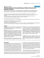
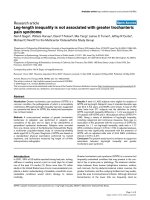
Inverse correlation of MYCN and c-MYC mRNA levels in neuroblastoma subtypes
Inverse correlation of MYCN and c-MYC mRNA levels in neuroblastoma subtypes. Relative mRNA expression is shown for MYCN and c-MYC as well as for
MDM2, DKC1, and PTMA, three direct targets of MYCN/c-MYC. Data are represented as box plots: horizontal boundaries of boxes represent the 25th
and 75th percentile. The 50th percentile (median) is denoted by a horizontal line in the box and whiskers above and below extend to the most extreme
data point, which is no more than 1.5 times the interquartile range from the box. A set of 251 primary neuroblastoma tumors was analyzed consisting of
138 localized-NA (stage 1/2/3), 30 stage 4s-NA, 52 stage 4-NA and 31 MYCN amplified (AMP) neuroblastoma tumors. Gene expression levels from stage
4s-NA, stage 4-NA, and MYCN amplified tumors were compared pair-wise with those of localized-NA tumors as reference. Differential gene expression
was assessed for each gene by using the Mann-Whitney test (cut-off of p < 0.05).
Genome Biology 2008, 9:R150
/>
Genome Biology 2008,
Because increased activity of MYCN in stage 4s-NA or c-MYC
in stage 4-NA tumors should both result in high expression of
shared target genes compared to localized-NA neuroblastomas, we analyzed known direct MYCN/c-MYC target genes,
namely MDM2 [21], DKC1 [22], and PTMA [23], in neuroblastoma subtypes. As expected, the highest expression of all
three transcripts was observed in MYCN amplified tumors
(Figure 1; p < 0.001 for all three transcripts, n = 31). MDM2
mRNA levels were higher in stage 4-NA (p = 0.005) and stage
4s-NA (p = 0.03) than in localized-NA tumors (the expression
range of MDM2 is large because of two MYCN amplified
tumors with non-syntenic co-amplification of MDM2 (data
not shown)). Similarly, DKC1 and PTMA expression was
higher in stage 4-NA (p < 0.001 for DKC1, p = 0.02 for PTMA)
and in stage 4s-NA (p = 0.03 for DKC1, p = 0.007 for PTMA)
than in localized-NA tumors. These results suggest an
increased MYCN/c-MYC activity also in stage 4s-NA (MYCN)
and in stage 4-NA (predominantly c-MYC) compared to localized-NA tumors. However, higher DKC1 mRNA levels in stage
4-NA tumors and higher PTMA mRNA levels in stage 4s-NA
tumors also suggest differential regulation of MYCN/c-MYC
target genes in these subtypes. To further analyze MYCN/cMYC activity as well as differential regulation of MYCN/cMYC target genes in neuroblastoma subtypes, we thought to
define a comprehensive set of target genes directly regulated
by MYCN and/or c-MYC in neuroblastoma cells.
Repression of endogenous c-MYC by targeted
expression of a MYCN transgene in SH-EPMYCN cells
defines c-MYC- and MYCN-regulated genes
To identify MYCN/c-MYC-regulated genes in neuroblastoma
cells, we employed the experimental system SH-EPMYCN,
which stably expresses a tetracycline-regulated MYCN transgene [23]. Exponentially growing SH-EPMYCN cells cultured
with tetracycline express c-MYC but almost no MYCN protein
(Figure 2a). Induction of MYCN by removing tetracycline
from the medium is associated with a rapid reduction of cMYC at the mRNA and protein levels. c-MYC reduction
occurs prior to the full expression of ectopically induced
MYCN protein (Figure 2a). Accordingly, mRNA levels of
direct MYCN/c-MYC targets, such as PTMA and DKC1, initially decline before accumulating MYCN protein leads to the
re-induction of these genes. Similar profiles were observed
with direct MYCN target genes, such as MDM2 and MCM7
(Additional data file 1).
We used SH-EPMYCN cells for a global search of MYCN and cMYC target genes in neuroblastoma cells using a customized
neuroblastoma oligonucleotide microarray (11K, Agilent) that
was enriched with probes for genes differentially expressed in
neuroblastoma subtypes and for direct MYCN/c-MYC target
genes [14,24]. Gene expression profiles of SH-EPMYCN cells at
2, 4, 8, 12, 24, and 48 hours after targeted MYCN expression
were generated. Self-organizing maps (SOMs) were used to
capture the predominant pattern of gene expression. This
analysis yielded 504 clusters (best matching units (BMUs))
Volume 9, Issue 10, Article R150
Westermann et al. R150.4
consisting, on average, of 20 clones per cluster (Additional
data file 1). We searched for clusters with characteristic gene
expression profiles of direct MYCN/c-MYC target genes. In
addition, known c-MYC target genes from a public database
[25] and known MYCN target genes from a literature search
were mapped to the 504 clusters (Additional data file 2). A
significant enrichment of known MYCN/c-MYC targets was
found in 6 clusters (clusters 140, 168, 195, 280, 308, and 336;
p < 0.05, adjusted for multiple testing), consisting of 167
genes. The genes in these six clusters were induced by MYCN
and c-MYC in SH-EPMYCN cells. Based on their average gene
expression profiles, we grouped the clusters into two subgroups, I and II. Subgroup I genes (clusters 140, 168, and 195)
were expressed at equal levels in SH-EPMYCN cells expressing
endogenous c-MYC (2 hours) and in those fully expressing
ectopic MYCN (24 and 48 hours), despite the fact that the
maximum protein level of MYCN was significantly higher
than that of endogenous c-MYC (Figure 2a; Additional data
file 1). This indicates that subgroup I genes are regulated by
MYCN, and also suggests that they are less responsive to
MYCN than to c-MYC in SH-EPMYCN cells. The mRNA levels
of subgroup II genes (clusters 280, 308, and 336) were highest in SH-EPMYCN cells fully expressing ectopic MYCN and followed the combined absolute c-MYC and MYCN protein
levels during the time course experiment. We also found clusters with MYCN and c-MYC repressed genes (for example,
subgroup III; Additional data file 1). However, enrichment of
known MYCN/c-MYC repressed genes from the literature/
database in defined clusters was not found using our statistical cut-off (after adjustment for multiple testing, no cluster
showed p < 0.05). This was at least partly due to the fact that
in SH-EPMYCN cells, some genes were repressed by MYCN but
not by c-MYC (subgroup IV). In addition, c-MYC repressed
genes from different experimental systems compiled in the cMYC target gene database were not necessarily repressed by
MYCN and/or c-MYC in SH-EPMYCN cells.
Therefore, we focused on genes for further validation that
were induced by both MYCN and c-MYC proteins in SHEPMYCN cells and grouped into subgroup I and II. We
extracted all available promoters from the genes represented
on the array and scanned for canonical E-boxes (CACGTG)
and for the 12 bp MYCN position-weight matrix [26] within
-2 kb and +2 kb of the transcriptional start site. We ranked all
504 clusters according to the relative number of putative
MYCN/c-MYC binding sites in each cluster. All clusters from
subgroups I and II were among the 15 top-ranked clusters
with enrichment of predicted MYCN/c-MYC binding sites
(data not shown).
To further validate target gene regulation by MYCN/c-MYC in
neuroblastoma cells, we performed ChIP-chip using a 244K
oligonucleotide promoter microarray (Agilent). We analyzed
the binding of MYCN and c-MYC to the promoters of the 147
subgroup I and II genes that were represented on the 244K
promoter microarray. We used five neuroblastoma cell lines
Genome Biology 2008, 9:R150
/>
Genome Biology 2008,
Volume 9, Issue 10, Article R150
Westermann et al. R150.5
(a)
2
1.2
c-MYC
1.5
DKC1
1
1
0.8
Ratio
Ratio
0.5
0
-0.5
0.6
0.4
-1
0.2
-1.5
0
-2
2
4
8
12
24
48
2
4
8
12
24
Hours
48
Hours
Western blot
0.8
MYCN
PTMA
0.6
0.4
2
4
8
12
24
48
Hours after MYCN induction
Ratio
c-MYC
0.2
0
-0.2
-0.4
-0.6
2
4
8
12
24
48
Hours
(b)
SJ-NB12
SY5Y
MYCN
SH-EP SH-EP
IMR5/75
Kelly
d
I entificationdof MYCN/c-MYC target genes in neuroblastoma cell lines
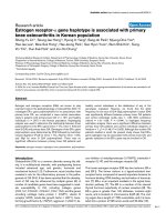
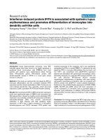
Figure 2
Identification and validation of MYCN/c-MYC target genes in neuroblastoma cell lines. (a) Repression of endogenous c-MYC by targeted expression of a
MYCN transgene in SH-EPMYCN cells defines MYCN/c-MYC-regulated genes. MYCN and c-MYC protein levels were monitored in a time series after
removing tetracycline in exponentially growing SH-EPMYCN cells that stably express a tetracycline-regulated MYCN transgene. Mean and standard deviation
of the relative mRNA levels of MYC, DKC1 and PTMA are given from two time series experiments as measured by a customized neuroblastoma oligo
microarray. (b) Hierarchical clustering of MYCN- and c-MYC binding to 140 target gene promoters as measured by ChIP-chip in 6 neuroblastoma cell
lines. ChIP-chip results of 140 MYCN/c-MYC target genes from 5 neuroblastoma cell lines that preferentially express either high levels of MYCN (SHEPMYCN, IMR5/75 (approximately 75 copies of MYCN) and Kelly (approximately 100-120 copies of MYCN)) or c-MYC (SJ-NB12 and SY5Y). Additionally, as
an intermediate type, parental SH-EP cells were analyzed. SH-EP cells preferentially express c-MYC, but also low levels of MYCN. ChIP-chip experiments
were performed with a monoclonal antibody against human MYCN and a polyclonal antibody against human c-MYC for each neuroblastoma cell line. A
cut-off for positive binding was set for both transcription factors to >4-fold enrichment for one and >2-fold enrichment of at least one of the two
neighboring probes. MYCN/c-MYC-binding is color-coded as follows: blue, c-MYC binding; red, MYCN/c-MYC binding; dark red, MYCN binding; light
yellow, lack of MYCN/c-MYC binding. Hierarchical clustering was used to group neuroblastoma cell lines according to their MYCN/c-MYC-binding
pattern. Differentiation between MYCN and c-MYC-binding was mainly achieved through the monoclonal MYCN antibody. The polyclonal antibody
against c-MYC also gave positive binding signals for a large set of analyzed target gene promoters in neuroblastoma cell lines with high MYCN that lack cMYC expression (SH-EPMYCN, IMR5/75 and Kelly).
Genome Biology 2008, 9:R150
/>
Genome Biology 2008,
that either preferentially express high levels of MYCN (SHEPMYCN, IMR5/75 (approximately 75 copies of MYCN), and
Kelly (approximately 100-120 copies of MYCN)) or c-MYC
(SJ-NB12 and SY5Y). Additionally, as an intermediate type,
parental SH-EP cells were analyzed, which preferentially
express c-MYC, but also MYCN at low level [20,23]. ChIPchip experiments were performed with a monoclonal antibody against human MYCN and a polyclonal antibody against
human c-MYC for each of the neuroblastoma cell lines. A cutoff for positive binding was defined as >4-fold enrichment for
one probe together with >2-fold enrichment for at least one of
the two neighboring probes compared to input control. In
addition, we manually inspected each of the MYCN and cMYC-binding profiles from the 147 genes. Seven genes were
excluded from the analysis because the probe sets for the
genes mapped within the genes but outside the target gene
promoter regions (all profiles for Kelly and SJ-NB12 cell lines
are given in Additional data files 3 and 4, respectively; MYCNand c-MYC-binding results are given in Additional data files
5-7). We also performed PCR-based ChIP for selected candidate genes (n = 13; Additional data file 8), which all showed
analogous results to ChIP-chip (data not shown). Almost all
140 target gene promoters showed binding of MYCN and/or
c-MYC in the six analyzed neuroblastoma cell lines as measured by ChIP-chip (Figure 2b). Intriguingly, hierarchical clustering of neuroblastoma cell lines according to the MYCN/cMYC-binding pattern clearly separated MYCN- and c-MYCexpressing neuroblastoma cell lines. Differentiation between
MYCN and c-MYC binding was mainly achieved through the
monoclonal anti-MYCN antibody. The polyclonal antibody
against c-MYC also gave positive binding signals for a large
set of target gene promoters in neuroblastoma cell lines with
high MYCN that lack detectable c-MYC expression (SHEPMYCN, IMR5/75 and Kelly). This was most likely due to
unspecific binding of the polyclonal c-MYC antibody to
MYCN in these cells. Nevertheless, the lack of binding of
MYCN to a large set of target gene promoters in the c-MYCexpressing cells, SJ-NB12 and SY5Y, and the positive binding
of c-MYC to almost all of these target gene promoters in these
cells allowed the distinction between MYCN and c-MYC.
Taken together, these results indicate that the genes from
subgroups I and II represent a core set of target genes directly
regulated by either MYCN or c-MYC in neuroblastoma cells
dependent on which MYC protein is expressed.
Gradual increase of MYCN/c-MYC target gene
expression from stage 4s-NA through stage 4-NA to
MYCN amplified tumors
To determine transcriptional activity of MYCN/c-MYC proteins in primary neuroblastomas (n = 251), we analyzed differential expression of subgroup I and II genes in
neuroblastoma subtypes using the Global test as proposed by
Goeman et al. [27]. Almost all these genes (154 of 167; 92%)
showed highest expression in MYCN amplified tumors, suggesting that regulation of these genes by MYCN is similar in
neuroblastoma cell lines and tumors. Compared to localized-
Volume 9, Issue 10, Article R150
Westermann et al. R150.6
NA tumors (stages 1, 2, 3), expression of subgroup I and II
genes was significantly associated with stage 4s-NA (p =
0.002), stage 4-NA (p < 0.001) and MYCN amplified tumors
(p < 0.001). Global test results further indicated that an
increasing number of MYCN/c-MYC target genes was
induced from stage 4s-NA through stage 4-NA to MYCN
amplified tumors (Additional data files 9-11). To further illustrate this, we grouped each of the 154 genes into one of four
classes based on pair-wise comparisons (Mann-Whitney test,
cut-off p < 0.05). These were, compared to localized-NA
tumors: overexpressed in MYCN amplified and in stage 4sNA tumors (class 1); overexpressed in MYCN amplified, stage
4-NA and stage 4s-NA tumors (class 2); overexpressed in
MYCN amplified tumors (class 3); overexpressed in MYCN
amplified and stage 4-NA tumors (class 4) (Figure 3). Compared to localized-NA tumors, 25 (16%) of the 154 MYCN/cMYC target genes, including CCT4, FBL, MDM2, NCL, NPM1,
PTMA, and TP53, were expressed at higher levels in stage 4sNA tumors (Table 1). Eighty-eight (57%) of the 154 MYCN/cMYC target genes, including 21 of those overexpressed also in
stage 4s-NA tumors, were expressed at higher levels in stage
4-NA than in localized-NA tumors (Table 1, class 2; Additional data file 5). Accordingly, stage 4-NA tumors shared
overexpression of 68 of 154 direct MYCN/c-MYC target genes
(44%), including AHCY, RUVBL1, PHB, CDK4, and MRPL3,
with MYCN amplified tumors. Together, this indicates that
besides MYCN amplified tumors, stage 4-NA tumors, and to
a lesser extent stage 4s-NA tumors, also show higher MYCN/
c-MYC activity compared to localized-NA tumors. In line with
this, we also found lower mRNA levels of an increasing
number of MYCN/c-MYC repressed genes from stage 4s-NA
(10 out of 68 (15%) in vitro validated repressed genes that are
also lower in MYCN amplified tumors) through stage 4-NA
(34 out of 68 (50%)) to MYCN amplified tumors (68 out of
102 in vitro validated repressed genes had the lowest expression levels in MYCN amplified tumors (67%)). Based on the
relative expression of MYCN and c-MYC in neuroblastoma
subtypes, we propose that elevated MYCN activity in stage 4sNA tumors induces only a restricted set of MYCN/c-MYC target genes, whereas elevated c-MYC activity in stage 4-NA
tumors induces a larger set of MYCN/c-MYC target genes.
High expression of MYCN/c-MYC target genes is a
robust marker of poor overall survival independent of
genomic MYCN status, age at diagnosis and disease
stage
Having shown that MYCN/c-MYC target gene activation is
also associated with distinct neuroblastoma subtypes, we
wanted to test whether MYCN/c-MYC activity as determined
by the expression levels of their target genes is associated with
overall survival and improves outcome prediction independent of known risk markers. We used the Global test to test the
influence of each of the 504 experimentally defined gene clusters on overall survival directly, without the intermediary of
single gene testing. The p-values for each cluster were
adjusted for multiple testing and ranked according to their
Genome Biology 2008, 9:R150
/>
Genome Biology 2008,
Volume 9, Issue 10, Article R150
Westermann et al. R150.7
Table 1
MYCN/c-MYC target genes overexpressed in stage 4s-NA compared to localized-NA tumors (classes 1 and 2)
Probe
Gene name
A_24_P311604
A_23_P102420
c-MYC target DB†
Validated by ChIP‡
Class
BMU
Group
MYCN/c-MYC-fold change*
C4orf28
1
195
I
1.38
+
CCT4
1
168
I
1.31
+
A_23_P5551
NCL
1
308
II
1.69
A_23_P44836
NT5DC2
1
140
I
1.40
+
A_32_P139196
C13ORF25V_1
2
308
II
3.83
ND
A_24_P133488
CDCA4
2
140
I
1.45
A_23_P137143
DKC1
2
308
II
1.93
A_23_P216396
EXOSC2
2
308
II
1.83
A_23_P78892
FBL
2
195
I
1.93
A_24_P228796
GAGE7B
2
195
I
1.27
A_23_P41025
GNL3
2
308
II
A_32_P8120
GNL3
2
308
A_23_P398460
HK2
2
280
Hs172673.9
Hs172673.9
2
A_23_P502750
MDM2
A_23_P92261
MGC2408
A_23_P50897
A_23_P214037
Up
+
+
Up
+
Up
+
1.80
Up
ND
II
1.81
Up
ND
II
1.71
Up
168
I
1.73
2
336
II
1.19
2
280
II
2.14
MKI67IP
2
280
II
1.97
Up
+
NPM1
2
140
I
1.61
Up
+
A_23_P57709
PCOLCE2
2
308
II
2.40
A_24_P34632
PTMA
2
308
II
2.21
Up
+
A_23_P126825
SLC16A1
2
195
I
1.22
+
A_23_P126291
SNRPE
2
336
II
1.49
+
A_23_P117068
SNRPF
2
336
II
1.44
+
A_23_P31536
SSBP1
2
336
II
1.24
A_23_P26810
TP53
2
140
I
1.44
+
ND
+
+
ChIP
+
+
+
+
Up
+
*Fold change expression in SH-EPMYCN cells after MYCN induction. †c-MYC target gene database entry [25]: Up, upregulated; ChIP, validated by ChIP.
‡Validation of MYCN/c-MYC binding using ChIP in this study (Additional data files 5-7). BMU, best matching unit; ND, not determined.
association with overall survival. Table 2 gives the association
with overall survival of the six MYCN/c-MYC target gene clusters and the rank in relation to all other clusters. In a separate
analysis, we determined the association with overall survival
for each of the 504 experimental gene clusters adjusted for
amplified MYCN, stage 4 versus stages 1, 2, 3, and 4s, and age
at diagnosis ≥1.5 years (Table 2). These well-established risk
markers highly correlated with poor outcome in univariate
analyses (p < 0.001 for each of these three markers). As
expected, the Global test without adjustment for co-variables
indicated that all MYCN/c-MYC target gene clusters were significantly associated with poor overall survival (p < 0.001).
Intriguingly, all six MYCN/c-MYC target gene clusters
remained significantly associated with overall survival after
adjusting for amplified MYCN, stage 4 versus stages 1, 2, 3,
and 4s, and age at diagnosis ≥1.5 years. Of note, two of the
MYCN/c-MYC target gene clusters (clusters 168 and 140,
both from subgroup I showing a higher responsiveness to cMYC than to MYCN in SH-EPMYCN) revealed the strongest
association with overall survival of all 504 clusters after
adjusting for co-variables (Table 2). Figure 4 shows the association with overall survival for each gene from cluster 168
with and without adjustment for co-variables. Most of the
genes within this cluster, such as AHCY, ARD1A, CDK4,
HSPD1, PHB, RUVBL1, and TRAP1, remained associated
with overall survival after adjustment for co-variables. A less
significant association with overall survival was observed for
clusters with MYCN/c-MYC repressed genes: clusters 454,
482, 484, and 486 were associated with poor overall survival
without adjustment for co-variables in the Global test (p <
0.001, adjusted for multiple testing), but they showed no significant association with poor overall survival when adjusting
for the co-variables amplified MYCN, stage 4 versus stages 1,
2, 3, and 4s, and age at diagnosis ≥1.5 years. We also asked
whether direct MYCN/c-MYC target genes as defined by our
analyses are represented in previously published gene expression-based classifiers that distinguish low-risk from high-risk
neuroblastomas independent of other risk markers. Gene
lists from these studies hardly overlapped, making interpretation difficult. The overlap with our MYCN/c-MYC target
gene list was defined by using the gene names as common
identifiers. Indeed, different genes defined by our study as
direct MYCN/c-MYC target genes were represented in the
gene expression classifier gene lists: from the 44 genes over-
Genome Biology 2008, 9:R150
/>
Genome Biology 2008,
−1.0
0.5
0.0
Relative expression
−0.5
−1.5
1.0
1.0
Relative expression
0.0
0.5
0.5
0.0
●
●
●
4
1/2/3 4s 4 AMP
MYCN-NA MYCN
●
●
−1.0
●
Relative expression
mRNA
MYCN-NA
0.0
●
●
4s
n=68
MYCN single-copy
●
●
●
n=20
●
●
●
●
●
1,2,3
−0.5
−1.0
●
−0.6
●
●
MRPL3
●
●
AMP
4
MRPL3
●
●
●
1,2,3
●
4
1
2
3
1,2,3
1/2/3 4s 4 AMP
MYCN-NA MYCN
AHCY
−1.5
●
●
●
−0.5
●
●
Relative expression
0.5
0.4
0.2
−0.4
n=21
●
●
●
AHCY
Class 4
1.0
0.6
MYCN-NA
FBL
●
●
0.0
1
2
3
AMP
Relative expression
4
−0.2
mRNA
4s
●
4
1/2/3 4s 4 AMP
MYCN-NA MYCN
FBL
TP53
●
●
−0.5
−0.5
TP53
Class 2
●
1,2,3
1,2,3
4
1/2/3 4s 4 AMP
MYCN-NA MYCN
●
●
●
●
1,2,3
4
1/2/3 4s 4 AMP
MYCN-NA MYCN
●
0.5
−1.0
●
●
●
●
●
●
●
●
n=61
●
1.0
1.0
4
●
●
●
●
●
●
●
0.0
4s
●
●
−0.5
0.5
0.0
0.5
Relative expression
n=4
EEF1E1
AMP
1
2
3
MYCN-NA
−0.5
MYCN-NA
●
●
EEF1E1
MTHFD2
Relative expression
mRNA
1.0
●
4
MTHFD2
Class 3
AMP
4s
0.0
1
2
3
Relative expression
mRNA
1.0
NCL
NCL
Westermann et al. R150.8
1.5
CCT4
CCT4
Class 1
Volume 9, Issue 10, Article R150
1,2,3
4
4
1/2/3 4s 4 AMP
MYCN-NA MYCN
1/2/3 4s 4 AMP
MYCN-NA MYCN
1,2,3
4
1/2/3 4s 4 AMP
MYCN-NA MYCN
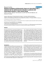
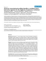
Figure 3
Expression of MYCN/c-MYC target genes in neuroblastoma subtypes
Expression of MYCN/c-MYC target genes in neuroblastoma subtypes. Differential expression was analyzed for each of the genes (n = 154) in MYCN
amplified (AMP), stage 4s-NA and stage 4-NA tumors using localized-NA (stage 1/2/3) tumors as reference in pair-wise comparisons (Mann-Whitney test,
cut-off p < 0.05, black). We grouped each of these 154 genes into one of four classes based on their relative expression in clinically relevant neuroblastoma
subtypes. These classes were, compared to localized-NA tumors: overexpressed in MYCN amplified and in stage 4s-NA tumors (class 1; CCT4 and NCL);
overexpressed in MYCN amplified, stage 4-NA and stage 4s-NA tumors (class 2; TP53 and FBL); overexpressed in MYCN amplified tumors (class 3;
MTHFD2 and EEF1E1); and overexpressed in MYCN amplified and stage 4-NA tumors (class 4; AHCY and MRPL3).
expressed in high-risk neuroblastomas independent of other
markers described by Schramm et al. [28], we identified 10
genes directly regulated by MYCN/c-MYC (DDX21, SCL25A3,
EIFA4A2, NME1, NME2, TKT, LDHA, LDHB, HSPD1,
HSPCB); from the 20 genes overexpressed in high-risk neuroblastomas independent of other markers described by
Ohira et al. [29], we identified 5 genes directly regulated by
MYCN/c-MYC (EEF1G, AHCY, TP53, ENO1, TKT); and from
the 66 genes overexpressed in high-risk neuroblastomas
independent of other markers described by Oberthuer et al.
[24], we identified 7 genes directly regulated by MYCN/cMYC (PRDX4, MRPL3, SNRPE, FBL, LOC200916, PAICS,
AHCY; Figure 5). Together, these results show that MYCN/cMYC activity as determined by the expression status of a subset of MYCN/c-MYC target genes is significantly associated
with poor overall survival independent of other established
markers and is a consistent element of gene expression-based
neuroblastoma risk classification systems.
Discussion
In this study, we analyzed MYCN and c-MYC activity as
reflected by the expression levels of a core set of direct
MYCN/c-MYC targets in neuroblastoma subtypes. As
expected, the highest expression levels of MYCN/c-MYC targets were observed in MYCN amplified tumors. However, we
found that besides MYCN amplified tumors, subtypes of
MYCN single-copy tumors, namely stage 4-NA and, to a lesser
extent, stage 4s-NA, also showed increased MYCN/c-MYC
target gene activation compared to localized-NA tumors. In
general, low MYCN mRNA and protein levels are found in
most stage 4-NA tumors [14-16], which does not explain the
high mRNA levels of MYCN/c-MYC target genes in this sub-
Genome Biology 2008, 9:R150
/>
Genome Biology 2008,
Volume 9, Issue 10, Article R150
Westermann et al. R150.9
Table 2
Association of MYCN/c-MYC target gene clusters with overall survival in primary neuroblastomas (n = 251)
Cluster
Number of genes
Rank OS*
p-value OS†
Rank OS with CV*
p-value OS with CV†
168 (I)
19
3
<0.0001
1
0.0004
140 (I)
38
4
<0.0001
2
0.0006
195 (I)
21
31
<0.0001
12
0.0060
308 (II)
33
18
<0.0001
26
0.0161
280 (II)
32
29
<0.0001
37
0.0232
336 (II)
26
51
<0.0001
45
0.0280
*Rank of all 504 clusters tested for association with overall survival (OS) using the Global test without and with adjustment for co-variables (CV;
amplified MYCN, stages 1, 2, 3, 4s versus 4, age at diagnosis ≥1.5 years). †p-value from Global test adjusted for multiple testing. In the Cluster column,
I or II gives the cluster group as defined by the SOM analysis using SH-EPMYCN cells.
type. Here, we describe an inverse correlation of MYCN and
c-MYC expression levels in stage 4-NA and stage 4s-NA
tumors. From experiments in neuroblastoma cell lines, it is
known that MYCN and c-MYC control their expression via
autoregulatory loops and via repressing each other at defined
promoter sites [20]. Neuroblastoma cell lines with high
expression of MYCN as a result of amplification lack c-MYC
expression. Whenever MYCN and c-MYC are co-expressed in
neuroblastoma cell lines, c-MYC expression predominates.
Together, this suggests that increased activity of c-MYC
represses MYCN in a substantial number of stage 4-NA
tumors. In contrast, an inverse regulation, namely the repression of c-MYC by MYCN, is found in MYCN amplified and, to
a lesser extent, in stage 4s-NA tumors. It is important to note
that localized-NA tumors also express MYCN as well as cMYC and it is likely that they are active because these tumors
frequently show high tumor cell proliferation indices [14].
Nevertheless, in localized-NA tumors, we did not observe that
one MYC transcription factor dominates over the other, such
as in the other neuroblastoma subtypes.
Influence of cluster number 168 genes on outcome
adjusted for genomic MYCN, stage, age at
diagnosis >1.5 years
Influence of cluster number 168 genes on outcome
0
Boxplot class
2
4
6
8
−1
10
Class 3
FAM128B
Class 1
CCT4
Class 4
CDK4
2
3
4
5
AHCY
ARD1A
1
Class 3
CYC1
Class 3
RPUSD4
Class 2 Hs172673.9
Class 3
HSP90AB1
Class 3
HSPD1
Class 3
PRKY
Class 3
MRPS27
Class 4
PHB
Class 4
POLR3D
Class 4
PRKDC
Class 4
RUBVL1
FAM128B
CCT4
CDK4
CYC1
RPUSD4
Hs172673.9
HSP90AB1
HSPD1
PRKY
MRPS27
PHB
Positively associated with survival
Negatively associated with survival
AHCY
ARD1A
Positively associated with survival
Negatively associated with survival
Class 4
Class 4
0
POLR3D
PRKDC 1
RUBVL1
Class 4
SFXN2
SFXN2
Class 3
C11ORF48
C11ORF48
Class 3
TRAP1
TRAP1
Figure 4
Association of cluster 168 genes with overall survival
Association of cluster 168 genes with overall survival. The two gene plots illustrate the influence on overall survival of each gene from cluster 168. The
gene plot gives the influence on overall survival without (left) and with (right) adjustment for the variables genomic MYCN status, age at diagnosis (≥1.5
years), and disease stage (stages 1, 2, 3, 4s versus stage 4). The gene plot shows a bar and a reference line for each gene tested. In a survival model, the
expected height is zero under the null hypothesis that the gene is not associated with the clinical outcome (= reference line). Marks in the bars indicate by
how many standard deviations the bar exceeds the reference line. The bars are colored to indicate a negative (red) association of a gene's expression with
overall survival. In addition, the boxplot class is given for each gene.
Genome Biology 2008, 9:R150
/>
Genome Biology 2008,
Volume 9, Issue 10, Article R150
Westermann et al. R150.10
Outcome
MYCN
1p deletion
11q deletion
Age at diagnosis
Stage
E2F signature
MYCN/c-MYC
signature
SCG2
PDE4DIP
FLJ39739
DST
PKIB
251 patients
Rank classifier
Symbol
Cluster
130
VRK1
447
108
FLJ10151
40
137
HIST1H1C
482
3
SLC25A5
383
90
PRDX4
221
58
HSPC163
247
139
MRPL3
336
57
SNRPE
336
107
ZNF525
25
105
RPL36A
1
223/195
110
FBL
100
BX119435
466
129
NOLA1
307
4
GMPS
445
364
95
LOC200916
1
PAICS
364
5
AHCY
196
13
AHCY
168
Ebox
MYCN/c-MYC-regulated
2 up
2.4 up
down/up
1.8 up
346
1.6 up
down/up
81
2.2 up
1.8 up
1.7 up
1.4 up
-4
2.6 up
5.6 up
284
1.5 up
1.8 up
2.5 up
(-283/18/617/685)
2.2 up
1.6 up
2.2 up
c-MYC TGDB
*UP
*UP
*UP
*UP
*UP
*UP
*UP
*UP
Figure 5
Representation of MYCN/c-MYC target genes in a gene expression-based neuroblastoma risk stratification system. Two-way hierarchical cluster analysis
using 144 oligonucleotide probes from the gene expression-based classifier and the 251 patients from the entire cohort. Clinical characteristics (outcome,
white = no event, gray = relapse/progression, black = death due to neuroblastoma; genomic MYCN status, white = NA, black = amplified; chromosome 1p
status, white = normal, black = 1p deleted, gray = not available; chromosome 11q status, white = normal, black = 11q deleted, gray = not available; age at
diagnosis, white <1.5 years, black ≥1.5 years; disease stage, white = stage 1, 2, gray = stage 3, yellow = stage 4s, black = stage 4) are added to the heatmap
of gene expression. The gene expression cluster with direct MYCN/c-MYC target genes is highlighted. The Rank Classifier column gives the classifier rank
found by the Prediction Analysis for Microarrays algorithm and a complete 10-times-repeated 10-fold cross validation. The Cluster column gives the
results from the SOM analysis using gene expression profiles from SH-EPMYCN cells. The MYCN/c-MYC regulated column gives the fold changes after
MYCN induction. The Ebox column gives the position of a canonical E-box in the promoter. The c-MYC TGDB column gives the entries in the public cMYC target gene database. *UP, upregulated.
Genome Biology 2008, 9:R150
/>
Genome Biology 2008,
Our findings further indicate that MYCN/c-MYC target gene
activation gradually increases from stage 4s-NA through
stage 4-NA to MYCN amplified tumors. High expression of a
large number of MYCN/c-MYC target genes was found in
stage 4-NA and MYCN amplified tumors, but not in stage 4sNA tumors, which is probably involved in the divergent clinical outcome of these subtypes. This also suggests that MYCN
in stage 4s tumors is a weaker transactivator than c-MYC in
stage 4-NA tumors. Whether this effect is due to the cellular
context in which they are expressed and/or due to different
functions of the two MYC proteins in neuroblastoma cells is
unclear. In favor of a cellular context factor, we observed that
promoter constructs from the PTMA gene, which is highly
expressed in stage 4s NA and MYCN amplified tumors,
showed a strong activation in N-type but not S-type neuroblastoma cell lines despite similar MYCN protein levels
(unpublished data). In favor of different functions of the two
MYC proteins, our analyses in SH-EPMYCN cells suggest that a
large number of MYCN/c-MYC target genes (subgroup I
genes) are less responsive to MYCN than to c-MYC. Another
unsolved question is which molecular mechanisms induce
elevated MYCN activity in stage 4s-NA tumors or elevated cMYC activity in stage 4-NA tumors. Candidate pathways
involved in differential regulation of MYC proteins are the
Sonic hedgehog pathway (Shh) for MYCN activation [30] and
the Wnt/beta-catenin pathway for c-MYC activation [31,32].
However, we observed that c-MYC mRNA levels are not
significantly higher in stage 4-NA than in localized-NA
tumors. This suggests that molecular mechanisms that
increase c-MYC protein abundance/stability or simply c-MYC
activity are involved in MYCN/c-MYC target gene activation
in stage 4 tumors.
Our data are in line with a model where stage 4s-NA tumors
exhibit a moderate MYCN function gain compared to localized-NA tumors. Both subtypes usually have favorable outcome. Most localized-NA tumors are cured by surgery alone
or even regress spontaneously. Stage 4s-NA tumors frequently regress spontaneously but regression can also be
induced by a 'mild' chemotherapy. We found that stage 4s-NA
tumors express, on average, the highest MYCN mRNA levels
of all non-amplified tumors [14]. From the experimentally
defined direct MYCN target genes, only a restricted set of 25
genes, including CCT4, FBL, MDM2, NCL, NPM1, PTMA, and
TP53, was overexpressed in stage 4s-NA compared to localized-NA tumors, indicating that elevated MYCN in stage 4sNA tumors only partially activates its downstream target
genes. On the one hand, this suggests that moderate MYCN
function gain in stage 4s-NA tumors is involved in the metastatic phenotype. On the other hand, moderate MYCN function gain in this subtype is still compatible with, or might even
favor, spontaneous regression. From the list of MYCN target
genes overexpressed in stage 4s-NA tumors, TP53 as a proapoptotic gene, and MDM2, coding for the direct inhibitor of
p53 and mediating pro-tumorigenic activities, are strong candidates to be involved in the unique phenotype of stage 4s-NA
Volume 9, Issue 10, Article R150
Westermann et al. R150.11
tumors. However, it is important to note that TP53 and
MDM2 are co-expressed at higher levels also in stage 4-NA
and MYCN amplified tumors. Both subtypes initially respond
to therapy, but rapidly acquire resistance and frequently
show progression/relapse, suggesting that additional conditions activating MDM2 and/or suppressing TP53 functions
are acquired. In line with this, alterations disrupting the p14MDM2-p53 pathway, such as MDM2 amplification, p14
methylation/deletion, and TP53 mutations are found in neuroblastoma cell lines that were established from relapsed
patients [33]. In this context, it remains to be shown whether
small compounds that selectively inhibit MDM2, such as nutlin-3, and that induce proliferation arrest and apoptosis in
neuroblastoma cell lines [34,35] represent a new therapeutic
option for high-risk neuroblastomas.
Conclusions
High expression of a defined subset of direct MYCN/c-MYC
target genes turned out to be a robust marker for poor overall
survival independent of the established markers, amplified
MYCN, disease stage (stage 4 versus stages 1, 2, 3, and 4s) and
age at diagnosis (≥1.5 years). Recently, several gene expression-based neuroblastoma risk stratification systems have
been developed that predict outcome more accurately than
established risk markers [24,28,29]. Unfortunately, the classifier gene lists emerging from these studies hardly overlap,
which has been ascribed to the different composition of the
investigated cohorts and the different high-throughput gene
expression platforms used. Our data show that markers of
increased MYCN/c-MYC activity are consistently represented
in these classifier gene lists, indicating that a gene expressionbased classifier that reflects MYCN/c-MYC function should
make an attractive tool for neuroblastoma classification and
risk prediction.
Materials and methods
Patients
All patients from this study (n = 251) were enrolled in the German Neuroblastoma Trials NB90-NB2004 with informed
consent and diagnosed between 1989 and 2004 (patient characteristics are in Additional data files 2 and 12). Tumor samples were collected prior to any cytoreductive treatment. The
only criterion for patient selection was availability of sufficient amounts of tumor material. Tumor specimens were
checked for at least 60% tumor content.
Neuroblastoma sample preparation and gene
expression analysis
Gene expression profiles from the tumors were generated as
dye-flipped dual-color replicates using customized 11K oligonucleotide microarrays as previously described [24]. The 11K
Agilent microarray was constructed in our laboratory based
on extensive neuroblastoma transcriptome information from
different whole-genome analyses from primary tumors and
Genome Biology 2008, 9:R150
/>
Genome Biology 2008,
neuroblastoma cell lines. These also include comparative
transcriptome analysis of MYCN amplified versus not amplified tumors as well as of neuroblastoma cell lines with variable/conditional MYCN/c-MYC expression that allowed the
enrichment with MYCN/c-MYC-regulated genes [14,24]
(unpublished data). The reference for each tumor RNA was
an RNA pool of 100 neuroblastoma tumor samples. Data normalization and quality control is described in Additional data
file 2. All raw and normalized microarray data are available at
the ArrayExpress database (Accession: E-TABM-38) [36].
Neuroblastoma cell line experiments and SOM analysis
The SH-EPMYCN cell line, previously also denoted as TET21N
[23], expressing a MYCN transgene under the control of a tetracycline-repressible element was used to generate gene
expression profiles from different time points after MYCN
induction showing variable MYCN and c-MYC levels. RNA
isolation from SH-EPMYCN cells was performed as previously
described [14]. Gene expression profiles were generated as
dye-flipped dual-color replicates using the same customized
11K oligonucleotide microarray platform used for the tumor
samples. The reference for RNA from SH-EPMYCN cells after
MYCN induction was RNA from SH-EPMYCN cells cultured in
parallel that lack MYCN expression. Gene expression profiles
from SH-EPMYCN cells with variable MYCN and c-MYC levels
were taken for a SOM analysis (Additional data file 2). Protein
expression was assessed by immunoblotting using 50 μg of
total cell lysates from the cell line experiments as previously
described [37]. Blots were probed with antibodies directed
against MYCN (SantaCruz, sc-53993, Santa Cruz, CA, USA)
and c-MYC (SantaCruz, sc-764, Santa Cruz, CA, USA).
ChIP, ChIP-chip and protein analysis
Chromatin immunoprecipitation was performed as described
previously [38,39] using 10 μg of MYCN (SantaCruz, sc53993), c-MYC (SantaCruz, sc-764) [40,41] and normal
mouse IgG (SantaCruz, sc-2025) antibodies and Dynabeads
ProteinG (Invitrogen, Carlsbad, CA, USA). Eluted and purified MYCN-ChIP-DNA (1 μl) of IMR5/75 and SH-EPMYCN was
used as a template in PCR reactions running for 35 cycles. The
primer sequences are given in Additional data file 8. In addition, ChIP-DNA templates from SH-EPMYCN, SH-EP, Kelly,
IMR5/75, SJNB-12 and SY5Y cells using MYCN and c-MYC
antibodies were amplified for DNA microarray analysis (Agilent Human Promoter ChIP-chip Set 244K) using the WGA
(Sigma-Aldrich, St. Louis, MO, USA) method [42]. DNA labeling, array hybridization and measurement were performed
according to Agilent mammalian ChIP-chip protocols. For
the visualization of ChIP-chip results, the cureos package
v0.2 for R was used (available upon request). The in silico
promoter analysis for the identification of putative MYC
binding sites (canonical and non-canonical E-boxes) is
described in Additional data file 2.
Volume 9, Issue 10, Article R150
Westermann et al. R150.12
Differential gene expression and survival analysis
Differential gene expression of MYCN/c-MYC and their target genes in neuroblastoma tumors was evaluated for stage
4s-NA, stage 4-NA and MYCN amplified using localized-NA
tumors (stages 1, 2, 3) as reference using Goeman's Global
test and the Wilcoxon rank sum test. A result was judged as
'statistically significant' at a p-value of 0.05 or smaller. Differential expression of MYCN was evaluated in two partially
overlapping cohorts, one measured by quantitative PCR [14]
and the other by oligo microarray (the overlap was 101
patients). To test the association of MYCN in vitro clusters
with overall survival (death due to neuroblastoma disease),
Goeman's Global test was used [27]. To evaluate the influence
of gene expression on outcome independent of established
markers, the Global test was adjusted for the following covariables: genomic MYCN status, stage of the disease (stage 4
versus stages 1, 2, 3, and 4s), and age at diagnosis (≥1.5 years
versus <1.5 years). Because of multiple testing of probably
dependent gene clusters, p-values were adjusted according to
Benjamini and Yekutieli [43] to control the false discovery
rate of 5%.
Abbreviations
ChIP, PCR-based chromatin immunoprecipitation; ChIPchip, array-based chromatin immunoprecipitation; NA, nonamplified; SOM, self-organizing map.
Authors' contributions
FW designed and coordinated the study. FW and DM interpreted results and drafted the manuscript. AO, MF, AB, BB
and FW carried out array-based expression profiling and data
analyses of neuroblastoma tumor samples and cell lines. BH
was responsible for clinical data management. TB and RK
performed in silico promoter analyses. JV and FP contributed
samples and performed literature searches of MYCN/c-MYC
target genes. DM performed chromatin immunoprecipitation
experiments. DM, TB and FW analyzed ChIP-chip data. AB,
BH and FW carried out global test and survival analyses. FW,
DM, KOH, JV, FP and MS contributed to the manuscript. All
authors read and approved the final manuscript.
Additional data files
The following additional data are available. Additional data
file 1 is a figure showing a Cluster map of genetic programs
regulated by conditional expression of c-MYC and MYCN proteins in SH-EPMYCN cells. Additional data file 2 is a document
describing in more detail the methods and materials.
Additional data files 3 and 4 are sets of figures showing ChIPchip results of MYCN/c-MYC target genes in the Kelly and SJNB12 cell lines. Additional data files 5, 6 and 7 are tables listing MYCN/c-MYC target genes overexpressed in stage 4s-NA,
stage 4-NA and MYCN amplified tumors, respectively, compared to localized-NA tumors. Additional data file 8 is a table
Genome Biology 2008, 9:R150
/>
Genome Biology 2008,
of genes and primers selected to confirm ChIP-chip results.
Additional data files 9, 10 and 11 are figures showing the association of MYCN/c-MYC induced genes with neuroblastoma
subtypes using the Global test. Additional data file 12 is a
table providing patient data.
16.
sion ofand results of1MYCN/c-MYCgrouped in classthe Kelly neuClusterc-MYC MYCN/c-MYC inducedSH-EPwithinresults. 2. neuAdditionalfor file ofselected totestregulated by conditional 2 cell
Patient mapof andandgenes, which in genes with cells 4-NA
Click here primersMYCN proteins target genes instage 4s-NA cell
neuroblastomasfilethe materials test. genes MYCNMYCN amplified
Associationdatatarget materials. test ChIP-chip cells.
roblastomas
Genes data.
MYCN/c-MYC using
line. data using 3 programs
line
ChIP-chip of genetic the Global
Detailed methods 2 Global test. target genes stage expres9
8
7
6
5
4
12
11
10
MYCN/c-MYC
confirm
the and
results
3
4.
4 SJ-NB12
3.
1
17.
Acknowledgements
We thank Steffen Bannert and Yvonne Kahlert for technical assistance. We
thank the German Neuroblastoma Tumor Bank for providing tumor samples, the German Neuroblastoma Study Group (study chair Frank
Berthold) for providing clinical data and the reference laboratories for providing molecular data. This work was supported by program project grants
from the Krebshilfe, BMBF (NGFN2 and Kompetenznetz Pediatric Oncology/Hematology) and the EU. The platform iCHIP (Integration Center of
HIgh throughPut experiments) has been used for the annotation of this
study. Jo Vandesompele is a postdoctoral researcher of the Research Foundation - Flanders (FWO-Vlaanderen). Filip Pattyn is supported by a grant of
the Ghent University Special Research Fund (BOF).
18.
19.
20.
21.
References
1.
2.
3.
4.
5.
6.
7.
8.
9.
10.
11.
12.
13.
14.
15.
Schwab M, Westermann F, Hero B, Berthold F: Neuroblastoma:
biology and molecular and chromosomal pathology. Lancet
Oncol 2003, 4:472-480.
Vandesompele J, Baudis M, De Preter K, Van Roy N, Ambros P, Bown
N, Brinkschmidt C, Christiansen H, Combaret V, Lastowska M,
Nicholson J, O'Meara A, Plantaz D, Stallings R, Brichard B, Broecke C
Van den, De Bie S, De Paepe A, Laureys G, Speleman F: Unequivocal
delineation of clinicogenetic subgroups and development of
a new model for improved outcome prediction in
neuroblastoma. J Clin Oncol 2005, 23:2280-2299.
Seeger RC, Brodeur GM, Sather H, Dalton A, Siegel SE, Wong KY,
Hammond D: Association of multiple copies of the N-myc
oncogene with rapid progression of neuroblastomas. N Engl J
Med 1985, 313:1111-1116.
Weiss WA, Aldape K, Mohapatra G, Feuerstein BG, Bishop JM: Targeted expression of MYCN causes neuroblastoma in transgenic mice. EMBO J 1997, 16:2985-2995.
Schwab M, Varmus HE, Bishop JM: Human N-myc gene contributes to neoplastic transformation of mammalian cells in
culture. Nature 1985, 316:160-162.
Adhikary S, Eilers M: Transcriptional regulation and transformation by Myc proteins. Nat Rev Mol Cell Biol 2005, 6:635-645.
Kleine-Kohlbrecher D, Adhikary S, Eilers M: Mechanisms of transcriptional repression by Myc. Curr Top Microbiol Immunol 2006,
302:51-62.
Prochownik EV, Li Y: The ever expanding role for c-Myc in promoting genomic instability. Cell Cycle 2007, 6:1024-1029.
Fulda S, Lutz W, Schwab M, Debatin KM: MycN sensitizes neuroblastoma cells for drug-induced apoptosis. Oncogene 1999,
18:1479-1486.
Pritchard J, Hickman JA: Why does stage 4s neuroblastoma
regress spontaneously? Lancet 1994, 344:869-870.
Sawada T, Hirayama M, Nakata T, Takeda T, Takasugi N, Mori T,
Maeda K, Koide R, Hanawa Y, Tsunoda A, et al.: Mass screening for
neuroblastoma in infants in Japan. Interim report of a mass
screening study group. Lancet 1984, 2:271-273.
Woods WG, Tuchman M, Robison LL, Bernstein M, Leclerc JM, Brisson LC, Brossard J, Hill G, Shuster J, Luepker R, Byrne T, Weitzman
S, Bunin G, Lemieux B: A population-based study of the usefulness of screening for neuroblastoma.
Lancet 1996,
348:1682-1687.
Schilling FH, Spix C, Berthold F, Erttmann R, Sander J, Treuner J,
Michaelis J: Children may not benefit from neuroblastoma
screening at 1 year of age. Updated results of the population
based controlled trial in Germany. Cancer Lett 2003, 197:19-28.
Westermann F, Henrich KO, Wei JS, Lutz W, Fischer M, Konig R,
Wiedemeyer R, Ehemann V, Brors B, Ernestus K, Leuschner I, Benner
A, Khan J, Schwab M: High Skp2 expression characterizes highrisk neuroblastomas independent of MYCN status. Clin Cancer
Res 2007, 13:4695-4703.
Cohn SL, London WB, Huang D, Katzenstein HM, Salwen HR, Rein-
22.
23.
24.
25.
26.
27.
28.
29.
30.
31.
32.
Volume 9, Issue 10, Article R150
Westermann et al. R150.13
hart T, Madafiglio J, Marshall GM, Norris MD, Haber M: MYCN
expression is not prognostic of adverse outcome in
advanced-stage neuroblastoma with nonamplified MYCN. J
Clin Oncol 2000, 18:3604-3613.
Tang XX, Zhao H, Kung B, Kim DY, Hicks SL, Cohn SL, Cheung NK,
Seeger RC, Evans AE, Ikegaki N: The MYCN enigma: significance
of MYCN expression in neuroblastoma. Cancer Res 2006,
66:2826-2833.
Lutz W, Fulda S, Jeremias I, Debatin KM, Schwab M: MycN and IFNgamma cooperate in apoptosis of human neuroblastoma
cells. Oncogene 1998, 17:339-346.
Edsjö A, Nilsson H, Vandesompele J, Karlsson J, Pattyn F, Culp LA,
Speleman F, Påhlman S: Neuroblastoma cells with overexpressed MYCN retain their capacity to undergo neuronal
differentiation. Lab Invest 2004, 84:406-417.
Sadée W, Yu VC, Richards ML, Preis PN, Schwab MR, Brodsky FM,
Biedler JL: Expression of neurotransmitter receptors and myc
protooncogenes in subclones of a human neuroblastoma cell
line. Cancer Res 1987, 47:5207-5212.
Breit S, Schwab M: Suppression of MYC by high expression of
NMYC in human neuroblastoma cells. J Neurosci Res 1989,
24:21-28.
Slack A, Chen Z, Tonelli R, Pule M, Hunt L, Pession A, Shohet JM: The
p53 regulatory gene MDM2 is a direct transcriptional target
of MYCN in neuroblastoma. Proc Natl Acad Sci USA 2005,
102:731-736.
Boon K, Caron HN, van Asperen R, Valentijn L, Hermus MC, van Sluis
P, Roobeek I, Weis I, Voute PA, Schwab M, Versteeg R: N-myc
enhances the expression of a large set of genes functioning in
ribosome biogenesis and protein synthesis. EMBO J 2001,
20:1383-1393.
Lutz W, Stöhr M, Schürmann J, Wenzel A, Löhr A, Schwab M: Conditional expression of N-myc in human neuroblastoma cells
increases expression of alpha-prothymosin and ornithine
decarboxylase and accelerates progression into S-phase
early after mitogenic stimulation of quiescent cells. Oncogene
1996, 13:803-812.
Oberthuer A, Berthold F, Warnat P, Hero B, Kahlert Y, Spitz R,
Ernestus K, König R, Haas S, Eils R, Schwab M, Brors B, Westermann
F, Fischer M: Customized oligonucleotide microarray gene
expression-based classification of neuroblastoma patients
outperforms current clinical risk stratification. J Clin Oncol
2006, 24:5070-5078.
Zeller KI, Jegga AG, Aronow BJ, O'Donnell KA, Dang CV: An integrated database of genes responsive to the Myc oncogenic
transcription factor: identification of direct genomic targets.
Genome Biol 2003, 4:R69.
Matys V, Kel-Margoulis OV, Fricke E, Liebich I, Land S, Barre-Dirrie
A, Reuter I, Chekmenev D, Krull M, Hornischer K, Voss N, Stegmaier
P, Lewicki-Potapov B, Saxel H, Kel AE, Wingender E: TRANSFAC
and its module TRANSCompel: transcriptional gene
regulation in eukaryotes. Nucleic Acids Res 2006, 34(Database
issue):D108-D110.
Goeman JJ, Oosting J, Cleton-Jansen AM, Anninga JK, van Houwelingen HC: Testing association of a pathway with survival using
gene expression data. Bioinformatics 2005, 21:1950-1957.
Schramm A, Schulte JH, Klein-Hitpass L, Havers W, Sieverts H, Berwanger B, Christiansen H, Warnat P, Brors B, Eils J, Eils R, Eggert A:
Prediction of clinical outcome and biological characterization of neuroblastoma by expression profiling. Oncogene 2005,
24:7902-7912.
Ohira M, Oba S, Nakamura Y, Isogai E, Kaneko S, Nakagawa A, Hirata
T, Kubo H, Goto T, Yamada S, Yoshida Y, Fuchioka M, Ishii S, Nakagawara A: Expression profiling using a tumor-specific cDNA
microarray predicts the prognosis of intermediate risk
neuroblastomas. Cancer Cell 2005, 7:337-350.
Hatton BA, Knoepfler PS, Kenney AM, Rowitch DH, de Alboran IM,
Olson JM, Eisenman RN: N-myc is an essential downstream
effector of Shh signaling during both normal and neoplastic
cerebellar growth. Cancer Res 2006, 66:8655-8661.
Liu X, Mazanek P, Dam V, Wang Q, Zhao H, Guo R, Jagannathan J,
Cnaan A, Maris JM, Hogarty MD: Deregulated Wnt/beta-catenin
program in high-risk neuroblastomas without MYCN
amplification. Oncogene 2008, 27:1478-1488.
van de Wetering M, Sancho E, Verweij C, de Lau W, Oving I, Hurlstone A, van der Horn K, Batlle E, Coudreuse D, Haramis AP, TjonPon-Fong M, Moerer P, van den Born M, Soete G, Pals S, Eilers M,
Medema R, Clevers H: The beta-catenin/TCF-4 complex
Genome Biology 2008, 9:R150
/>
33.
34.
35.
36.
37.
38.
39.
40.
41.
42.
43.
44.
45.
46.
47.
48.
49.
50.
51.
52.
53.
Genome Biology 2008,
imposes a crypt progenitor phenotype on colorectal cancer
cells. Cell 2002, 111:241-250.
Carr J, Bell E, Pearson AD, Kees UR, Beris H, Lunec J, Tweddle DA:
Increased frequency of aberrations in the p53/MDM2/
p14(ARF) pathway in neuroblastoma cell lines established at
relapse. Cancer Res 2006, 66:2138-2145.
Van Maerken T, Speleman F, Vermeulen J, Lambertz I, De Clercq S,
De Smet E, Yigit N, Coppens V, Philippé J, De Paepe A, Marine JC,
Vandesompele J: Small-molecule MDM2 antagonists as a new
therapy concept for neuroblastoma.
Cancer Res 2006,
66:9646-9655.
Barbieri E, Mehta P, Chen Z, Zhang L, Slack A, Berg S, Shohet JM:
MDM2 inhibition sensitizes neuroblastoma to chemotherapy-induced apoptotic cell death. Mol Cancer Ther 2006,
5:2358-2365.
Brazma A, Parkinson H, Sarkans U, Shojatalab M, Vilo J, Abeygunawardena N, Holloway E, Kapushesky M, Kemmeren P, Lara GG,
Oezcimen A, Rocca-Serra P, Sansone SA: ArrayExpress—a public
repository for microarray gene expression data at the EBI.
Nucleic Acids Res 2003, 31:68-71.
Wiedemeyer R, Westermann F, Wittke I, Nowock J, Schwab M:
Ataxin-2 promotes apoptosis of human neuroblastoma cells.
Oncogene 2003, 22:401-411.
Strieder V, Lutz W: E2F proteins regulate MYCN expression in
neuroblastomas. J Biol Chem 2003, 278:2983-2989.
Lee TI, Johnstone SE, Young RA: Chromatin immunoprecipitation and microarray-based analysis of protein location. Nat
Protoc 2006, 1:729-748.
Knoepfler PS, Zhang XY, Cheng PF, Gafken PR, McMahon SB, Eisenman RN: Myc influences global chromatin structure. EMBO J
2006, 25:2723-2734.
Guccione E, Martinato F, Finocchiaro G, Luzi L, Tizzoni L, Dall' Olio
V, Zardo G, Nervi C, Bernard L, Amati B: Myc-binding-site recognition in the human genome is determined by chromatin
context. Nat Cell Biol 2006, 8:764-770.
O'Geen H, Nicolet CM, Blahnik K, Green R, Farnham PJ: Comparison of sample preparation methods for ChIP-chip assays. Biotechniques 2006, 41:577-580.
Benjamini Y, Yekutieli D: The control of the false discovery rate
in multiple testing under dependency.
Ann Stat 2001,
29:1165-1188.
Brodeur GM, Pritchard J, Berthold F, Carlsen NL, Castel V, Castelberry RP, De Bernardi B, Evans AE, Favrot M, Hedborg F, et al.: Revisions of the international criteria for neuroblastoma
diagnosis, staging, and response to treatment. J Clin Oncol
1993, 11:1466-1477.
Ambros PF, Ambros IM, SIOP Europe Neuroblastoma Pathology,
Biology, and Bone Marrow Group: Pathology and biology guidelines for resectable and unresectable neuroblastic tumors
and bone marrow examination guidelines. Med Pediatr Oncol
2001, 37:492-504.
CRAN []
Bioconductor []
Buness A, Huber W, Steiner K, Sultmann H, Poustka A: arrayMagic:
two-colour cDNA microarray quality control and
preprocessing. Bioinformatics 2005, 21:554-556.
Huber W, von Heydebreck A, Sültmann H, Poustka A, Vingron M:
Variance stabilization applied to microarray data calibration
and to the quantification of differential expression. Bioinformatics 2002, 18(Suppl 1):S96-S104.
Hubbard TJ, Aken BL, Beal K, Ballester B, Caccamo M, Chen Y, Clarke
L, Coates G, Cunningham F, Cutts T, Down T, Dyer SC, Fitzgerald S,
Fernandez-Banet J, Graf S, Haider S, Hammond M, Herrero J, Holland
R, Howe K, Howe K, Johnson N, Kahari A, Keefe D, Kokocinski F,
Kulesha E, Lawson D, Longden I, Melsopp C, Megy K, et al.: Ensembl
2007. Nucleic Acids Res 2007, 35(Database issue):D610-D617.
MYCNot [ />Kohonen T: Self-organizing Maps Second edition. Heidelberg: Springer;
1997. [Springer Series in Information Sciences, volume 30]
SOM Toolbox [ />
Genome Biology 2008, 9:R150
Volume 9, Issue 10, Article R150
Westermann et al. R150.14