Báo cáo y học: "Ribosomal footprints on a transcriptome landscape" potx
Bạn đang xem bản rút gọn của tài liệu. Xem và tải ngay bản đầy đủ của tài liệu tại đây (100.29 KB, 3 trang )
Genome
BBiioollooggyy
2009,
1100::
215
Minireview
RRiibboossoommaall ffoooottpprriinnttss oonn aa ttrraannssccrriippttoommee llaannddssccaappee
David R Morris
Address: Department of Biochemistry, University of Washington, Seattle, WA 98195-7350, USA. Email:
AAbbssttrraacctt
Next-generation massively parallel sequencing technology provides a powerful new means of
assessing rates and regulation of translation across an entire transcriptome.
Published: 28 April 2009
Genome
BBiioollooggyy
2009,
1100::
215 (doi:10.1186/gb-2009-10-4-215)
The electronic version of this article is the complete one and can be
found online at />© 2009 BioMed Central Ltd
The introduction of massively parallel DNA sequencing
platforms over the past five years - so-called ‘next-generation’
sequencing technology - has created the capacity to generate
tens of millions of short sequence reads in a single run. These
sequences can be identified by alignment to the known
genomes of the all-important model organisms, including
Homo sapiens [1]. The information garnered from this
technology is providing new insights into important areas of
genome, chromatin and transcriptome biology.
One of the applications of next-generation sequencing -
short-read cDNA analysis or ‘RNAseq’ [2,3] - has its
conceptual roots in serial analysis of gene expression (SAGE)
[4]. Whereas SAGE provides thousands of sequences of
short sequence tags that have been cloned as concatemers,
RNAseq ups the ante to tens of millions of independently
derived sequences per experiment. For RNA biology,
transcriptome analysis by RNAseq provides robust
quantitative reproducibility, dynamic range of many orders-
of-magnitude, transcript directionality, analysis of repetitive
sequences, independent measurement of highly similar
sequences and detection of post-transcriptional processing
at the single nucleotide level. Using RNAseq methodology, a
recent study from Jonathan Weissman’s laboratory (Ingolia
et al. [5]) yielded a snapshot of the steady-state linear distri-
bution of ribosomes on RNA transcripts in cells of Saccharo-
myces cerevisiae, providing a new powerful experimental
tool for analysis of translational control and co-translational
processes.
RRiibboossoommee pprrooffiilliinngg bbyy RRNNAAsseeqq
Following the early demonstration by Steitz of ribosome
footprints at the initiation codons of bacteriophage R17 RNA
[6], Wolin and Walter showed that eukaryotic ribosomes
carrying out translation protected around 30 nucleotides of
mRNA sequence from digestion by RNase [7]. Exploiting
this observation, they demonstrated clusters of ribosome
protection at discrete sites in the preprolactin transcript.
These clusters were interpreted as reflecting rate-limiting
steps at translation initiation and termination, as well as
ribosome pausing at the site of interaction of the nascent
signal peptide with the signal recognition particle.
Ingolia et al. [5] have now extended analysis of these
ribosome-protected fragments to the genome-wide scale
through RNAseq technology. They implemented an
imaginative intramolecular ligation strategy to generate
directional, unbiased cDNA libraries for sequencing
ribosome-protected RNA fragments. Despite significant
contamination by ribosomal RNA, they were able to assign
7×10
6
RNAseq reads to more than 4,500 yeast genes. These
ribosome ‘footprints’ were mapped with a high degree of
precision and revealed a remarkable three-base periodicity
corresponding to the codons within protein-coding sequences
across the transcriptome. The abundance of ribosome-
protected fragments from a given gene was used to predict
the level of the encoded protein and was shown to be a
significantly better predictor than mRNA level (multiple
regression correlation coefficient R
2
= 0.42 versus R
2
= 0.17).
This study also demonstrated how patterns of ribosome
footprints could be used to provide insights into trans-
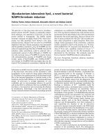
lational regulatory mechanisms. Figure 1 illustrates potential
sites of ribosome localization on a generic mRNA. From the
Wolin and Walter study [7], one would anticipate footprints
at initiation codons and perhaps enhanced ribosome density
at termination sites.
Ribosomes would be expected to distribute randomly across
coding sequences, with the exception of the codon
periodicity noted above. Non-random occurrences of foot-
prints within coding sequences are interpreted as sites of
translational pausing, for example those associated with rare
codons or co-translational activities. Within the untrans-
lated terminal regions (UTRs) of mRNA, footprints might be
expected in association with functional upstream open
reading frames (uORFs). Indeed, as expected, Ingolia et al.
[5] find that 98.8% of the footprints mapped to coding
sequences, with the remainder predominantly associated
with uORFs in the 5’ UTRs.
Although uORFs are known to participate in translational
control [8], the extent of their translation across a
transcriptome has never been evaluated. To attempt this,
Ingolia et al. [5] annotated a total of 1,048 candidate uORFs
with AUG starts in the yeast transcriptome and found that
153 of these showed evidence of ribosome association under
the growth conditions examined. Among these ribosome-
associated uORFs was the gene GCN4. Ribosome footprints
over the four uORFs in GCN4 behaved upon amino acid
starvation as predicted by the generally accepted model [9]
for regulation of this gene - uORF 1 is constitutively trans-
lated and there is a reciprocal relationship between
translation of uORFs 2-4 and the main coding sequence that
is controlled by amino acid starvation.
Interestingly, regulated ribosome loading, apparently origi-
nating from two non-AUG starts, was observed upstream of
the known uORFs in the GCN4 5’ UTR. Although the
existence of uORFs with non-AUG initiation codons has
been the subject of speculation, the presence of these in
GCN4, as well as in more than 1,600 other candidates high-
lighted by Ingolia et al. [5], gives fascinating hints of
previously unrecognized modes of translational control.
PPeerrssppeeccttiivveess aanndd ccaauuttiioonnss
Ribosome profiling by RNAseq is certain to uncover many
new and unexpected aspects of mRNA translation and its
regulation. The most straightforward application will result
from more robust prediction of protein levels than can be
obtained from transcript abundance alone [5]. Even more
significant will be new insights into the events that occur as a
ribosome traverses an mRNA from the cap to the poly(A)
tail. A striking example of this in the work of Ingolia et al. is
the apparent abundance of uORFs with non-AUG starts
throughout the yeast transcriptome. The implications of
these new insights for both translational control and con-
stitutive translation efficiency are tremendous. New clues
regarding the events that occur as ribosomes pause along the
coding sequences are likely to emerge after more extensive
analysis of the existing data and/or increasing the sequence
depth. Such co-translational processes might include folding
or insertion of nascent peptides into cellular structures, as
well as non-standard decoding mechanisms such as
frameshifting or readthrough of termination codons.
As with any powerful new methodology, the results should
be interpreted with caution; there are undoubtedly pitfalls
awaiting the unwary. For example, one should be prepared
for regulated changes in 5’ UTR structure, which may occur
commonly in yeast [10,11] and perhaps other species. These
changes in UTR structure could drastically alter patterns of
ribosome footprints. Likewise, the mere presence of a
ribosome on a coding sequence does not mean that it is
elongating its nascent polypeptide chain. A polyribosome
with all ribosomes arrested at random would show
footprints indistinguishable from those of an actively
translating polysome. Regulation at the level of elongation is
particularly relevant in the context of current controversy
over the mechanisms by which microRNAs inhibit trans-
lation [12-15].
/>Genome
BBiioollooggyy
2009, Volume 10, Issue 4, Article 215 Morris 215.2
Genome
BBiioollooggyy
2009,
1100::
215
FFiigguurree 11
Positioning of ribosomes on a messenger RNA. The 5’ cap is to the left and the poly(A) tail is to the right. The red symbols depict non-random
accumulation of ribosomes at an uORF, the initiation codon, a site of ribosome pausing within the coding sequence (CDS), and at termination. The green
symbols represent freely translating ribosomes at random sites along the coding sequence.
3′ UTR5′ UTR
AAAAA
Ribosome arrest sites
5
′ cap
CDS
Termination
Pause
Initiation
uORF
A technical issue could also drastically influence the inter-
pretation of results. Before preparing extracts, it is routine
procedure in many labs to ‘freeze’ the ribosomes on trans-
cripts with high concentrations of the elongation inhibitor
cycloheximide. If the concentration of the inhibitor is not
sufficient, elongation is preferentially inhibited over initia-
tion (at least in mammalian cells) and ribosomes are loaded
onto transcripts [16], an artifact that the resolving power of
RNAseq profiling would easily detect. Considering that
ribosomes ‘read’ mRNA at a rate of about ten codons per
second [17], exposure to intermediate concentrations of
cycloheximide for only a few seconds (as a result of
inefficient uptake or delivery of the inhibitor), would
severely distort the distribution of ribosomes on transcripts,
resulting in a higher density at the 5’ end of the coding
sequence. This technical problem should be particularly
noted in experiments with intact animals, where delivery of
the inhibitor is less controllable. The foregoing are simply
words of caution, however, and should not detract from the
power and elegance of this new experimental approach.
When it comes to defining mechanisms of translational
control, the results of ribosome profiling by RNAseq comple-
ment the information obtained by analysis of polyribosomes
using techniques involving physical separation. A simple
example illustrates this point. If the ribosome “density” (as
defined by Ingolia et al. [5]) is found to decrease by a factor
of ten for a particular transcript, two interpretations come to
mind: all of the transcripts are being translated at 10% the
rate (that is, the rate of initiation has dropped by 90%); or
10% of the transcripts are being translated with the
remainder in untranslated messenger ribonucleoprotein
particles. RNAseq profiling does not distinguish between
these alternatives. With currently available technologies,
precise mechanisms of translational control can only be
defined by combining the extraordinary power of RNAseq
profiling with the kinds of information obtained from
traditional polysome profiles generated by sucrose gradient
centrifugation or other physical separation methods.
AAcckknnoowwlleeddggeemmeennttss
I would like to thank Alan Weiner, Adam Geballe and Vivian MacKay for
critically reading the manuscript and providing insightful suggestions.
RReeffeerreenncceess
1. Shendure J, Ji H:
NNeexxtt ggeenneerraattiioonn DDNNAA sseeqquueenncciinngg
Nat Biotechnol
2008,
2266::
1135-1145.
2. Shendure J:
TThhee bbeeggiinnnniinngg ooff tthhee eenndd ffoorr mmiiccrrooaarrrraayyss??
Nat Methods
2008,
55::
585-587.
3. Cloonan N, Grimmond SM:
TTrraannssccrriippttoommee ccoonntteenntt aanndd ddyynnaammiiccss aatt
ssiinnggllee nnuucclleeoottiiddee rreessoolluuttiioonn
Genome Biol
2008,
99::
234.
4. Velculescu VE, Zhang L, Vogelstein B, Kinzler KW:
SSeerriiaall aannaallyyssiiss ooff
ggeennee eexxpprreessssiioonn
Science
1995,
227700::
484-487.
5. Ingolia NT, Ghaemmaghami S, Newman JR, Weissman JS:
GGeennoommee
wwiiddee aannaallyyssiiss
iinn vviivvoo
ooff ttrraannssllaattiioonn wwiitthh nnuucclleeoottiiddee rreessoolluuttiioonn uussiinngg
rriibboossoommee pprrooffiilliinngg
Science
, 2009,
332244::
218-223.
6. Steitz JA:
DDiissccrriimmiinnaattoorryy rriibboossoommee rreebbiinnddiinngg ooff iissoollaatteedd rreeggiioonnss ooff
pprrootteeiinn ssyynntthheessiiss iinniittiiaattiioonn ffrroomm tthhee rriibboonnuucclleeiicc aacciidd ooff bbaacctteerriioo
pphhaaggee RR1177
Proc Natl Acad Sci USA
1973,
7700::
2605-2609.
7. Wolin SL, Walter P:
RRiibboossoommee ppaauussiinngg aanndd ssttaacckkiinngg dduurriinngg ttrraannssllaa
ttiioonn ooff aa eeuukkaarryyoottiicc mmRRNNAA
EMBO J
1988,
77::
3559-3569.
8. Morris DR, Geballe AP:
UUppssttrreeaamm ooppeenn rreeaaddiinngg ffrraammeess aass rreegguullaattoorrss
ooff mmRRNNAA ttrraannssllaattiioonn
Mol Cell Biol
2000,
2200::
8635-8642.
9. Hinnebusch AG:
TTrraannssllaattiioonnaall rreegguullaattiioonn ooff GGCCNN44 aanndd tthhee ggeenneerraall
aammiinnoo aacciidd ccoonnttrrooll ooff yyeeaasstt
Annu Rev Microbiol
2005,
5599::
407-450.
10. Law GL, Bickel KS, MacKay VL, Morris DR:
TThhee uunnddeerr ttrraannssllaatteedd
ttrraannssccrriippttoommee rreevveeaallss wwiiddeesspprreeaadd ttrraannssllaattiioonnaall ssiilleenncciinngg bbyy aalltteerrnnaa
ttiivvee 55’’ ttrraannssccrriipptt lleeaaddeerrss
Genome Biol
2005,
66::
R111.
11. Bickel KS, Morris DR:
RRoollee ooff tthhee ttrraannssccrriippttiioonn aaccttiivvaattoorr SSttee1122pp aass aa
rreepprreessssoorr ooff PPRRYY33 eexxpprreessssiioonn
Mol Cell Biol
2006,
2266::
7901-7912.
12. Olsen PH, Ambros V:
TThhee
lliinn 44
rreegguullaattoorryy RRNNAA ccoonnttrroollss ddeevveelloopp
mmeennttaall ttiimmiinngg iinn
CCaaeennoorrhhaabbddiittiiss eelleeggaannss
bbyy bblloocckkiinngg LLIINN 1144 pprrootteeiinn
ssyynntthheessiiss aafftteerr tthhee iinniittiiaattiioonn ooff ttrraannssllaattiioonn
Dev Biol
1999,
221166::
671-
680.
13. Seggerson K, Tang L, Moss EG:
TTwwoo ggeenneettiicc cciirrccuuiittss rreepprreessss tthhee
CCaaeennoorrhhaabbddiittiiss eelleeggaannss
hheetteerroocchhrroonniicc ggeennee
lliinn 2288
aafftteerr ttrraannssllaattiioonn
iinniittiiaattiioonn
Dev Biol
2002,
224433::
215-225.
14. Nottrott S, Simard MJ, Richter JD:
HHuummaann
lleett 77aa
mmiiRRNNAA bblloocckkss
pprrootteeiinn pprroodduuccttiioonn oonn aaccttiivveellyy ttrraannssllaattiinngg ppoollyyrriibboossoommeess
Nat Struct
Mol Biol
2006,
1133::
1108-1114.
15. Ding XC, Grosshans H:
RReepprreessssiioonn ooff
CC eelleeggaannss
mmiiccrrooRRNNAA ttaarrggeettss
aatt tthhee iinniittiiaattiioonn lleevveell ooff ttrraannssllaattiioonn rreeqquuiirreess GGWW118822 pprrootteeiinnss
EMBO
J
2009,
2288::
213-222.
16. Lodish HF:
AAllpphhaa aanndd bbeettaa gglloobbiinn mmeesssseennggeerr rriibboonnuucclleeiicc aacciidd DDiiffffeerr
eenntt aammoouunnttss aanndd rraatteess ooff iinniittiiaattiioonn ooff ttrraannssllaattiioonn
J Biol Chem
1971,
224466::
7131-7138.
17. Mathews MB, Sonenberg N, Hershey JWB:
OOrriiggiinnss aanndd ttaarrggeettss ooff
ttrraannssllaattiioonnaall ccoonnttrrooll
In
Translational Control
. Edited by Hershey
JWB, Mathews MB, Sonenberg N. Cold Spring Harbor, NY: Cold
Spring Harbor Press; 1996:1-29.
/>Genome
BBiioollooggyy
2009, Volume 10, Issue 4, Article 215 Morris 215.3
Genome
BBiioollooggyy
2009,
1100::
215