sidman's neuroanatomy
Bạn đang xem bản rút gọn của tài liệu. Xem và tải ngay bản đầy đủ của tài liệu tại đây (24.42 MB, 814 trang )
LWBK942-FM.qxd 6/25/11 8:45 AM Page x
Sidman’s Neuroanatomy
A Programmed Learning Tool
SECOND EDITION
GRBT251-3375G-FM[i-xiv].qxd 10/10/07 15:08 Page i Aptara Inc.
GRBT251-3375G-FM[i-xiv].qxd 10/10/07 15:08 Page ii Aptara Inc.
Douglas J. Gould, PhD
Associate Professor, Division of Anatomy
The Ohio State University College of Medicine
Columbus, Ohio
Jennifer K. Brueckner, PhD
Associate Professor
Department of Anatomy and Neurobiology
University of Kentucky College of Medicine
Lexington, Kentucky
Sidman’s Neuroanatomy
A Programmed Learning Tool
SECOND EDITION
GRBT251-3375G-FM[i-xiv].qxd 10/10/07 15:08 Page iii Aptara Inc.
Acquisitions Editor: Crystal Taylor
Managing Editor: Kelley Squazzo
Senior Marketing Manager: Valerie Sanders
Production Editor: Beth Martz
Design Coordinator: Stephen Druding
Compositor: Aptara
Second Edition
Copyright © 2008 Lippincott Williams & Wilkins, a Wolters Kluwer business.
351 West Camden Street 530 Walnut Street
Baltimore, MD 21201 Philadelphia, PA 19106
Printed in the United States of America
All rights reserved. This book is protected by copyright. No part of this book may be
reproduced or transmitted in any form or by any means, including as photocopies or
scanned-in or other electronic copies, or utilized by any information storage and
retrieval system without written permission from the copyright owner, except for brief
quotations embodied in critical articles and reviews. Materials appearing in this book
prepared by individuals as part of their official duties as U.S. government employees are
not covered by the above-mentioned copyright. To request permission, please contact
Lippincott Williams & Wilkins at 530 Walnut Street, Philadelphia, PA 19106, via email at
per
, or via website at lww.com (products and services).
9 8 7 6 5 4 3 2 1
Library of Congress Cataloging-in-Publication Data
Gould, Douglas J.
Sidman’s neuroanatomy : a programmed learning tool / Douglas J. Gould, Jennifer K.
Brueckner.—2nd ed.
p. cm.
ISBN-13: 978-0-7817-6568-8 (alk. paper)
ISBN-10: 0-7817-6568-4 (alk. paper)
1. Neuroanatomy—Programmed instruction. I. Brueckner, Jennifer K., 1970-
II. Sidman, Richard L. Neuroanatomy. III. Title.
QM451.S5 2008
611’.8—dc22
2007033285
DISCLAIMER
Care has been taken to confirm the accuracy of the information present and to describe gen-
erally accepted practices. However, the authors, editors, and publisher are not responsible for
errors or omissions or for any consequences from application of the information in this book and
make no warranty, expressed or implied, with respect to the currency, completeness, or accuracy of
the contents of the publication. Application of this information in a particular situation remains the
professional responsibility of the practitioner; the clinical treatments described and recommended
may not be considered absolute and universal recommendations.
The authors, editors, and publisher have exerted every effort to ensure that drug selection
and dosage set forth in this text are in accordance with the current recommendations and practice
at the time of publication. However, in view of ongoing research, changes in government regula-
tions, and the constant flow of information relating to drug therapy and drug reactions, the reader
is urged to check the package insert for each drug for any change in indications and dosage and for
added warnings and precautions. This is particularly important when the recommended agent is a
new or infrequently employed drug.
Some drugs and medical devices presented in this publication have Food and Drug
Administration (FDA) clearance for limited use in restricted research settings. It is the responsibility
of the health care provider to ascertain the FDA status of each drug or device planned for use in
their clinical practice.
To purchase additional copies of this book, call our customer service department at (800) 638-3030
or fax orders to (301) 223-2320. International customers should call (301) 223-2300.
Visit Lippincott Williams & Wilkins on the Internet: . Lippincott Williams &
Wilkins customer service representatives are available from 8:30
AM to 6:00 PM, EST.
GRBT251-3375G-FM[i-xiv].qxd 10/10/07 15:08 Page iv Aptara Inc.
To my wife, Marie, for her love, encouragement, support,
and above all, her patience; to my daughters, Maggie
and Lulu, for all of the joy they bring. (DJG)
To my dear parents, John and Rheba, whose love,
encouragement, and support have sustained me
both professionally and personally. (JKB)
GRBT251-3375G-FM[i-xiv].qxd 10/10/07 15:08 Page v Aptara Inc.
GRBT251-3375G-FM[i-xiv].qxd 10/10/07 15:08 Page vi Aptara Inc.
vii
The first edition of Neuroanatomy: A Programmed Text, Volume 1, was published over
40 years ago by Drs. Richard and Murray Sidman, with the goal of creating a two-volume
set that would function as a neuroanatomy tutorial using a programmed-learning strategy.
Although Volume 2 was never completed, the longevity of Volume 1 stands as a testament
to the legendary and innovative work of those involved in the original project.
This second edition of the text, Sidman’s Neuroanatomy: A Programmed Learning
Tool, extends the content of the first edition to include critical topics such as the basal
nuclei (ganglia), extrapyramidal pathways, cerebellum, diencephalon, and special senses
and completes the authors’ original goal of creating a comprehensive neuroanatomy tuto-
rial. Much of the original material has been retained with minor modifications, including
updated terminology, appropriate clinical scenarios, and fresh artwork. We have worked
diligently to integrate new material seamlessly, with the goal of remaining true to the pro-
grammed-learning philosophy and style. The book will lead the reader through the funda-
mentals of neuroanatomy in a step-by-step fashion, as might be done by an actual instruc-
tor, with each step building on the one before. To use the book most effectively, you
should proceed in numbered sequence, working your way through at your own pace.
While we have worked hard to ensure accuracy, we appreciate that some errors and
omissions may have escaped our attention. We would welcome your comments and sug-
gestions to improve this book in subsequent editions.
Preface
GRBT251-3375G-FM[i-xiv].qxd 10/10/07 15:08 Page vii Aptara Inc.
GRBT251-3375G-FM[i-xiv].qxd 10/10/07 15:08 Page viii Aptara Inc.
ix
The authors would like to thank:
The original authors of and contributors to Neuroanatomy: A Programmed Text,
Volume 1. They created a time-tested staple of neuroanatomy education that we hope we
have done justice in revision.
The faculty and student reviewers of the book proposal and manuscript:
Faculty: David Lopes Cardozo, PhD; James Culberson, PhD; Kathleen M. Klueber, PhD;
Farrel Robinson, PhD; Anthony Salvatore, PhD; Mark Seifert, PhD; R. Shane Tubbs, PhD;
Michael Yard, BS, PhD (cand)
Students: Gopika Banker; Amir Ghaferi; Erica Grimm; James Kong; James Pinckney, II;
Shilpa Shah; Simant Shah
The individuals at Lippincott Williams & Wilkins who were involved in this revision,
including Crystal Taylor, Kelley Squazzo, and Valerie Sanders. Their patience, organiza-
tion, and cooperation made the creation of this book an outstanding experience.
All of the senior neuroanatomy experts who we have informally consulted with in
hallways, on the telephone, and via email. Without your expertise, this revision would not
have been possible.
Acknowledgments
GRBT251-3375G-FM[i-xiv].qxd 10/10/07 15:08 Page ix Aptara Inc.
GRBT251-3375G-FM[i-xiv].qxd 10/10/07 15:08 Page x Aptara Inc.
Dedication v
Preface vii
Acknowledgments ix
Part I: Cerebrum
CHAPTER 1 Surface of the Brain I 1
CHAPTER
2 Surface of the Brain II 15
CHAPTER 3 Divisions of the Brain and Brodmann's Areas 39
CHAPTER 4 Membranes of the Brain 59
CHAPTER
5 Blood Supply to the Brain 67
CHAPTER 6 Ventricular System 85
CHAPTER
7 The Corticospinal System: Course of Fibers from Cortex to Basal Ganglia 93
CHAPTER 8 The Corticospinal System: Medial View–Internal Capsule 117
CHAPTER 9 The Corticospinal System: Introduction to Horizontal Sections 129
CHAPTER
10 The Corticospinal System: White Matter Structures in Relation to Basal Nuclei 143
CHAPTER 11 The Corticospinal System: Additional Relations to Deeper Structures 153
CHAPTER 12 The Corticospinal System: Relations Among the Insula, Claustrum, and White Matter
Structures 169
CHAPTER 13 The Corticospinal System: Relations of Basal Nuclei to Thalamus 181
CHAPTER 14 The Corticospinal System, Basal Nuclei, and Thalamus 191
CHAPTER 15 Relations Between the Thalamus and Cortex: Location of the Hypothalamus 211
CHAPTER 16 Relations Between Cortex and Upper Brainstem: Sagittal Views 217
CHAPTER 17 The Corticospinal System and Brainstem Structures: Midbrain 229
CHAPTER 18 The Corticospinal System and Brainstem Structures: Pons and Medulla 243
CHAPTER 19 The Decussation of the Pyramids 255
CHAPTER 20 Representation of Body Parts in the Corticospinal System: Cortex and Brainstem 271
CHAPTER 21 Lesions in the Corticospinal System 283
CHAPTER 22 Clinical Signs of Corticospinal Damage 291
Part II: Basal Nuclei
CHAPTER 23 Identification of the Basal Nuclei 303
CHAPTER 24 Connections of the Basal Nuclei 309
CHAPTER 25 Circuits within and with the Cerebrum, Cerebellum, and Spinal Cord 317
CHAPTER 26 Pathways of the Extrapyramidal System 321
CHAPTER 27 Blood Supply to the Basal Nuclei 329
CHAPTER 28 Clinical Signs of Basal Nuclei Damage 335
CHAPTER 29 Lesions of the Basal Nuclei 339
Part III: Cerebellum
CHAPTER 30 The Structure of the Cerebellum 343
CHAPTER 31 Cerebellar Peduncles 353
Contents
xi
GRBT251-3375G-FM[i-xiv].qxd 10/10/07 15:08 Page xi Aptara Inc.
CHAPTER
32 Cerebellar Pathways 357
CHAPTER 33 Blood Supply to the Cerebellum 361
CHAPTER 34 Cerebellar Lesions 365
CHAPTER
35 Clinical Signs of Cerebellar Damage 369
Part IV: Diencephalon
CHAPTER 36 Divisions of the Diencephalon 375
CHAPTER 37 Connections of the Diencephalon 389
CHAPTER 38 Thalamic Nuclei and Relationships 403
CHAPTER 39 Hypothalamic Nuclei and Relationships 415
CHAPTER 40 Blood Supply to the Diencephalon 423
CHAPTER
41 Lesions of the Diencephalon 427
CHAPTER 42 Clinical Signs of Damage to the Diencephalon 429
Part V: Brainstem
CHAPTER 43 Surface of the Brainstem 433
CHAPTER
44 Intrinsic Anatomy of the Brainstem 445
CHAPTER 45 Blood Supply to the Brainstem 455
CHAPTER
46 Brainstem: Spinocerebellar Components of the Lower Medulla 463
CHAPTER 47 Brainstem: Transition from Spinal Cord to Medulla, with Emphasis on the Kinesthetic
Pathway 483
CHAPTER
48 Brainstem: Spinal Trigeminal System in the Transition Zone from Spinal Cord to Medulla
503
CHAPTER 49 Introduction to Motor Nuclei of the Brainstem 517
CHAPTER
50 The Somatic Group of Brainstem Nuclei 525
CHAPTER 51 The Branchial and Visceral Groups of Brainstem Motor Nuclei 535
CHAPTER 52 Contributions of Brainstem Motor Nuclei to the Cranial Nerves 543
CHAPTER 53 Location of Motor Nuclei on Phantom Views of Brainstem 553
CHAPTER 54 Motor Nuclei and Other Landmarks in Transverse Sections Through the Brainstem 559
CHAPTER 55 Somatic and Visceral Sensory Components of the Brainstem: Trigeminal System 577
CHAPTER 56 Somatic and Visceral Sensory Components of the Brainstem, Concluded: Mesencephalic
V and Solitarius Systems 593
CHAPTER 57 Review of Cranial Nerve Components 607
CHAPTER 58 Lesions of the Brainstem 619
CHAPTER 59 Peripheral Lesions of the Cranial Nerves 629
Part VI: Spinal Cord
CHAPTER 60 Spinal Cord: Relations Between Spine and Spinal Cord 635
CHAPTER 61 Blood Supply to the Spinal Cord 651
CHAPTER
62 Identification of Cord Levels 653
CHAPTER 63 White and Gray Matter of the Spinal Cord and Location of Corticospinal Tracts 665
CHAPTER 64 The Types of Somesthetic Sensation: Relations Between Peripheral Sensory Pathways
and Spinal Cord 675
CHAPTER 65 Location of Sensory Pathways in the White Matter of the Spinal Cord 683
CHAPTER 66 Synapses in Sensory Pathways: Lateral and Anterior Spinothalamic Tracts 701
CHAPTER 67 Synapses in Sensory Pathways: Posterior Columns and Spinocerebellar Tracts 715
CHAPTER 68 Reflexes as Indications of Motor Disease 727
Contents
xii
GRBT251-3375G-FM[i-xiv].qxd 10/10/07 15:08 Page xii Aptara Inc.
CHAPTER
69 The Sympathetic Nervous System in the Spinal Cord 735
CHAPTER 70 Review of Spinal Pathways: Clinical Examples 741
CHAPTER 71 Review of Spinal Pathways: Further Clinical Examples 753
CHAPTER
72 Lesions of the Spinal Cord 767
CHAPTER 73 Membranes of the Spinal Cord 773
Part VII: Special Senses
CHAPTER 74 Visual System 779
CHAPTER 75 Chemical Senses 787
CHAPTER 76 Auditory and Vestibular Systems 791
Figure Credits 797
Contents
xiii
Animations
Online neuroanatomical animations listed at the end of each Part.
GRBT251-3375G-FM[i-xiv].qxd 10/10/07 15:08 Page xiii Aptara Inc.
GRBT251-3375G-FM[i-xiv].qxd 10/10/07 15:08 Page xiv Aptara Inc.
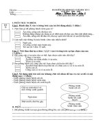
Temporal lobe
Lateral sulcus
I
Surface of the Brain I
1.1. Examine the surface of the cerebral cortex and note the grooves, or
sulci. The deepest and longest groove on the lateral surface of the
cerebral cortex is the .
1.2. In the 17th century, the French anatomist Francois Sylvius first
recognized the sulcus as a horizontal groove separating
the lobe inferiorly from the frontal and parietal lobes
superiorly.
1.3. On the diagram provided, locate and label the major groove
running horizontally just superior to the temporal lobe.
CEREBRUM
1
1
GRBT251-3375G-C01[01-14].qxd 10/10/07 15:29 Page 1 Aptara Inc.
Lateral sulcus
(Sylvian fissure)
I
CEREBRUM
2
1.1A. lateral sulcus (also known as the Sylvian fissure)
1.2A. lateral; temporal
1.3A.
GRBT251-3375G-C01[01-14].qxd 10/10/07 15:29 Page 2 Aptara Inc.
1.4. Identify and label the superior temporal sulcus, located parallel and
inferior to the lateral sulcus.
1.5. To locate the superior temporal sulcus, connect the three dots on the
diagram and label the sulcus.
1.6. The preceding diagrams showed the left side of the cerebral cortex;
this diagram shows the right side. Draw and label the superior temporal
sulcus.
1.7. The superior sulcus takes its name from its location on the part of
the temporal lobe.
1.8. The lateral sulcus is deeper than the sulcus immediately inferior
and parallel to it, termed the sulcus.
1 ■ Surface of the Brain I
3
GRBT251-3375G-C01[01-14].qxd 10/10/07 15:29 Page 3 Aptara Inc.
Superior temporal sulcus
I
CEREBRUM
4
1.4A.
1.5A.
1.6A.
1.7A. temporal
; superior
1.8A. superior temporal
Superior temporal sulcus
Superior temporal sulcus
GRBT251-3375G-C01[01-14].qxd 10/10/07 15:29 Page 4 Aptara Inc.
1.9. The sulcus is deeper than the superior sulcus.
1.10. The worm-like convolutions of the cortex are gyri (singular is gyrus).
Each is bounded by sulci.
1.11. The grooves on the cortex are called ; the convolutions between the grooves are
called .
1.12. The lateral sulcus and the superior temporal sulcus enclose a between them.
1.13. Adjacent gyri appear to be separated on the surface of the
cortex, but as the diagram shows, the surface is actually infolded,
and adjacent gyri are at the base of each sulcus.
1.14. More than half of the surface of a gyrus is hidden from view in
the walls of two adjacent . On the cut surface, blacken the
hidden portions of the gyrus labeled “G.” The most superior region of
the area you blackened is hidden in the wall of the
sulcus; label this sulcus on the diagram.
1 ■ Surface of the Brain I
5
G
GRBT251-3375G-C01[01-14].qxd 10/10/07 15:29 Page 5 Aptara Inc.
I
CEREBRUM
6
1.9A. lateral; temporal
1.10A. gyrus
1.11A. sulci
; gyri
1.12A. gyrus
1.13A. continuous
1.14A. sulci; superior temporal
Superior temporal sulcus
G
GRBT251-3375G-C01[01-14].qxd 10/10/07 15:29 Page 6 Aptara Inc.
1.15. Inferior to the lateral sulcus, the superior temporal gyrus forms the
superior part of the lobe. Label the gyrus with its name.
1.16. On the diagram, label the:
lateral sulcus
superior temporal sulcus
superior temporal gyrus.
1.17. Inferior to the lateral sulcus is the lobe.
1.18. The cerebral cortex has other lobes in addition to the temporal lobe.
One of these other lobes, named for its location in the frontal portion of the
brain, is the lobe.
1.19. Three parallel sulci run from the superior surface of the cortex
inferiorly to the lateral sulcus. Locate these three sulci, and draw a single
circle around the group of them on the diagram (they are located between
the broken lines).
1 ■ Surface of the Brain I
7
GRBT251-3375G-C01[01-14].qxd 10/10/07 15:29 Page 7 Aptara Inc.
Lateral
sulcus
Superior
temporal
gyrus
Superior
temporal
sulcus
Superior temporal
g
yrus
I
CEREBRUM
8
1.15A. temporal
1.16A.
1.17A. temporal
1.18A. frontal
1.19A.
GRBT251-3375G-C01[01-14].qxd 10/10/07 15:29 Page 8 Aptara Inc.
1.20. The central sulcus forms the posterior boundary of the lobe.
1.21. The three sulci enclose two .
1.22. The two gyri on either side of the central sulcus are logically named the precentral and
central sulci.
1.23. The gyrus between the central and precentral sulci is the central gyrus.
1.24. Of the two gyri bordering the central sulcus, the one in the frontal lobe is
the gyrus.
1.25. The gyrus posterior to the central sulcus is the gyrus. The gyrus just
anterior to the central sulcus is the gyrus.
1.26. The gyrus farthest posterior on the frontal lobe is the gyrus.
1.27. The lobe directly posterior to the frontal lobe is the parietal lobe.
The postcentral gyrus is in the lobe.
1 ■ Surface of the Brain I
9
GRBT251-3375G-C01[01-14].qxd 10/10/07 15:29 Page 9 Aptara Inc.