nghiên cứu hiệu quả điều trị tẩy trắng răng sống nhiễm sắc tetracycline bản tóm tắt tiếng anh
Bạn đang xem bản rút gọn của tài liệu. Xem và tải ngay bản đầy đủ của tài liệu tại đây (458.44 KB, 32 trang )
1
THESIS INTRODUCTION
ACKNOWLEDGEMENT
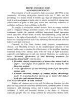
Discoloration of teeth occupied a high percentage (86.90%) in the
community, including tetracycline stained teeth was 22.86% and this
percentage was mainly found in middle age. Sign of tetracycline stained
teeth is serious changes of teeth color in which, normal teeth change into
yellow-brown or green or dark grey colors, this obviously influences to
aesthetics and causes less confidence in the life.
To treat to a tetracycline stained denture, there are many methods
such as composite fillings, porcelain veneers, full-coverage crowns. These
treatments require the patients suffering intervened dental operations
which cause loss of hard tissue of teeth, time-consuming, costly and this
acquires to be replaced periodically. A protocol of less invasive for hard
tissue but changing color of teeth is by whitening discolored teeth with the
help of cold light activation.
Currently in Viet Nam, further study projects, comprehensive
clinical, safe bleaching protocol on the morphological structure of the
enamel surface and evaluation the effectiveness of the in-office bleaching
treatment tetracycline stained teeth with the help of light activation;
especially cold light source is still very limited. Therefore, we conducted a
thesis: “Research effectiveness of vital bleaching treatment for
tetracycline stained teeth” with the aims:
1. Describle clinical characteristics of tetracycline stained teeth at
degree I and degree II on patients who were treated by vital
bleaching method.
2. Assess effectiveness of vital bleaching method for tetracycline
stained teeth.
3. Evaluate structural changes of enamel surface morphology
under the scanning electron microscopy on tetracycline stained
teeth after experimental bleaching.
THE NECESSARIES OF THE THESIS
Denture of a person serves not only for chewing function, but also
plays an important role in his/her pronunciation, communication in social
community with charming smiles, and it makes people become more
confident. A white and beautiful denture is the desire of everyone,
1
1
2
especially people who are being suffered by tetracycline stained teeth.
How is clinical characteristics of degree I and degree II tetracycline
stained teeth? How efficacy do tetracycline stained teeth bleach? How do
enamel surface morphology change under the scanning electron
microscopy on tetracycline stained teeth after experimental bleaching?
Those need to be examined, identified, in order to contribute to a safe and
effective bleaching protocol for tetracycline stained teeth relying on
surface of enamel structure.
PRACTICAL SIGNIFICANCE & NEW CONTRIBUTION
1. Description of the clinical characteristics of degree I and degree II
tetracycline stained teeth. Identified indicators of the Munsell color
spectrum and CIE La*b* color space for each degree of tetracycline stain.
2. Affirm vital bleaching treatment for tetracycline stained teeth achieve high
effectiveness and safe on structure of enamel surface morphology.
3. There are specific evidences of the bleaching agents based on trials
performed in different environments on enamel surface morphology.
4. Establish a safe and effective bleaching protocol for surface of enamel
structure of tetracycline stained teeth.
5. Apply a colorimetric technique by Vita Easyshade compact
spectroradiometers in diagnosis and treatment of a stained denture.
6. Affirm effectiveness of the warning of teeth staining by tetracycline in the
community.
THESIS STRUCTURE
Thesis consists 146 pages, apart from the acknowledgement,
conclusion, and proposal consist 5 pages, the thesis consists of 4 chapters:
Chapter 1: General study issue (including 34 pages), Chapter 2: Research
objectives and methodologies (including 30 pages), Chapter 3: Research
results, (including 37 pages), Chapter 4: Discussion, (including 40 pages).
The thesis comprises 40 tables, 28 charts, 40 photos, 156 references (16 of
which were in Vietnamese, and other 140 were in English).
B. THESIS CONTENTS
Chapter 1. OVERVIEW
1.1. Histological structure of tooth enamel definition.
1.1.1. Histological structure of tooth enamel
2
2
3
Organic forms: Mature enamel consists mainly soluble and insoluble
proteins and a small amount of fat and carbohydrates. Teeth staining are
pigments combined with fatty acids, proteins, enzymes, on the enamel
surface, it contains long chain links, alternating single link chains and
double link chains. Bleaching has used hydrogen peroxide (H
2
O
2
) to
remove proteins. Observation under scanning electron microscopy (SEM),
H
2
O
2
decomposes into free radicals causing oxidation of fatty acids and
proteins, cleavaging long - chain links into the small branches, and the
proteins on the enamel surface are removed.
1.1.2. Metabolic process of tooth enamel: Enamel has a role as a semi
permeable membrane and an ion - exchange.
1.2. Color of teeth and causes of teeth staining.
1.2.1. Color of teeth.
Munsell color spectrum: Hue (h) is a property that allows distinguishing
colors. Value (V) indicates the sensitivity or brightness of a color range
from pure black to pure white. Chroma (C) is only the level of color
saturation and intensity description, the intense or glare of the color.
CIELa*b* color space: Light and dark axis value (L) is a unit of
brightness of the object (L: 0 - 100), green - red axis value (a*) is the unit
of measurement red (+a*) or green (-a*), blue - yellow axis value (b*) is
the unit of measurement yellow (+b*) or blue (-b*).
1.2.2. The causes of tooth staining.
1.2.2.1. Extrinsic stain.
1.2.2.2. Intrinsic stain.
1.2.3. Tetracycline stained teeth.
1.2.3.1. Epidemiology: Tetracycline staining in the world community as
well as in Vietnam is quite high.
1.2.3.2. Mechanism of tooth discoloration caused by tetracycline staining:
Tetracycline molecules combine organic chassis to create a insoluble and
stable compound which leads to discoloration of enamel and dentin
structure. Light causes oxidasation to yellow in tetreacycline stained teeth
and forms purple color.
1.2.3.3. Clinical characteristics of tetracycline stained teeth.
Jordan and Boksman (1984) showed that lesions are yellow striped lines,
corresponding to development lines in dentin scale and creating yellow
3
3
4
fluorescence trips under ultraviolet light. Tetracycline stained teeth were
divided into 4 degrees.
1.3. Effectiveness of vital bleaching.
1.3.1. Whitening agent.
1.3.1.3. Mechanism of whitening agent: In bleaching process, H
2
O
2
is a
final product. H
2
O
2
diffuses through the organic matrix of the enamel,
dentin and decomposing into free radicals (HO
2
*, HO*, O*) with single
electrons, they have enormous energy and instability and they will
combine with organic molecules to achieve stability. So pigments can be
broken from large molecules into small molecules and they could be
removed easily by diffusion of substances into simpler molecules that
reflect less light therefore they obtain bleaching effectiveness.
1.3.2. Vital bleaching methods
1.3.3. Studies of vital bleaching results in the world and in Vietnam
1.3.3.1. In the world: Summary of clinical studies and in vitro experiments
has shown that: Safety and efficacy of general bleaching teeth and
particular tetracycline stained teeth focuses on at-home bleaching
techniques. Several case reports in-office tetracycline stained teeth
bleaching gave good results at the end of treatment period, but time
tracking was not long enough, there was not any systematic study and
there was not any safe and effective protocol applied for a vital teeth by
tetracycline staining bleaching protocol on the enamel morphological
issued. Therefore, the research required to clarify this issue.
1.3.3.2. Vietnam: Until now, in Vietnam there were not any reports on
effective vital bleaching tetracycline stained teeth systematically and
comprehensively on clinic and experiment.
Chapter 2. STUDY OBJECTS and METHODOLOGIES
The study consists of 2 components: Clinal and experimental components.
2.1. Clinical design
2.1.1. Research object
2.1.1.1. Inclusion criteria: The patients with degree I and II tetracycline
stained teeth (Classification I by Jordan and Boksman: Uniform light
yellow, brown, or gray stain confined to incise three quarters of the crown,
B3 - B4 corresponding. Classification II: Deep yellow, brown or gray
4
4
5
stain, without banding in cervical tooth, C3 - C4 corresponding). Incisors
and premolars are not enamel hypoplasia. Irrespective of sex and age, age:
over 18. History of using tetracycline before 12 years. As citizens of
Vietnam. Patients who agreed to cooperate in the study.
2.1.1.2. Exclusion criteria: More sensitive teeth. Incisors have been falling
gingival and cervical opening. Allergy to any component of bleaching
agents. The patients have teeth staining but filled composite, cosmetic
prosthesis for incisors. Teeth stains due to other causes. Degree III and IV
tetracycline stined teeth. Enamel hypoplasia. More teeth decay. Pregnancy
and breast feeding. The patients who have acute systemic diseases.
Children under 18 years old. History of prior tooth whitening. Incisors had
endodontic treatment.
2.1.2. Research location & time
Research location: Institute of Odonto - Stomatology under Ha Noi
Medical University, National hospital of Odonto - Stomatology. Dental of
108 Military Central hospital.
Time: From 01/ 2010 to 01/ 2013.
2.1.3. Research methodologies
2.1.3.1. Research design
Intervention of uncontrolled clinical trials, the following before - after
comparison time series at 8 times.
2.1.3.2. Research sample
Using formula for comparison of 2 rates. p
1
value is the proportion of
patients with good results at the begin of treatment (p
1
= 0 for all patients
in a state that not color well), prognosis for rate change after each
intervention was 20% of patient will improve your teeth situation (p
2
=
0,2), the intervention is 3 times.
z
(1- α/2)
: The trusted coefficient at probability rate 95% (= 1,96).
1-β
: Sample force (= 90%)
5
5
6
p : (p1+p2)/2.
n = 37. The research conducted stratified analysis of treatment
effectiveness in 2 groups of degree I and II tetracycline stain (sample size
for each degree tetracycline stain), so sample size is counted as 74,
actually, the sample size of this study was 78.
2.1.3.3. Sample selection
In each group of degree I and II tetracycline stain, the study selected
randomly patients who satisfied with above criteria and consented to
participate in this research.
2.1.4. The study protocol
2.1.4.1. Making information gathering forms
2.1.4.2. Clinical examination
Classification of patients with degree I and II tetracycline stained teeth
according to above criteria.
2.1.4.3. Steps of bleaching
Step 1: Against sensitive dentin by sucking toothpaste with 5% KNO
3
(Sensodynefresh Mint) before 30 minutes. Step 2: Prepare (protect lip,
cheek, eyes, and mucosa). Step 3: Enamel opening 5 minutes. Step 4:
(cycle 1) Put the gel 35% H
2
O
2
(9 minutes). Step 5: Repeat step 4 except
the default projector lamp for 8 minutes in cycle 2 and 3. Step 6: Finishing
of treatment cycle. Step 7: Evaluate results immediately after treatment.
Step 8: Directive patients. Performing is 3 phases, 1 week after the other
each (phase 2 and 3 do not use enamel opening). Finishing of bleaching
protocol: Brushing your teeth with toothpaste demineralization (Colgate
sensitive Pro - Relief) 4 weeks.
2.1.5. Assessing the effectiveness of treatment
2.1.5.1. Evaluation of color change
The indicators change color in the Munsell color spectrum and CIELa*b*
color space: ∆C, ∆h, ∆V, ∆b*, ∆L, ∆a*, ∆E. According to American
Dental Association, the gold standard for assessing the effectiveness of
whitening products is ∆E ≥ 4.
2.1.5.2. Assessing the result of treatment
Based on these criteria: Color change according to Vita and the side effects
of the bleaching products (sensitive, mucosal and gingival lesions) and
level of satisfaction of patients, this study divided the good, rather and
6
6
7
average results. Each patient has a follow-up vote bleaching protocol and
recorded after each follow - up visit.
2.1.6. Research variables: The independent variable is the individual
characteristics of the patient. Dependent variable: Munsell color spectrum
(V, C, h) and CIELa*b* color space (L, a*, b*) in the first votes. The color
change in the Munsell color spectrum and CIELa*b* color space ∆C, ∆h,
∆V, ∆L, ∆a*, ∆b* và ∆E in the nexts. 7 evaluations. Evaluation treatment
outcome: Good, rather, average.
2.1.7. Patient observation, management & study data collection
Data collection through 8 evaluations: Before treatment, treatment 1
st
, 2
nd
,
3
rd
, 3 weeks, 3 months, 6 months, 12 months through measure indicators
repeated by Vita Easyshade compact. Observation and examination were
done according to the above criteria. Data were recorded in details on
monitoring forms.
2.2. Experimental research
2.2.1. Research objects
2.2.1.1. Inclusion criteria: Tetracycline stained teeth, premolars and
incisors were extracted for orthodontic reason, inflammation periodontal,
the root teeth completely closed, the teeth are not decay, not cracked, not
filled, age between 18 to 45 years.
2.2.1.2. Exclusion criteria: Teeth stain due to other causes, the teeth are
decay, cracked, filled, enamel hypoplasia, the root teeth imcompletely
closed.
2.2.2. Research location & time
Research location: Institute of Odonto - Stomatology under Ha Noi
Medical University, National hospital of Odonto – Stomatology, Dental of
108 Military Central hospital, 69 Hospital - The Steering Command.
Time: From 01/ 2010 to 01/ 2013.
2.2.3. Research method
Research design: Experimental study, morphology description under the
SEM.
Sample size: Using all of teeth was extracted and achieved above criteria.
This study was 95 teeth.
Sample choice: Randomized divide into 3 groups.
2.2.4. Study process
7
7
8
Encryption 3 groups: Group 1: Bleaching with 35% H
2
O
2
, and immersion
in artificial saliva. Group 2: Bleaching with 35% H
2
O
2
, immersion in
artificial saliva, brushing your teeth with SensodyneFresh Mint toothpaste
daily. Group 3: Bleaching with 35% H
2
O
2
, immersion in artificial saliva,
brushing your teeth with SensodyneFresh Mint toothpaste daily and
bleaching finish brushing your teeth with Colgate sensitive Preo –Relief 2
weeks.
Research on SEM: After an experiment done, teeth samples were fixed in
aluminum column for SEM evaluations and then dehydrated, dried in the
environment at 37ºC (room temperature) in a closed tank with silica gel for
about 12 hours, samples then were fixed into a base and covered with gold
(deskII, Dentor Moorestown, NJ, United States) for 80 seconds and
examined under SEM (JEOL, Tokyo, Japan) with voltage 15 KV.
Analysis of the results, comparison between the bleached enamel and
control group in the magnification x750, x1500, x2000.
2.2.5. Assessing the result
According to Le Van Son et al (2011) change in the enamel surface
following lesion degree into 4 types: No lesion (degree 0): No change in
the enamel surface in the control group. Mild lesion (degree 1):
Dominantly, enamel surface morphology changed slightly, alternating as
the change of middle and rare severity. Injury medium (degree 2):
Dominantly, the teeth surface morphology changed middle, alternating as
the change of light and severity changes. Severe lesion (degree 3):
Dominantly, the teeth surface morphology changed severity, alternating as
the change of light and moderate.
Treatment efficacy: Efficacy: Enamel surface morphology unchanged or
slightly (degree 0 and degree 1). No efficacy: Enamel surface morphology
changed with moderate and severe (degree 2 and degree 3).
2.2.6. Study variables: Dependent variables: Change in the enamel surface
following lesion degree into 4 types: No lesion (degree 0), Mild lesion
(degree 1), Injury medium (degree 2), Severe lesion (degree 3). Treatment
efficacy: Efficacy and no efficacy.
2.3. Remedies error: Measures to be applied to limit errors from
sampling, using Easyshade compact colorimetric, and data processing.
8
8
9
2.4. Data processing: Data were collected and analyzed by the method of
biostatistics, using SPSS 16.0 software and statistical algorithms.
2.5. Ethic research: Before enrolled in this study, all patients were fully
explained, thorough. Patients were understood the results and side effects
that might occur. Patients were understood the bleaching protocol, and
agreed to receive treatment and to follow procedures. Patients voluntarily
signed for the study participation. The information collected by the
patients was kept confidentially and was only used for research purposes.
This research applied to improve the aesthetics and health protection for
patients but not for other purposes.
9
9
10
Chapter 3. RESEARCH OUTCOMES
3.1. Clinical characteristics of degree I and II tetracycline stained
teeth
3.1.1. General characteristics of study subjects
Total of 78 patients were studied: Percentage of female patients (78.2%)
was higher than male patients (21.8%). A group of age 20 - 29 (20.5%)
was lower than a group of age 30 - 45 (79.5%). The rate of degree II
tetracycline stained teeth in a group of age 30 - 45 was 97.4%. Degree II
tetracycline stained teeth was mainly in a group of age 20 - 29 (15/16
cases).
3.1.2. Clinical characteristics degree I and II tetracycline stained teeth
Table 3.5. Distribution of colors in the Munsell (C, h, V) color
spectrum according to dental groups
Dental group n
V C h
± SD ± SD ± SD
Incisor 78 13,2 ± 1,8 27,8 ± 4,3 79,4 ± 4,9
Canine 78 15,5 ± 0,5 31,6 ± 5,8 75,9 ± 4,2
Premolar 78 12,1 ± 1,7 23,8 ± 3,7 81,7 ± 2,9
p (ANOVA test) < 0,01 < 0,01 < 0,01
General 78 13,6±1,2 27,7±4,3 80,0±3,8
Vita V, Chroma C: Canines were highest and premolares were lowest,
there was statistical significant (p<0,01). Hue h: Canine were lowest and
premolar were highest, there was statistical significant (p<0,01).
Table 3.6. Distribution of colors in the CIELa*b* color space
according to dental groups
Dental group n
L a* b*
± SD ± SD ± SD
Incisor 78 71,0 ± 4,2 5,0 ± 1,9 27,3 ± 4,4
Canine 78 65,4 ± 6,5 7,2 ± 1,4 30,6 ± 6,0
Premolar 78 73,2 ± 3,6 3,3 ± 0,9 23,5 ± 3,8
p (ANOVA test) < 0,01 < 0,01 < 0,01
General 78 69,9±4,6 5,2±1,3 27,1±4,4
10
10
11
Light L: Canines were lowest and premolars lars were highest, there was
statistical significant (p < 0,01). Redness a*, yellowness b*: Canines were
highest and premolar were lowest, there was statistical significant (p <
0,01).
11
11
12
Table 3.7. Distribution of colors L, a*, b*, C, h, V according to degree
tetracycline stain
Degree n
L a* b* C h V
±
SD
± SD ±
SD
±
SD
± SD ± SD
Degree I
3
9
73,4±1,
2
4,1±0,
4
26,8±1,
0
27,1±1,
0
81,4±0,
9
12,4±0,
2
Degree II
3
9
66,3±3,
9
6,2±1,
0
27,4±6,
2
28,4±5,
9
76,6±4,
0
14,8±0,
5
p(Mann-Whitney
test)
< 0,05 <0,05 >0,05 >0,05 <0,05 <0,05
L, h: Degree II was always lower than degree I, there was statistical
significant
(p < 0,05). V, a*: Degree II was always higher than degree I, there was
statistical significant (p < 0,05). C, b*: There was no difference between
the 2 degrees
(p > 0,05).
3.2. Assessment of the effectiveness of the vital bleaching treatment for
tetracycline stain
3.2.1. Evaluation of tooth color change
3.2.1.1. Evaluation of tooth color change in the Munsell color spectrum
The average value of ∆C, ∆h, ∆V at 1
st
, 2
nd
, 3
rd
treatment: C, V reduced
and h rised between 3 groups in which the change of the premolars were
lowest, there was statistical significant (p < 0,01).
The change in Chroma C: General for research subjects and for all 3
groups of teeth (incisors, canines, premolars): C reduced over time
intervention and follow-up, there was statistical significant (p < 0,01).
Over 12 months of follow-up, C reduced average: General for all 3 dental
groups were 11.3 units; incisors were 12.9 units; canines were 14.3 units;
premolars were 6.8 units. Average value of C at evaluation 8 times: from
12
12
13
1
st
to 4
th
: There was significant reduction (average reduction 12.1 units).
Next times: Average value was relatively stable.
Table 3.12. The change of Vita V (∆V)
Dental
group
n =78
Before
treatment
1
st
2
nd
3
rd
3
weeks
3
months
6
months
12
month
s
p(A
NO
VA
test)
± SD
±SD
±SD
± SD ±
SD
±
SD
±
SD
± SD
Incisor
0,0
-
3,3±2,
1
-7,2
±2,1
-
10,3±2,
0
-
10,3±2
,0
-10,2
±2,2
-10,2±
2,2
-
10,1±2,
2
<
0,01
Canine
0,0
-
2,2±1,
6
-
6,7±2,
1
-
11,3±2,
1
-
11,3±2
,1
-
10,5±2
,1
-
10,1±2,
1
-
9,7±2,2
<
0,01
Premola
r 0,0
-
1,9±1,
8
-5,6
±2,2
-8,7 ±
1,9
-8,7 ±
1,9
-7,5
±2,1
-7,1
±2,2
-
6,8±2,2
<
0,01
General
0,0
-2,5
±0,9
-6,5
±1,8
-10,1
±1,6
-10,1±
1,6
-9,4
±1,7
-9,1
±1,7
-
8,9±1,7
<
0,01
General for all research subjects and for all 3 groups of teeth (incisors,
canines, premolars): V reduced over time intervention and follow-up, there
was statistical significant (p < 0,01). Over 12 months of follow-up, general
V index was average reduction of 8.9 units, incisors were average
reduction 10.1 units, canines were average reduction 9.7 units, premolars
were average reduction 6.8 units. Average value of V at evaluation of 8
times: from 1
st
to 4
th
: There was significant reduction (average reduction
10.1 units). Next times: Average value was relatively stable.
The change of hue (∆h): General for all research subjects and for all 3
groups of teeth (incisors, canines, premolars): h increased over time
intervention and follow-up, there was statistical significant (p < 0,01).
Over 12 months of follow-up, general h index was average increase of 8.4
units, incisors were average increase of 9.4 units, canines were average
increase of 10.7 units, premolars were average increase of 5.3 units, there
was statistical significant (p < 0,01). Average value of h at evaluation of 8
13
13
14
times: From 1
st
to 4
th
: There was significant increase (average reduction
10.2 units). Next times: Average value was relatively stable.
3.2.1.2. Evaluation of tooth color change according to CIELa*b* color
space
The average value of ∆L, ∆ a*, ∆b* at 1
st
, 2
nd
, 3
rd
treatment: There was
change in the light L, the redness a*, and the yellowness b* between 3
dental groups (incisors, canines, premolars) during 1
st
, 2
nd
, 3
rd
treatment,
statistically there was statistical significant (p < 0,01). The change of
premolars was lowest and the change of canines was highest, there was
statistical significant (p < 0,01).
The change of light (∆L): for all 3 groups of teeth (incisors, canines,
premolars): L increased over time intervention and follow - up, there was
statistical significant (p < 0,01). Over 12 months of follow-up, general L
index was average increase 7.7 units, incisors were average increase of 7.1
units, canines were average increase of 11.9 units, premolars were average
increase of 4.2 units, there was statistical significant (p < 0,01). Average
value of V at evaluation of 8 times: From 1
st
to 4
th
: There was significant
increase (average increase 8.7 units). Next times: Average value was
relatively stable.
The change of yellowness (∆b*): For all 3 groups of teeth (incisors,
canines, premolars): b* reduced over time intervention and follow - up,
there was statistical significant (p < 0,01). Over 12 months of follow - up,
general b* index was average reduction of 10.7 units, incisors were
average reduction of 12.1 units, canines are average reduction of 13.4
units, premolars were average reduction of 6.5 units, there was statistical
significant (p < 0,01). Average value of b* at evaluation of 8 times: from
1
st
to 4
th
: There was significant reduction (average reduction of 11.5 units).
Next times: Average value was relatively stable.
The change of redness (∆a*): For all 3 groups of teeth (incisors, canines,
premolars): a* reduced over time intervention and follow - up, there was
statistical significant (p < 0,01). Over 12 months of follow - up, general a*
index was average reduction of 4.4 units, incisors were average reduction
of 4.8 units, canines were average reduction of 6.1 units, premolars were
average reduction of 2.4 units, there was statistical significant (p < 0,01).
Average value of a* at evaluation of 8 times: From 1
st
to 4
th
: There was
14
14
15
significant reduction (average reduction of 4.9 units). Next times: Average
value was relatively stable.
Average value of ∆E at 1
st
, 2
nd
, 3
rd
treatment: ∆E in 3 dental groups
(incisors, canines, premolars), there was different at each time of treatment
(at 1
st
, 2
nd
, 3
rd
), there was statistical significant (p < 0,01) in which the
increase of the premolars were lower and the canines were highest.
Table 3.19. The color change according to ∆E color space
Dental
group
n = 78
Before
treatm
ent
1
st
2
nd
3
rd
3
weeks
3
mont
hs
6
mont
hs
12
months
p(AN
OVA
test)
±
SD
± SD
± SD
±
SD
± SD
± SD
±
SD
±
SD
Incisor
0,0 8,8±2,6 12,7±3,3 16,0±2,3 16,1±2,316,2±2,3
16,0±2,
3
15,6±2,4 < 0,01
Canine
0,0 10,3±2,916,6±3,7 21,0±3,8 20,9±3,820,7±3,7
20,3±3,
6
19,7±4,0 < 0,01
Premola
r
0,0 4,5±2,3 7,8 ±2,7
11,4 ±
2,5
11,4±2,411,9±2,49,6 ±3,2 8,9±3,3 < 0,01
General
0,0 7,9 ±1,712,3 ±2,716,1 ±2,316,1± 2,2
16,2
±2,4
15,3
±2,5
14,7
±2,7
< 0,01
General for all research subjects and for all 3 groups of teeth (incisors,
canines, premolars): ∆E increased over time intervention and follow - up,
there was statistical significant (p < 0,01). Over 12 months of follow - up,
general ∆E was average increase of 14.7 units, incisors were average
increase of 15.6 units, canines were average increase of 19.7 units,
premolars were average increase of 8.9 units, there was statistical
significant (p < 0,01). Average value of ∆E at evaluation of 8 times: From
1
st
to 4
th
: There was significant increase (average increase of 16.1 units).
Next times: Average value was relatively stable, 100% achieve to efficacy
color change, efficacy of bleaching teeth.
3.2.2. Evaluation of treatment result
3.2.2.1. Results 1 time treatment: After 1
st
treatment only achieve rather
and average results, in which rather result was 44,9%, an average result
15
15
16
was 55.1%. Degree I tetracycline stain: rather result was 71.8%. This
result was higher than degree II (17.1%). The rather result of female was
50.8%, this result was higher than male (23.5%). The rather result of group
of age 20 - 29 was 87.5%. This result was higher than group of age 30 - 45
(85.9%).
3.2.2.2. Results of 2nd time treatment: After 2
nd
treatment: Good result was
85.9%, rather result was 12.8%, and average result was 1.3%. Degree I
tetracycline stain: good result was 89.7%. This result was higher than
degree II (82.1%). The good result of female was 85.2%, this result was
lower than male (88.2%). The good result of group of age 20 - 29 was
100%. This result was higher than group of age 30 - 45 (82.3%).
3.2.2.3. Results 3 time treatment: After 3
rd
treatment: Good result was
96.1%, rather result was 3.9%. Degree I tetracycline stain: good result was
94.9%. This result was lower than degree II (97.4%). The good result of
female was 98.4%, this result was higher than male (88.2%). The good
result of group of age 20 - 29 was 100%. This result was higher than group
of age 30 - 45 (95.2%).
Good result (96.1%) was maintained at time of evaluations: 3 weeks, 3
months, 6 months, 12 months. There was a male patient in group of age 30
- 45, degree II tetracycline stain achieved rather result, after 3 months
came back average result.
3.3. Evaluation of morphological changes in the surface enamel under
scanning electron microscope on tetracycline stained teeth following
experiment bleaching
Table 3.34. The morphological changes in the surface structure of
enamel after bleaching according to degree lesion to surface enamel
Lesion
Group
Degree
0
Degree
1
Degree
2
Degree
3
Total
p
(testχ
2
)
n % n % n % n % n %
Group 1
2 6,5 17 54,
8
9 29,
0
3 9,7 31 100,
0
<0,05
Group 2
4 12,
9
18 58,
1
7 22,
6
2 6,4 31 100,
0
Group 3
11 33,
3
19 57,
6
2 6,1 1 3,0 33 100,
0
Comment:
16
16
17
Lesion of enamel surface are severity and middle in group 1 (38.7%) was
highest and lowest in group 3 (9.1%). The different was statistical
significant, p < 0,05.
Table 3.35. Effective assessment of the enamel surface morphology
after experimental bleaching
Effective
Group
Effective Ineffective Total p (test χ
2
)
n % n % n %
Group 1 19 61,
3
12 38,7 31 100,
0 <0,05
Group 2 22 71,
0
9 29,0 31 100,
0
Group 3 30 90,
9
3 9,1 33 100,
0
Comment:
Efficacy on enamel surface morphology of group 3 is highest, occupying
90,9%, followed by group 1 and group 2 with 61,3% and 71%,
respectively the difference is statistically significant, p < 0,05.
Comments featured several photos of each research group
Control sample: There were Tomes pits on the enamel surface with
scratches.
Group 1: A part of the enamel surface bleached (magnification x1500), the
image: Papillary enamels were formed after bleaching. Papillary enamels
had mineralization seeds. There was a conjunction between papillary
enamels.
Group 2: The enamel surface was evident Tome concave but sharp blunt,
there were irregular spherical particles.
Group 3: Enamel surface morphology was similar to its of the control
sample but Tome holes were shallower, not clear with smooth and shiny
surface (magnificantion x750).
CHAPTER 4: DISCUSSION
4.1. Clinical significance of I, II degree tetracycline discolored teeth
4.1.1. General characteristics of the study objects
Research on 78 patients, among them the female patients were four
times higher than that of the male patients. Probably due to the aesthetic
needs of women, this result was similar to the study by Pham Thi Thu
17
17
18
Hien (2012), Nguyen Thi Phong Lan (2004). According to Phan Le Thu
Hang (2004), the female patients with tetracycline stained teeth was 1.5
times higher than that of male patients. Thus the results of this study fitted
completely.
Percentage of patients in the group of age 30 - 45 were 4 times as
many as the group of age 20 - 29. Tetrcycline stained teeth rate of type II
in the group of age 30 - 45 accounted for the highest percentage of 97.4%.
Group of age 20 - 29 mainly tetracycline contamination level of I. The
results of this study were similar to results of studies Pham Thi Thu Hien
(2012), Tran Thi Huong Giang (2008), Nguyen Thi Phong Lan (2004), this
can be explained as follows: Tetracycline antibiotics appeared in 1948 and
was used in Vietnam, popular in the late 60s and 70s. So tetracycline
prevalence in the group of age over 30 was high. After 80 decades of the
20th century there was a warning about the color of tetracycline
contamination should doctors and patients more aware when using
tetracycline limit for children under age 12 years should have fewer than
30 and to a lesser extent. Thus, the warning of tetracycline stains
community has been effective.
4.1.2. Clinical significance of tetracycline discolored teeth type I, II
Distribution of colors in the Munsell color spectrum: The Vita V,
Chroma (C): The canines were the highest and the premolar were the
lowest, the difference was statistically significant. Hue (h): the canines
were the lowest and the premolars were the highest, with statistical
significance. Colors in incisors (B4 respectively) was light gray - brown
yellow. Canines (corresponding C4 - A4) was red gray - brown yellow.
Premolars (respectively B3 - A3.5) with yellow orange red. In determining
the parameter h color on polar coordinates canines (75.9) corresponding
gray yellow red, incisors (79.4) corresponding red and yellow teeth small
(81.7) corresponding orange, was consistent with the Vita color. Thus, the
color that heterogeneity between incisors, canines and premolars, of which
the front teeth were darker than the back teeth, the difference was
statistically significant. This result is also smilar to the study of Kwon,
2012, Venkateswarlu M et al (2009). This can be explained as follows:
Tetracycline and its peer agents capable of forming complexes with
calcium ions on the surface of the hydroxy apatite crystals in bone and
18
18
19
tooth tissue. Darker dentin enamel discoloration. Light tetracycline
oxidized quinone create red. Group teeth before exposure to light easily
and easily change colors than the group that later.
Distribution of tetracycline discolored teeth in the CIELa*b* color
space: L average brightness of the canines were the lowest and of the
premolars were the highest, with statistical significance. Redness a*,
yellowness b*, average of the canine were highest and of the premolars
were lowest, with statistical significance. This result was similar to
findings of Kwon (2012) and Venkateswarlu M et al (2009), Kugel (2002).
In this study yellowness b* were higher than the average of yellowness in
Kwon’s (2012) study conducted in Japan and Kugel made in the U.S. in
2002. This difference was probably due to different races. Thus, when the
dentists whiten the teeth they need to be regardful of these parameters. To
have beautiful white teeth for patients with tetracycline teeth the dentist
should be noted when applied to teeth bleaching time for each tooth to
bring the best whitening results after bleaching to achieve similar
parameters.
Distribution colors in the Munsell color spectrum and color space
based on the degree of tetracycline stain: Degree I tetracycline teeth
have the lighter color and higher hue than degree II. Degree I have redness
lower than degree II, the difference was statistically significant. Also, the
yellowness and the chroma was no difference. The colors Vita V: Degree I
tetracycine stained teeth: yellow - gray - light brown level. Degree II
tetracycline stained teeth: yellow - gray - darker sepia level, the difference
was statistical significant. Chroma of degree I tetracycline stained teeth
(81.4) correspond to orange, tetracycline teeth degree II (76.6) correspond
to red gray yellow. This result was similar to that of Jordan and Boksman
study (1984), but not to the specific parameters for each chromosome
level, while this study gave specific indicators for each level of
chromosomal tetracycline. The clinical features will help the dentist to
have a treatment plan specific to tetracycline exposure.
4.2. Assess the effectiveness of the vital bleaching tetracycline stained
teeth 4.2.1. Evaluation of tooth color change
4.2.1.1. Rating changes color according to Munsell color spectrum
19
19
20
Chroma (C) and hue (h) increased during the treatment and follow-up with
statistical significance. The average decrease of C, h average index
increased from 1st to 4th, 4th to 8th from relatively stable. Around the
world and Vietnam was not much research on the changing bleached color
chroma and hue. Research by Joe C (2009) to assess chroma tended to
decrease after bleaching, and Hue tended to increase after bleaching, the
study only evaluated at one point after bleaching treatment white. Chroma
of less than 4.4 Joe C reduce research and hue average 3.2 units. Maybe
it's because the starting point was the choice of different patients and
different treatment times. This study selected patients with tetracycline
should be higher chroma, on the other hand treatment done 3 times to
change the above results. Furthermore, this study also analyzed the
changes in the chroma of each group of teeth (incisors, canines and
premolars). At each treatment time point 1st, 2nd and 3rd, the difference
was statistical significant changes in chroma C and the hue (h). Vita V
between the 3 treatment groups namely that premolars always have the
lowest change. This result can be explained as follows: The initial starting
point of the small teeth have low chroma, hue third highest in the group
that should have the effect of bleach was little change than the group that
has the darker color index.
The Vita V colors were reduced over time, and monitoring of treatment
was statistical significant. Over 12 months of follow - up Vita V pixels
average reduction of 8.9 common units, incisor of average reduction was
10.1 units, canine of reduction average was 9.7 units, premolar of
reduction average was 6.8 units. Case report of Kinoshita et al (2009) for
the laser teeth whitening for cases tetracycline stained teeth which color
change from C4 to B2 corresponding change compared to 12 units for the
first time, according to Tavares et al (2003), improved color compared to
13 units for the first time. While this study was to perform 3 new
bleaching treatment achieved an average change of 10 color units. The
results of many studies on vital bleaching tetracycline stain also vary
considerably, according to Matis et al (2006) bleaching method used in the
last 6 months to be able to achieve the color change from C4 to B1 15
color units, respectively. According to Nguyen Thi Phong Lan (2004) after
bleaching improves average 9 - 10 units tetracycline stained teeth to bring
20
20
21
new satisfaction for dentists and patients. In this study, 2
nd
post-treatment
colors mean reduction of 6.5 units should continue 3
rd
therapy and results
were expected by dentists and patients. According to Bowler et al (1997)
that the reason chromosomal tetracycline to treat prolonged or repeated
treatment at the clinic was able to generate by tetracycline with calcium
ions and chelating into cartilage, teeth and bones to form a tetracycline
complex - especially calcium orthophos-phate on dentin. Because of the
deposition of pigment in the deep dentin should take time and multiple
treatments to achieve desired results.
4.2.1.2. Evaluation of color change according to the CIELa *b * color
space
L increased brightness over the duration of treatment and follow - up with
statistical significance. Over 12 months of follow-up overall brightness
increased on average 7.7 unit. Yellowness b*, redness a* decreased over
the treatment period and follow - up with statistical significance. Over the
12 - month follow - up showed yellowness b* overall decreased average of
10.7 units. Redness a* general average decreased 4.4 units. The average
value of L brightness, yellowness b*, redness a* at 8 times of evaluation,
there was a marked change from the first evaluation to the 4th evaluation
(average increase of 8.7 L units, b* values decreased on average 11.5
units, thus redness a* reduced average 4.9 units. From the 4th to the 8th,
was relatively stable. According to Matis et al after 6 months of treatment
indicators change as follows: verage L change was 15 units, the average
yellowness b* reduced 2.3 units, a* reduced average 2.5 units. Results of
this study differ from the result of Matis et al can be explained that derives
color (brightness, yellowness and redness levels) in this study was higher
than the initial color index Matis et al. This was also found at in this study
to analyze each particular show that each point in the treatment of 1st, 2nd
and 3rd, the difference was statistically significant. Changes brightness L,
the redness a* and b* yellowness than before dental treatment between the
3 groups (inciscors, canines, premolars), premolars change (L is increase,
and a*, b* are decrease) the lowest and the highest were canines. Such that
the teeth have lower brightness will change more brightness, the teeth have
high redness a* and high yellow b* will decrease even more. Similar to
results as reported Kwon cases in (2012) while performing combination
21
21
22
method in - office bleaching and at - home bleaching. Past may be
explained by the following mechanism: H
2
O
2
and diffuse through the
enamel to the dentin, reacts with organic pigments was the key factor to
form the tooth color. According to Lee et al (2009) Bleaching by light
plasma was improved significantly. In - office bleaching using 35% H
2
O
2
concentrations and sources different light. Lighting accelerates
decomposition of H
2
O
2
generated free radicals OH* (double after
phototherapy for 1 minute) molecules react with surrounding color as their
structure was broken and whitening takes place. This result was a
testament to the rapid changes seen when using whitening lamp.
Results of treatment of stable only after 12 months of research similar to
Matis et al (2002) and Leonardo et al (2003). This result may be achieved
by chromosomal tetracycline was endogenous chromosomes of infected
ivory, less affected external environmental factors. End bleaching
treatment process we have implemented a data mining process for patients
brush your teeth by Colgate Sensitive Pro-Relief toothpaste enamel surface
should be shiny and smooth over, contributing to maintain the treatment
effect after whitening.
4.2.1.3. Evaluation of color change in ΔE
For all three groups of teeth (incisors, canines and premolars) ΔE increases
over the duration of treatment and follow-up with statistical significance.
Over 12 months of follow - up, ΔE overall average increase of 14.7 units,
incisors were average increase 15.6 units, canine were average increase
19.7 units, premolars average increase of 8.9 units. The average value of
ΔE at 8 points of evaluation increased, increased significantly from the 1st
to the 4th (average increase of 16.1 units). From the 4th to the 8th, the
average value of ΔE was relatively stable. Even the first time we have
treated 100% efficiency as recommended by the ADA. For tetracycline
stained teeth this change has not achieved the desired aesthetic. Therefore,
this study continued 3
rd
treatment to achieve optimal aesthetic patients.
After 12 months, the study was 100% effective as recommended by the
ADA. Between the three groups that (inciscors, canines, premolars) in
each treatment time point 1st, 2nd and 3rd, with differences statistically
significant, namely premolars increased the lowest and canines increase
the highest. The change ΔE end of treatment 3 times remarkably similar
22
22
23
Kwon results of 2012, specifically canines were 24.58, incisors were
15.45. Matis et al's study (2006) 6 months after treatment ends ΔE mean
change 16 units. Such findings were similar to results of teeth whitening
treatment chromosomal tetracycline live at home or in the clinic in
combination with at home. The color changes in patients have tetracycline
teeth higher (3 times) than bleach stains due to other causes. With a
conventional bleaching results often follow Meireles et al (2009) ΔE was
4.3 to 4.7 units by
Joe C (2009) gained 6.1 ΔE units, according to Costa (2011) Δ E 6 units
achieved.
Recently, some new research shows that in - office bleaching can give
optimal results by Kinoshita et al (2009) in Japan, he used the laser teeth
whitening treatment using 35% H
2
O
2
was once an appointment can
improve color from C4 to B2. According to Lee's (2009) plasma whitening
lamp color change ΔE 19.7 overall. This was only sporadic studies, not
only evaluate the system and at the end of treatment, so this study
conducted in 8 track time in the 12 - month evaluation in a systematic
manner and has brought efficient in the short term for patients with
tetracycline stain. Short duration of treatment for optimal results, patients
can expect a maximum change at week 2 after treatment 3 times with 16.1
ΔE units achieved similar results bleaching treatment teeth at home for 6
months. The results of this treatment can be the basis for advising patients
infected with tetracycline teeth of time can change color to achieve desired
performance after 2 weeks and 3 waves at the clinic. This result can help
dentists build bracket appropriate treatment for patients with planned
arrangements and time to comply with treatment.
According to Haywood (1997) at - home bleaching method was the
difficulty: Treatment depends entirely on the patient, dentist can not force
patients to comply with treatment, with treatment lasting several weeks or
months. The process of bleaching treatment was difficult that was sensitive
dentin. Results of treatment depends on the cooperation and compliance of
patient treatment. In fact, there were many difficulties for patients required
to comply with the bleaching process, especially for patients with tight
timescale. So at the clinic bleaching meet anticipated demand for both
patients and aesthetically time.
23
23
24
4.2.2. Evaluation of treatment outcomes
Results the 1st times treatment: results primarily fall quite tetracycline
contamination levels I, for women and group of age 20 - 29. This also
makes sense because the group of age 20 - 29 in this study mainly
tetracycline contamination level of I. The color changes in tetracycline
teeth type I moderate can bring satisfaction to patients easier than type II.
Such a result was entirely consistent.
Results the 2
nd
times treatment: Good results focus on group tetracycline
teeth type I, group of age 20 - 29 years old. For tetracycline teeth type I
and younger patients easily accept change color just right. The color
change of the average Vita II levels decreased more than the I. In
considering the criteria synthetic color changes, medication side effects
and satisfaction of patients and found that good results in a higher
tetracycline type I than type II in the treatment second. This result was
explained as follows: tetracycline stained type I have the ball and the
surface enamel golden brown lighter gray levels so that small changes can
easily make patients more satisfied than the improvement seen the eye
(can be considered significant) level II of tetracycline contamination. This
study also found the first bleaching tetracycline stain 2nd most drab
yellow, while the second tetracycline infection despite many changes color
but still light brown gray red yellow tetracycline. So with tetracycline
tooth infection, the degree of color change did not meet the treatment
needs of patients. According to Nguyen Thi Phong Lan (2004), to improve
the 9 - 10 units colors with the tetracycline stained tooth really bring
satisfaction to the patient. So we continue to treat the 3rd times to meet the
treatment needs of patients.
Results of the 3
rd
times treatment: Results of the general good research
subjects was 96.1%, 3.9% was pretty. Tetracycline stained teeth type I
have good results (94.9%) lower than tetracycline stained teeth type II
(97.4%). Female achieve better results than men. Group of age 30 - 45 less
fruitful group of age 20 - 29. This study follows three treatments reduced
the average Vita 10.1 colors. This result was similar to the results of
Nguyen Thi Phong Lan (2004). A very satisfied (96.2%) after the 3
rd
times
treatment so this study obtained a result quite ideal (well 96.1%). After
two treatments were created to facilitate the path to drug dentin impact
24
24
25
should ease in treatment the 3
rd
times to achieve outstanding results. With
three times the whitening treatment was a powerful influence on the color
molecule so infected the tetracycline stained type II significant color
change than type I.
According to Chandrasekhar et al (2011) using cold light source, 35%
H
2
O
2
system agents and Beyond light after a treatment has improved from
D3 to A1, A3 to B1 from level 9 to change the Vita. According to
Kinoshita et al (2009), using 35% H
2
O
2
with light laser whitening for
tetracycline teeth type II case for the Japanese show changes from C4 to
B2 after a course of therapy. The research has contributed confirmed
tetracycline vital teeth bleaching method at clinic was effective. Results of
this study was to clarify this issue. Vital bleaching tetracycline stained
teeth by means of in - office was efficient and time - saving treatment.
Patients with tetracycline stained teeth can achieve expectations
expectations after 3 treatments, improved smile improve the quality of life,
help them integrate into the community and communicate confidently.
Results of treatment of 3 weeks, 3 months, 6 months and 12 months: This
result was maintained through the votes 3 months, 6 months and 12
months. The positive results of this study after the end of treatment and
higher color stability similar to previous studies such as: Pham Thi Thu
Hien et al (2012) results 90% better color stability after 6 months Nguyen
Thi Phong Lan (2004) well after 1 year was 80%. Chromosomal
tetracycline teeth due to tetracycline chelating create dentin complex
creates a deep pigment in dentin to do so at the clinic bleaching 3 times to
bring good results, a longer course of therapy compared with other cases
of color. Some studies on chromosomal tetracycline teeth whitening done
at home to extend to 6 months as Leonardo et al (2003), Matis et al (2006)
and showed stable results achieved within 5 years. The results of this study
were similar stable with previous studies. To determine the stability of the
clinic bleaching on tetracycline chromosomes patients need to continue to
monitor these patients for a longer period. For consistent results, this study
re-made mineral enamel surface after the end of treatment bleaching
creams have re mineral brush (Colgate Sensitive Pro-Relief). Mineral
toothpaste with re-use technology arginine ivory pipe to clog causing
surface enamel whitening smooth, impervious to color food, so they have
25
25