Generation and analysis of KRAS v12 in driving liver tumorigenesis using transgenic zebrafish models
Bạn đang xem bản rút gọn của tài liệu. Xem và tải ngay bản đầy đủ của tài liệu tại đây (8.64 MB, 154 trang )
GENERATION AND ANALYSIS OF KRAS
V12
IN DRIVING
LIVER TUMORIGENESIS USING TRANSGENIC
ZEBRAFISH MODELS
NGUYEN ANH TUAN
(B.Sc., Vietnam National University;
University of Natural Sciences, Ho Chi Minh City)
A THESIS SUBMITTED
FOR THE DEGREE OF DOCTOR OF PHILOSOPHY
DEPARTMENT OF BIOLOGICAL SCIENCES
NATIONAL UNIVERSITY OF SINGAPORE
2011
I
ACKNOWLEDGEMENTS
The work presented in this thesis was accomplished at the Department of Biological
Sciences (DBS) and Temasek Life Sciences Labolatory (TLL), National University of
Singapore, from August 2007 to August 2011. It gives me great pleasure to acknowledge
all who made this thesis possible.
First and foremost, I would like to express my deepest and most sincere gratitude
to my supervisors, Prof. Gong Zhiyuan (DBS), Dr. Serguei Parinov (TLL) and Dr.
Alexandre Emelyanov (TLL) for their innovative insights, valuable guidance, unending
support, perpetual encouragements throughout my study. Their enormous scientific
experience together with logical way of thinking have been of great value for me and also
provided a good basis for the present thesis.
I wish to appreciate Prof. Chou Loke Ming (DBS) for his coming to Vietnam to
interview and offer me an invaluable opportunity to be NUS scholar.
I owe my most sincere thanks to Dr. Koh Chor Hui Vivien (DBS) for her truthful
friendship and great help that have remained consistent during my study.
I warmly thank Dr. Jan Spitsbergen (University of Oregon) and Dr. Lam Siew
Hong (DBS) for their professional advices and technical instructions in my research. I
also appreciate the helps from many friends and colleges, including Yan Tie, Huiqing
Zhan, Cecilia Winata, Lana Korzh, Zhen Li, Tiweng Chew, Bing Liang, Lili Sun, Helen
Quach, Long Tran, Shimin Lim, Kumari Pooja, Thiet Vu and Lam Dang during my
study. I would also like to thank DBS Graduate Officers, especially Ms. Reena and Ms.
Priscilla, for their dedicated helps during the course of my study. Many thanks to all
members in Prof. Gong laboratory (DBS), Dr. Karuna laboratory (TLL), Dr. Yue
II
labolatory (TLL), TLL fish facility, GIS microarray facility and Biopolis shared facilities
for all their helps and support in this thesis.
My most heartfelt gratitude goes to my dearest Family members. Thanks to my
grandmother as my great mentor, together with my parents, aunts and sisters for their
loving considerations and great believe in me all through these years. Most importantly, I
feel so proud and lucky to have my most faithful partner, Quang Duc Dao, whose loving,
understanding, supporting and accompanying me throughout all these years have given
me strength to finish this study. Therefore, I wish to dedicate this thesis to my Family.
Last but not least, I greatly acknowledge the National University of Singapore
for awarding me the Graduate Research Scholarship.
Singapore, 27
th
June 2011
III
TABLE OF CONTENTS
Acknowledgements………………………………………………………………. I
Table of contents………………………………………………………………… III
Summary………………………………………………………………………… VIII
List of tables………………………………………………………………………. XI
List of figures …………………………………………………………………… XII
List of common abbreviations…………………………………………………… XIV
Chapter 1: Introduction………………………………………………………… 1
1.1 Introduction to human liver cancer…………………………………… 2
1.1.1 Incidence, epidemiology and risk factors……………………… 2
1.1.2 Current trends in therapeutic strategies of human HCC…… … 5
1.2 Zebrafish as a liver cancer model……………………………………… 6
1.2.1 Advantageous use of the zebrafish in research…………………… 7
1.2.2 Modeling human diseases using zebrafish…………………….…… 10
1.2.3 Zebrafish models of human liver cancer: Chemical
carcinogenesis and transgenic approaches…………… 13
1.2.4 Application of conditional expression systems in transgenic
zebrafish …… 14
1.3 Oncogenic Ras in human liver cancer…………………………………… 17
1.3.1 Molecular perspective of Ras in cancer biology………………… 17
1.3.2 Association of Ras with HCC………………………………….…. 20
1.4 Objectives and significance of the study………………………………… 22
IV
Chapter 2: Materials and Methods …………………………………………… 24
2.1 General molecular biology techniques and plasmid construction… 25
2.1.1 Polymerase chain reaction (PCR) …………………………………. 25
2.1.2 Cloning…………………………………………………………… 25
2.1.3 Isolation of zebrafish kras oncogene and construction of
Tg(fabp10:EGFP-kras
V12
) plasmid…………… ……….…… 26
2.1.4 Construction of inducible transgenic systems …………………… 29
2.2 Generation of transgenic zebrafish …………………………………… 30
2.2.1 Zebrafish maintenance……………….…………………………… 30
2.2.2 RNA synthesis, microinjection and screening of transgenic fish… 30
2.3 Gross morphological and histopathological analyses of
zebrafish tumor….….….….….….….….….….….….….….….….….… 31
2.4 Tumor screening for the inducible systems….….….….….….….….… 33
2.5 Transplantation of liver tumors into wild-type zebrafish ….….….….…33
2.6 Isolation of total RNA/genomic DNA and
reverse transcriptase/quantitative real-time/genotyping PCR….….……… 34
2.6.1 Isolation of total mRNA….….….….….….….….….….….….…… 34
2.6.2 Reverse transcriptase PCR….….….…….….….…….….….……… 34
2.6.3 Quantitative real-time PCR….….….…….….….…….….….…… 35
2.6.4 Isolation of genomic DNA and genotyping PCR….….….……… 38
2.7 Western blot analysis….….….…….….….…….….….…….….….…… 39
2.8 Immunohistochemistry….….….…….….….…….….….…….….….…… 40
2.9 Cellular senescence and cell death analyses….….….…….….….………… 40
V
2.10 Zebrafish oligonucleotide microarray construction and hybridization…… 41
2.11 Transcriptomic analyses….….….…….….….…….….….…….….….…….42
2.12 Inhibitor treatment….….….…….….….…….….….…….….….…………. 44
2.13 Statistical analyses….….….…….….….…….….….…….….….……… 45
Chapter 3: Results….….….…….….….…….….….…….….….…….….….…….46
3.1 Analysis of constitutive liver-specific expression of oncogenic
kras
V12
in driving liver tumorigenesis in transgenic zebrafish….………… 47
3.1.1 Generation of Tg(fabp10:EGFP-kras
V12
) transgenic zebrafish……. 47
3.1.2 High level of kras
V12
expression led to early lethality and
induced HCC….….….…….….….…….….….…….….….……… 51
3.1.3 Transplantability of kras
V12
liver tumors in WT recipients………… 56
3.1.4 Differential activation of ERK, JNK and p38 mitogen-activated
protein kinase pathways during kras
V12
liver tumorigenesis…… 58
3.1.5 Activation of the Wnt/β-catenin pathway during kras
V12
liver tumorigenesis….….….…….….….…….….….…….….….…. 62
3.1.6 Acceleration of Liver tumor onset by loss of p53-mediated
senescence….….….…….….….…….….….…….….….…….……. 64
3.1.7 Transcriptomic analyses of kras
V12
liver tumorigenesis…… …… 68
3.1.8 Identification of a HCC-specific signature and a liver cancer
progression signature….….….…….….….… …… 71
3.2 Development and analysis of mifepristone-inducible and -reversible
kras
V12
liver tumorigenesis in transgenic zebrafish……… 79
VI
3.2.1 System design ….….….…….….….…….….….…….….….…… 79
3.2.2 Control of liver tumor progression and regression in kras
V12
transgenic zebrafish by mifepristone administration….….….…… 82
3.2.3 Activation of ERK and AKT pathways required for
kras
V12
-driven liver tumorigenesis and tumor maintenance………. 88
3.2.4 Prevention of kras
V12
liver tumorigenesis by inhibiting ERK
and/or AKT pathways ….….….…….….….…….….….… …… 91
3.3 Development and analysis of mifepristone-inducible Cre/loxP
recombination to conditionally control kras
V12
liver tumorigenesis
in transgenic zebrafish….….….…….….….…….….….…….….….…… 94
3.3.1 System design ….….….…….….….…….….….…….….….…… 94
3.3.2 Determination of concentration- and time-dependent
mifepristone induction of Cre expression….….….…….….….…… 97
3.3.3 Mosaicism of EGFP-kras
V12
expression in Triple-Tg fish causing
hepatocellular carcinoma and other types of liver tumor……… 100
3.3.4 Deregulation of ERK and Wnt/β-catenin pathways during
kras
V12
-induced liver tumor progression….….….…….….….…… 106
Chapter 4: Discussions….….….…….….….…….….….…….….… …….….…. 108
4.1 A high level of kras
V12
expression leading to HCC in transgenic
zebrafish….….….…….….….…….….….…….….….…….….….……… 109
4.2 Conserved gene expression signatures underlying liver tumorigenesis
in humans and kras
V12
transgenic zebrafish….….….…….….….…….…… 113
VII
4.3 Mifepristone-inducible and -reversible kras
V12
system potential
for high throughput anti-cancer drug screens….….….…….….….…….…. 114
4.4 Mifepristone-inducible Cre/loxP regulating kras
V12
system induces
various liver tumors and closely mimics spontaneous cancer
development….….….…….….….…….….….…….….….…….….….…… 118
4.5 Summary and conclusions….….….…….….….…….….….…….….….…. 120
Bibliography.….….…….….….…….….….…….….….…….….….…….….… 125
Appendices
VIII
SUMMARY
Human liver cancer is one of the deadliest cancers worldwide, with hepatocellular
carcinoma (HCC) being the most common type. The neoplastic development of human
HCCs is a complex multistage process, with heterogeneity in morphology and genetics
that makes its ultimate clinical benefit negligible. Despite the relevance of HCC
malignancy, a fundamental understanding of the molecular mechanisms of
hepatocarcinogenesis is currently rather limited. As a potent proto-oncogene and bona
fide central regulator of signal transduction pathways in many human cancers, Ras is at
the leading edge of most tumorigenic events and is activated in nearly all HCC cases.
Thus, targeting Ras signaling has emerged as a potential strategy to treat advanced HCC.
However, the mechanism of Ras-induced liver cancer remains elusive and in vivo models
that enable investigations of the important role of Ras in liver tumorigenesis are lacking.
To address these problems, a constitutive transgenic zebrafish liver cancer model
was first generated using a hepatocyte-specific promoter (fabp10) to target oncogenic
kras
V12
expression to the liver. Fusion with EGFP allowed visualization of the process of
tumor development from early stages. Only high level of kras
V12
expression initiated liver
tumorigenesis. The kras
V12
tumors showed progressive features from hyperplasia to
invasive HCC which was accompanied by a loss of p53-dependent senescence response.
HCC cells derived from this line also displayed transplantability. Transcriptomic analyses
delineated several pathways and identified two conserved gene signatures accounting for
HCC specificity and HCC progression in both zebrafish and human. These findings
validated the potential of kras
V12
transgenic fish in modeling human liver cancer.
However, several limitations were found in this model such as low HCC penetrance and
IX
premature lethality due to early Ras activation.
Motivated by previous findings, another model allowing for liver-specific and
inducible EGFP-kras
V12
expression was generated using mifepristone-inducible strategy,
which allowed to induce oncogene expression at any desirable time and to accelerate
tumor onset. Robust and homogeneous HCC growth was achieved in 100% transgenics
after 1 month induction. HCC was found to be “addicted” to Ras signaling for tumor
maintenance as mifepristone withdrawal led to tumor regression via cell death. Targeting
Kras
V12
liver tumorigeneis via its downstream effectors, Raf/MEK/ERK and
PI3K/AKT/mTOR, by chemical inhibitors significantly suppressed the over-growth of
hyperplastic liver in EGFP-kras
V12
larvae. Collectively, this model offered an effective
and predictable liver cancer model for large-scale studies.
It is well known that human cancer is usually initiated by a sporadic event of
mutations occurring in a single or group of cells. Therefore, a third kras
V12
liver cancer
model was established using the mifepristone-inducible Cre/loxP approach. By exposure
to mifepristone, Cre recombination was induced to permanently activate the liver-specific
EGFP-kras
V12
expression. Due to incomplete Cre-mediated recombination, a mosaic
pattern of kras
V12
expression resulted in broad liver tumor spectrum. Clonal proliferation
of neoplastic cells expressing EGFP-kras
V12
in normal-appearing liver can be observed in
transgenic fish, offering a unique model to study spontaneous oncogenic mutations in
humans.
In summary, the kras
V12
transgenic zebrafish is the first in vivo model unveilling
molecular mechanisms underlying Ras-induced liver tumorigenesis that recapitulates
typical hallmarks of human HCC. The two conserved HCC gene signatures identified in
X
this study might be useful as prognostic markers and potential therapeutic targets in
human liver cancer. Adopting these kras
V12
transgenic zebrafish model systems in which
high incidence and consistent pattern of cancer progression are coupled with low
maintenance costs of zebrafish would allow systematic study of liver cancer progression
and regression as well as provide novel platforms for high-throughput screening of anti-
cancer drugs.
XI
LIST OF TABLES
Table 2.1 (p.37) Primer sequences used in quantitative real-time PCR
Table 2.2 (p.38) Primer sequences used in genotyping PCR
Table 3.1 (p.74) Potential HCC-specific gene signature restricted only to human
HCC
Table 3.2 (p.77) Potential liver cancer progression-associated gene signature
Table 3.3 (p.78) Validation of differential gene expression in kras
V12
transgenic fish
by qRT-PCR
Table 3.4 (p.105) Histopathologic findings in Triple-Tg zebrafish overexpressing
kras
V12
since 1-month-old
Table 4.1 (p.124) Comparison of the three kras
V12
-induced liver cancer models using
transgenic zebrafish in this project
XII
LIST OF FIGURES
Figure 1.1 (p.4) Multi-stage process of hepatocarcinogenesis
Figure 1.2 (p.9) Advantages of zebrafish as a powerful model organism for cancer
research
Figure 1.3 (p.19) Distribution of KRAS somatic mutation frequency in human
cancers
Figure 2.1 (p.28) Alignment of human and zebrafish Kras
V12
amino acid sequences
Figure 3.1 (p.49) Generation and characterization of Tg(fabp10:EGFP-kras
V12
)
transgenic zebrafish
Figure 3.2 (p.50) Morphological development of liver in transgenic fish expressing
EGFP-kras
V12
Figure 3.3 (p.53) Premature lethality caused by high level of kras
V12
expression in
transgenic zebrafish.
Figure 3.4 (p.54) Liver tumors progression in kras
V12
transgenic zebrafish
Figure 3.5 (p.57) Growth of transplanted kras
V12
liver tumors in WT recipients
Figure 3.6 (p.60) Hyperactivation of the mitogen-activated protein kinase (MAPK)
signaling pathway in kras
V12
transgenic zebrafish
Figure 3.7 (p.63) Activation of the Wnt/β-catenin pathway during kras
V12
liver
tumorigenesis
Figure 3.8 (p.66) Kras
V12
-induced p53-dependent senescence in the pre-neoplastic
liver
Figure 3.9 (p.69) Flowchart of microarray data analysis
Figure 3.10 (p.72) GSEA identification of conserved gene signatures common
between zebrafish and human HCC
Figure 3.11 (p.81) Mifepristone-inducible liver-specific oncogenic kras
V12
expression
in transgenic zebrafish
Figure 3.12 (p.85) Dosage-dependent induction of kras
V12
expression and liver tumor
induction and regression
Figure 3.13 (p.87) Advanced liver cancer in kras
V12
transgenic fish
XIII
Figure 3.14 (p.90) Roles of Raf/MEK/ERK and PI3K/AKT/mTOR pathways during
kras
V12
tumor progression and regression
Figure 3.15 (p.93) Suppression of liver tumorigenesis by inhibition of Raf/MEK/ERK
and PI3K/AKT/mTOR pathways
Figure 3.16 (p.96) Strategies for the mifepristone-induced Cre-mediated conditional
expression of kras
V12
in transgenic zebrafish
Figure 3.17 (p.99) Optimization of Cre expression mediated by mifepristone in 1-
month-old transgenic fish
Figure 3.18 (p.102) Mosaic pattern of Cre-mediated activation of EGFP-Kras
V12
in
Triple-Tg fish
Figure 3.19 (p.103) Heterogeneous liver tumors induced by oncogenic kras
V12
Figure 3.20 (p.104) Early induction of kras
V12
caused high penetrance of liver tumors
Figure 3.21 (p.107) Deregulation of ERK and Wnt/β-catenin pathways during kras
V12
-
induced liver tumor progression
Figure 4.1 (p.112) Proposed mechanism of Ras-induced liver tumorigenesis in
transgenic zebrafish model
Figure 4.2 (p.117) Tumorigenesis and tumor regression in the mifepristone-inducible
kras
V12
liver tumor model
XIV
LIST OF COMMON ABBREVIATIONS
Ac/Ds Activator/Dissociation transposon system
bp base pair
cryB crystallin beta B
DMSO dimethylsulphoxide
DNA deoxyribonucleic acid
Driver/Cre-effector double transgenic zebrafish harboring the Liver-driver and Cre-
effector constructs
Driver/Ras-effector double transgenic zebrafish harboring the Liver-driver and
Ras-effector constructs
dpi day(s) post-injection
dpf day(s) post-fertilization
EGFP enhanced green fluorescent protein
EGFP-Kras
V12
fusion protein of N-terminal EGFP and C-terminal zebrafish
Kras
V12
ENU ethylnitrosourea
fabp10 fatty-acid binding protein 10
FWER family-wise error rate
GSEA gene set enrichment analysis
h hour(s)
HB hepatoblastoma
HCA hepatocellular adenoma
HCC hepatocellular carcinoma
XV
HL hyperplastic liver
kb kilobase pair
mpf month(s) post-fertilization
mRNA messenger ribonucleic acid
NES normalized enrichment score
N
Tg
transgenic zebrafish with normal liver
N
WT
wild-type zebrafish with normal liver
OIS oncogene-induced senescence
PCR polymerasechain reaction
qRT-PCR quantitative real-time PCR
RNA ribonucleic acid
RT-PCR reverse transcriptase PCR
TILLING targeting-induced local lesions in genomes
Triple-Tg triple transgenic zebrafish harboring three different constructs
including Liver-driver, Cre-effector and LChL-Ras
TUNEL terminal deoxynucleotidyl transferase dUTP nick end labeling
wpf week(s) post-fertilization
wpi week(s) post-induction
WT wild-type zebrafish
1
CHAPTER 1: INTRODUCTION
Introduction
2
1.1 Introduction to human liver cancer
1.1.1 Incidence, epidemiology and risk factors
Human liver cancer ranks as the fifth most prevalent malignancy and the third leading
cause of cancer mortalities worldwide with only 10% five-year survival rates (Villanueva
and Llovet, 2011). Liver cancer comprises of diverse, histologically distinct primary
hepatic neoplasms, with hepatocellular carcinoma (HCC) as the most common type
accounting for approximately 83% of all cases. The incidence of new HCC cases is
estimated to be 0.5-1 million globally per year which causes approximately 0.6 million
annual deaths (Gomaa et al., 2008). HCC incidence increases with age and also generally
affects men more frequently than women. Although HCC affects all segments of the
world population, over 80% of HCC occurs in developing countries which lack
infrastructure for the management of this disease. Indeed, the highest HCC incidence was
reported in Asia and Africa (>20 cases per 100,000 of the population) (Nordenstedt et al.,
2010). The incidence of HCC also varies between different geographic regions, as well as
countries that reflect regional differences in the prevalence of specific etiological factors
and ethnicity. In Europe and the USA, HCC has recently gained major attention due to its
doubling incidence during the past two decades
(El-Serag and Rudolph, 2007). The major
known factors associated with HCC development include hepatitis B viral (HBV) and/or
C (HCV) infection, alcoholic abuse, aflatoxin B1 exposure and cirrhosis-inducing
conditions. As such, over 80% of HCC are attributed to chronic HBV and HCV
infections (Yang and Roberts, 2010). HBV-induced hepatocarcinogenesis contributes to
most HCC in certain regions of Asia and Africa where HBV is epidemic. On the other
hand, HCV is the most significant risk factor for HCC in Western Europe and North
American areas.
Introduction
3
Due to various etiological factors, human HCCs are morphologically and
genetically heterogeneous, which makes its molecular pathogenesis complex involving
genetic and epigenetic events. Therefore, the molecular mechanisms underlying
hepatocellular carcinogenesis are still poorly understood. As for most types of cancer,
liver tumorigenesis is a multi-stage process starting from hyperplastic nodules to
dysplasia, and eventually benign and malignant full-blown HCC (Figure 1.1) (Farazi and
DePinho, 2006). Despite its severity, there are limited therapeutic options for HCC and
the ultimate clinical benefits remain negligible. Thus, more research needs to be
conducted to fully understand HCC for improvement of enhanced treatments to control
the growing trend of HCC mortality cases.
Introduction
4
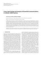
Figure 1.1 Multi-stage process of hepatocarcinogenesis. The proposed
histopathological progression and common molecular features of HCC caused by
different etiologies including hepatitis B or C virus (HBV or HCV), aflatoxin B1 and
alcohol were shown. After hepatic injury was triggered by any one of risk factors, there
was necrosis followed by hepatocyte proliferation. Continuous cycles of this process led
to liver cirrhosis with the formation of abnormal liver nodules. Consequently, these
nodules progressed to hyperplasia, dysplasia and ultimately hepatocellular carcinoma
(HCC), which could be further classified into subgroups containing well differentiated,
moderately differentiated and poorly differentiated tumor cells. Loss-of-function p53 and
genomic instability were found to involve during HCC progression. This figure was
adapted from (Farazi and DePinho, 2006).
Introduction
5
1.1.2 Current trends in therapeutic strategies of human HCC
Treatment options for early HCC include liver transplantation, resection or local radiation
therapies. Although the main curative treatment for HCC is surgical resection, there is
limited improvement to the availability of alternative treatments (Llovet and Bruix,
2008). In fact, the tumor recurrence rate is frequently high and most HCC patients are
diagnosed at relative late stages when the above treatment and chemotherapeutic options
are inapplicable. Another major obstacle for treatment of this cancer is the fact that HCC
is regularly resistant to conventional chemotherapy and radiotherapy. Furthermore, there
is significant clinical and genetic heterogeneity among HCCs of different etiologies and
standard treatments may therefore not work for all HCC cases (Villanueva and Llovet,
2011). Thus, HCC intervention is still a big challenge and a complete understanding of
the common molecular events leading to the initiation and progression of HCC is a
prerequisite to the prognosis and discovery of early treatment for this cancer. In the past
few years, considerable progress has been made in elucidating some of the molecular
steps leading to the development of HCC. Currently, two main pathogenic mechanisms
prevail during hepatocarcinogenesis, including cirrhosis associated with sustained cycles
of hepatic necrosis–inflammation–regeneration caused by hepatitis infection or toxins,
and mutations occurring in single or multiple oncogenes or tumor suppressor genes
(Farazi and DePinho, 2006). Both mechanisms have been linked with alterations in
several important signaling pathways. These key signal transduction pathways that have
been implicated in the development and progression of HCC include those mediated by
VEGF, IGF and EGFR, and the Raf/MEK/ERK, PI3K/AKT, Wnt/β-catenin and
JAK/STAT pathways. The identification of common molecular changes among the
Introduction
6
different etiological factors has created a potential avenue for anticancer drug discovery
or molecular targeted therapies for HCC. Unlike conventional cytotoxic chemotherapy,
targeted therapies are designed to inhibit tumor-specific molecular structures or activation
of pathways that are involved in the development of HCC. Two main classes of targeted
therapies are currently available, namely monoclonal antibodies and small-molecular
inhibitors (Spangenberg et al., 2009). Strikingly, Sorafenib, a multi-target compound
which effectively blocks both Ras/Raf/MEK/ERK and VEGF pathways, is the only drug
approved for the treatment of advanced HCC (Villanueva and Llovet, 2011). The survival
benefit obtained in advanced HCC patients treated with Sorafenib was 10.7 months
versus 7.9 months in the placebo group. The advent of Sorafenib and molecular targeted
therapies represented the dawn of a new era in the complex management of HCC, which
should be complemented with other molecular approaches. Future research is expected in
the development of more model systems as well as to study HCC progression and
identify new oncogenes as targets for therapies, and to test new compounds to block
currently undruggable pathways or several other simultaneous pathways through high-
throughput screening.
1.2 Zebrafish as a liver cancer model
Animal models have been widely used in biomedical research to define the pathogenesis
of cancer and as in vivo systems for developing and testing new therapies. Indeed, drug
discovery involves a complex process of biomedical and cellular assays, with final
validation in mammalian models before ultimate test in humans (Zon and Peterson,
2005). The laboratory mouse is one of the best models to study liver cancer in vivo due to
Introduction
7
various features, such as its entirely sequenced genome and the genetic and biological
similarities to human (Fausto and Campbell, 2010). Furthermore, the mouse together with
other mammalian models including rats has made the prediction of drug efficacy and
toxicity more reliable. However, these animal models tend to be costly, laborious, require
large quantities of precious compounds and are unfeasible for large-scale studies
(Sharpless and DePinho, 2006). In this context, the zebrafish (Danio rerio) has come to
attention as an economic model which generally mimics human diseases and offers the
ability to quickly and inexpensively test the efficacy and safety of compound libraries
(Amatruda and Patton, 2008; Liu and Leach, 2011; Zon and Peterson, 2005).
1.2.1 Advantageous use of the zebrafish in research
The zebrafish is a powerful vertebrate model system not only in developmental biology,
but also in biomedical research (Lieschke and Currie, 2007). This small (3-4 cm)
freshwater tropical teleost vertebrate is originally from the Ganges River in India. The
history of zebrafish as an experimental model began as early as 1980s when George
Streisinger and colleagues established pure strains of zebrafish and pioneered its utility as
a model organism to study embryogenesis (Streisinger et al., 1981). The initial focus of
zebrafish research was on developmental biology reflecting its unique advantages such as
short life cycle, optical clarity of embryos and larvae, and embryological manipulability.
Zebrafish reaches sexual maturity by three months of age with high fecundity. A breeding
pair can produce large numbers (100-200) of embryos in one morning. The growth and
development of embryonic zebrafish are rapid, finishing gastrulation within 10 hours and
hatching by 2 days after fertilization with most organs already well-developed. Another
Introduction
8
attractive feature to the developmental biologist is that the transparent embryos develop
outside the mother, thus allowing noninvasive visualization and ploidy manipulation for
genetic analysis from the point of fertilization (Lieschke and Currie, 2007). Zebrafish
embryos are also permeable to many small molecules and hence become a potential
whole-animal vertebrate model for chemical genomics. In the past decade, several
additional tools have been developed that greatly increases the utility of the zebrafish as
an experimental model. Indeed, the zebrafish genome sequencing project is almost-
completed, which facilitated genomic studies for gene expression profiling (Lieschke and
Currie, 2007). On the other hand, whole-mount in situ hybridization permits the analysis
of gene transcription, whereas injection of morpholino antisense oligonucleotides
(morpholinos) allows the study of gene function robustly in zebrafish embryos. In
addition, techniques for generating transgenic zebrafish, such as cloning, mutagenesis,
transgenesis and microinjection, further strengthen the use of zebrafish. Recently, there is
increased generation and analysis of zebrafish models of human diseases (Amatruda and
Patton, 2008; Liu and Leach, 2011). Owing to the ease of housing maintenance, short
generation time and fecundity, zebrafish studies are cost-effective and provide
advantages over other models in high-throughput small molecule screening. All these key
attributes underpin the use of zebrafish as an excellent experimental model (Figure 1.2).
Introduction
9
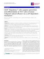
Figure 1.2 Advantages of zebrafish as a powerful model organism for cancer
research. Similar to flies and worms, zebrafish embryos are transparent and produced in
large numbers which are suitable for genetic and chemical screens as well as
experimental manipulation. In addition, its small size and rapid generation time facilitates
genetic studies and offers economic benefit. Like mammals, zebrafish possesses
vertebrate anatomy, physiology, and tumor biology. This figure was adapted from
(Amatruda et al., 2002).