Genetic analysis of the role of ARP2 3 complex in border cell migration in drosophila melanogaster
Bạn đang xem bản rút gọn của tài liệu. Xem và tải ngay bản đầy đủ của tài liệu tại đây (3.13 MB, 133 trang )
GENETIC ANALYSIS OF THE ROLE
OF ARP2/3 COMPLEX IN BORDER CELL MIGRATION
IN DROSOPHILA MELANOGASTER
LU RUIFENG
(Ph.D of Science), NUS
A THESIS SUBMITTED FOR
THE DEGREE OF PhD OF SCIENCE
DEPARTMENT OF BIOLOGICAL SCIENCE
NATIONAL UNIVERSITY OF SINGAPORE
2010
Arp2/3 complex in border cell migration Lu Ruifeng
2010
a
Acknowledgement
First of all, I’d like to take this opportunity to express my gratitude to Dr. Pernille
Rørth for her supervision and guidance on the project. Her enthusiasm on science has
been affecting the group every day. Help from many people contribute to the work. I give
my warmest thanks to Dr. Hsin-Ho Sung. Hsin-Ho designed the scheme of the screens
and generated fly stock for the screens but he was glad to pass the project to me. I also
highly appreciate the daily help from Dr. Adam Cliffe. He has written thousands lines of
macros for ImageJ, which liberates us from hours of routine works on movie processing
and data sorting. My thanks also go to Dr. Minna Poukkula. She and Dr. Cliffe developed
and optimized the protocol for live imaging. Minna taught me live imaging technique and
quality control of movies. All the best for her future academic career. Thank Dr. Inaki
Mikiko for sharing of stocks and movies on cytochalasin-D drug treated border cells.
Thank Dr. Cliffe and Dr. Smitha Vishnu for critical reading and suggestions on the
manuscript. Thank Rishta Changede for her discussion on ELMO RNAi data. Thank
Nachen Yang for exchanging ideas and discussion on experiments. Thanks also go to Dr.
He Yuehui for his continuous encouragement and caring for my research progress. Last
but not least, I thank Dr. Stephen Cohen for funding me to finish the thesis at the last year
of my PhD study.
Arp2/3 complex in border cell migration Lu Ruifeng
2010
b
Table of contents
Summary·······························································································i
List of figures·························································································ii
List of symbols·······················································································iv
I. Introduction······································································1
1.1 Cell migration········ ·······································································2
1.2 Chemotaxis···················································································4
1.3 Protrusions in migrating cells······························································5
1.4 Actin biochemistry··········································································6
1.5 Biological processes that depend on actin···············································7
1.6 Regulation of actin filament remodeling··················································9
1.7 Arp2/3 complex and formins are actin filament nucleators···························11
1.7.1 Arp2/3 complex······································································11
1.7.2 Formins···············································································13
1.7.3 Arp2/3 complex is essential for many cellular processes······················14
1.8 Regulation of the Arp2/3 complex and Diaphanous···································17
1.9 Regulation of WASP and WAVEs·······················································21
1.10 Regulation of formins·····································································23
1.11 Dendritic nucleation model of actin filament network······························23
1.12 Collective cell migration·································································24
1.13 Border cell provides a good model to study collective migration in vivo········25
1.13.1 Drosophila oogenesis·····························································25
1.13.2 Molecular requirement for border cell migration in Drosophila
oogenesis ···········································································28
1.13.3 Live imaging opens doors for studying the dynamics of cell migration in
vivo···················································································31
II. Results ···········································································33
2.1 FLP/FRT mosaic screen in border cell··················································33
2.2 mbm germline mutants showed strong border cell migration delay················36
2.3 FRT-l(2)SH 1750 homozygous border cells show migration defect···············39
2.4 MARCM clone analysis of Arp2/3 subunits············································44
2.5 AFG RNAi of Arp2/3 subunits and SCAR·············································49
2.6 Live image analysis of Arp2/3 and SCAR RNAi······································52
2.7 Arp2/3 depleted border cells move slowly·············································54
2.8 Cell motility is intact in Arp2/3 depleted border cells································57
2.9 Extensions formed less in Arp2/3 and SCAR reduction border cells···············58
2.10 Extension lifetime is less affected by Arp2/3 complex······························60
2.11 The productivity of extensions in Arp2/3 depleted border cells···················61
2.12 Early phase and late phase are guided by different mechanisms···················62
2.13 Diaphanous is also important for border cell migration·····························69
Arp2/3 complex in border cell migration Lu Ruifeng
2010
c
2.14 Arp2/3 does not affect E-cadherin adhesion··········································73
2.15 Arp2/3 is not required for internalization of membrane material··················75
III. Discussion·······································································78
3.1 FLP/FRT screen············································································79
3.2 Arp2/3 affects border cell migration in cell-autonomous and non cell-autonomous
patterns······················································································79
3.3 Both Arp2/3 and Dia are required for border cell to initiate migration·············80
3.4 Arp2/3 is required for cluster movement but not cell motility·······················82
3.5 Arp2/3 and Dia control protrusion formation··········································83
3.6 Arp2/3 complex is required differently for early phase and late phase of
migration····················································································83
3.7 Physiological function of protrusions in border cells·································88
3.8 Physiological function of actin dynamics in border cell migration·················91
3.9 Arp2/3 and Dia act in different ways in border cell migration······················93
3.10 Mesanchymal movement and amoeboid movement·································95
IV. Material and methods······················································100
4.1 Fly husbandry·············································································101
4.2 X-Gal staining············································································101
4.3 Somatic mutant clone generation······················································102
4.4 Germ line mutant clone generation····················································103
4.5 Immunofluorescence·····································································104
4.6 Additional clonal analysis·······························································105
4.7 MARCM clone generation······························································105
4.8 Flipout Expression of UAS-driven Genetic Constructs·····························105
4.9 Preparation of egg chambers for live imaging········································106
4.10 Confocal microscopy imaging························································107
4.11 Determination of migration speed ···················································108
References·······················································································109
Publication······················································································125
Arp2/3 complex in border cell migration Lu Ruifeng
2010
i
Summary
During oogenesis of Drosophila, one group of cells called border cells delaminate
from the anterior epithelium and migrate to the oocyte in a stereotypic way. Border
cells provide a good system to study cell migration in vivo due to their genetic
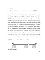
tractability. arc-p34 was isolated from a border cell mutant clone screen due to its
strong effect on border cell migration. arc-p34 encodes the Drosophila homolog of
mammalian ARPC2, a component of Arp2/3 complex. When the level of various
Arp2/3 components is reduced by RNAi, many border cell clusters fail to initiate the
migration. If they initiate migration, these border cell clusters move much slower at
first, but migrate normally later, suggesting distinct mechanisms differentially depend
on Arp2/3. Single cell tracking shows that Arp2/3-impaired border cells are still
motile, but show less directional movement. Thus Arp2/3 may be acting upstream or
downstream of guidance cues to steer border cell migration.
Arp2/3 complex in border cell migration Lu Ruifeng
2010
ii
List of Figures
Figure 1.1 Function of chemokines is to induce cell migration.
Figure 1.2 Migrating cell send out lamellipodia and filopodia.
Figure 1.3 Actin is important for various biological processes.
Figure 1.4 Models of actin filament nucleation by Arp2/3 and formins
Figure 1.5 Arp2/3 is important for the expansion of trichome in Arabidopsis thaliana
Figure 1.6 Domains organization in WASPs and WAVEs and the regulation
mechanisms.
Figure 1.7 Dendritic nucleation model of actin assembly
Figure 1.8 Border cells are specified in stage 9 of oogenesis.
Figure 1.9 RTK signaling guides border cell migration.
Figure 2.1 Germ line mutant for mbm
1819
affects oocyte patterning.
Figure 2.2 The gene disrupted in FRT-l(2)SH1750 is important for border cell
migration.
Figure 2.3 Figure 2.3 Border cell migration shows delayed phenotype in FRT-l(2)
SH1750 germline mutant clones.
Figure 2.4 Figure 2.4 Complementation test between FRT-l(2)SH1750 and small
deletions uncovering 38A-38D.
Figure 2.5 Abnormal oogenesis in FRT-l(2) SH1750 mutants.
Figure 2.6 MARCM clonal analysis of Arc-p34.
Figure 2.7 Reduction of Arp2/3 complex subunits or SCAR protein level delayed
border cell migration
Figure 2.8 AFG RNAi of Arc-p34 showed initiation defect and dramatic migration
defect.
Figure 2.9 Arp2/3 activity affect border cell migration via reducing directionality.
Arp2/3 complex in border cell migration Lu Ruifeng
2010
iii
Figure 2.10 The number, size and life time of extensions.
Figure 2.11 The productivity of cellular protrusions is reduced if Arp2/3 is depleted
from border cells.
Figure 2.12 Knocking down Arp2/3 complex causes migration phenotype.
Figure 2.13 Knocking down Arp2/3 complex decreased border cell migration speed.
Figure 2.14 Analysis of number, length, size and life time of cellular extensions in
slbo-Gal4 RNAi border cells.
Figure 2.15 Diaphanous affects border cell migration independent of its role in
cytokinesis.
Figure 2.16 Diaphanous exerts distinct function in early phase and late phase of the
migration.
Figure 2.17 Quantification of Arp2/3 RNAi or Dia RNAi border cell migration in
background of one copy of E-Cadherin.
Figure 2.18 Arp2/3 is not essential for internalization of FM4-64 in border cells.
Figure 4.1 Scheme of generation of mutant clone with labeled marker.
Figure 4.2 Scheme of germline mutant screen.
Arp2/3 complex in border cell migration Lu Ruifeng
2010
iv
List of Symbols
ARP2/3, actin related protein 2/3
EGFR, epidermal growth factor receptor
ELMO, engulfment and cell motility
EMT, epithelium to mesenchymal transition
Ena/WASP, Enabled/vasodilator-stimulated phosphoprotein
Mbc, myoblast city
Mbm, mushroom body miniature
PICK1, protein interacting with Cα-kinase
PVR, PDGF and VEGF receptors
SCAR, suppressor of cyclic AMP receptor
Slbo, slow border cell
SOP2, suppressor of profilin
WASP, Wiskott-Aldrich syndrome family proteins
WAVE, Wiskott-Aldrich verprolin homologous protein
Arp2/3 complex in border cell migration Lu Ruifeng
2010
1
I. Introduction
Arp2/3 complex in border cell migration Lu Ruifeng
2010
2
1.1 Cell migration
Cell migration is a defining feature of animal cells (Pollard and Cooper 2009),
which is crucial for both single cellular organisms and multicellular organisms. Single
cell organisms migrate to reach nutrients and to escape from dangers, as well as to
facilitate dispersal. In multicellular organism, cell migration is required for embryonic
morphogenesis, wound healing and immune surveillance (Pollard and Borisy 2003).
One of the earliest examples of migration in development is gastrulation (Montero
and Heisenberg 2004). During gastrulation, large groups of cells migrate collectively as
sheets to form three embryonic layers: ectoderm, mesoderm, and endoderm. Subsequent-
ly, cells migrate out from various epithelial layers to specific location. Interactions with
new microenvironment induce them to differentiate to form the specialized cells that
make up different tissues and organs.
In vertebrates, after gastrulation, neural crest cells are specified at the border of the
neural plate and the non-neural ectoderm. The neural crest is a population of migrating,
pluripotent cells which appears transiently in the dorsal neuroectoderm. During
neurulation, the borders of the neural plate converge at the dorsal midline to form the
neural tube. Subsequently, neural crest cells from the roof plate of the neural tube
undergo an epithelial to mesenchymal transition (EMT), delaminating from the
neuroepithelium and migrating as loosely associated strands or streams throughout the
entire embryo and give rise to different tissues, including craniofacial bones and
cartilage, the enteric and peripheral nervous systems and pigment cells.
Migration is also a prominent component of tissue repair and immune surveillance.
In the renewal of skin and intestine, fresh epithelial cells migrate up from the basal layer
Arp2/3 complex in border cell migration Lu Ruifeng
2010
3
and the crypts, respectively. A simple in vitro model of this is process is the scratch assay
which has been used extensively to study cell migration in tissue repair in vitro. This
assay involves creation of a new artificial gap, by scratching a confluent cell monolayer.
Shortly after the generation of the “scratch” gap, the rows of cells on the edge of the gap
will reorient and polarized themselves followed by a collective migration of the whole
sheet of cells in a direction perpendicular to the wound edge. Finally new cell–cell
contacts are established again and the “scratch” opening is closed. The unoccupied space
might be used as a spatial signal to guide the migration.
All white blood cells (WBC) are known as leukocytes, the major players in
immune surveillance. Leukocytes are not tightly associated with a particular organ or
tissue, which allows them to move freely, similar to independent, single-celled
organisms. During an immune response, leukocytes from the circulation migrate into the
surrounding tissue to destroy invading microorganisms and infected cells and to clear
debris (Peri 2010).
Migration also contributes to pathological conditions such as tumor metastasis,
vascular disease and chronic inflammatory disorders (Pals et al., 1989). The most deadly
aspect of cancer is its ability to spread, or metastasize. Metastasis refers to the process by
which malignant cells break off from primary tumor and travel to other parts of the body.
To begin the process of metastasis, a malignant cell must first break away the adhesion
both to surrounding cells and extracellular matrix. Cancer cells release enzymes called
metalloproteinases (MMPs) to dissolve basement membranes and other extracellular
matrices (Roy et al. 2009; Groblewska et al., 2010; Kessenbrock et al., 2010), allowing
its penetration of surrounding tissues. Once a cancer cell has detached, it invades the
Arp2/3 complex in border cell migration Lu Ruifeng
2010
4
surrounding tissue and makes its way into blood or lymphatic vessels, so giving the
cancer cells access to other parts of the body. Once at a new site, the cells must again
penetrate the basement membrane of the blood vessel and colonize in the new tissue (Pals
et al., 1989). Metastasized tumors usually indicate a later stage disease, and treatment
becomes more difficult with poorer outcomes. Metastasis is a complicated process and
the underlying mechanisms are not completely understood. Therefore, understanding the
fundamental mechanisms underlying cell migration is essential for effective therapeutic
approach for treating disease.
1.2 Chemotaxis
The overall migration speed is dependent on both the linear movement speed and the
extent to which that movement is in a persistent direction (Lauffenburger and Horwitz
1996). To move in a specific direction, a cell must be guided and often in the absence of a
guidance cue, motile cells will migrate randomly. Motile cells are able to sense
extracellular signals from their environment and direct their movement along the
concentration gradient of these signals. This process is called chemotaxis (Figure 1.1).
Substances that induce a chemotactic response are known as chemoattractants.
Chemotaxis is positive if cells move towards a higher concentration of a chemical, and
negative if the direction is opposite. Chemotaxis is used by bacteria to find nutrients (for
example, glucose) by swimming towards the highest concentration of nutrients
molecules, or to escape from poisons (for example, phenol). In multicellular organisms,
chemotaxis is critical to early (e.g. movement of sperm towards the egg during
fertilization) and subsequent phases of development (e.g. migration of neurons or
lymphocytes) as well as in normal function (Stephens et al. 2008). Chemotaxis is often
Arp2/3 complex in border cell migration Lu Ruifeng
2010
5
essential for cell survival in development, as cells that failed to reach their expected
destination on time die. It is also a highly sensitive mechanism, as eukaryotic cells are
able to sense concentration gradients as shallow as a 2-10% difference (Parent and
Devreotes 1999; Firtel and Chung 2000).
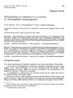
Figure 1.1 Function of chemokines is to induce cell migration. Cells will move toward the
direction of increment of continuous chemokine concentration gradient. In other
words, cells migrate toward the source of chemokine.
1.3 Protrusions in migrating cells
Directed cell migration is a cyclical process often characterized in four steps
(Figure 1.2A): protrusion of the front membrane extension; adhesion formation and
stabilization at the extended extension; adhesion sites serve as tractions sites for acto–
myosin-based contraction, which pulls the cell body forward; and disassembly of
adhesion sites at the cell rear. Continuous directed cell migration requires balanced
adhesions formation at the leading edge and disassembly of adhesion at the trailing edge.
In eukaryotes, the outcome of perceiving signal gradient is the protrusion of cell
membrane at the leading edge. Cellular protrusions can range from large flat sheets of
lamellipodia or spike-like filopodia (Figure 1.2 B). Lamellipodia provide the major force
Arp2/3 complex in border cell migration Lu Ruifeng
2010
6
to push the membrane forward at the leading edge, while filopodia are responsible for
detecting extracellular chemoattractants. As a cell migrates through a gradient of
chemoattractant, the polarity of the cell increases (Parent and Devreotes 1999). The
leading edge becomes more sensitive to chemoattractant, while the formation of lateral
protrusions is suppressed. Cell migration is critically dependent on this localized
signaling (Jekely and Rorth 2003).
Figure 1.2 Migrating cell send out lamellipodia and filopodia. A. migrating cell is
characterized with a front and a back. Actin polymerization at the front pushes the
membrane forward. Cell-substratum adhesion assembly at the front and
disassembly at the trailing tail are coordinated. From Nature Reviews Molecular
Cell Biology. B. Two fluorescently-labeled growth cones. The growth cone (green)
on the left is an example of a “filopodial” growth cone, while the one on the right is
a “lamellipodial” growth cone. From Gordon-Weeks, P. R. 2005.
1.4 Actin Biochemistry
The dominant structural components of protrusions are actin filaments, which are
arranged in networks in lamellipodia and bundles in filopodia. Formation and extension
of lamellipodia is driven by assembly of actin networks (Figure 1.2A) (Bugyi and Carlier
2010). The core constituent of the actin cytoskeleton is the actin filaments, which are
formed from double helical polymers of globular actin (G-actin). G-actin is a 43-kDa
Arp2/3 complex in border cell migration Lu Ruifeng
2010
7
ATPase that can assemble into filamentous actin (F-actin) via catalyzing ATP hydrolysis.
ATP hydrolysis by actin is coupled closely with polymerization and regulates the
assembly and disassembly of actin filaments. G-actin is able to assembly at both ends of
one actin filament at different rates (Pollard 1986). Actin filaments are polarized with a
fast growing barbed end and a slow growing pointed end. In protrusions, actin filaments
are oriented with the barbed end toward membrane (Small et al. 1978) allowing the rapid
growth at the barbed end drives protrusion of cell membrane and thus cell motility
(Pollard et al. 1982).
Actin remodeling has an essential role in various processes, including cell
migration, endocytosis, vesicle trafficking and cytokinesis (Goley and Welch 2006).
Many of them are essential for the survival of the cell; therefore general loss of actin has
lethal effect. To study actin function in a specific process in a certain tissue, we need to
modulate tissue specific regulators of actin to avoid early lethality.
1.5 Biological processes that depend on actin
Actin filaments as part of the cytoskeleton
The actin cytoskeleton is responsible for the mechanical support and geometry of cells
(Figure 1.3A), which are important for their functions.
Arp2/3 complex in border cell migration Lu Ruifeng
2010
8
Figure 1.3 Actin is important for various processes. A. Actin cytoskeleton structures (in red)
in fibroblast cells. B. Budding yeast (Saccharomyces cerevisiae) expressing a
actin patch protein Abp1::GFP. C. Listeria monocytogenes (in red) are
polymerizing host cell actin into comet tails (in green) to push them across the
cytoplasm. Inset in C shows magnified Listeria. From (Pollard and Berro 2009)
Clathrin-dependent endocytosis
In yeast, “actin patches” are formed at sites of endocytosis on plasma membrane
(Figure 1.3B). Assembly of actin filaments at these sites facilitates the clathrin-mediated
internalization of endocytic vesicles and subsequent intracellular transportation.
Bacterial assembly of actin rich comet tails
The intracellular “rocketing” motility of Listeria shows links between movement and
actin polymerization (Figure 1.3 C) (Tilney et al. 1992). After invading the host cell,
Listeria utilizes the motility machinery of the host cells to assemble a comet tail of actin
filaments. Continuous assembly of actin filament at tail pushes them through cytoplasm
(Dramsi and Cossart 1998). Accumulated evidence showed that viruses (Frischknecht et
al. 1999), endosomes (Merrifield et al. 1999) and vesicles (Rozelle et al. 2000) also
employ comet tails for intracellular motility.
Contractile ring in cytokinesis
At the last step of cell division, a contractile ring of actin filament and myosin II
assemble between two daughter cells. Myosin II can produce contraction by pulling actin
filaments, resulting in a cleavage furrow at the cell membrane. The two daughter cells are
separated by pinching of the contractile ring and membrane fusion.
Track for organelles transportation
Many cells use myosin motors for transportation of vesicle and organelles along actin
filaments.
Arp2/3 complex in border cell migration Lu Ruifeng
2010
9
Cell motility
Actin filaments are essential for cell migration. In migrating cells, actin filaments are
polarized with the plus ends toward the membrane. This inherent polarity of actin
filaments is used to drive membrane protrusions, which is often the first step in cell
migration. During cell locomotion, myosin interacts with actin filaments to pull the rear
of cell forward.
1.6 Regulation of actin filament remodeling
Since various developmental processes, which are essential for the survival of cell,
utilize actin cytoskeleton, the polymerization and depolymerization of actin filament are
under tight controls by over a hundred actin binding proteins. Once nucleated, actin can
polymerize at the barbed end at a rate proportional to the G-actin concentration. Actin is
one of the most abundant proteins in eukaryotic cells with a cellular concentration of 100
µM. To prevent actin polymerization from running amok, the large pool of actin
monomers are buffered by monomer binding proteins, such as profilin and thymosin β4.
These factors suppress spontaneous nucleation of new filaments, but enable actin
polymerization at the barbed ends. Thus, the rate limiting step is the formation of free
barbed ends. Three mechanisms contribute to the generation of free barbed ends:
uncapping the capped filaments; severing existing filaments; and forming actin filaments
de novo. Though G-actin is able to self assemble, spontaneous nucleation is kinetically
unfavorable because the process involves the formation of the intermediate dimer and
trimer (Pollard and Borisy 2003), which are extremely unstable and dissociate rapidly.
However, a variety of cellular processed require a responsive, rapid burst of actin
Arp2/3 complex in border cell migration Lu Ruifeng
2010
10
assembly at specific subcellular locations. To circumvent the limitation of spontaneous
nucleation, cells use factors that promote actin nucleation. A nucleator is defined as a
factor that stimulates formation of an actin filament that grows rapidly at its barbed end.
Two roles of actin nucleators are defined: First, to regulate the time and position of actin
filament formation. Second, to protect the barbed end from being bound by capping
proteins. Arp2/3 is one major and the best studied nucleator of branched actin
(Vartiainen and Machesky 2004). Formins bind barbed end of actin filaments to promote
linear (unbranched) actin filament elongation, antagonizing both capping and branching.
Though de novo nucleation of new actin filaments has been considered as the dominant
mechanism in the leading edge, the contribution from other two mechanisms should not
be neglected. Cofilin/actin-depolymerizing factor (ADF) is an actin binding protein that
is required for actin-filament disassembly, cytokinesis and the organization of muscle
actin filaments (Bamburg 1999). Cofilin/ADF severs actin filaments and promotes actin
dissociation from the pointed end in vivo to generate free barbed ends (Bamburg 1999).
Surprisingly, depletion of cofilin reduces the rate of lamellipodia formation rather than
increasing it (Akin and Mullins 2008). It was therefore speculated that cofilin severing
activity is essential for generating free actin barbed end for actin polymerization, hence
accelerates actin treadmilling, possibly in cooperation with the Arp2/3 complex (Akin
and Mullins 2008).
Actin polymerization at barbed ends depletes the G-actin pool rapidly. For a cell to
respond quickly to environmental stimuli it requires a large G-actin pool which is
polymerization competent. At steady state, capping proteins bind to the barbed end of
actin filaments and inhibit elongation to maintain the G-actin pool. Therefore, actin
Arp2/3 complex in border cell migration Lu Ruifeng
2010
11
filament growth depends on the competition between nucleators and capping factors.
High-affinity binding of capping factors determines the length of F-actin and limits the
number of free barbed ends, which reduces the rate of G-actin monomer depletion.
Capping proteins therefore reduce the drain on the G-actin pool, allowing more uncapped
F-actin growth. Enabled/vasodilator-stimulated phosphoprotein (Ena/VASP) is the other
known actin nucleator. Like formins, Ena/VASP proteins bind barbed ends of actin
filaments. However, Ena/VASP promotes actin filament elongation when VASP is bound
to beads but not in solution, suggesting that the activity of VASP requires some form of
attachment. Generally, the net dynamics of actin filament are determined by nucleation,
branching, elongating at one hand and severing and capping at the other.
1.7 Arp2/3 complex and formins are actin filament nucleators
1.7.1 Arp2/3 complex
Arp2/3 was first isolated from Acanthamoeba castellanii based on its affinity for the
actin binding protein profilin (Machesky et al. 1994). Soon after, the nucleation activity
of the Arp2/3 complex was identified (Mullins et al. 1997). Since then, biochemical,
electron microscopic studies have focused on the mechanism of Arp2/3 mediated actin
filament nucleation and branching. Arp2/3 is a 220 kDa complex consisting of seven
subunits. The two core subunits are actin-related protein (ARP) Arp2 and Arp3. The
remaining five subunits are named ARPC1 (40 kDa), ARPC2 (34 kDa), ARPC3 (21
kDa), ARPC4 (20 kDa), ARPC5 (16 kDa). These subunits are evolutionarily conserved
and have been found in plants, fungi, amoeba, flies, and vertebrates (Pollard and Borisy
2003). Biochemical and microscopic data suggest that Arp2/3 complex binds to the side
Arp2/3 complex in border cell migration Lu Ruifeng
2010
12
of an existing filament and initiatea a new filament at a 70 ° y-branch in vitro (Pollard
and Borisy 2003). Arp2/3 binds to the pointed end of the nascent actin filament and
leaves the barbed end free for elongation. So far Arp2/3 is the only known actin nucleator
that mediates branched networks.
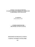
Figure 1.4 Models of actin filament nucleation by Arp2/3 and formins. Electron
tomography shows several branches (boxes), Arp2/3 complex (circles) and gold
markers (arrowheads) in a 3D reconstruction of the actin filament branches. From
(Rouiller et al. 2008) B. Arp2/3 nucleates actin filament on existing filaments and
binds to pointed end of newly formed filament. Formins form doughnut shape
and nucleate linear filament. After nucleation, Arp2/3 remains associated with
pointed end while formins processively move at the barbed end as the filament
elongates. From(Insall and Machesky 2009).
Biochemical and electron microscopic studies have revealed the structural base of
Arp2/3 complex, which suggests that ARPC2 and ARPC4 form the structural core of the
complex, with the remaining subunits surrounding them. ARPC2 and ARPC4 contact the
mother filament, whereas ARP2 and ARP3 associated with the pointed end of the nascent
filament (Rouiller et al. 2008). The structural organization of Arp2 and Arp3 are similar
Arp2/3 complex in border cell migration Lu Ruifeng
2010
13
to actin, so it is supposed that Arp2/3 complex acts as template to mediate initiation of
actin polymerization. Like actin, Arp2 and Arp3 bind ATP. ATP binding causes
conformation change and is important for their nucleation activity in vitro. The proposed
model (Figure 1.4) suggests that upon binding to actin filament, a conformation change
of the whole complex reorganizes ARP2 and ARP3 into a dimer which acts as template
of subsequent actin assembly. The Arp2/3 complex remains bound to the pointed end of
F-actin leaving a new barbed end free for subsequent elongation.
1.7.2 Formins
Formin is another actin nucleator, catalyzing the formation of linear (unbranched) actin
filaments in vitro (Pruyne et al. 2002) and assembles diverse actin structures, including
stress fibers, cytokinetic contractile rings, and actin cables in vivo (Kovar et al. 2006;
Goode and Eck 2007). In contrast to the Arp2/3 complex, which nucleate a novel actin
filament on existing filaments and remains associated with pointed end of the nucleated
filament after nucleation, formins remain associated with the barbed end after nucleation,
moving processively as the filament elongates (Pollard 2007). The mechanism underlying
processive movement remains unclear.
Formin family proteins contain two formin homology domain (FH):FH1 and FH2.
FH2 domains form donut-shape head-to-tail homodimers and are responsible for the
association with the barbed end of the nucleated filament. FH2 processive association
was believed to prevent the binding of capping factors to the barbed end (Kovar et al.
2003). FH1 contains polyproline sequences and interacts with profilin (Chang et al.
1997). Since profilin binds to actin monomer, FH1 domains can bind the profilin-G-actin
Arp2/3 complex in border cell migration Lu Ruifeng
2010
14
complex near the barbed end of actin filaments. It was postulated that formin nucleates
new filaments by binding and stabilizing the intermediate actin dimer and trimer (Pruyne
et al. 2002). Drosophila Diaphanous belongs to the formin protein family, which is
highly conserved and has been implicated in the formation of linear (unbranched) actin
filaments. Crystal structure shows that N-terminal domains of mDia1 form a dimer and
inhibit the actin nucleation activity of FH2 by intramolecular interaction.
1.7.3 Arp2/3 complex is essential for many cellular processes
After Arp2/3 complex was identified to nucleate and branch actin filament in vitro
study, extensive efforts have been put on investigating its function in vivo. Knockout and
knockdown experiments showed that the Arp2/3 complex is essential for the viability of
many cell types. The Arp2/3 complex appears important in a variety of specialized cell
functions that involve the actin cytoskeleton. Arp2/3 mutant mammalian cells often have
lower levels of actin filaments, consistent with the role of Arp2/3 in actin filament
nucleation. Arp2/3 is also necessary for phagocytosis in mammals and the social amoeba
Dictyostelium discoideum (Insall et al. 2001; Warren et al. 2002)
Endocytosis in yeast
In yeast, a family of proline-rich proteins named verprolin is known to bind WASP
(Kaksonen et al. 2006). Verprolins coordinate WASP with type I myosin to activate actin
assembly at actin patch during clathrin dependent endocytosis (Galletta and Cooper
2009).
Arp2/3 complex in border cell migration Lu Ruifeng
2010
15
Ventral closure of C.elegans
Arp2/3 plays essential role in cell migration during ventral enclosure in Caenorhabditis
elegans (Sawa et al. 2003). During ventral closure, the embryonic epidermis migrates
from the dorsal surface towards the ventral surface, ending up sealing the ventral surface
(Sawa et al. 2003). Disruption of any one of Arp2/3 subunit results in the loss of
migration in the epidermal cells (Sawa et al. 2003). The leading edge of the migrating
epidermis in Arp2/3 depleted C. elegans embryos shows a lack of filamentous actin, and
the finger like protrusions that normally form are absent (Sawa et al. 2003). One report
has revealed the involvement of Arp2/3 in guiding longitudinal migration of excretory
cells in C. elegans (Sanz-Moreno et al. 2008). Arp2/3 is also required cell-autonomously
for axon guidance and initiation of filopodia on growth cones, but not important for the
growth of axon growth cone. In C. elegans development, gastrulation is initiated by the
internalization of two endodermal precursor cells (Severson et al. 2002). If Arp2/3 is
depleted, the endodermal precursor cells fail to be fully internalized (Severson et al.
2002; Roh-Johnson and Goldstein 2009).
Roles of Arp2/3 complex in Drosophila
Rogers et al. used RNAi to systematically study the molecules required for lamella
formation in Drosophila S2 cells. They found that RNAi knockdown of components of
the Arp2/3 complex or SCAR/WACE impaired the formation of lamella (Rogers et al.
2003). It has been found that the role of Arp2/3 in endocytosis is important in the
remodeling of epithelia adhesion junctions (Georgiou et al. 2008). Actin nucleators are
also crucial for remodeling the actin cytoskeleton in response to extrinsic or intrinsic
Arp2/3 complex in border cell migration Lu Ruifeng
2010
16
cues. In Drosophila, the Arp2/3 complex is required for a variety of processes, including
blastoderm organization, axon development, eye morphogensis, and egg chamber
morphology (Zallen et al. 2002). One prominent actin structure in the egg chamber is the
ring canal. These intercellular channels connect nurse cells to the oocyte. Cytoplasm of
nurse cells is transferred into oocyte through ring canals to provide nutrients for
development. Arp2/3 is important for the growth, maturation and maintenance of ring
canals (Hudson and Cooley 2002; Zallen et al. 2002). If Arp2/3 activity is affected, ring
canals decrease in diameter dramatically, sometimes even collapse (Hudson and Cooley
2002). In oogenesis, depletion of Arp2/3 in germline cells leading to multinucleate nurse
cells with the absence of nurse cell membrane (Zallen et al. 2002).
Cell shape of Trichome in Arabidopsis thaliana
A genetic screen in Arabidopsis thaliana for genes affecting cell shape of leaf
epidermal cells called trichomes, one complementation group of mutations called
“distorted” were isolated (Hulskamp et al. 1994). These were later identified as homolog
of Arp2/3 subunits (Mathur 2005). Arp2/3 is important for cell expansion during
trichome development. In Arp2/3 mutant plants, the trichomes display a general
distortion, and cotyledon cells failed to develop their usual lobed, jigsaw-puzzle shape
(Mathur 2005). Compared to the uniform distribution of F-Acin in wild type trichomes,
F-actin in Arp2/3 mutant trichome forms randomly localized dense aggregates, highly
bundled F-actin and randomly located cortical actin patches (Mathur et al. 1999;
Szymanski et al. 1999), again consistent with the important role of Arp2/3 complex in
actin dynamics regulation.
Arp2/3 complex in border cell migration Lu Ruifeng
2010
17
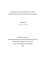
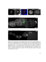
Figure 1.5 Arp2/3 is important for the expansion of trichome in Arabidopsis thaliana.
Compared to wild type control (A), the shape of thichomes is distorted in ARPC2
mutant (B).
1.8 Regulation of the Arp2/3 complex and Diaphanous
On its own, Arp2/3 complex is inactive and requires additional nucleation promoting
factors (NPFs) to nucleate actin filaments (Pollard and Borisy 2003). The main NPFs of
Arp2/3 complex are Wiskott-Aldrich syndrome family proteins (WASP), which serve as
scaffolds for Arp2/3 complex. WASP protein was first discovered in Wiskott-Aldrich
syndrome (WAS) victims (Bosticardo et al. 2009). WAS is an X-linked genetic
immunodeficiency, characterized by recurrent infection, eczema, and thrombocytopenia
(Bosticardo et al. 2009). The disease is associated with mutations in the WASP gene.
WASP proteins are multidomain and grouped into three categories based on primary
sequence homology and functional data:
i) WASP and generally expressed WASP (N-WASP);
ii) Wiskott-Aldrich verprolin homologous protein (WAVE) 1, 2 and 3 or
suppressor of cyclic AMP receptor (SCAR);
iii) Recently identified WISH, WHAMM and JMY.
Control
Arpc2
-
/-