Functional analysis of the nuage, a unique germline organelle, in drosophila melanogaster 6
Bạn đang xem bản rút gọn của tài liệu. Xem và tải ngay bản đầy đủ của tài liệu tại đây (1.94 MB, 6 trang )
86
3.3 Nuage and P-bodies regulate post-transcriptional retroelement silencing
3.3.1 Nuage cytoplasmic bodies overlap with mRNA degradation components
On careful examination of two Piwi subfamily proteins AUB and AGO3, and a tudor-
domain protein KRIMP, these nuage components also existed in cytoplasmic foci that
were 0.1 to 1 μm in diameter (green arrows, Figure 3.3.1; Harris and Macdonald, 2001).
These cytoplasmic foci became progressively prominent from stage 4 onwards during
oogenesis and were ubiquitously distributed as discrete puncta throughout the nurse cell
cytoplasm at stage 4-5 (Figure 3.3.1).
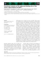
Figure 3.3.1 Nuage/piRNA pathway components exhibit both perinuclear and
cytoplasmic foci. AUB-GFP (green), AGO3 (red), and KRIMP (magenta) cytoplasmic
foci co-localise (green arrows) in stage 4-5 egg chamber. Bars are 20 µm and 10 μm for
egg chambers and nurse cells, respectively.
The spatial and temporal distributions of these cytoplasmic foci resemble the P-bodies
described in the Drosophila germline (Lin et al., 2008). Hence, wild-type ovary was co-
stained for the P-body components dDCP1, dDCP2 (Lin et al., 2006), Me31B (a homolog
87
of yeast decapping activator Dhh1p; Coller et al., 2001), and the Drosophila homolog of
yeast Xrn1p, PCM (Barbee et al., 2006; Till et al., 1998; Zabolotskaya et al., 2008).
Consistent with a recent report that mouse AGO proteins Piwi-like 4 (MIWI2) and Piwi-
like 2 (MILI) associate with P-bodies (Aravin et al., 2009), 40-57%, 38-51%, and 31-
79% of the P-bodies were found to overlap or dock AUB, AGO3, and KRIMP foci,
respectively (Figure 3.3.2, indicated by white arrows and Figure 3.3.3a). This large
percentage variation suggests that the association of cytoplasmic nuage with P-bodies is
highly dynamic. Furthermore, P-body foci that lacked the piRNA pathway components
were observed (Figure 3.3.2 and Figure 3.3.3c, indicated by white arrowheads),
suggesting that a subset of P-bodies contains piRNA pathway components, while others
do not. These observations imply that cytoplasmic foci identifiable as the P-bodies
include molecular complexes with distinct functions, as reflected by their different
compositions.
88
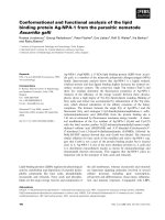
Figure 3.3.2 Nuage cytoplasmic foci overlap mRNA degradation proteins of the P-bodies. AUB, AGO3, and KRIMP cytoplasmic
bodies (red) overlap with mRNA degradation proteins dDCP1, dDCP2, Me31B, and PCM (green; white arrows). A subset of P-body
foci does not overlap with nuage cytoplasmic foci (white arrowheads). All images represent a single confocal section. Bar is 10 μm.
89
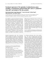
Figure 3.3.3 The association of the cytoplasmic nuage and P-body foci is highly
dynamic. (a) Overlaps of cytoplasmic nuage and P-body foci. Overlaps that are
quantified in (b) include complete overlaps and partial overlaps that consist of nuage
cytoplasmic foci docking partially around the mRNA degradation components.
Overlapping nuage/P-body foci are expressed as percentages of the total number of
overlapping and non-overlapping P-body foci. The range of overlaps (complete or partial)
appears to be independent of the foci sizes and nuage/P-body pairs. (b) Immunostaining
of overlapping cytoplasmic AGO3 (red) and Me31B-GFP (green) foci. A complete
overlap and partial overlap are indicated by a white arrow and arrowhead respectively.
Bar is 4 μm. (c) Immunostaining of non-overlapping Me31B. A Me31B focus (green)
that lacks the cytoplasmic KRIMP (red) is indicated by a white arrowhead. Bar is 4 μm.
3.3.2 Retroelement transcripts are localised to the nuage cytoplasmic bodies
Nuage components are reported to mediate retroelement repression in the germline (Lim
and Kai, 2007; Pane et al., 2007). To ask whether the cytoplasmic foci containing the
nuage and P-body components are involved in retroelement silencing, I looked for the
presence of the retroelement transcripts using the ms2/MCP-GFP labeling system
(Appendix VI; Forrest and Gavis, 2003). In order to do so, flies harbouring two heat-
shock-inducible transgenes were generated. One contained HeT-A or I-element CDS,
90
devoid of the 5’-UTR and promoter region, and fused to six tandem stem-loop binding
sites for bacteriophage MCP at the 3’-UTR. The other encoded for the fusion protein
MCP-GFP. Upon induction, MCP-GFP binds the recognition motif on HeT-A-(ms2)
6
or
I-element-(ms2)
6
transcripts, and these mRNAs were visualised as GFP signal.
In control (aub or krimp heterozygote) ovaries, GFP signal was found in cytoplasmic foci
that were also stained for the 5’- to 3’- exoribonuclease PCM, and the piRNA pathway
protein KRIMP (green arrows, Figure 3.3.4a-b). These GFP-labeled foci were not
detected in the ovary expressing MCP-GFP alone (Figure 3.3.4a), indicating that GFP
signals represent full-length HeT-A-(ms2)
6
transcripts or the decay intermediates
harbouring MCP binding sites. Similarly, the localisation of GFP-labeled I-element
transcript to KRIMP/PCM foci was observed in the control ovary (green arrows, Figure
3.3.4a). Using a non-retroelement control nos, no obvious localisation of nos-(ms2)
6
to
distinct cytoplasmic bodies was observed (Figure 3.3.4c; Forrest and Gavis, 2003),
confirming that the localisation of the retroelement transcripts is not artifactual. In aub
and krimp mutant ovaries, the GFP-labeled HeT-A transcript no longer localised to the
cytoplasmic KRIMP foci (Figure 3.3.4a). Instead, it appeared to be diffuse in the
cytoplasm and nucleus, indicating that the granular localisation of the transcripts
observed in the control ovaries depends on AUB and KRIMP functions.
91
Figure 3.3.4 Retroelement transcripts co-localise with nuage cytoplasmic foci and
mRNA degradation proteins. (a) Immunostaining of ovaries expressing MCP-GFP
fusion protein and the transgene of retroelements harbouring MCP binding sites [HeT-
A(ms2)
6
or I-element(ms2)
6
] in control (aub or krimp heterozygotes) and aub or krimp
mutants. HeT-A(ms2)
6
or I-element(ms2)
6
tagged with GFP co-localises with the piRNA
pathway component KRIMP (magenta) and 5’→3’ exoribonuclease PCM (red) in the
same cytoplasmic foci (green arrows in b). In aub and krimp mutants, GFP-labeled HeT-
A
mRNA appears to be largely cytoplasmic and nuclear, and no longer localises to the
cytoplasmic KRIMP foci. (c) A non-retroelement control nos(ms2)
6
recapitulates the
endogenous mRNA localisation at the oocyte posterior but no localisation to distinct