Functional analysis of the nuage, a unique germline organelle, in drosophila melanogaster 10
Bạn đang xem bản rút gọn của tài liệu. Xem và tải ngay bản đầy đủ của tài liệu tại đây (1.32 MB, 46 trang )
106
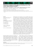
Figure 3.4.2 Cytoplasmic KRIMP do not overlap with PDI-GFP. KRIMP (red)
cytoplasmic foci do not overlap with PDI-GFP (green), which is a disulfide isomerase
that mediates protein folding in the lumen of ER. Bar is 10 μm.
The overlaps of pi-bodies with the vesicular bodies suggest that piRNA-mediated
retroelement silencing and the endosomal pathway are connected. Hence, it will be
interesting to understand in the future how the formation of such cytoplasmic
compartments regulates retroelement silencing.
107
4 Discussion
The nuage is a unique, electron-dense structure that is found on the perinuclear regions of
many animal germline cells. The evolutionarily conserved nature of the nuage
emphasizes its importance and essentiality in the germline. Although the structure of the
nuage has been extensively studied since the 18
th
century, its composition and
contribution(s) to the germline are still not well-apprehended. In this thesis, I have
described a function for the nuage in mediating post-transcriptional retroelement
silencing. The repression of retroelements in the germ cells, which are the founder cells
of future generations, is imperative since rampant transposition can inflict deleterious
mutations on the genome and compromise gene functions such as those that regulate the
host fitness and fertility.
An interesting host-derived retroelement function in the Drosophila germline is telomere
length maintenance. The telomere length is preserved in the Drosophila germline by the
adequate repression of the expression and transposition of telomeric retroelements such
as HeT-A, TART, and TAHRE (Savitsky et al., 2006; Vagin et al., 2004). In this course of
study, I have demonstrated that the nuage components SPN-E, VAS, AUB, KRIMP,
MAEL, and ARMI play a significant role in repressing some telomeric retroelements, as
well as other non-LTR and non-telomeric counterparts such as mst40 and I-element.
Repression of the retrolements to a physiological level appears to be mediated by a
unique class of small RNAs, known as piRNAs. piRNAs are reported to interact with the
AGO proteins such as AUB, AGO3 and Piwi, which harbour endoribonucleolytic/slicer
108
functions to promote mRNA cleavage (Brennecke et al., 2007; Gunawardane et al., 2007;
Saito et al., 2007). These findings therefore suggest that the nuage silences retroelement
expression post-transcriptionally.
4.1 Nuage role in post-transcriptional regulation
Using the Drosophila ovary as an in vivo system, I have demonstrated the localisation of
the nuage proteins AUB, KRIMP and AGO3, and mRNA degradation enzymes dDCP1/2,
Me31B, and PCM in the pi-bodies. The integrity of the pi-bodies appears to be piRNA-
dependent and correlates with retroelement silencing. This involves contributions from
both the nuage and mRNA degradation proteins. By inducing the transcription of an
exogenous HeT-A and then examining for decay/stabilisation of the transcript, I further
conclude that piRNA-mediated retroelement silencing is in part post-transcriptional in
vivo. Moreover, mRNA degradation components DCP1 and SKI3 repress the expression
of retroelement HeT-A, without exhibiting noticeable piRNA biogenesis defect. This
implies a defect in processes downstream of piRNA production, possibly the removal of
the retroelement transcripts by mRNA degradation.
In view of past and recent findings, my work suggests that the mRNA degradation
machinery mediates the post-transcriptional removal of the retroelement transcripts or
decay intermediates, possibly upon piRNA-mediated cleavage (Brennecke et al., 2007;
Findley et al., 2003; Harris and Macdonald, 2001; Kennerdell et al., 2002; Li et al., 2009;
Lim et al., 2009; Malone et al., 2009). The 5’ and 3’ moieties of the decay intermediates
generated by RISC-mediated endoribonucleolytic cleavage are removed by the XRN1
109
and SKI/exosome complexes respectively in S2 cells (Orban and Izaurralde, 2005).
However, retroelement decay intermediates are not detected in vivo with the mRNA
degradation mutants pcm and ski3 at steady-state. This may reflect the redundancy of
other enzymes in the single mRNA degradation mutants in mediating degradation.
Alternatively, mRNA degradation genes may contribute to the post-transcriptional
silencing of retroelements via a piRNA-independent pathway.
The exogenous HeT-A transcript that was expressed by a single heat-shock induction is
efficiently silenced in the control ovary, but remains stabilised in the piRNA pathway
mutant aub. This suggests that post-transcriptional retroelement silencing by piRNAs
occurs in trans. Indeed, the introduction of antisense I-element transgene into the
Drosophila female germline results in the silencing of the sense transcript (Gauthier et
al., 2000; Jensen et al., 1999a; Jensen et al., 1999b; Malinsky et al., 2000; Robin et al.,
2003). Furthermore, trans-silencing of homologous transposons by telomere-associated
piRNAs has been reported in D. melanogaster female germline (Josse et al., 2007). Since
the HeT-A transgene is placed under the control of an inducible promoter, possible
contributions from natural promoters or UTRs in mediating silencing are also ruled out.
However, it remains possible that piRNAs are targeted to the nascent transcript and HeT-
A is silenced co-transcriptionally.
4.2 Nuage role in transcriptional regulation
The examination of steady-state retroelement mRNAs shows more substantial
accumulation of full-length HeT-A transcript, when compared to the stabilised exogenous
110
HeT-A in aub mutant. This suggests that the destabilisation of HeT-A in wild-type ovary
involves an additional hierarchy of regulation besides post-transcriptional control.
Indeed, several evidences have suggested and shown that retroelements are silenced
transcriptionally (Costa et al., 2006; Kim et al., 2006; Klenov et al., 2007; Pal-Bhadra et
al., 2004).
In Drosophila ovary, it has been reported that SPN-E represses germline, but not somatic,
expression of HeT-A by regulating the chromatin state of retroelement promoter region in
a piRNA-dependent manner (Klenov et al., 2007). Mutations in spn-E and aub also
impact the de-localisation of HP1 and HP2 from the chromatins (Pal-Bhadra et al., 2004).
Moreover, Drosophila MAEL shuttles between the nucleus and cytoplasm (Findley et al.,
2003) and mouse MAEL associates with the chromatin remodeler SNF5/INI1 (Costa et
al., 2006). Some Drosophila nuage components such as KRIMP and SPN-E contain tudor
domains that are implicated to associate with the methylated peptides of histones H3 and
H4 (Kim et al., 2006). Hence, piRNA-RISCs may regulate the chromatin state by
influencing the localisation or de-localisation of modifying factors to repress
unfavourable gene expression in the germline cells. Taken together, at least two
hierarchies of retroelement surveillance appear to function in the fly germline, possibly
post-transcriptional regulation in the cytoplasm and transcriptional control in the nucleus.
4.3 pi-bodies are linked to endosomal trafficking
The association of the cytoplasmic nuage with the mRNA degradation proteins in the
Drosophila ovary hints that a macromolecular RNP complex is implicated in the post-
111
transcriptional retroelement silencing at the pi-bodies. Indeed, other nuage components
besides AUB, AGO3, and KRIMP, also localise to the same cytoplasmic nuage bodies
(unpublished). Intriguingly, the pi-body function appears to be coupled to the secretory
and/or endosomal pathways as observed from the abnormalities between the association
of TER94 and endosomal markers with nuage/P-body foci in the piRNA pathway
mutants. Moreover, recent interesting works have implicated the interdependency of
RNAi and endosomal trafficking (Gibbings et al., 2009; Lee et al., 2009), and TER94 is
also found to associate with the nuage component VAS and P-body protein Me31B
(Thomson et al., 2008). One of the endosomal markers ARF6 is a monomeric GTP-
binding protein that promotes the internalisation of G-protein coupled receptors
(Houndolo et al., 2005). Hence, I speculate that specific signaling cascade(s) is activated
to target piRNA-RISCs and/or P-bodies upon receptor internalisation. To put endosomal
trafficking into the perspective of pi-bodies, this phenomenon may reflect a form of host
defense against retroelement infection by localising RNAi machinery to these
cytoplasmic sites containing endosomal compartments since retroelement-derived
counterparts, RNA viruses, are known to deploy the endocytic pathway for entry and
spreading (Lee et al., 2009).
Besides sharing a similar morphology and architecture with the vesicular bodies,
perinuclear and cytoplasmic nuage also resemble other germline features such as the
sponge bodies and pole plasm. Sponge bodies consist of elongated elements formed by
ER-like cisternae or vesicles, interspersed in an electron-dense amorphous material
(Wilsch-Brauninger et al., 1997). Pole plasm represents a specialised, cytoplasmic region
112
that contains the polar granules, which are posterior determinants of the future PGCs or
pole cells. Like the nuage, sponge bodies and pole plasm lack surrounding membranes,
contain RNAs, and is frequently associated with the ER and mitochondria. Some nuage
components such as VAS and AUB are also detected in the pole plasm (Snee and
Macdonald, 2004). The nuage, sponge bodies, and pole plasm may therefore represent
intracellular compartments for the assembly and transport of cis- and trans-acting
elements involved in RNA silencing.
4.4 The nuage is a multi-protein structure
Since the nuage components appear to participate in retroelement silencing as a multi-
protein structure, the elucidation of individual gene function(s) will provide insights to
how these proteins function mutually as a RNP complex. The mechanistic functions of
some nuage components have already been reported: AUB and AGO3 possess
endoribonucleolytic activities to cleave mRNA in vitro (Gunawardane et al., 2007);
MAEL has promoter binding capability to exert regulation at the transcriptional level
(Pek et al., 2009); the intron of VAS encodes for a protein, VAS intronic gene (VIG), that
constitutes a component of the RISC (Caudy et al., 2002).
One molecule of interest in this thesis is KRIMP, a nuage component that is identified in
the laboratory. KRIMP protein contains a CCCH-type zinc finger motif, a coiled-coil
domain, and a tudor domain. In the current study, I have characterised krimp mutant
phenotypes and shown that it shares similar defects as the other nuage component
113
mutants. These defects include oocyte polarity specification, oocyte karyosome
compaction, timely osk mRNA translation during oogenesis, and piRNA-dependent
retroelement silencing. The motif and domains of KRIMP exhibit distinct functions as
observed from the phenotypic rescue of krimp mutant ovary harbouring different
truncated KRIMP transgenes. The expression of tudor domain alone is sufficient to
ensure the timely expression of OSK protein. On the other hand, the simultaneous
expression of coiled-coil domain and CCCH-type zinc finger motif restores the oocyte
polarity defect, as well as KRIMP genetic interaction with AGO3 and MAEL. All of the
modules on KRIMP appear to participate in retroelement repression, either singly or in
combination, to different extents. To further distinguish the contributions of the coiled-
coil domain and CCCH-type zinc finger motif, I have already generated another two
transgenes, each harbouring either only the coil-coiled domain or zinc finger motif.
The CCCH-type zinc finger motif and tudor domain have been extensively studied in
multiple organisms. Proteins with CCCH-type zinc finger motif(s) are thought to exhibit
RNA-binding properties and are predominantly described in AU-rich element (ARE)-
mediated mRNA decay. mRNAs habouring AREs are characterised by the presence of
AUUUA motifs within the sequence and are targeted by RNA-binding proteins (Murray
and Schoenberg, 2007). For instance, the presence of AREs within tumour necrosis
factor alpha (TNFα) mRNA renders its susceptibility to deadenylation by a CCCH-zinc
finger protein, Tristetraprolin during inflammation in C. elegans (Lai et al., 1999).
Interestingly, two copies and one copy of AUUUA motifs are detected in the 5’- and 3’-
UTRs of the retroelement HeT-A sequence (unpublished). Hence, it is probable that
114
KRIMP functions as a RNA-binding protein to trigger ARE-mediated retroelement
decay.
Tudor domains of several proteins such as TDRD1, TDRD2, TDRD4, TDRD6, TDRD7,
and TDRD9, are reported to bind the mouse Piwi homologues MIWI and MILI in the
germline cells (Vagin et al., 2009). The specificity of tudor-MIWI/MILI interaction
depends on the presence of dimethylated arginine residues in MIWI/MILI. Besides
mouse Piwi, arginine residues are also dimethylated in Drosophila AUB and AGO3
(Kirino et al., 2009). Protein arginine methyltransferase 5 (PRMT5) is found to associate
with Piwi, AUB and AGO3, and is necessary to promote arginine dimethylation, as well
as retroelement silencing and piRNA production. This suggests that arginine
dimethylation of the AGO proteins by PRMT5 is critical to mediate retroelement
repression (Kirino et al., 2009; Vagin et al., 2009). Hence, it will be interesting to
determine if KRIMP-AUB/AGO3 interaction is dependent on arginine dimethylation.
Lastly, a yeast-2-hybrid screen has identified a E3 ubiquitin ligase complex factor,
Speckle-type POZ protein SPOP (also known as Roadkill in D. melanogaster), as a
potential interactor of KRIMP (Liu et al., 2009), suggesting that ubiquitinylation has
regulatory role(s) in retroelement silencing. Indeed, two recent works have shown that
ubiquitinylation is essential to aid in miRNA loading onto the RISC (Gibbings et al.,
2009; Lee et al., 2009). In addition, SPOP is highly expressed in a number of cancer cell
types, which include liver, kidney, prostate, testes, and uterus (Liu et al., 2009),
indicating that KRIMP potentially regulates tumourigenesis.
115
4.5 Future perspectives
In my thesis work, I have focused on understanding the nuage’s contributions to
retroelement silencing in the female germline of D. melanogaster. Other interesting open
questions include the functional conservation of the nuage in DNA transposon silencing,
as well as the existence of an analogous somatic nuage counterpart.
4.5.1 Nuage potential role in RNAi of DNA elements
It is now evident that the nuage, as well as P-body components, contribute to the
silencing of retroelements in the germline of D. melanogaster (Brennecke et al., 2007;
Chen et al., 2007; Gunawardane et al., 2007; Lim and Kai, 2007; Lim et al., 2009; Pane et
al., 2007; Vagin et al., 2004; Vagin et al., 2006). Since DNA transposons also manifest in
the germline (Laski et al., 1986; Rio et al., 1986), it is also exciting to speculate the
involvement of the nuage in the silencing of DNA elements. In fact, one of the nuage
components AUB has been implicated in the P-M hybrid dysgenesis, where P-element
repression in the euchromatin is sensitive to aub mutation and appears to be mediated by
RNA molecules that are derived from the heterochromatin (Reiss et al., 2004; Simmons
et al., 2007). Unequivocally, a recent study by Brennecke et al (2008) demonstrates that
the repression of P-element in the female germline of D. melanogaster is mediated by
maternally-inherited piRNAs, suggesting the involvement of small RNAs in the silencing
of DNA transposons.
116
4.5.2 Does the nuage function in the soma?
Two complementary works that are published recently have provided evidences of an
ovarian somatic piRNA pathway that functions in a ping pong cycle and AGO3-
independent manner to generate somatic piRNAs (Li et al., 2009; Malone et al., 2009).
Among all the nuage components that the authors have examined, only ZUC appears to
function in the soma to produce flamenco-derived piRNAs. Other nuage components
such as SPN-E, VAS, AUB, KRIMP, and ARMI, in turn, contribute differentially to the
biogenesis of germline piRNAs derived from the 42AB cluster (Malone et al., 2009).
Besides, the nuage/RNAi machinery SPN-E and AUB have been reported to exert
functions on heterochromatin silencing in the eye and salivary glands of D. melanogaster
(Pal-Bhadra et al., 2004).
A preliminary finding in our laboratory has indicated the presence of VAS, KRIMP, and
MAEL transcripts in the wild-type adult heads (unpublished). This predicts the nuage
contribution in the nervous tissues, possibly in the silencing of retroelements. Indeed, the
non-LTR retroelement L1 is reported to manifest in the human neural progenitor cells
(Coufal et al., 2009). In D. melanogaster, the P-body proteins Me31B, PCM, and dDCP1
are expressed in cultured motor neurons derived from the larval brain (Barbee et al.,
2006).
Taken together, somatic tissues appear to utilise similar forms of silencing machinery.
Hence, it will be exciting to discern the assembly of pi-body-like RNP complexes, as well
as probable nuage somatic function(s) with relation to retroelement expression in a
117
piRNA- and P-body-dependent manner. Finally, it will also be interesting to elucidate the
importance of retroelement silencing in the progenitor cells of multiple tissues.
118
5 Bibliography
Akasaki, K., M. Fukuzawa, H. Kinoshita, K. Furuno, and H. Tsuji. 1993. Cycling of two
endogenous lysosomal membrane proteins LAMP2 and Acid Phosphatase,
between the cell surface and lysosomes in cultured rat hepatocytes. J Biochem.
114:598-604.
Amikura, R., K. Hanyu, M. Kashikawa, and S. Kobayashi. 2001. Tudor Protein is
essential for the localisation of mitrochrondrial RNAs in polar granules of
Drosophila embryos. Mech Dev. 107:97-104.
Anderson, P. 2005. A place for RNAi. Dev Cell. 9:311-312.
Aravin, A.A., G.W. Heijden, J. Castaneda, V.V. Vagin, G.J. Hannon, and A. Bortvin.
2009. Cytoplasmic compartmentalisation of the fetal piRNA pathway in mice.
PLoS Genet. in press.
Aravin, A.A., M.S. Klenov, V.V. Vagin, F. Bantignies, G. Cavalli, and V.A. Gvozdev.
2004. Dissection of a natural RNA silencing process in the Drosophila
melanogaster germline. Mol Cell Biol. a24:6742-6750.
Aravin, A.A., M. Lagos-Quintana, A. Yalcin, M. Zavolan, D. Marks, B. Snyder, T.
Gaasterland, J. Meyer, and T. Tuschl. 2003. The small RNA profile during
Drosophila melanogaster development. Dev Cell. 5:337-350.
Aravin, A.A., N.M. Naumova, A.V. Tulin, V.V. Vagin, Y. Rozovsky, and V. Gvozdev.
2001. Double-stranded RNA-mediated silencing of genomic tandem repeats and
transposable elements in the D. melanogaster germline. Curr Biol. 11:1017-1027.
119
Aravin, A.A., R. Sachidanandam, A. Girard, K. Fejes-Toth, and G.J. Hannon. 2007.
Developmentally regulated piRNA clusters implicate MILI in transposon control.
Science. 316:744-747.
Bachmann, A., and E. Knust. 2008. The use of P-element transposons to generate
transgenic flies. Methods Mol Biol. 420:61-77.
Balakireva, M.D., Y. Shevelyov, D.I. Nurminsky, K.J. Livak, and V.A. Gvozdev. 1992.
Structural organisation and diversification of Y-linked sequences comprising
su(ste) genes in Drosophila melanogaster. Nucleic acids res. 20:3731-3736.
Barbee, S.A., P.S. Estes, A.M. Cziko, J. Hillebrand, R.A. Luedeman, J.M. Coller, N.
Johnson, I.C. Howlett, C. Geng, R. Ueda, A.H. Brand, S.F. Newbury, J.E.
Wilhelm, R.B. Levine, A. Nakamura, R. Parker, and M. Ramaswami. 2006.
Staufen- and FMRP-containing neuronal RNPs are structurally and functionally
related to somatic P-bodies. Neuron. 52:997-1009.
Barbosa, V., N. Kimm, and R. Lehmann. 2007. A maternal screen for genes regulating
Drosophila oocyte polarity uncovers new steps in meiotic progression. Genetics.
176:1967-1977.
Bartel, D.P. 2004. MicroRNAs: genomics, biogenesis, mechanism, and function. Cell.
116:281-297.
Bashkirov, V.I., H. Scherthan, J.A. Solinger, J.M. Buerstedde, and W.D. Heyer. 1997. A
mouse cytoplasmic exoribonuclease (mXRN1p) with preference for G4 tetraplex
substrates. J Cell Biol. 136:761-773.
Beaudoin, S., B. Vanderperre, C. Grenier, I. Tremblay, F. Leduc, and X. Roucou. 2008.
A large ribonucleoprotein particle induced by cytoplasmic PrP shares striking
120
similarities with the chromatoid body, an RNA granule predicted to function in
post-transcriptional gene regulation. Biochima Biophys Acta.
Becalska, A.N., and E.R. Gavis. 2009. Lighting up mRNA localisation in Drosophila
oogenesis. Development. 136:2493-2503.
Behm-Ansmant, I., J. Rehwinkel, T. Doerks, A. Stark, P. Bork, and E. Izaurralde. 2006.
mRNA degradation by miRNAs and GW182 requires both CCR4:NOT
deadenylase and DCP1:DCP2 decapping complexes. Genes Dev. 20:1885-1898.
Berleth, T., M. Burri, G. Thoma, D. Bopp, S. Richstein, G. Frigerio, M. Noll, and C.
Nusslein-Volhard. 1988. The role of localisation of bicoid RNA in organising the
anterior pattern of the Drosophila embryo. EMBO J. 7:1749-1756.
Bernard, P., and M. Couturier. 1992. Cell killing by the F plasmid CcdB protein involves
poisoning of DNA-Topoisomerase II complexes. J Mol Biol. 226:735-745.
Bertrand, E., P. Chartrand, M. Schaefer, S.M. Shenoy, R.H. Singer, and R.M. Long.
1998. Localisation of ash1 mRNA particles in living yeast. Mol Cell. 2:437-445.
Boswell, R.E., and A.P. Mahowald. 1985. Tudor, a gene required for assembly of the
germ plasm in Drosophila melanogaster. Cell. 43:97-104.
Bowen, N.J. 2001. Drosophila euchromatic LTR retrotransposons are much younger than
the host species in which they reside. Genome Res. 11:1527-1540.
Bregliano, J.C., G. Picard, A. Bucheton, A. Pelisson, J.M. Lavige, and P. L'Heritier.
1980. Hybrid dysgenesis in Drosophila melanogaster. Science. 207:606-611.
Brennecke, J., A.A. Aravin, A. Stark, M. Dus, M. Kellis, R. Sachidanandam, and G.J.
Hannon. 2007. Discrete small RNA-generating loci as master regulators of
transposon activity in Drosophila. Cell. 128:1-15.
121
Brennecke, J., C.D. Malone, A.A. Aravin, R. Sachidanandam, A. Stark, and G.J. Hannon.
2008. An epigenetic role for maternally inherited piRNAs in transposon silencing.
Science. 322:1387-1392.
Bucheton, A. 1990. I transposable elements and I-R hybrid dysgenesis in Drosophila.
Trends Genet. 6:16-21.
Bucheton, A. 1995. The relationship between the flamenco gene and gypsy in
Drosophila: How to tame a retrovirus. Trends Genet. 11:349-353.
Buszczak, M., S. Paterno, D. Lighthouse, J. Bachman, J. Planck, S. Owen, A.D. Skora,
T.G. Nystul, B. Ohlstein, A. Allen, J.E. Wilhelm, T. Murphy, R.W. Levis, E.
Matunis, N. Srivali, R.A. Horskins, and A. Spradling. 2007. The Carnegie protein
trap library: A versatile tool for Drosophila developmental studies. Genetics.
175:1505-1531.
Casacuberta, E., and M.L. Pardue. 2006. RNA interference has a role in regulating
Drosophila telomeres. Genome Biol. 7:220.1-220.5.
Caudy, A.A., M. Myers, G.J. Hannon, and S.M. Hammond. 2002. Fragile X-related
protein and VIG associate with the RNA interference machinery. Genes Dev.
16:2491-2496.
Cerutti, L., N. Mian, and A. Baterman. 2000. Domains in gene silencing and cell
differentiation proteins: the novel PAZ domain and redefinition of the PIWI
domain. Trends Biochem Sci. 25:481-482.
Chavrier, P., and B. Goud. 1999. The role of ARF and Rab GTPases in membrane
transport. Curr Opin Cell Biol. 11:466-475.
122
Chen, Y., A. Pane, and T. Schupbach. 2007. cutoff and aubergine mutations result in
retrotransposon upregulation and checkpoint activation in Drosophila. Curr Biol.
17:1-6.
Chernukhin, I.V., J.E. Seago, and S.F. Newbury. 2001. Drosophila 5'-to-3'
exoribonuclease Pacman. Methods Enzymol. 342:293-302.
Chuma, S., M. Hiyoshi, A. Yamamoto, M. Hosokawa, K. Takamura, and N. Nakasuji.
2003. Mouse Tudor Repeat-1 (MTR-1) is a novel component of chromatoid
bodies/nuage in male germ cells and forms a complex with snRNPs. Mech Dev.
120:979-990.
Chuma, S., M. Hosokawa, K. Kitamura, S. Kasai, M. Fujioka, M. Hiyoshi, K. Takamune,
T. Noce, and N. Nakatsuji. 2006. TDRD1/MTR-1, a tudor-related gene, is
essential for male germ cell differentiation and nuage/germinal granule formation
in mice. Proc Nat Acad Sci USA. 103:15894-15899.
Clegg, N.J., D.M. Frost, M.K. Larkin, L. Subrahmanyan, Z. Bryant, and H. Ruohola-
Baker. 1997. Maelstrom is required for an early step in the establishment of
Drosophila oocyte polarity: Posterior localisation of grk mRNA. Development.
124:4661-4671.
Coller, J.M., M. Tucker, U. Sheth, M.A. Valencia-Sanchez, and R. Parker. 2001. The
DEAD box helicase Dhh1p functions in mRNA decapping and interacts with both
the decapping and deadenylase complexes. RNA. 7:1717-1727.
Cook, H.A., B.S. Koppetsch, J. Wu, and W.E. Theurkauf. 2004. The Drosophila SDE3
Homolog Armitage is required for oskar mRNA silencing and embryonic axis
specification. Cell. 116:817-829.
123
Costa, Y., R.M. Speed, P. Gautler, C.A. Semple, K. Maratou, J.M.A. Turner, and H.J.
Cooke. 2006. Mouse Maelstrom: the link between meiotic silencing of
unsynapsed chromatin and microRNA pathway? Hum Mol Genet. 15:2324-2334.
Coufal, N.G., J.L. Garcia-Perez, G.E. Peng, G.W. Yeo, Y. Mu, M.T. Lovci, M. Morell,
K.S. O'Shea, J.V. Moran, and F.H. Gage. 2009. L1 retrotransposition in human
neural progenitor cells. Nature. 460:1127-1131.
Cougot, N., S. Babajko, and B. Seraphin. 2004. Cytoplasmic foci are sites of mRNA
decay in human cells. J Cell Biol. 165:31-40.
Ding, L., A. Spencer, K. Morita, and M. Han. 2005. The developmental timing regulator
AIN-1 interacts with miRISCs and may target the Argonaute protein ALG-1 to
cytoplasmic P-bodies in C. elegans. Mol Cell. 19:437-447.
Donaldson, J.G. 2003. Multiple roles for ARF6: Sorting, structuring, and signaling at the
plasma membrane. J Biol Chem. 278:41573-41576.
Drummond-Barbosa, D., and A. Spradling. 2004. Alpha-endosulfine, a potential regulator
of insulin secretion is required for adult tissue growth control in Drosophila. Dev
Biol. 266:310-321.
Eddy, E.M. 1975. Germ plasm and the differentiation of the germ cell line. Int Rev Cytol.
43:229-281.
Eeden, F., and D. St Johnston. 1999. The polarisation of the anterior-posterior and dorsal-
ventral axes during Drosophila oogenesis. Curr Opin Genet Dev. 9:396-404.
Ephrussi, A., and R. Lehmann. 1991. Oskar organises the germ plasm and directs
localisation of the posterior determinant Nanos. Cell. 66:37-50.
124
Eulalio, A., I. Behm-Ansmant, and E. Izaurralde. 2007. P-bodies: at the crossroads of
post-transcriptional pathways. Nature Rev. 8:9-22.
Fillpowicz, W. 2005. RNAi: the nuts and bolts of the RISC machine. Cell. 122:17-20.
Findley, S.D., M. Tamanaha, N.J. Clegg, and H. Ruohola-Baker. 2003. maelstrom, a
Drosophila spindle-class gene, encodes a protein that colocalises with Vasa and
RDE1/AGO1 homolog Aubergine in nuage. Development. 130:859-871.
Fischer, J.A., E. Giniger, T. Maniatis, and M. Ptashne. 1988. GAL4 activates
transcription in Drosophila. Nature. 332:853-856.
Forrest, K.M., and E.R. Gavis. 2003. Live imaging of endogeneous RNA reveals a
diffusion and entrapment mechanism for nanos mRNA localisation in Drosophila.
Curr Biol. 13:1159-1168.
Frydrychova, R.C., H. Biessmann, and J.M. Mason. 2009. Regulation of telomere length
in Drosophila. Cytogenet Genome Res. 122:356-364.
Gallo, C.M., E. Munro, D. Rasoloson, C. Merritt, and G. Seydoux. 2008. Processing
bodies and germ granules are distinct RNA granules that interact in C. elegans
embryos. Dev Biol. 323:76-87.
Gauthier, E., C. Tatout, and H. Pinon. 2000. Artificial and epigenetic regulation of the I
factor, a non-viral retrotransposon of Drosophila melanogaster. Genetics.
156:1867-1878.
Ghabrial, A., R.P. Ray, and T. Schupbach. 1998. okra and spindle-B encode components
of the RAD52 DNA repair pathway and affect meiosis and patterning in
Drosophila oogenesis. Genes Dev. 12:2711-2723.
125
Gibbings, D.J., C. Ciaudo, M. Erhardt, and O. Voinnet. 2009. Multivesicular bodies
associate with components of miRNA effector complexes and modulate miRNA
activity. Nat Cell Biol. 11:1143-1149.
Gillespie, D.E., and C.A. Berg. 1995. Homeless is required for RNA Localisation in
Drosophila oogenesis and encodes a new member of the DE-H family of RNA-
dependent ATPases. Genes Dev. 9:2495-2508.
Gloor, G.B., and W.R. Engels. 1992. Single fly DNA preps for PCR. Dros Inform Ser.
71:148-149.
Gloor, G.B., C.R. Preston, D.M. Johnson-Schlitz, N.A. Nassif, R.W. Phillis, W.K. Benz,
H.M. Robertson, and W.R. Engels. 1993. Type I repressors of P-element mobility.
Genetics. 135:81-95.
Gonzalez-Reyes, A., H. Elliott, and D. St Johnston. 1997. Oocyte determination and the
origin of polarity in Drosophila: the role of spindle genes. Development.
124:4927-4937.
Goulet, I., S. Boisvenue, S. Mokas, R. Mazroui, and J. Cote. 2008. TDRD3, a novel
Tudor domain-containing protein, localises to cytoplasmic stress granules. Hum
Mole Genet. 17:3055-3074.
Gruidl, M.E., P.A. Smith, K.A. Kuznicki, J.S. McCrone, J. Kirchner, D.L. Roussell, S.
Strome, and K.L. Bennett. 1996. Multiple potential germline helicases are
components of the germline-specific P-granules of Caenorhabditis elegans. Proc
Nat Acad Sci USA. 93:13837-13842.
126
Gubitz, A.K., Z. Mourelatos, L. Abel, J. Rappsilber, M. Mann, and G. Dreyfuss. 2002.
Gemin5, a novel WD repeat protein component of the SMN complex that binds
SM proteins. J Biol Chem. 277:5631-5636.
Gunawardane, L.S., K. Saito, K.M. Nishida, K. Miyoshi, Y. Kawamura, T. Nagami, H.
Siomi, and M.C. Siomi. 2007. A slicer-mediated mechanism for repeat-associated
siRNA 5' end formation in Drosophila. Science. 315:1587-1590.
Harris, A.N., and P.M. Macdonald. 2001. aubergine encodes a Drosophila polar granule
component required for pole cell formation and is related to eIF2C. Development.
128:2823-2832.
Hartenstein, V., and Y.N. Jan. 1992. Studying Drosophila embryogenesis with P-lacZ
enhancer trap lines. Roux's Arch Dev Biol. 201:194-220.
Hartig, J.V., Y. Tomari, and K. Forstemann. 2007. piRNAs - the ancient hunters of
genome invaders. Genes Dev. 21:1707-1713.
Hatfield, S.D., H.R. Shcherbata, K.A. Fischer, K. Nakahara, R.W. Carthew, and H.
Ruohola-Baker. 2005. Stem cell division is regulated by the microRNA pathway.
Nature. 435:974-978.
Hay, B., L. Ackerman, S. Barbel, L.Y. Jan, and L.N. Jan. 1988. Identification of a
component of Drosophila polar granules. Development. 103:625-640.
He, L., and G.J. Hannon. 2004. MicroRNAs: Small RNAs with big role in gene
regulation. Nat Rev Genet. 5:522-531.
Hegner, R.W. 1914. The germ cell cycle in animals. Macmillan, New York.
127
Hemler, M.E. 2003. Tetraspanin proteins mediate celluar penetration, invasion, and
fusion events and define a novel type of membrane microdomain. Ann Rev Cell
Dev Biol. 19:397-422.
Hoogeveen, A.T., and B.A. Oostra. 1997. The fragile X syndrome. J Inherit Metab Dis.
20:139-151.
Houndolo, T., P.L. Boulay, and A. Claing. 2005. G Protein-coupled receptor endocytosis
in ADP-ribosylation factor 6-depleted cells. J Biol Chem. 280:5598-5604.
Houseley, J., J. La Cava, and D. Tollervey. 2006. RNA-quality control by the exosome.
Nat Rev Mol Cell Biol. 7:529-539.
Houwing, S., L.M. Kamminga, E. Berezikov, D. Cronembold, A. Girard, H. van den Elst,
D.V. Filippov, H. Blaser, E. Raz, C.B. Moens, R.H.A. Plasterk, G.J. Hannon,
B.W. Draper, and R.F. Ketting. 2007. A Role for Piwi and piRNAs in germ cell
maintenance and transposon silencing in zebrafish. Cell. 129:69-82.
Ingelfinger, D., D.J. Arndt-Jovin, L. R, R., and T. Achsel. 2002. The human LSm1-7
proteins co-localise with the mRNA degrading enzymes DCP1/2 and XRN1 in
distinct cytoplasmic foci. RNA. 8:1489-1501.
Jagannath, A., and M.J. Wood. 2008. Localisation of double-stranded siRNA to
cytoplasmic P-bodies is AGO2-dependent and results in upregulation of GW182
and AGO2. Mol Biol Cell.
Jensen, S., M.P. Gassama, and T. Heidmann. 1999a. Co-suppression of I transposon
activity in Drosophila by I-containing sense and antisense transgenes. Genetics.
153:1767-1774.
128
Jensen, S., M.P. Gassama, and T. Heidmann. 1999b. Taming of transposable element by
homology-dependent gene silencing. Nat Genet. 21:209-212.
Johnstone, O., and P. Lasko. 2004. Interaction with eIF5B is essential for Vasa function
during development. Development. 131:4167-4178.
Josse, T., L. Teysset, A.L. Todeschini, C.M. Sidor, D. Anxolabehere, and S. Ronsseray.
2007. Telomeric trans-silencing: An epigenetic repression containing RNA
silencing and heterochromatin formation. PLoS Genet. 3:e158.
Kai, T., and A. Spradling. 2004. Differentiating germ cells can revert into functional stem
cells in Drosophila melanogaster ovaries. Nature. 428:564-569.
Kai, T., D. Williams, and A. Spradling. 2005. The expression profile of purified
Drosophila germline stem cells. Dev Biol. 283:486-503.
Kennerdell, J.R., S. Yamaguchi, and R.W. Carthew. 2002. RNAi is activated during
Drosophila oocyte maturation in a manner dependent on Aubergine and Spindle-
E. Genes Dev. 16:1884-1889.
Keyes, L.N., and A. Spradling. 1997. The Drosophila gene fs(2)cup interacts with otu to
define a cytoplasmic pathway required for the structure and function of germline
chromosomes. Development. 124:1419-1431.
Kidwell, M.G., and J.F. Kidwell. 1977. Hybrid dysgenesis in Drosophila melanogaster:
A syndrome of aberrant traits including mutation, sterility, and male
recombination. Genetics. 86:813-833.
Kim-Ha, J., J.L. Smith, and P.M. Macdonald. 1991. oskar mRNA is localised to the
posterior pole of the Drosophila oocyte. Cell. 66:23-25.
129
Kim, J., J. Daniel, A. Espejo, A. Lake, M. Krishna, L. Xia, Y. Zhang, and M.T. Bedford.
2006. Tudor, MBT and Chromo domains gauge the degree of lysine methylation.
EMBO Rep. 7:397-403.
Kirino, Y., N. Kim, M. Planell-Saguer, E. Khandros, S. Chiorean, P.S. Klein, I.
Rigoutsos, T.A. Jongens, and Z. Mourelatos. 2009. Arginine methylation of Piwi
proteins catalysed by dPRMT5 is required for AGO3 and AUB stability. Nat Cell
Biol. 11:652-658.
Klattenhoff, C., and W.E. Theurkauf. 2008. Biogenesis and germline functions of
piRNAs. Development. 135:3-9.
Klenov, M.S., S.A. Lavrov, A.D. Stolyarenko, S.S. Ryazansky, A.A. Aravin, T. Tuschl,
and V. Gvozdev. 2007. Repeat-associated siRNAs cause chromatin silencing of
retrotransposons in the Drosophila melanogaster germline. Nucleic acids res.
35:5430-5438.
Komiya, T., K. Itoh, K. Ikenishi, and M. Furusawa. 1994. Isolation and characterisation
of a novel gene of the DEAD box protein family which is specifically expressed
in germ cells of Xenopus laevis. Dev Biol. 162:354-363.
Koonin, E.V. 1996. A duplicated catalytic motif in a new superfamily of
phosphohydrolases and phospholipid synthases that includes Poxvirus envelope
proteins. Trends Biochem Sci. 21:242-243.
Kotaja, N., S.N. Bhattacharyya, L. Jaskiewicz, S. Kimmins, M. Parvinen, W. Filipowicz,
and C. Sassone, P. 2006. The chromatoid body of male germ cells: Similarity with
Processing bodies and presence of Dicer and microRNA pathway components.
Proc Nat Acad Sci USA. 103:2647-2652.
130
Kotaja, N., and C. Sassone, P. 2007. The chromatoid body: a germ-cell-specific RNA-
processing centre. Nat Rev. 8:85-90.
Kotelnikov, R.N., M.S. Klenov, Y. Rozovsky, L.V. Olenina, M.V. Kibanov, and V.
Gvozdev. 2009. Peculiarities of piRNA-mediated post-transcriptional silencing of
stellate repeats in testes of Drosophila melanogaster. Nucleic acids res. 37:3254-
3263.
Kugler, J.M., J. Chicoine, and P. Lasko. 2009. Bicaudal-C associates with a Trailer
Hitch/Me31B complex and is required for efficient Gurken secretion. Dev Biol.
328:160-172.
Lai, W.S., and P.J. Blackshear. 2001. Interactions of CCCH zinc finger proteins with
mRNA. J Biol Chem. 276:23144-23154.
Lai, W.S., E. Carballo, J.R. Strum, E.A. Kennington, R.S. Phillips, and P.J. Blackshear.
1999. Evidence that Tristetraprolin binds to AU-rich elements and promotes the
deadenylation and destablisation of tumour necrosis factor alpha mRNA. Mol
Cell Biol. 19:4311-4323.
Lai, W.S., E. Carballo, J.M. Thorn, E.A. Kennington, and P.J. Blackshear. 2000.
Interactions of CCCH zinc finger proteins with mRNA. J Biol Chem. 275:17827-
17837.
Lai, W.S., E.A. Kennington, and P.J. Blackshear. 2002. Interactions of CCCH zinc finger
proteins with mRNA. J Biol Chem. 277:9606-9613.
Lall, S., F. Piano, and R.E. Davis. 2005. Caenorhabditis elegans Decapping proteins:
Localisation and functional analysis of DCP1, DCP2, and DCPS during
embryogenesis. Mol Biol Cell. 16:5880-5890.