Functional characterization of giant killer in flower development and meristem regulation in arabidopsis thaliana 2
Bạn đang xem bản rút gọn của tài liệu. Xem và tải ngay bản đầy đủ của tài liệu tại đây (6.81 MB, 152 trang )
1
CHAPTER 1: Introduction
1.1 General Introduction
In the course of human civilization, we are constantly exploring ways to harness and
domesticate various plant species for our use as food sources, raw materials and bio-
energy. In view of our current pressing issues on global warming and climate changes,
extensive understanding of our sessile co-habitant of Mother Earth has become
increasingly crucial in the development of a sustainable environment to live in.
There are plenty of examples showing that plant research has significant impact on
humankind. Research studies on our staple crops like oryza sativa (rice) have
contributed in reducing poverty and hunger (beta.irri.org/news/). Since the imminent
threat on fossil fuels depletion would have immediate consequences on global
economic and political stability, scientific studies on Elaeis guineensis (oil palm) and
Jatropha curcas (Barbados nut) for biodiesel development will certainly help in
soothing the jitters triggered by the global oil crisis (Fairless, 2007; Kennedy, 2007).
In general, scientific findings generated from the plant research will ultimately help in
transforming human society and will provide numerous incentives for the future
development of the mankind. Nevertheless, scientific research that uses economic or
agricultural plant species often has a murky outlook. It is commonly suffering from
various technical glitches, which may be caused by the species undefined genome
sequences, their long generation time, and their intractability to molecular and genetic
2
approaches. In light of this, extended and detailed analysis on a model plant like
Arabidopsis thaliana would invariably provide useful insights and lay down a
concrete foundation for the understanding of other plant species. For instance, study
of floral organ identity genes in Arabidopsis thaliana has expedited the process of
identifying their conserved counterparts in Dendrobium crumenatum (orchid), an
ornamental plant that has significant economic implications (Xu et al., 2006).
Arabidopsis thaliana is a small-size flowering plant (also known as mouse-ear cress)
belonging to the mustard family. It is amenable to various molecular and genetic
manipulations for scientific research (Meyerowitz and Pruitt, 1985). Its genome had
been fully sequenced in year 2000 (Arabidopsis Genome Initiative, 2000) and
documented to be at a relatively small size of 120 Mb with about 25, 000 genes
sprawling across 5 chromosomes.
More than two decades ago, several phenotypically striking floral homeotic mutants
had been identified by Meyerowitz and colleagues (Bowman et al., 1989; Yanofsky et
al., 1990; Bowman et al., 1991). Detailed characterization and analysis of these
mutant Arabidopsis flowers, together with the works done in Antirrhinum majus, lead
to the formulation of the well-known ABC model in flower development (Coen and
Meyerowitz, 1991). Arabidopsis thaliana has since eased its way through to become
the most popular model in studying plant development, biochemistry, and various
molecular pathways including stem cell regulation, some of which were later found to
be conserved even in animal kingdom. With its relative short generation time of 7-8
3
weeks, it has out-competed other plants like Antirrhinum majus, Petunia hybrida,
Nicotiana tabacum (tobacco), and oryza sativa in the understanding of plant-specific
events, such as complex hormonal regulation, flowering, pathogenesis, etceteras.
Transgenic Arabidopsis plants can be obtained by several transformation methods
like Agrobacterium tumefaciens-mediated transformation by the transferred DNA of
the tumor-inducing Ti plasmid (T-DNA) and gene gun bombardment (Clough and
Bent, 1998; Taylor and Fauquet, 2002; Clough, 2005; Zhang et al., 2006; Ueki et al.,
2009). A large collection of T-DNA mutagenesis lines is available from well-
established Arabidopsis databases and stock centers to study the knock-out or knock-
down effect of genes (www.arabidopsis.org; o; Raina et al., 2002;
Pan et al., 2003). Common gene silencing methods using double stranded RNA and
microRNA work well in Arabidopsis and have contributed significantly in the
understanding of numerous gene functions (Baulcombe, 2004; Schwab et al., 2005;
Mansoor et al., 2006; Schwab et al., 2006; Ossowski et al., 2008; Bartel, 2009). In
addition, it is easy to perform genetic analysis in Arabidopsis thaliana with its diploid
genome and heterosexual reproduction mode.
Furthermore, detailed analysis of many cellular and developmental processes is made
possible in Arabidopsis by utilizing inducible systems like heat shock promoter,
ethanol inducible system, and steroid hormone receptors inducible systems. In the
glucocorticoid receptor (GR) post-translational activation system (Aoyama and Chua,
1997), hormone binding domain of rat glucocorticoid receptor is fused with a gene of
4
interest, which normally encodes for a DNA binding factor. The translated protein
fusion is retained in cytosol due to the binding of GR by heat shock protein. Upon
receiving synthetic steroid dexamethasone (DEX), the fusion protein would then be
translocated into the nucleus to provide the gene activity of interest.
1.2 Regulation of shoot apical meristem, inflorescence meristem and floral
meristem in Arabidopsis thaliana
In plants, there are two distinct apical stem cell pools governing the growth and
differentiation at the shoot tip and root tip. The shoot apical meristem (SAM) and root
apical meristem (RAM) can give rise to various organs and tissues to sustain
postembryonic growth and differentiation in plants (Veit, 2004). During the
vegetative growth phase, the cells in shoot apical meristem can divide and
differentiate into leaf primordia. Upon receiving flowering transition signal (for
transition to reproductive phase), the SAM turns into an inflorescence meristem
(Figure 1), which divides and gives rise to flower primordia or secondary
inflorescence meristems subtended by bracts. Each of these floral primordia is
developed from a floral meristem (FM) and can be further developed into four
specific floral organs, which are perianths (sepals and petals) and floral reproductive
organs (stamens and carpels). The FM is terminated after the pre-determined organ
primordia are generated. A normal Arabidopsis flower consists of four sepals (calyx),
four petals (corolla), six stamens, and a pistil formed by two fused carpels (Zik and
Irish, 2003).
5
In contrast to floral meristems, shoot apical meristems and inflorescence meristems
are indeterminate, which means it can continue to grow and differentiate into
different organs depending on environmental cues it receives. The stem cells in shoot
apical meristems reside in the L1 and L2 layers of the dome-shaped apical region
termed central zone (CZ) (Carles and Fletcher, 2003; Sharma et al., 2003). The cells
flanking the lateral sides of the central zone form the peripheral zone (PZ). These
cells at the peripheral zone differentiate and give rise to new organ primordia. The rib
zone (RZ) is located beneath the central zone and consists of cells that sustain the
growth of stem.
The regulation of stem cell niche in shoot apical meristem is a well-coordinated
process in Arabidopsis (Sablowski, 2007). WUSCHEL (WUS) and SHOOT
MERISTEMLESS (STM) are two key regulators for shoot apical meristem
development (Clark et al., 1996; Gallois et al., 2002; Lenhard et al., 2002). WUS is
expressed in a small population of cells at L3 layer beneath the L1 and L2 layers
where the stem cells reside. It encodes for a homeodomain-containing transcription
factor that plays a pivotal role in maintaining the stem cell pools. This population of
WUS-expressing cells is commonly regarded as the organizing centre of shoot
meristem. In wus mutants, the stem cell pools in shoot apical meristem is depleted
precociously, which leads to premature termination of meristem activity (Mayer et al.,
1998). STM is also a homeodomain-containing protein which is expressed in the
6
meristem. It is suggested to play a less prominent role in meristem maintenance in
comparison to WUS.
WUS expression in the organizing centre is restricted by CLAVATA3 (CLV3). CLV3
is a secreted polypeptide that is expressed in L1 and L2 layers of the central zone
(Rojo et al., 2002). The domain of cells that are expressing CLV3 is commonly
regarded as stem cell pools in the meristem. CLV3 moves between cells and serves as
a ligand for a receptor complex consists of CLV1 and CLV2 (Clark et al., 1993;
Fletcher et al., 1999). Ectopic CLV3 activity causes instant suppression of WUS
expression and a decrease of meristem activity (Reddy and Meyerowitz, 2005). In
clavata mutants, the WUS expression domain is expanded and the meristem is
enlarged. Interestingly, WUS has an instrumental role in the activation of CLV3
expression. This WUS-CLV regulatory feedback loop is instrumental in keeping the
meristem size in check (Brand et al., 2000; Schoof et al., 2000).
The growth of the indeterminate shoot apical meristem is eventually replaced by the
growth of inflorescence meristem upon transition to reproductive development. The
process of flowering transition (from vegetative meristem to inflorescence meristem)
is accomplished by both internal and external signals, which converge on primary
regulators for floral identity. Recently, the protein product of FLOWERING LOCUS
T (FT) gene has been strongly suggested to be the much sought-after flowering signal
(Huang et al., 2005; Corbesier et al., 2007). FT is expressed in leaves and its
translated product is found to be transported from leaves to shoot apex though phloem
7
to trigger flowering upon long-day exposure. In addition to FT, phytohormones like
gibberellins (GAs) also play an important role in the induction of flowering transition
(Blazquez et al., 1998). These flowering signals then activate a number of major
regulators that are important for inflorescence meristem identity which include
LEAFY (LFY), APETALA1 (AP1) and CAULIFLOWER (CAL). LFY is a
transcription factor that has an important role in promoting floral determinacy, as well
as inflorescence meristem determinacy (Schultz and Haughn, 1991; Weigel et al.,
1992; Weigel and Nilsson, 1995). In lfy mutant, the floral meristems are transformed
into inflorescence shoots. Conversely, overexpression of LFY transforms the
inflorescence meristem into a terminal flower. AP1 belongs to a MADS (MCM-1,
AGAMOUS, DEFICIENS, SRF)-domain protein family. It is a key determinant for
both inflorescence meristem identity and floral organ identity (Irish and Sussex, 1990;
Mandel et al., 1992; Mandel and Yanofsky, 1995). CAL is another MADS-domain
protein closely related to AP1 (Kempin et al., 1995) . In ap1 cal double mutant, the
floral meristem becomes indeterminate and produces ectopic meristems to form a
cauliflower-like structure at the inflorescence apex. In contrary, AP1 overexpression
transforms the inflorescence meristem into a terminal flower.
8
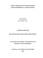
Figure 1: Inflorescence meristem and floral meristem in Arabidopsis thaliana
Upper panel: schematic diagram of inflorescence meristem and floral primordium in
Arabidopsis thaliana. L1, L2, and L3 layer of the meristem is shown. CLV3
expression domain is highlighted in pink, and WUS expression domain is highlighted
in green. Lower panel: a section of the meristem obtained from Ler wild-type plant.
9
1.3 Regulation of flower development in Arabidopsis thaliana
During the processes of floral organ identity specification and floral organ
differentiation, there are well-coordinated interplays of transcription factors. The
classical ABC model was proposed nearly two decades ago to explain the organ
identity determination in flower development (Coen and Meyerowitz, 1991). The
ABC model predicts that the combinatorial action from ABC floral homeotic genes is
largely responsible for the specification of the floral organ identity. The ABC genes,
A class for APETALA1 (AP1) and APETALA2 (AP2), B class for APETALA3 (AP3)
and PISTILLATA (PI), and C class for AGAMOUS (AG), have been extensively
studied and have been shown to encode transcription factors (Bowman et al., 1989;
Yanofsky et al., 1990; Bowman et al., 1991; Jack et al., 1992; Goto and Meyerowitz,
1994; Gustafson-Brown et al., 1994; Jofuku et al., 1994).
Apart from its role in inflorescence meristem identity, AP1 also serves as an A-class
gene for organ identity determination. AP1, together with another A-class gene AP2,
is responsible for the specification of the first whorl of floral primordia into sepals
(Irish and Sussex, 1990; Jofuku et al., 1994). AP2 encodes for a member of
AP2/EREBP class of transcription factors. In combination with B-class genes activity,
AP1 and AP2 promote development of the second whorl of floral primordia into
petals. In ap1 mutants, the first whorl organs undergo homeotic conversion, which
turns sepals into bracts subtended by floral meristems. In the meantime, the second
10
whorl organs in ap1 mutant are lost. In ap2 mutants, sepals are converted into carpels
or missing, whereas the petals in the second whorl are lost.
B-class genes such as AP3 and PI act synergistically with both A-class and C-class
genes to specify the development of second whorl and third whorl into petals and
stamens, respectively (Jack et al., 1992; Goto and Meyerowitz, 1994) . In the absence
of B-class genes, the second whorl organs are converted into sepals (in place of petals)
and the third whorl organs are transformed into carpels (in place of stamens). Both
AP3 and PI encode for members of MADS domain transcription factors.
The C-class gene AG also encodes a member of the MADS domain transcription
factors, and AG is necessary for the specification of stamens and carpels, the floral
reproductive organs (Bowman et al., 1989; Yanofsky et al., 1990). In ag-1 mutants,
the flower undergoes homeotic conversion to adopt a sepal-petal-petal reiteration
instead of the normal sepal-petal-stamen-carpel structure. The complete lack of
reproductive organs in ag-1 flowers places AG at the top of hierarchy of genes
controlling reproductive development. This conclusion is supported by microarray
expression profiling of wild-type and ag mutant flowers showing that more than 1,
000 genes are expressed downstream of AG (Wellmer et al., 2004). In addition to its
function in organ identity specification, AG also plays a prominent role in floral
determinacy. In the absence of AG, WUS activity is prolonged in the indeterminate
floral meristem (Lenhard et al., 2001; Lohmann et al., 2001). Our recent study has
11
revealed that AG acts through a zinc finger repressor, KNUCKLES (KNU) to
negatively regulate WUS for floral determinacy (Sun et al., 2009).
Studies performed on SEPALLATA (SEP) genes in Arabidopsis have added an
interesting twist into the classical ABC model for flower development (Pelaz et al.,
2000). The three SEP genes, namely SEP1, SEP2 and SEP3 are now widely regarded
as E-class genes in ABCE model of flower development (Figure 2). These E-class
genes act as important regulators for the specification of petals (second whorl),
stamens (third whorl) and carpels (fourth whorl) during flower development. In sep1
sep2 sep3 triple mutants, the flowers produced consist only of sepals. Recently, SEP3
has been shown to act as an upstream activator of the B and C class genes activity
(Kaufmann et al., 2009). Another SEP gene, SEP4 is also involved in the
development of sepals, petals, stamens, and carpels (Ditta et al., 2004). In sep1 sep2
sep3 sep4 quadruple mutans, all flower organs are converted into leaf-like organs.
Following the unraveling of SEP proteins function, the ‘quartet model’ of flower
organ identity has been proposed to explain the combinatorial effects of ABCE genes
(Theissen, 2001; Theissen and Saedler, 2001). In this model, it is suggested that there
are four floral homeotic proteins forming protein complexes to regulate and specify
the development of floral organs. In each whorl, there will be a distinct combination
of the four homeotic proteins, henceforth gives rise to different organ identities. For
instance, in whorl 2, there might be a combination of AP1, PI, AP3 and SEP proteins
to specify petals. Whereas in whorl 4, the protein complex might be a combination of
AG and SEP proteins (two molecules for each protein) in the specification of carpels.
12
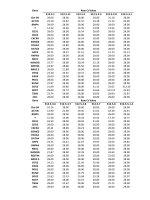
Figure 2: The ABCE model of flower development
Schematic diagram showing the ABCE model of flower development. Combinatorial
action of A, B, and E-class genes specify the development of perianth organ. Whereas,
the B, C, and E-class genes determine the identity of reproductive organs.
13
1.4 Patterning and differentiation of lateral organs in Arabidopsis thaliana
1.4.1 The patterning of lateral organs
Lateral organs are produced from the flanks of shoot apical meristem. During the
initiation of lateral organ primordia, it is suggested that auxin efflux carrier like
PINFORMED (PIN) mediates a directional flow of auxin into a localized region that
will mark the site for the emergence of a new primordium (Reinhardt et al., 2000;
Benkova et al., 2003). In this site where auxin reaches its maximal concentration, the
expression of class-I KNOTTED1-LIKE HOMEOBOX (KNOX) genes are
substantially downregulated. The KNOX genes encode for homeobox transcription
factors that are involved in the maintenance of shoot apical meristem (Hake et al.,
2004). The repression of KNOX genes in the emerging primordia is substantiated by
ASSYMETRIC LEAVES1 (AS1) and ASSYMETRIC LEAVES2 (AS2) (Semiarti et
al., 2001; Iwakawa et al., 2007; Ueno et al., 2007). AS1 encodes a MYB-domain
transcription factor, whereas AS2 encodes an AS2-LOB-domain protein. Mutations in
AS1 and AS2 genes will lead to the production of asymmetric and lobed rosette leaves
(Byrne et al., 2000; Byrne, 2005). Both AS1 and AS2 form a heterodimer to execute
their functions. AS1/AS2 heterodimer binds directly to promoters of the KNOX genes,
BREVIPEDICELLUS (BP) and KNAT1 to downregulate their expression (Guo et al.,
2008). In the meantime, the expression of AS1 and AS2 in the shoot apical meristem
is, in turn, negatively regulated by the KNOX gene, STM. The boundary specification
between the shoot apical meristem and the new lateral organ primordium is mediated
14
by a set of redundant, but partially distinct CUP-SHAPED COTYLEDONS (CUC)
genes (Takada et al., 2001; Vroemen et al., 2003). CUC genes encode for NAC-
family transcription factors. Mutations in CUC genes lead to the fusion of cotyledons,
which results in cup-shaped cotyledons.
1.4.2 Regulation of abaxial-adaxial polarity
The abaxial-adaxial polarity (Figure 3A) in the lateral organ is promptly established
after the emergence of the new primordium. There are two sets of genes responsible
for the specification of the abaxial and adaxial cell fates. The KANADI (KAN) genes
and the YABBY (YAB) genes (Bowman, 2000; Bowman et al., 2002) are required for
the establishment of abaxial axis in the lateral organs. The KAN genes like KAN1,
KAN2, and KAN3 are transcription factors of the GARP family. In kan1 kan2 double
mutant plant, the abaxial cell types of most of the lateral organs are replaced by the
adaxial cell types (Eshed et al., 2001). The YAB genes like FILAMENTOUS
FLOWER (FIL), YAB2, YAB3, INNER NO OUTER (INO), and CRABS CLAW (CRC)
encode the YAB-family transcription factors. FIL, YAB2 and YAB3 are expressed in
the abaxial domain of all of the lateral organs. These three genes act redundantly to
promote abaxial cell fate in the lateral organs. In fil-5 yab3-1 double mutant, there is a
loss of polar development of most of the floral organs (Siegfried et al., 1999). INO
expression is restricted to the abaxial side of the ovule’s outer integument. In ino
mutant, there is a loss of polar differentiation of the outer integument (Baker et al.,
1997; Villanueva et al., 1999; Bowman, 2000). CRC is expressed exclusively in
15
carpels and nectaries (Bowman and Smyth, 1999). In crc mutant, there is only a
partial loss of abaxial identity. However, in crc kan double mutant, there is an ectopic
production of adaxial tissue in the abaxial region (Alvarez and Smyth, 1999; Eshed et
al., 1999).
The adaxial axis of the lateral organs in Arabidopsis is primarily established by the
genes PHABULOSA (PHB), PHAVOLUTA (PHV), and REVOLUTA (REV) (Talbert
et al., 1995; McConnell et al., 2001; Otsuga et al., 2001; Emery et al., 2003). They
belong to a gene family encoding the class III homeodomain-leucine zipper (HD-
ZipIII) transcription factors. Ectopic expression of either PHB or PHV is sufficient
for abaxial-to-adaxial conversion. In rev mutant, flowers are converted into
filamentous structures, signifying a loss of organ polarity.
The interaction between the abaxial- and the adaxial-specifying genes has further
contributed to the establishment of polarity in the lateral organs. The AP2/ERF-type
transcription factor, AINTEGUMENTA (ANT), acts with the YABBY-family protein
FIL, to positively regulate the expression of the adaxial-specifying gene, PHB (Nole-
Wilson and Krizek, 2006). The expression of HD-ZipIII homeobox genes such as
PHB, PHV, and REV are also closely regulated by miRNA165/166 (Mallory et al.,
2004; Zhou et al., 2007). The KAN genes repress the expression of REV and PHV at
the abaxial site. In kan1 kan2 double mutant, the confinement of REV and PHV
expression to the adaxial domain is delayed. In addition, the expression level of REV
and PHV is also higher in the double mutant (Eshed et al., 2001).
16
In parallel with the KAN genes, ETTIN (ETT, AUXIN RESPONSE FACTOR 3) and
AUXIN RESPONSE FACTOR 4 (ARF4), have also been implicated in the regulation
of abaxial cell fate (Pekker et al., 2005). In ett arf4 double mutant, the abaxial tissues
are transformed into the adaxial one, and the phenotype of the double mutant is
closely related to that of KAN mutations. The expression of ETT has been found to be
closely regulated by trans-acting siRNA (ta-siRNA) (Adenot et al., 2006; Fahlgren et
al., 2006; Garcia et al., 2006).
During the development of reproductive organs, NUBBIN (NUB) and JAGGED (JAG)
genes have been proposed to act in parallel to the HD-ZipIII pathway to promote the
adaxial cell fate (Dinneny et al., 2006). NUB and JAG encode C
2
H
2
zinc-finger
transcription factors. NUB is expressed in the adaxial side of stamens and carpels,
whereas JAG is expressed in a non-polar way in the reproductive organs. In jag nub
double mutant, stamens and carpels are abaxialized, and the organ growth is severely
impaired.
17
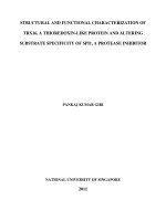
Figure 3: The axes of polarity in lateral organs and the gynoecium
(A) Schematic diagrams showing the abaxial-adaxial axis in lateral organs. IM:
inflorescence meristem; FM: floral meristem. (B) Schematic diagrams showing
apical-basal axis in the gynoecium.
A
B
18
1.4.3 Regulation of apical-basal polarity
During the development of female reproductive organ in Arabidopsis, the apical-basal
polarity (Figure 3B) is established and maintained by a number of genes, whose
mutations result in impaired development of the gynoecium. Major regulators for the
differentiation of gynoecium’s apical tissues is a basic helix-loop-helix transcription
factor, SPATULA (SPT) (Heisler et al., 2001) and ETT. In spt mutant, there is
reduced growth of the style, stigma, and septum. The transmitting tract is also absent
in the spt mutant. In addition to its function in abaxial-adaxial patterning, ETT has a
greater role in apical-basal patterning of the gynoecium (Sessions and Zambryski,
1995; Sessions et al., 1997). In ett mutant, there is an expansion of the apical (style
and stigma) and basal (the gynophore) tissues with a reduction of the ovary region. It
is suggested that ETT represses the expression of SPT to promote the formation of the
ovary. STYLISH (STY) genes have also been implicated in the regulation of the
Arabidopsis gynoecium development (Kuusk et al., 2002; Sohlberg et al., 2006).
STY1 and STY2 are partially redundant genes encoding proteins with a RING finger-
like motif. In sty1 mutant, there is aberrant formation of the style of the gynoecium.
In sty1 sty2 double mutant, there is an enhanced phenotype of sty1 mutant with
decrease proliferation of stylar xylem, and reduction of stylar and stigmatic tissues.
A strong presence of an auxin response factor like ETT in the regulation of the apical-
basal polarity has suggested an instrumental role for auxin in the patterning of the
gynoecium. Indeed, auxin has been proposed to be the key phytohormone that is
19
responsible for setting up the apical-basal polarity in the gynoecium (Nemhauser et
al., 2000). It is hypothesized that there is a decreasing gradient of auxin from the
apical part of the gynoecial primordium to the basal. According to this hypothesis,
high levels of auxin induces style and stigma differentiation, intermediate levels of
auxin leads to formation of ovary, and low levels of auxin is responsible for the
specification of the basal gynophore. Recently, STY1 is reported to activate the
expression of YUCCA4, a gene encodes a flavin monooxygenase involved in auxin
biosythesis (Sohlberg et al., 2006). Overexpression of YUCCA genes has been shown
to lead to the overproduction of auxin (Cheng et al., 2006).
1.4.4 Downstream target genes of AGAMOUS
Despite AG’s prominent role in reproductive organs development, very few target
genes of AG have been characterized in detail. AG has been implicated in the
regulation of other MADS-box genes like AP1 and SHATTERPROOF2 (SHP2)
(Gustafson-Brown et al., 1994; Savidge et al., 1995). AG prevents the accumulation
of AP1 transcripts in the two inner whorls of the floral organs (whorl 3 and whorl 4)
that will give rise to reproductive organs (Gustafson-Brown et al., 1994). A more
comprehensive study has pointed out that SHP2 is a direct downstream target of AG
(Savidge et al., 1995). SHP2 encodes a MADS-box protein sharing substantial
similarity with AG. It is formerly known as AGAMOUS-LIKE 5 (AGL5). SHP2 and
its close member, SHP1 have partial redundant function with AG. Both SHP1 and
SHP2 are involved in fruit dehiscence (Liljegren et al., 2000). SHP2 is expressed
20
during early carpel development after the onset of AG expression. Ectopic expression
of AG effectively activates the expression SHP2. In ag mutant, there is a loss of SHP
expression. Furthermore, AG is shown to bind specifically to a promoter element of
SHP2.
AG has been shown to positively regulate the expression of the SPOROCYTELESS
(SPL) gene (Ito et al., 2004). SPL is involved in microsporogenesis, a process leading
to pollen formation. AG binds to the 3’ region of the SPL gene and activates its
expression. In another of our study, AG has been demonstrated to mediate late-stage
stamen development (anther morphogenesis and dehiscence, and filament formation
and elongation) (Ito et al., 2007). It does so by controlling the expression of
DEFECTIVE IN ANTHER DEHISCENCE1 (DAD1). DAD1 encodes a gene involved
in the biosynthesis of a lipid-derived phytohormone, jasmonic acid. AG binds to the
5’ coding region of the DAD1 gene, and activates its expression at later stages of
floral development (days 7-8 after the onset of AG expression).
1.5 Functions of AT-hook DNA binding proteins during development
AT-hook DNA binding proteins may contribute to a functional nuclear architecture
by binding to the nuclear matrix, and may also be structural components that remain
inside the nucleus after removal of basic proteins and histones (Strick and Laemmli,
1995; Aravind and Landsman, 1998; Morisawa et al., 2000). AT-hook motifs bind to
the minor grooves of duplex DNA in matrix attachment regions (MARs) of target
21
DNA sequences (Reeves and Nissen, 1990; Reeves, 2001), an unique property that
distinguishes them from common transcription factors that primarily bind to the
major grooves of duplex DNA.
MARs are stretches of characteristic AT-rich DNA sequences that tend to attain a
single-stranded conformation through base unpairing. In this regard, MARs are also
regarded as base unpairing regions (BURs). This greater tendency of forming
unpairing region in MAR is likely to be the result of sequence-induced torsional
stress propagated from the surrounding DNA (Boulikas, 1995). MARs and AT-hook
DNA binding proteins are suggested to be the key determinants in anchoring specific
DNA sequences to nuclear matrix, a process that helps to generate chromatin loop
domain and also to introduce structural changes in the chromatin (Reeves, 2001).
In animals, the MAR binding protein SATB1, which contains an AT-hook DNA
binding motif, has been implicated in tissue- or cell type-specific regulation of
multiple genes (Alvarez et al., 2000; Cai et al., 2003; Britanova et al., 2005; Dobreva
et al., 2006; Han et al., 2008). SATB1 is suggested to play a prominent role in
chromatin assembly and histone modification. It binds in the minor grooves of duplex
DNA (Nakagomi et al., 1994) and is able to regulate the transcription of multiple
genes over long distances (Yasui et al., 2002). It is demonstrated that SATB1 can
recruit histone deacetylase to a specific locus and mediates a large-domain histone
deacetylation within that locus (Yasui et al., 2002).
22
A close member of SATB1, SATB2 is shown to act as a molecular node in a
transcriptional network regulating skeletogenesis (Dobreva et al., 2006). In Satb2
-/-
mice, there are severe defects in craniofacial patterning and osteoblast differentiation.
SATB2 regulates the expression of multiple genes in osteoblasts, which includes
Bone Sialoprotein (Bsp), Osteocalcin (Ocn), and several Hox genes like Hoxa2.
In plants, several AT-hook DNA binding proteins have been identified and are
implicated in various aspects of plant development (Weigel et al., 2000; Lim et al.,
2007; Matsushita et al., 2007; Vom Endt et al., 2007; Street et al., 2008). In
Arabidopsis, ESCAROLA (ESC/ORE7) has been isolated from an activation-tagging
screen and is suggested to play a role in leaf longevity (Lim et al., 2007). A closely
related SOB3 (SUPPRESSOR OF PHYB-4#3) gene is involved in the negative
modulation of hypocotyl growth (Street et al., 2008). AGF1 (AT-HOOK PROTEIN
OF GA FEEDBACK REGULATION 1) has been identified from a yeast one-hybrid
screen as a negative regulator of GA signaling through binding to cis-acting sequence
of GA 3-oxidase (Matsushita et al., 2007). In addition, AT-hook proteins are also
likely to participate in the regulation of gene expression in response to jasmonic acids
(JA) in suspension culture of Catharanthus roseus cells (Vom Endt et al., 2007).
Recently, AHL22 (AT-HOOK MOTIF NUCLEAR LOCALIZED PROTEIN22)
has been reported to be involved in the regulation of flowering and hypocotyl
elongation (Xiao et al., 2009). Overexpression of AHL22 effectively delays the
flowering process in Arabidopsis. In spite of these reports, the function of AT-hook
23
DNA binding protein has yet to be demonstrated in flower development and plant
meristem regulation.
1.6 Objective of the study
The aim of this study is to augment the understanding of flower development and
meristem regulation in Arabidopsis thaliana through functional study of a novel
regulator of gene expression, GIANT KILLER. Specifically, I wish to address the
following questions in the study: First, is GIK a direct target of AG? Second, what is
the function of GIK in reproductive development? Third, what are the downstream
targets of GIK in reproductive development? Fourth, how does GIK mediate gene
regulation? Fifth, does GIK have any roles in meristem regulation?
1.7 Significance of this study
In this study, I have presented findings showing that GIK serves as a new way of
control employed by the floral homeotic gene AGAMOUS to regulate multiple genes
expression. I have identified several key regulators in reproductive patterning and
differentiation as downstream targets of GIK. In particular, I have shown that ETT is
negatively modulated by GIK, and that the GIK-mediated ETT regulation is closely
associated with epigenetic changes of the chromatin. In addition, my study has also
provided new insight for the understanding of meristem integrity in Arabidopsis
thaliana. I have shown that ectopic GIK activity effectively disrupts stem cell
24
regulation, and causes aberrant expression of WUS in inflorescence and floral
meristems.
25
CHAPTER 2: Materials and Methods
2.1 Materials
2.1.1 Plant materials
All Arabidopsis thaliana plants used in this study are in the Landsberg erecta
background unless otherwise stated. They were grown at 22 °C under continuous
light condition in plant growth chambers or the plant growth room. The 35S::GIK and
35S::GIK-GR-6HA seeds were provided by Dr Toshiro Ito. A GIK insertion line was
obtained from the TRAPPER collection (
(stock number, ET14389). The enhancer trap was inserted at the middle of the coding
region, 450 bp downstream from the start codon. Homozygous lines were verified by
PCR-based genotyping.
2.1.2 Bacterial strains
Escherichia coli strains used for the molecular cloning in this study were DH5α and
XL1-BLUE unless otherwise stated. The One Shot ccdB Survival-T1 (Invitrogen)
was used specifically to amplify and maintain plasmids containing cytotoxic ccdB
gene. E. coli strains were grown in Luria-Bertani (LB) medium at 37 °C with
appropriate selective antibiotics. The Agrobacterium tumefaciens strain used for the
plant transformation in this study was C58C1 (GV3101). The C58C1 strain carrying