Analysis of regulator of g protein signaling (RGS) function in growth, development and pathogenicity of magnaporthe grisea 1
Bạn đang xem bản rút gọn của tài liệu. Xem và tải ngay bản đầy đủ của tài liệu tại đây (19.52 MB, 220 trang )
ANALYSIS OF REGULATOR OF G-PROTEIN SIGNALING
(RGS) FUNCTION IN GROWTH, DEVELOPMENT AND
PATHOGENICITY OF MAGNAPORTHE GRISEA
HAO LIU
NATIONAL UNIVERSITY OF SINGAPORE
2006
ANALYSIS OF REGULATOR OF G-PROTEIN SIGNALING
(RGS) FUNCTION IN GROWTH, DEVELOPMENT AND
PATHOGENICITY OF MAGNAPORTHE GRISEA
HAO LIU
A THESIS SUBMITTED
FOR THE DEGREE OF DOCTOR OF PHILOSOPHY
TEMASEK LIFE SCIENCES LABORATORY
NATIONAL UNIVERSITY OF SINGAPORE
ii
ACKNOWLEDGEMENTS
I would like to express my heartfelt thanks to my thesis supervisor Dr. Naweed Naqvi for
giving me the opportunity to work on M. grisea and for his constant support and guidance
through out the course of this work. I thank all the members of my thesis committee: Prof
William Chia, A/Prof Mohan Balasubramanian and A/Prof Yue Wang for helpful
suggestions. Special thanks to Dr. Hongyan Wang, who introduced IMA to me. I also
thank Dr. Fengwei Yu for his fruitful discussion and suggestions.
Many thanks to Angayarkanni Suresh for excellent technical assistance, and to all
members of the Fungal Genomics Group for suggestions and discussions. I also extend
my thanks to all the members of the Cell Biology Forum for constructive criticism. I
thank TLL administrative and support staff for all the help. Financial support from the
Temasek Life Sciences Laboratory is duly acknowledged.
iii
TABLE OF CONTENTS
Page
Summary…………………………………………………………………………………ix
List of figures……………………………………………………………………………xii
List of abbreviations……………………………………………………………… ….xv
Chapter I Introduction…………………………………………………………………. 1
1.1 General introduction to fungal development…………………………. 1
1.1.1 Fungal mating…………………………………… …5
1.1.2 Morphological switch in Fungi……………………… …… .7
1.1.3 Fungal asexual development ………………………………… ….11
1.1.4 Fungal pathogenicity…… …………………………… … 14
1.2 General introduction of G-protein-mediated signaling cascade …… 17
1.2.1 Heterometric G proteins………………………………………………… 18
1.2.2 Model organisms to study G proteins…………………………………… 21
1.2.2.1 G proteins in plants…………………………………………… 21
1.2.2.2 G proteins in yeast…………………………………………… 22
1.2.2.3 G proteins in mammals………………………………………… 23
1.2.3 Desensitization of G protein Signaling…………………………… …… 24
1.2.3.1 The discovery of Regulator of G-protein signaling (RGS)………24
1.2.3.2 The mechanism of RGS function……………………… …… 26
1.3 G proteins in fungal pathogens………………………………………………………28
iv
1.3.1 G proteins in Aspergillus………………………………………………… 28
1.3.2 G proteins in Candida albicans……………………………………………29
1.3.3 G proteins in Ustilago maydis……………………………………….…… 29
1.3.4 G proteins in Cryphonectria parasitica……………………………………30
1.4 Magnaporthe grisea, the rice blast pathogen……………………………….……… 31
1.4.1 General introduction to Magnaporthe………………………………… …31
1.4.2 Magnaporthe conidiation … ……33
1.4.3 Morphology of appressorium …………………………………………… 33
1.4.4 Signal perception and transduction for appressorium development…….…35
1.4.4.1 Surface signal perception……………………………………… 35
1.4.4.2 Intracellular signal transduction………………………………….37
1.4.5 G proteins in Magnaporthe………………… 38
1.5 Aims and objectives of this thesis……………………………………………………39
1.6 The significance of this study……………………………………………………… 40
Chapter II Materials and Methods……………………………………………………42
2.1 Strains, Growth, Infection Assays and Reagents…………………………………….42
2.1.1 Magnaporthe grisea strains and growth conditions…………….………….42
2.1.2 E. coli strains and growth conditions…………………………………… 42
2.1.3 Agrobacterium tumefaciens strains and growth conditions…………… …43
2.1.4 Appressorium formation assay with Manaporthe conidia…………………43
2.1.5 Barley related methods and infection with Magnaporthe conidia……… 44
2.1.6 Rice related methods and infection with Magnaporthe conidia………… 44
v
2.2 Molecular Methods………………………………………………………………… 44
2.2.1 DNA techniques……………………………………………………………44
2.2.1.1 PCR amplification……………………………………………… 44
2.2.1.2 Agarose gel electrophoresis and gel purification of nucleic acid
fragments………………………………………………………….…… 45
2.2.1.3 Recombinant DNA techniques………………………………… 45
2.2.1.4 Genomic DNA extraction from Magnaporthe…………….…… 46
2.2.1.5 Southern Blot…………………………………………………….47
2.2.1.6 Ligation-mediated PCR ……………………………………… 49
2.2.1.7 Transformation of E. coli by heat shock method………… …….49
2.2.1.8 Transformation of Agrobacterium by electroporation methods…50
2.2.2 RNA techniques……………………………………………………………50
2.2.3 T-DNA random insertion, gene targeting and genetic complementation….51
2.2.3.1 Gene disruption strategy……………………………………… 51
2.2.3.2 Targeted gene replacement………………………………………52
2.2.3.3 Genetic complementation of rgs1∆…………………………… 52
2.3.3.4 RGS1 overexpression……………………………………….……53
2.2.3.5 Site-directed mutation in MAGA, MAGB and MAGC……… …53
2.3 Protein and immunology related methods………………………………………… 54
2.3.1 Total protein lysates from Magnaporthe (Denatured and Native)……… 54
2.3.2 Expression and purification of fusion protein in E. coli………………… 54
2.3.3 Rgs1 antiserum and its specificity……………………………………… 55
2.3.4 Protein electrophoresis, immunoblots and reprobing protocals………… 56
vi
2.3.5 Rgs1 and Gα protein interaction in vitro………………………………… 57
2.3.6 Endogenous Rgs1 interacts with recombinant Gα proteins……………….58
2.4 cAMP extraction and analysis……………………………………………………….58
2.4.1 Extraction of cAMP from Magnaporthe mycelium and germ tubes… ….59
2.4.2 Analysis of cAMP with cAMP Biotrak Enzymeimmunoassay System… 59
2.5 Hardness Assay………………………………………………………………………60
2.6 Light Microscopy…………………………………………………………………….60
Chapter III Identification and Characterization of RGS1 deficient mutant in M.
grisea 62
3.1 Introduction………………………………………………………………………… 62
3.2 Results……………………………………………………………………………… 63
3.2.1 Agrobacterium T-DNA mediated insertional mutagenesis in
Magnaporthe…………………………………………………………………… 63
3.2.2 Identification of the disrupted locus…………………………………… 64
3.2.3 Characterization of TMT1398 mutant…………………………………… 71
3.2.4 Cloning of RGS1 in Magnaporthe………………………………… …… 76
3.2.5 Creation and Characterization of rgs1
∆
mutant……………………………81
3.2.6 Genetic complementation of rgs1
∆
81
3.2.7 Excessive Rgs1 reduces conidiation but not appressorium development….82
3.3 Discussion……………………………………………………………………………82
vii
Chapter IV Mechanism of Rgs1 function…………………………………………… 94
4.1 Introduction………………………………………………………………………… 94
4.2 Results……………………………………………………………………………… 95
4.2.1 RGS-domain containing proteins in Magnaporthe……………………… 95
4.2.2 Appressorium formation in Gα deletion mutants………………………….96
4.2.3 Appressorium formation in rgs1∆ G
α
∆ mutants………………………….98
4.2.4 Appressorium formation in RGS-insensitive Gα mutants…………………98
4.2.5 Rgs1 dependent regulation of cyclic AMP level……………………… 104
4.2.6 Rgs1 physically interacts with MagA…………………………………….104
4.2.7 Gα
i
subunit MagB is critical for conidiogenesis………………… …….106
4.2.8 Mgb1, the Gβ subunit, is required for conidiogenesis in
Magnaporthe……………………………………………………………………109
4.2.9 Water soaking phenotype of Gα mutant colonies……………………… 109
4.2.10 The expressions of candidate hydrophobin genes in rgs1∆ 112
4.2.11 Physical interaction between Rgs1 and MagB………………………… 114
4.2.12 Physical interaction between Rgs1 and MagC…………………… ……114
4.3 Discussion……………………………………………………………………… 117
4.3.1 Rgs1 regulates MagA for appressorium development………………… 117
4.3.2 Rgs1 regulates MagB for conidiogenesis and surface hydrophobicity… 118
viii
Chapter V Thigmotropic signaling in M. grisea pathogenesis…………………… 121
5.1 Introduction…………………………………………………………………………121
5.2 Results………………………………………………………………………………121
5.2.1 Surface hardness stimulus is essential for appressorium differentiation in
Magnaporthe……………………………………………………………………121
5.2.2 Timing of the thigmotropic signal sensing……………………………….125
5.2.3 Relationship between thigmotropism and cAMP levels………………….127
5.2.4 Mechanosensitive channels in Magnaporthe……………………… ……131
5.3 Discussion………………………………………………………………………… 134
Chapter VI General Discussion………………………………………………………137
Appendix 1…………………………………………………………………… ………144
References…………………………………………………………………………… 149
ix
SUMMARY
The Magnaporthe-rice interaction is a major model for understanding plant disease,
largely because of its economic importance, and also due to the molecular genetic
tractability of the blast fungus. Rice-blast disease caused by Magnaporthe is initiated
when conidia germinate upon attachment to the host surface. In response to surface and
environmental cues, the resultant germ tubes undergo infection-specific differentiation to
form the dome-shaped appressoria, which are employed to forcibly penetrate host cuticle.
Signaling between Magnaporthe and rice is therefore predicted to be critical for initiating
the pathogenesis cycle. However, the molecular mechanisms underlying this parasite-host
communication are not fully understood.
This study initially describes an Agrobacterium Transferred-DNA mediated insertional
mutagenesis in Magnaporthe, aimed at identifying genes required for fungal
pathogenicity. This led to the identification of an insertional mutant TMT1398, in which
a gene (RGS1) encoding an RGS-domain containing protein was disrupted. TMT1398
and an rgs1∆ strain showed pleiotropic defects such as soaking phenotype,
hyperconidiation, appressoria formation on non-inductive surfaces and reduced
pathogenicity. Through genetic complementation it was ascertained that the defects
displayed by TMT1398 and rgs1∆ were due to the loss of Rgs1 function.
In Magnaporthe, appressorium formation has been shown to be induced by surface
hydrophobicity. Unlike the wild-type, TMT1398 and rgs1∆ did not depend on surface
x
hydrophobicity signals for appressorium development and could form appressoria on
hydrophilic or hydrophobic surfaces with equal efficiency. However, neither the wild-
type nor the rgs1∆ could form appressoria on soft surfaces, regardless of the
hydrophobicity, suggesting that surface hardness signal could be crucial for appressorium
development. A major highlight of the study presented here is the identification of such a
thigmotropic response, which was found to be essential for initiating appressorium
formation in Magnaporthe. The critical surface hardness sufficient to induce
appressorium formation was estimated through biophysical analysis of host leaf surface
as well as the inductive artificial membranes.
Regulators of G-protein signaling (RGS) accelerate the intrinsic GTPase activity of the
constituent Gα subunits and thus negatively regulate the heterotrimeric G-protein
signaling cascades. The mechanism of Rgs1 function was therefore investigated and
elucidated by analyzing the function(s) of its potential Gα subunit targets (Gα
s
subunit
MagA, Gα
i
subunit MagB, and Gα
II
subunit MagC) in Magnaporthe. Characterization of
individual Gα-deletion strains and RGS1-insensitive mutants (magA
G187S
, magB
G183S
, and
magC
G184S
) revealed that Rgs1 directly regulates MagA during appressorium initiation.
Interestingly, rgs1∆ and magA
G187S
accumulated higher levels of cAMP compared to the
wild-type further suggesting that cAMP-mediated downstream signaling is important for
appressorium formation. The magB∆ failed to conidiate, whereas magB
G183S
hyperconidiated like rgs1∆, suggesting that Rgs1 regulates MagB during conidiogenesis.
Further characterization of the soaking phenotype of the rgs1∆ and the magB
G183S
colonies
showed that Rgs1 and MagB function together to regulate mycelial
xi
hydrophobicity. In biochemical analyses, Rgs1 physically interacted with and
individually accelerated the GTPase activity of each of the three Gα subunits (MagA,
MagB and MagC) in Magnaporthe. Thus, as a unique and multifunctional regulator of G
protein signaling, Rgs1 regulates mycelial surface hydrophobicity, asexual reproduction,
appressorium development, pathogenicity and thigmotropism in Magnaporthe.
xii
LIST OF FIGURES
Figure……………………………………………………………………………… Page
Figure 1 Morphism in fungi……………… 3
Figure 2 Conidiogenesis in fungi …… ……… 13
Figure 3 Schematic representation of canonical G protein signaling ………………… 21
Figure 4 The RGS4– G
i
α
1
Complex…………………………………………….………28
Figure 5 Two life cycles in Magnaporthe……………………………………………….33
Figure 6 Morphology of appressorium………………………………………………….35
Figure 7 Infection assay on barley leaves……………………………………………….65
Figure 8 Appresorium formation assay………………………………………………….66
Figure 9 Ligation-mediated PCR for identifying flanking sequences for Agrobacterium
T-DNA insertions in Magnaporthe . ……………………………………….……………67
Figure 10 Identification of the T-DNA insertion sites……………… …………… … 70
Figure 11 Appressorium formation defect in TMT1398… …………………………… 72
Figure 12 Accelerated appressorium formation in TMT1398………………………… 74
Figure 13 Defect in conidiogenesis of TMT1398……………………………………….75
Figure 14 Defect in pathogenicity of TMT1398……………………………………… 77
Figure 15 Penetration peg formation of TMT1398…………………………………… 78
Figure 16 Easily Wettable phenotype of TMT1398…………………………………….79
Figure 17 Cloning of RGS1 83
xiii
Figure 18 Alignment of Rgs1 with fungal orthologs……………………………………84
Figure 19 Analysis of RGS domain of Rgs1 with multiple sequence alignments………85
Figure 20 Creation of RGS1 deletion mutant……………………………………………86
Figure 21 Rgs1 negatively regulates conidiation……………………………………… 87
Figure 22 Rgs1 negatively regulates appressorium formation on non-inductive /
hydrophilic surfaces…………………………………………………………………… 88
Figure 23 RGS1 is required for full pathogenicity in Maganporthe…………………….89
Figure 24 RGS1 is involved in surface hydrophobicity……………… 90
Figure 25 RGS1 overexpression in Magnaporthe……………………………………….91
Figure 26 RGS proteins in Magnaporthe………………………………………….…….97
Figure 27 Appressorium formation in Gα deletion mutants………………………… 99
Figure 28 Appressorium formation assays with rgs1∆Gα∆ mutants…………… … 100
Figure 29 Conserved switch region I among Magnaporthe Gα proteins…………… 102
Figure 30 Appressorium formation assays in RGS-insensitive Gα mutants……… …103
Figure 31 Rgs1 and MagA regulate intracellular cAMP levels………………… ……105
Figure 32 Rgs1 interacts with MagA…………………………………………… … 107
Figure 33 Rgs1 acts in concert with MagB during conidiogenesis……………….… 108
Figure 34 Gβ subunit Mgb1 is required for conidiogenesis in Magnaporthe…………110
Figure 35 Rgs1 acts coordinately with MagB to regulate of mycelial hydrophobicity 111
Figure 36 Hydrophobin gene expression in wild-type and rgs1∆ 113
Figure 37 Rgs1 interacts with MagB………………………………………………… 115
Figure 38 Rgs1 physically interacts with MagC……………………………………….116
Figure 39 Soft surfaces do not induce appressorium formation……………………….123
xiv
Figure 40 Contact with hard surface induce appressorium development…………… 124
Figure 41 The increased induction ability of appressorium formation on dried agar.…126
Figure 42 Surface-hardness signal is perceived and integrated within two hours of
conidia germination….…………………………………………………………………128
Figure 43 Inhibition of appressorium formation with Gadolinium…………………….129
Figure 44 Hardness signaling and intracellular cAMP……………………………… 130
Figure 45 Mechanosensitive channels are not required for appressorium formation….133
Figure 46 Growth characteristics of mechanosensitive channel mutants under stress
conditions……………………………………………………………………………….135
xv
LIST OF ABBREVIATIONS
aa Amino acid
ABA Abscisic acid
BAC Bacterial artificial chromosome
cAMP Cyclic adenosine monophosphate
CM Complete medium
d Day
DEP Disheveled, Egl-10 and Pleckstrin
DEPC Diethyl pyrocarbonate
Gα G protein alpha subunit
Gβ G protein beta subunit
Gγ G protein gamma subunit
GAP Guanosine triphosphatase activating proteins
GDP Guanosine diphosphate
GEF Guanine-nucleotide exchange factors
GIRK G protein-coupled inwardly rectifying potassium
G protein Guanine nucleotide binding protein
GPCR G protein-coupled receptor
GSH Glutathione
GST Glutathione-s-transferase
GTP Guanosine triphosphate
GTPase Guanosine triphosphatase
xvi
h Hour
HPH Hygromycin phosphotransferase
LB Left board of T-DNA
MAPK Mitogen-activated protein kinase
MBP Maltose binding protein
m Minute
MscL Mechanosensitive channels large current
MscS Mechanosensitive channels small current
ORF Open reading frame
PA Prune agar
PAGE Polyacrylamide gel electropheresis
PCR Polymerase chain reaction
RB Right board of T-DNA
RGS Regulator of G-protein signaling
RPM Revolution per minute
RT Room temperature
RT-PCR Reverse transcription polymerase chain reaction
s Second
SDS Sodium dodecyl sulphate
T-DNA Transfer DNA
Tris Tris(hydroxymethyl)aminomethane
WT Wild-type
1
Chapter I INTRODUCTION
1.1 General introduction to fungal growth and development
The eukaryotic kingdom of fungi encompasses a tremendously diverse and enormously versatile
range of organisms, including yeasts, molds, or a combination of both forms. Fungi have adapted
to different modes of growth and development in evolution to spread in the environment, or to
survive unfavorable conditions. Yeasts are true fungi whose usual and dominant growth form is
unicellular (Scherr and Weaver 1953). Budding or fission is the favorite mechanism for yeast
reproduction (Chant and Pringle 1991; Chang and Nurse 1996). Budding is asexual reproduction
of a new organism, or daughter cell, by polarized protrusion of part of another organism, or
mother cell (Chant and Pringle 1991). A bud can first develop on different part of the mother
cell. The nucleus of the mother cell splits into two daughter nuclei, one of which migrates into
the bud cell (Chant and Pringle 1991; Chang and Nurse 1996; Chant 1999). The bud continues to
grow until it separates from mother cell, forming a new cell, which is genetically identical to the
mother cell. In contrast, growth by fission is a form of symmetric yeast reproduction. As seen in
Schizosaccharomyces pombe, the cell enters mitosis, followed by elongation of the cell and
formation of a septum that divides the cell into halves, separating the two nuclei into two cells of
the same size (Feierbach and Chang 2001). Molds, in contrast to unicellular yeasts, occur in long
filaments known as hyphae consisting of one of more cells surrounded by a tubular cell wall,
which grow by hyphal apical extension (Gow 1994). Apical growth in filamentous fungi is a
polarized cellular mitotic growth in which nearly all cell growth occurs only at the hyphal tip,
followed by fission of cells through the formation of incomplete septa (Horio and Oakley 2005).
Hyphal tip growth is characterized
by the initial establishment of one growth site, which is
2
followed
by its continuous maintenance (Steinberg 2007). Hyphal tip growth, branching, and
hyphal fusion results in the formation of a complex tri-dimensional hyphal network in fungi
(Gow 1994).
The unicellular growth form is the preferred mode of reproduction of yeasts. However some
yeasts can grow in a variety of morphological forms, ranging from budding cell (unicellular
yeast) to pseudohyphae (chains of elongated cells with visible constrictions at the sites of septa)
and true hyphae (linear filaments without visible constrictions at the septa) (Figure 1) (Kron and
Gow 1995; Liu 2001; Berman 2006). This kind of reversible growth type switch and transition in
fungi is designated as dimorphism (Bolker 2001). The yeast to pseudohyphal or hyphal switch is
triggered by environmental signals, such as temperature or pH or nutritional starvation, which
usually imposes the stress response in fungi (Brown and Gow 1999). The molecular mechanism
involved in this morphogenesis has been well investigated and revealed to be well conserved in
yeast and fungi. The morphogenetic switch in Saccharomyces cerevisiae requires the cooperation
of two different intracellular signaling pathways, a MAP kinase cascade and a cAMP-dependent
signal network (Gancedo 2001).
1.1.1 Fungal mating
The potential benefits of fungal sex are to purge the genome of deleterious mutations, to generate
diversity, or both (Heitman 2006). Saccharomyces cerevisiae is a model organism used to study
fungal mating. S. cerevisiae is a single celled eukaryote reproducing by mitosis, with daughter
cells budding off of mother cells. S. cerevisiae can survive and grow in two forms, diploid and
haploid. The haploid cells undergo a simple life cycle of mitosis and growth, and under
3
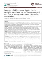
Figure 1. Vegetative morphology of fungal cells.
(A) Yeast cells can form both (B) pseudohyphae and (C) true hyphae. Switching between the
pseudohyphal and hyphal morphologies is less frequent.
(Adapted from Berman J. Morphogenesis and cell cycle progression in Candida albicans. Curr
Opin Microbiol. 2006 Dec; 9(6):595-601)
4
conditions of high stress will generally simply die (Hirschberg and Simchen 1977). The diploid
cells undergo a simple life cycle of mitosis and growth as well, but under conditions of stress can
undergo sporulation, entering meiosis and producing a variety of haploid spores (Hirschberg and
Simchen 1977; Schild and Byers 1980). Thus, diploid cells can undergo both mitosis and
meiosis, but haploid cells can only undergo mitosis. S. cerevisiae has both the asexual and sexual
life phases. Haploid cells are one of two mating types (a or α), and are capable of responding to
the mating pheromone, a short peptide produced by other haploid cells of the opposite mating
type and mate with them to produce stable diploid cells (Dohlman 1993). The mating of yeast
only occurs between haploids, which can be either the a or α mating type and thus display simple
sexual differentiation (Dohlman 1993).
S. cerevisiae mating type is determined by a single locus, MAT, which in turn governs the sexual
behavior of both haploid and diploid cells (Nasmyth 1982; Harashima et al. 1989). Two alleles
of MAT (MATa in a cells and MAT α in α cells, respectively) govern transcriptional repression
and activation of mating type specific genes (Nasmyth 1982; Haber 1992; Bardwell et al. 1994;
Haber 1998). In brief, the MATa allele of MAT encodes a pair of genes called a1 and a2, which in
haploids direct the transcription of the a-specific transcriptional program, such as producing the
mating pheromone a-factor and expressing α-factor cell surface receptor STE2 (Nasmyth 1982).
Moreover, MATa allele also represses the transcription of the α-specific transcriptional program
by inhibiting the producing the mating pheromone α-factor and a-factor cell surface receptor
STE3 (Nasmyth 1982). This defines an “a” cell. The MAT α alleles of MAT encodes the α1 and
α2 genes, which in haploids direct the transcription of the α-specific program (such as producing
α-factor, expressing STE3, and repressing STE2), which causes the cell to be an α-cells. a-cells
5
respond to α-factor and α-cells respond to a-factor, respectively, by growing a projection for cell
conjugation (Nasmyth 1982). The response of haploid cells only to the mating pheromone of the
opposite mating type allows mating between a and α cells, but not between cells of the same
mating type (Hirsch and Cross 1992; Elion 2000). Diploid cells do not produce or respond to
either mating pheromone and do not mate (Hirsch and Cross 1992; Elion 2000).
Cellular responses to mating pheromone ultimately elicit important changes including (1):
cytoskeletal structural reorganization leading to polarized cell growth; (2) induction of gene
transcription; (3) arrest of cell cycle progression in G1 phase; (4) changes in nuclear architecture
(Kurjan 1993; Elion 2000; Dohlman 2002). All these cellular changes are critical to the
successful progression of mating. Briefly, polarized cell growth is required to establish the site
for cell fusion. New gene transcription is required to produce, for example, proteins that mediate
cell adhesion and cell fusion. Growth arrest is required to synchronize the cell cycles of the two
opposite mating partner. Nuclear changes are required in preparation for nuclear fusion and the
completion of zygote formation (Kurjan 1993; Elion 2000; Dohlman 2002).
The molecular mechanism involved in mating has been well investigated. The pheromone (a or
α factor) signal is perceived and relayed to the intracellular heterotrimeric G protein by the cell
surface receptor (Ste3 or Ste2, respectively) to initiate the mating program (Dohlman 1993). The
role of G protein in S. cerevisiae mating will be discussed in chapter 1.2.2.2 in detail. Study of S.
cerevisiae mating leaded to the identification of the first mitogen-activated protein kinase
(MAPK) signaling cascade, a three-tired signal transduction module known as MAPK cascade is
conserved ubiquitously throughout the eukaryotic kingdom (Posas et al. 1998). A canonical
6
MAPK pathway is composed of MAP kinase, MAP kinase kinase (MAPKK) and MAP kinase
kinase kinase (MAPKKK). G-protein mediated mating signaling activates Ste11, a MAPKKK in
S. cerevisiae. Ste11, in turn, phosphorylates and activates Ste7, a MAPKK, which likewise,
phosphorylates and activates Fus3, a MAPK (Gustin et al. 1998). Fus3 is a Ser/Thr-specific
protein kinase which has multiple targets in both the cytosol and the nucleus. The
phosphorylation of Fus3 leads to activation of transcription and other mating specific events in
nucleus (Dohlman 2002).
1.1.2 Morphological switch in fungi
Fungal dimorphism is the ability to produce either separated yeast cells or filamentous forms
(Sanchez-Martinez and Perez-Martin 2001). This morphological switch represents an important
developmental stage of some fungal pathogens, such as Candida albicans (Ernst 2000). C.
albicans (sometimes referred to as monilia) is the major fungal pathogen that colonizes medical
implants and cause device-associated infections with exceptionally high mortality (Douglas
2002; Filler 2006; Pfaller and Diekema 2007). C. albicans is a diploid asexual fungus that is
normally present on the skin and in mucous membranes such as the vagina, mouth, or rectum
(Romani et al. 2003). The fungus can travel through the blood stream and affect the throat,
intestines, and heart valves (Whiteway and Oberholzer 2004). C. albicans is also a causal agent
of opportunistic oral and vaginal infections in humans (Loh and Sivalingam 2003). Under normal
circumstances, C. albicans lives in 80% of human population with no harmful effects, although
overgrowth results in Candidiasis (Chapman 2003). However, in immunocompromised patients
(e.g. AIDS, cancer chemotherapy, organ or bone marrow transplantation) C. albicans infection
has emerged as important causes of morbidity (Altamura et al. 2001). In addition, hospital-
7
related infection with C. albicans in patients not previously considered at risk (e.g. patients on an
intensive care unit) has become a cause of major health concern (Lim and Stern 1986; Mukherjee
et al. 2005).
C. albicans can grow in a variety of morphological forms, ranging from budding yeast to
pseudohyphae and hyphae (Mitchell 1998). To infect host tissue, the usual unicellular yeast-like
form of C. albicans reacts to environmental cues, such as serum and increasing temperature, and
switches into an invasive multicellular filamentous form (Mitchell 1998). This transition is
important for the development of pathogenicity because the mutant strain ‘locked’ in the yeast
form is avirulent (Ernst 2000). This switching between two cell-types is known as dimorphism.
Molecular study has revealed the underlying mechanism of C. albicans dimorphism (Ernst
2000). Through differential expression screen, some filamentation response genes are found to
be expressed at higher levels in filamentous cells than in yeast cells (Liu 2001). Two parallel
pathways involved in regulation of filamentation have been identified (Ernst 2000). The first is
C. albicans MAPK cascade, including the upstream kinases Cst20p and Hst7p and the
downstream transcription factor Cph1p (Leberer et al. 1996). Mutants lacking any of these
components are non-filamentous under some inducing conditions, but produce filaments in the
presence of serum (Navarro-Garcia et al. 1998). The simple inference from this finding is that an
alternate pathway must act in parallel with the MAPK components to govern filamentation. Each
pathway may respond to a subset of inducing signals, or that may be a graded response in which
both pathways are required for weak signals but a single pathway is sufficient for strong signals.
The identification of Efg1-deficient mutant (Efg1 is homologue to S. cerevisiae Phd1, a
transcriptional regulatory protein involved in regulation of pseudohyphal growth) confirmed this
8
hypothesis and revealed the second pathway in C. albicans dimorphism. An Efg1-deficient
mutant produces filaments with aberrant morphology, suggesting Efg1p has some role in
filamentation (Stoldt et al. 1997). Moreover, an Efg1
-
Cph
-
double mutant produces no filaments,
even in the presence of serum (Lo et al. 1997).
In addition to the bud-filament transition, C. albicans is capable of undergoing a different type of
morphological change (superficially resembling dimorphism) that has been termed ‘phenotypic
switching’ (Slutsky et al. 1987). This switching is most easily observed in the morphology of
colonies. A single cell can divide, and in the absence of environmental signals give rise to
several distinct types of colonies (Bergen et al. 1990). This switching occurs spontaneously at
frequencies well above those produced by point mutation and has been reported to be reversible.
In addition, cells isolated from each type of colony usually produce the same type of colony on
replating, indicating that a variant colony morphology, once formed, is heritable. One of the
classically studied strains that undergoes phenotypic switching is WO-1, which consists of two
phases, one that grows as smooth white colonies and the other that grows as flat gray colonies
(Slutsky et al. 1987; Soll et al. 1994). The other strain known to undergo switching is 3153A,
which produces at least seven different colony morphologies (Soll and Kraft 1988). The
molecular basis of phenotypic switching in C. albicans is not well understood. While several
gene that are expressed differently in different colony morphologies have been identified, and
several possible mechanisms are suggested. In the 3153A strain, a gene called SIR2 (for Silent
Information Regulator) has been found that seems to be important for phenotypic switching,
SIR2 was originally identified in S. cerevisiae where it is involved in chromosomal silencing, a
form of the genome is reversibly inactivated by changes in chromatin structures (Pillus and Rine